Cell-derived microparticles and the lung
Dario Nieri
1,2, Tommaso Neri
1,2, Silvia Petrini
1, Barbara Vagaggini
1,
Pierluigi Paggiaro
1and Alessandro Celi
1Affiliations:1Laboratorio di Biologia Cellulare Respiratoria, SVD Fisiopatologia Respiratoria e Riabilitazione, Dipartimento di Patologia Chirurgica, Medica, Molecolare e dell’Area Critica, University of Pisa, Pisa, Italy.
2Both authors contributed equally.
Correspondence: Alessandro Celi, Università di Pisa, Dipartimento Cardiotoracico e Vascolare, Via Paradisa 2, 56124, Pisa, Italy. E-mail: [email protected]
ABSTRACT Cell-derived microparticles are small (0.1–1μm) vesicles shed by most eukaryotic cells upon activation or during apoptosis. Microparticles carry on their surface, and enclose within their cytoplasm, molecules derived from the parental cell, including proteins, DNA, RNA, microRNA and phospholipids. Microparticles are now considered functional units that represent a disseminated storage pool of bioactive effectors and participate both in the maintenance of homeostasis and in the pathogenesis of diseases. The mechanisms involved in microparticle generation include intracellular calcium mobilisation, cytoskeleton rearrangement, kinase phosphorylation and activation of the nuclear factor-κB. The role of microparticles in blood coagulation and inflammation, including airway inflammation, is well established inin vitroand animal models. The role of microparticles in human pulmonary diseases, both as pathogenic determinants and biomarkers, is being actively investigated. Microparticles of endothelial origin, suggestive of apoptosis, have been demonstrated in the peripheral blood of patients with emphysema, lending support to the hypothesis that endothelial dysfunction and apoptosis are involved in the pathogenesis of the disease and represent a link with cardiovascular comorbidities. Microparticles also have potential roles in patients with asthma, diffuse parenchymal lung disease, thromboembolism, lung cancer and pulmonary arterial hypertension.
@ERSpublications
Microparticles are potential biomarkers and targets for therapeutic interventions in respiratory medicinehttp://ow.ly/ZTCp6
Introduction
Virtually all eukaryotic cells shed submicron vesicles constitutively, upon activation or during apoptosis. While their existence has been known for a long time, these structures were initially considered laboratory artefacts or, at most, cell debris devoid of physiological significance. Only relatively recently have researchers begun to appreciate their potential roles in physiology and pathophysiology, and their involvement in processes as diverse as, for example, blood coagulation, inflammation, intercellular signalling and tumour cell growth. Cell-derived vesicles may vary in size, composition and mechanisms of generation, and depending on these characteristics they are usually called exosomes, microparticles (or ectosomes) and apoptotic bodies; however, their precise identification and characterisation has proven difficult, and a shared nomenclature for the different types of vesicles is still lacking.
In this review we will first discuss some basic aspects of microparticle function and generation and then describe the roles of these vesicles as potential mediators and biomarkers in respiratory diseases. While we will try to focus on the structures usually referred to as microparticles, it is not possible to avoid that some of the data presented here are in fact related to other types of vesicles.
Copyright ©ERS 2016. ERR articles are open access and distributed under the terms of the Creative Commons Attribution Non-Commercial Licence 4.0.
Received: Feb 09 2016 | Accepted after revision: March 19 2016
Conflict of interest: Disclosures can be found alongside the online version of this article at err.ersjournals.com
Differential characteristics of extracellular vesicles
Cell-derived vesicles can be defined as spherical particles enclosed by a phospholipid bilayer [1]. The dimensions of these structures vary and represent one of the characteristics that distinguish the three types of vesicles previously mentioned: exosomes typically range in size between 30 and 100 nm, microparticles range between 100 and 1000 nm, while apoptotic bodies are larger, with diameters between 1 and 3 µm [2]. Exosomes are generated by inward budding of the membrane that leads to the generation of multivesicular bodies; these structures then fuse with the cell membrane and are released as exosomes [3]. Apoptotic bodies are generated upon cell shrinkage and apoptotic death [3]. Microparticles, which as previously stated represent the focus of this review, are formed by the outward blebbing of the plasma membrane with subsequent release after proteolytic cleavage of the cytoskeleton [4]. Figure 1a shows the mechanisms of formation of these three types of vesicles, while figure 1b represents a more detailed schematic of microparticle generation.
Historical perspective: microparticles as
“
platelet dust
”
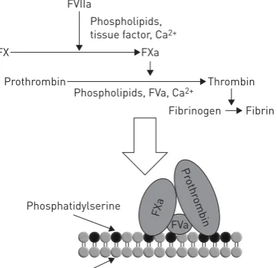
In 1955, O’BRIEN [5] described “platelet-like activity” in normal human serum. 12 years later, WOLF [6] came to similar conclusions, demonstrating the presence of “platelet dust” in plasma. These two reports are probably the first ever to describe what we now call microparticles. To understand what O’BRIEN [5] and WOLF [6] meant by the expressions “platelet-like activity” and “platelet dust”, we must refer to the classical view of the coagulation cascade, a simplified version of which, limited to the so-called extrinsic pathway, is depicted in figure 2. The coagulation cascade comprises a series of enzymatic reactions, each activating a proenzyme that becomes able to activate the next. Eventually, the enzyme thrombin (also known as FIIa) cleaves fibrinogen, generating fibrin that represents the insoluble matrix of the clot. It has been known for decades that several steps of the cascade require negatively charged phospholipids for the reaction to take place with the appropriate kinetics. Activated platelets have long been considered the main source of such membranes due to their small size, and therefore high surface/volume ratio, and to the fact
a) b)
Exosomes 30–100 nm
Microparticles 100–1000 nm
Apoptotic bodies 1–3 µm
FIGURE 1Schematic representations ofa)the three types of extracellular vesicles andb)microparticle generation in more detail.
FIGURE 2Simplified scheme of the“extrinsic” pathway of blood coagulation. F: factor.
FVIIa
Prothrombin Thrombin
Fibrinogen Fibrin
Pr othr
ombin
Phospholipids, FVa, Ca2+
FX FXa
Phospholipids, tissue factor, Ca2+
FXa
Phosphatidylserine
Phosphatidylcholine
that upon activation platelets externalise the negatively charged phospholipid, phosphatidylserine [7]. What O’BRIEN[5] and WOLF[6] described was the presence, in both serum and plasma, of structures able to support the aforementioned reactions; these fluids, by definition cell-free environments, must therefore contain negatively charged phospholipids that are not cell-associated. These considerations led to the concept that negatively charged membrane fragments are released from platelets.
Vascular microparticles beyond platelet dust
Besides platelets, other vascular cells shed microparticles, including endothelial cells [8], leukocytes [9] and erythrocytes [10]. As previously mentioned, these microparticles have a procoagulant potential due to the presence of phosphatidylserine on their surface [11]. However, the presence of tissue factor (TF), the initiator of the so-called extrinsic coagulation pathway (figure 2), on the surface of some microparticles adds to their procoagulant potential [11]. Accordingly, numerous studies have demonstrated a role for microparticles of different cell origin in blood coagulation and thrombosis, bothin vitroandin vivo[12–16]. However, besides TF, microparticles have the potential to carry on their surface, and to enclose within their cytoplasm, other molecules derived from the parental cells, including proteins, DNA, RNA and microRNAs. Over the years, a picture has emerged that indicates that microparticles represent functional units with the potential of representing a disseminated storage pool of bioactive effectors, neither soluble nor cell-associated, that participate both in the maintenance of homeostasis and in the pathogenesis of disease [17].
Mechanisms involved in microparticle formation
The mechanisms involved in microparticle formation have only partially been elucidated; however, two distinct, well-characterised pathways have been identified, namely cell activation and apoptosis [18]. Microparticle shedding caused by activation starts within minutes after the addition of the relevant agonist [19] and is characterised by an increase in cytosolic calcium concentration [20]. In contrast, in apoptosis-dependent microparticle formation, dynamic membrane blebbing follows cell contraction and DNA fragmentation, and its time-course is usually measured in hours [21]. Several studies have demonstrated that inhibition of the increase in cytosolic calcium prevents microparticle formation. CAMPBELLet al.[22] demonstrated that the release of microparticles by S49 mouse lymphoma cells induced by ionomycin was inhibited by EGTA, a calcium chelator. We have shown that EGTA inhibits peroxisome proliferator-activated receptor (PPAR)-γ-induced release of microparticles by human monocytes/ macrophages (HMMs) [23]. In a similar model, using cigarette smoke extract as a stimulus for microparticle release, we also inhibited microparticle generation with the L-type calcium channel blocker verapamil [24]. Finally, the demonstration that calmodulin inhibition prevents cigarette smoke extract-induced microparticle generation indicates a role for this calcium-binding messenger protein [24].
Different key regulators of the cytoskeleton have been identified in microparticle generation. BURGERet al.[8] demonstrated that Rho kinases are involved in endothelial cell-derived microparticle (EMP) formation, since Rho kinase inhibition blocked angiotensin II-induced release of microparticles. Furthermore, increased Rho kinase activity causes the release of circulating EMPs [25]. In addition, the small GTPase RhoA triggers a specific signalling pathway essential for microparticle biogenesis in various human cancer cells [26].
Other groups have demonstrated the involvement of mitogen-activated protein kinases (MAPKs) in microparticle generation. For example, activating autoantibodies to the angiotensin II receptor type 1 obtained from hypertensive patients promote EMP generation through the activation of p38 MAPK signalling, and p38 inhibitors effectively block this phenomenon [27]. Two groups have independently shown that the p38 pathway is involved in EMP generation upon stimulation with indoxyl sulphate [28] and tumour necrosis factor (TNF)-α[29]. In macrophage-derived microparticles obtained after stimulation with tobacco smoke extract, the production of microparticles relies on a series of dynamic, regulated steps that include activation of the c-Jun N-terminal kinase ( JNK) and p38 MAPKs [30]. In the same model, other researchers have demonstrated that tobacco smoke extract exposure causes activation of JNK, p38 and extracellular signal-regulated kinase (ERK) MAPKs, as well as apoptosis, a major mechanism for microparticle generation, and that only the inhibition of ERK, but not p38 or JNK, significantly blunted tobacco smoke extract-induced microparticle generation [31]. In addition, we have demonstrated the involvement of the ERK pathway in the generation of microparticles by human monocytes stimulated with PPAR-γagonists [23].
Potential role of microparticles in airway inflammation
The recruitment of blood-borne leukocytes into the airways represents a critical step in inflammatory reactions and requires the orchestrated action of cytokines, chemokines and cell–cell adhesion molecules [34]. Microparticles contribute to the production of various pro-inflammatory mediators by lung cells, thus becoming potential key factors in the airway inflammatory process. We have demonstrated that HMMs stimulated with the calcium ionophore A23187 and histamine generate microparticles with a pro-inflammatory potential for human lung cells. These microparticles, incubated with bronchial and alveolar epithelial cells, upregulate interleukin (IL)-8, monocyte chemotactic protein-1 and intercellular adhesion molecule-1 synthesis [35]. We investigated the mechanisms leading to increased transcription of pro-inflammatory mediators induced by HMM-derived microparticles. First, we demonstrated that HMM-derived microparticles cause the translocation of NF-κB into the nucleus [36]. We then showed that PPAR-γagonists, such as rosiglitazone and 15-deoxy-Δ12,14-prostaglandin-J2, inhibit the phenomenon [36], in keeping with the postulated anti-inflammatory role of these molecules that have in fact been proposed as potential therapeutic targets in lung diseases [37]. Microparticles with a similar pro-inflammatory potential on lung epithelial cells are also generated by HMMs exposed to cigarette smoke extract [24]. In addition, FOGLIet al.[38] demonstrated that HMM-derived microparticles induce a pro-inflammatory phenotype in human bronchial smooth muscle cells and that montelukast, a cysteinyl leukotriene receptor antagonist, reverses the phenomenon. In particular, montelukast prevented nuclear translocation of NF-κB, blocked IL-8 and prostaglandin E2 release and restored salbutamol response in microparticle-stimulated human bronchial smooth muscle cells [38]. Finally, another group has demonstrated that human lymphoblastoma T-cell-derived microparticles induce the production of inflammatory cytokines, including TNF-α, IL-6 and IL-8, in a dose- and time-dependent manner by human bronchial cells [39].
Complementary experimental approaches involving animal models have provided many insights that contribute to the understanding of the relationships between airway inflammation and microparticle generation. PORRO et al.[40] demonstrated that microparticles isolated from the sputum of cystic fibrosis patients are pro-inflammatory when injected into the murine lung, determine a strong neutrophilic infiltrate in the parenchyma and at the perivascular/peribronchial level, and that, among the possible mediators involved, lipopolysaccharide has a putative role in this phenomenon. In addition, exogenous EMPs injected into C57BL/6 mice caused a significant rise in pulmonary capillary permeability both as a primary and secondary injury [41].
Microparticles in human lung diseases
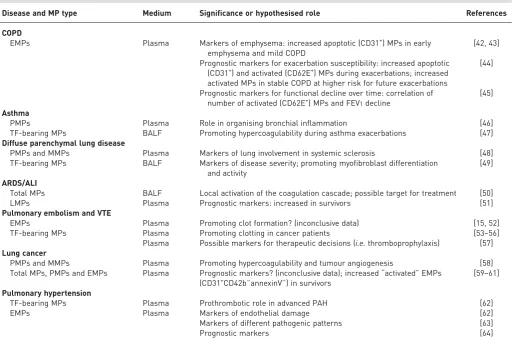
Microparticles have been investigated in several human lung diseases as possible pathogenic elements, prognostic markers and therapeutic targets. The data reported in the following sections are summarised in table 1.
Chronic obstructive pulmonary disease
Chronic obstructive pulmonary disease (COPD) is associated with several relevant comorbidities, possibly linked together through systemic inflammation [65–67]; among such comorbidities, cardiovascular diseases are probably the most significant, especially in prognostic terms [68–73]. It is of note that the presence of cardiovascular abnormalities has been demonstrated in a significant proportion of patients affected by COPD, even in the absence of clinically evident cardiac disease [74–76]. These close associations might be mediated by both endothelial dysfunction and activation, indicating an active pathogenic role played by endothelial cells [77, 78] as well as by lung vasculature damage and apoptosis. The latter is particularly relevant in the pathogenesis of emphysema and attributed, at least in part, to reduced function and/or levels of vascular endothelial growth factor [79–81].
THOMASHOWet al. [43] analysed EMPs from 104 COPD patients and 76 controls, and found a significant increase in CD31+ EMP levels (identified as apoptotic microparticles) in mild COPD, whereas CD62E+ EMPs (identified as activated microparticles) were higher in severe COPD. Additionally, apoptotic EMP levels were positively correlated with the percentage of emphysema, measured by computed tomography scan, and negatively correlated with pulmonary microvasculature perfusion, as assessed by magnetic resonance imaging. Again, these observations suggest that pulmonary endothelial apoptosis could represent an early step in emphysema development.
TAKAHASHIet al.[44] found a significant increase in both apoptotic and activated (CD31+and CD62E+, respectively) EMPs during exacerbations compared with stable COPD. Although expression of angiotensin-converting enzyme was not assessed in this study, since the large majority of EMPs did not express von Willebrand factor (a marker of systemic vasculature not expressed by pulmonary capillaries), it was concluded that during an exacerbation pulmonary capillaries were mostly affected, both in terms of apoptosis and activation/inflammation. Furthermore, the study showed that elevated baseline levels of CD62E+ EMPs, suggestive of a sustained endothelial activation, even in stable COPD patients could predict a susceptibility to exacerbations, thus representing a possible prognostic marker. In a later study by the same investigators, a significant correlation between baseline CD62E+EMP levels and annual change in forced expiratory volume in 1 s (FEV1) was found: higher EMP levels predicted a more relevant decline in FEV1, suggesting that a persistent endothelial activation and inflammation could play a role in the functional decline of COPD patients [45].
Thus, analysis of pulmonary endothelial cell activation and apoptosis through EMP evaluation is proving instrumental in generating data that lend further support to the hypothesis that COPD is a disease with a
TABLE 1Summary of the known roles of microparticles (MPs) in human lung diseases
Disease and MP type Medium Significance or hypothesised role References
COPD
EMPs Plasma Markers of emphysema: increased apoptotic (CD31+) MPs in early
emphysema and mild COPD
[42, 43]
Prognostic markers for exacerbation susceptibility: increased apoptotic (CD31+) and activated (CD62E+) MPs during exacerbations; increased
activated MPs in stable COPD at higher risk for future exacerbations
[44]
Prognostic markers for functional decline over time: correlation of number of activated (CD62E+) MPs and FEV1decline
[45]
Asthma
PMPs Plasma Role in organising bronchial inflammation [46]
TF-bearing MPs BALF Promoting hypercoagulability during asthma exacerbations [47] Diffuse parenchymal lung disease
PMPs and MMPs Plasma Markers of lung involvement in systemic sclerosis [48]
TF-bearing MPs BALF Markers of disease severity; promoting myofibroblast differentiation and activity
[49]
ARDS/ALI
Total MPs BALF Local activation of the coagulation cascade; possible target for treatment [50]
LMPs Plasma Prognostic markers: increased in survivors [51]
Pulmonary embolism and VTE
EMPs Plasma Promoting clot formation? (inconclusive data) [15, 52]
TF-bearing MPs Plasma Promoting clotting in cancer patients [53–56]
Plasma Possible markers for therapeutic decisions (i.e.thromboprophylaxis) [57] Lung cancer
PMPs and MMPs Plasma Promoting hypercoagulability and tumour angiogenesis [58] Total MPs, PMPs and EMPs Plasma Prognostic markers? (inconclusive data); increased“activated”EMPs
(CD31+CD42b−annexinV−) in survivors [59–61]
Pulmonary hypertension
TF-bearing MPs Plasma Prothrombotic role in advanced PAH [62]
EMPs Plasma Markers of endothelial damage [62]
Markers of different pathogenic patterns [63]
Prognostic markers [64]
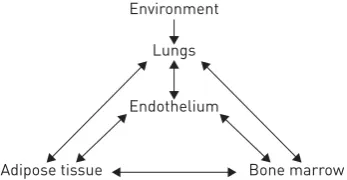
significant endothelial component, which could link lung disease and its systemic (and especially cardiovascular) comorbidities [78]. AGUSTÍ et al. [82] recently proposed a general model whereby the lungs, endothelium, bone marrow and adipose tissue form a network, in which the lung represents an external sensor, the endothelium an internal sensor and the bone marrow and adipose tissue the responsive elements. The model, summarised in figure 3, is largely speculative and AGUSTÍ et al. [82] acknowledged that much experimental work is needed in order to thoroughly identify the links between the lungs and the other components. Microparticles may represent one such link. Notably, we have reported that both leptin, an adipokine secreted by adipose tissue [83], and airborne pollutants [84] cause microparticle generation in macrophages and endothelial cells.
Asthma
Asthma is usually characterised by different patterns of airway inflammation, with a complex network of cellular and molecular mediators [85–87]. In contrast to COPD, very little is known about the possible role of microparticles in asthma and the scarce available data are preliminary, so that further research in the field, through properly designed studies, is needed. In a recent study conducted on 35 subjects (20 individuals with asthma and 15 healthy controls), asthmatic patients showed significantly higher baseline levels of platelet-derived microparticles (PMPs) than controls [46]. It is worth noting that all asthmatic patients were taking inhaled steroids. This finding could mark a potential pathogenic role for PMPs in airways inflammation, especially in organising cellular (in particular leukocyte) trafficking between blood and bronchi.
The interplay between inflammation and blood coagulation is well recognised; accordingly, an increased risk of pulmonary embolism in asthmatics, with an association with exacerbations, has been described [88, 89]. MAJOORet al.[47] investigated the role of viral infections in the haemostatic balance in asthmatic subjects. They showed an increase in TF-bearing microparticles in the bronchoalveolar lavage fluid (BALF) of asthmatics after experimental rhinovirus infection. They hypothesised that TF-bearing microparticles could play a pathogenic role in the local activation of coagulation during asthma infectious exacerbations, although the population sample was too small (14 individuals with asthma and 14 healthy controls) to allow for definite conclusions.
Diffuse parenchymal lung diseases
Diffuse parenchymal lung diseases represent a very inhomogeneous group of pulmonary diseases [90], with several different aetiologies and pathogenic patterns, most of them not clearly understood or, in fact, even almost completely unknown. Moreover, many of these conditions are quite rare. Very few studies have addressed the potential role of microparticles in diffuse parenchymal lung diseases. NOMURAet al. [48] showed, in a small sample of 42 patients with progressive systemic sclerosis, that levels of PMPs and monocyte-derived microparticles (MMPs) were increased compared with 30 healthy controls. More interestingly, significantly higher levels of PMPs and MMPs were found in patients with pulmonary involvement (i.e.with interstitial lung disease), compared with patients without lung disease. Such elevated levels of microparticles might therefore represent a possible marker of more advanced disease and a putative marker to stratify patients when choosing a treatment strategy, such as anti-platelet drugs.
A link between blood coagulation and pulmonary fibrosis is well recognised [91]. Based on the role of microparticles in blood coagulation, we investigated their presence in diffuse parenchymal lung disease patients [49]. We showed an increased number of microparticles in the BALF of 19 patients with interstitial lung diseases compared with 11 control subjects. Furthermore, the microparticle-bound TF (MP-TF) procoagulant activity was significantly higher in patients than in controls. When patients were further divided into idiopathic pulmonary fibrosis (IPF) and non-IPF groups, the MP-TF procoagulant activity was significantly higher in the former group. Additionally, a statistically significant negative correlation was found between TF-bearing microparticles and both forced vital capacity andDLCOin IPF patients [49]. In the same study,in vitroassays showed that an oxidative stimulus, namely H2O2, increased the production of procoagulant microparticles by alveolar epithelial cells in culture [49]. Local synthesis of FIGURE 3 Proposed interactions among the
environment, lungs, endothelium, bone marrow and adipose tissue in the pathogenesis of chronic obstructive pulmonary disease and its comorbidities. Reproduced and modified from [82] with permission.
Environment
Lungs
Endothelium
coagulation factor X and its activation to factor Xa in the lung has been implicated in the pathogenesis of IPF. Factor Xa is a protease that, besides its well-known role in cleaving prothrombin to generate thrombin, is also capable of cleaving the protease-activated receptor (PAR)-1 on fibroblasts, signalling their differentiation into myofibroblasts and therefore potentially contributing to the fibrotic process [92]. Increased local synthesis of factor X and local expression of factor Xa has been demonstrated in IPF lungs [92]. Our data are consistent with the existence of a pathway whereby an oxidative stress induces the generation of procoagulant, TF-bearing microparticles that activate locally synthesised factor X to Xa, thus inducing a PAR-1-mediated activation of fibroblasts and a profibrotic response.
Acute respiratory distress syndrome
In acute respiratory distress syndrome (ARDS) a burst of local inflammation is present, associated with an activation of the coagulation cascade and a significant deposition of fibrin in the alveolar spaces. Based on previous evidence of the activation of the TF pathway in ARDS [93], BASTARACHE et al. [50] found a significantly higher concentration of microparticles in pulmonary oedema fluid from ARDS subjects than from patients with cardiogenic pulmonary oedema. Moreover, microparticles from ARDS patients had a higher procoagulant activity compared with those from patients with cardiogenic oedema. This procoagulant activity was mainly due to the presence of increased concentration of TF in the fluid, which strongly correlated with microparticle levels, indicating the presence of TF-bearing microparticles. As suggested by subsequentin vitro analysis, the alveolar epithelium probably represented the main source of microparticles in ARDS. Furthermore, the study showed a trend towards lower microparticle concentrations in ARDS patients who survived compared with those who died of the disease. BASTARACHEet al.[50] speculated that microparticles could play a key role in the activation of blood coagulation and in the deposition of fibrin in the alveolar spaces that characterises ARDS, and that they might represent a potential target for future treatment of ARDS.
Apparently discordant results emerged from another study that analysed plasma and BALF levels of microparticles of different cellular origin (leukocytes, neutrophils, endothelium and platelets) in 52 patients with ARDS and 22 controls [51]. Higher plasmatic levels of leukocyte-derived microparticles (LMPs) were associated with a better prognosis in the ARDS group, a result apparently at odds with that of BASTARACHEet al.[50]; in fact, these results only suggest that circulating LMPs, as opposed to PMPs and MMPs, might be protective, possibly through their positive effects on vascular tone, as speculated in previous works on sepsis and septic shock [94].
Finally, in a murine model, the injection of EMPs into mice induced a significant release of the pro-inflammatory cytokines TNF-αand IL-1β, with a subsequent recruitment of neutrophils [95]. These effects were further increased by the concomitant or sequential administration of bacterial lipopolysaccharide, indicating that EMPs might represent a signal that primes the lung for the following inflammatory response to an external injury. Again, from this point of view, EMPs could represent a potential therapeutic target for ARDS. In conclusion, these studies have mainly hypothesised a role for microparticles (especially for EMPs) in the pathogenesis of ARDS: in particular, EMPs could represent a link between alveolar inflammation and coagulation (two key steps in the pathobiology of ARDS), and perhaps even a target for future treatment.
Pulmonary embolism
The potential role of procoagulant microparticles in venous thromboembolism (VTE) has been extensively investigated. A first series of studies measured the level of circulating microparticles, irrespective of their MP-TF procoagulant activity. CHIRINOS et al. [15] found an increase in plasma levels of EMPs and of EMP–monocyte conjugates in 25 patients with VTE compared with 25 healthy controls, suggesting that during VTE episodes EMPs and their interactions with leukocytes could play a role in clot formation. From this point of view, EMPs might represent a potential therapeutic target for preventing and treating thromboembolic events.
In contrast, in another study, EMP and PMP levels were similar between patients with acute pulmonary embolism and control subjects [52]. A difference in circulating EMP and PMP levels was found when the pulmonary embolism group was compared with the control subjects without cardiovascular risk factors, but disappeared when controls with cardiovascular risk factors were included [52], suggesting that circulating microparticles simply represent markers of such cardiovascular risk factors.
While these reports did not investigate the possible functional role of MP-TF in clot formation, other studies have mainly focused onin vitrotests for MP-TF procoagulant activity of circulating microparticles, to better elucidate the pathophysiological relationship between microparticles and VTE. In a retrospective study by GARCIA RODRIGUEZ et al. [97], the procoagulant activity of TF-bearing microparticles (MP-TF activity) was higher in patients with suspected pulmonary embolism than in controls; however, there were no significant differences between MP-TF activity in patients with confirmed pulmonary embolism and patients without the disease. Since an exploratory analysis in the group with suspected pulmonary embolism revealed higher levels of MP-TF activity in patients affected by cancer, GARCIARODRIGUEZet al. [97] concluded that MP-TF activity was not part of the VTE event, but rather reflected the presence of cancer. The prospective study by THALER et al. [98], conducted on patients with deep vein thrombosis (with or without pulmonary embolism), came to similar conclusions and did not support the hypothesis that TF-bearing microparticles play a clear role in the pathogenesis of thromboembolic events. In conclusion, functional studies on MP-TF activity failed to definitively confirm the hypothesis that emerged from previous studies that investigated the levels of circulating microparticles.
The association of VTE with cancer is well established [99] and the role of microparticles in this setting has also been investigated. In a study by TESSELAARet al.[53], in patients with VTE and metastatic cancer, MP-TF activity was higher than in patients with cancer but no VTE, in healthy subjects or in patients with idiopathic VTE. These findings suggest the possibility that tumour-derived microparticles might initiate the clotting cascadein vivoin cancer patients, thus playing a pathogenic role. Similar results derive from other studies that confirm that MP-TF activity is associated with cancer, with an“overactivity”in the presence of a VTE event [54–56]. These findings support the hypothesis that these microparticles are produced by tumour cells, play a pathogenic role in the development of VTE and may represent a potential biomarker for critical clinical challenges such as, for example, the choice of the ideal prophylactic treatment in oncology patients. ZWICKER et al. [57], in a phase II randomised trial, divided a cohort of 66 patients with a nonresectable cancer, according to the levels of MP-TF activity (low and high levels); they further split the high-level group into two arms, according to the administration of a prophylactic dose of enoxaparin. Patients with high levels of MP-TF activity treated with enoxaparin showed a significantly lower cumulative incidence of VTE at 2 months compared with those not treated and with high levels of MP-TF activity, while the incidence was similar to the low-level group, suggesting that levels of MP-TF activity could be used as a potential biomarker to stratify the risk of VTE and drive the choice of a prophylactic anticoagulation strategy in cancer patients.
Lung cancer
Lung cancer represents the main cause of cancer death worldwide [100]. In order to reach an earlier diagnosis and to better select patients for individual treatments, several biomarkers have been studied, including microparticles, but with inconsistent results regarding the possible biological roles of microparticles in lung cancer. In an early study by KANAZAWA et al. [58], PMP and MMP levels were higher in 64 patients with lung cancer (both small and nonsmall cell lung cancer) compared with 30 controls. Additionally, in cancer patients, platelet activation markers were also higher than in controls, suggesting a potential biological relationship between microparticles (both MMPs and PMPs) and vascular complications (both hypercoagulability and tumour angiogenesis) in these subjects.
Other studies have confirmed the higher levels of different types of microparticles in patients with lung cancer compared with healthy controls. These have mostly focused on the possible prognostic value of microparticles in lung cancer but the results were discordant. FLEITASet al.[59] found higher levels of total circulating microparticles in 60 patients with nonsmall cell lung cancer compared with 60 controls, but the most relevant finding was that, in cancer patients, higher baseline levels of microparticles were associated with a better progression-free and overall survival. Discordant results emerged from another study conducted in 107 end-stage nonsmall cell lung cancer patients, divided into 1-year survivors and nonsurvivors, measuring PMPs and EMPs [60]. While there was no difference in PMPs and “apoptotic” EMPs (defined as CD31+CD42b−annexinV+microparticles) between the two groups,“activated”EMP levels (defined as CD31+CD42b−annexinV−microparticles) were significantly higher in nonsurvivors [60]. After a multivariate regression analysis, these higher levels were an independent predictor of 1-year mortality. This result was clearly in contrast with the study by FLEITASet al.[59]. Moreover, in a previous study on the topic by the same investigators [61], both PMP and EMP levels were higher in 130 lung cancer patients compared with 30 healthy controls, but they were not associated with the presence of metastasis or with cancer stage (earlyversusadvanced). All these data underline the need for further studies in this field, in order to better elucidate the possible prognostic and therapeutic role of microparticles in lung cancer.
Pulmonary arterial hypertension
and thrombosis, and endothelial dysfunction [102–106]. Microparticles have been investigated in animal models of PAH [107–109]. The results support the hypothesis that microparticles might act directly on endothelial function by altering vascular balance, through a reduction of nitric oxide production, an increase in oxidative stress and as a circulating source of vasoconstrictor agents such as thromboxane A2.
In human pulmonary hypertension, microparticles could represent both a pathogenic element and a prognostic marker. A pathogenic role for microparticles in humans has been proposed in a study on 20 patients affected by PAH [62]. These subjects showed higher levels of endoglin+EMPs compared with 23 controls. Moreover, patients had higher MP-TF activity than controls, with the highest values in more severe hypertensive subjects (World Health Organization (WHO) functional classes III and IV and 6-min walking distance <380 m). These data supported a theoretical relationship between endothelial damage and PAH severity. Furthermore, the highest levels of TF-bearing microparticle activity in the most severe subgroup of patients might underline the possible prothrombotic contribution of microparticles in the advanced stage of disease. Finally, since endoglin is an accessory receptor for transforming growth factor-β, involved in cell proliferation and in neoangiogenesis, EMPs could participate in the pathogenesis of plexiform lesions and vascular remodelling.
Another study supported a role for microparticles in the pathogenesis of PAH [110]. In this study, 10 patients with idiopathic PAH showed a significant increase of CD39 nucleotidase expression and function on microparticle membranes compared with 10 controls. Since CD39 has an ATP nucleotidase activity, and since ATP has an endothelium-dependent vasodilatory effect on pulmonary arteries, the CD39 increased activity could contribute to further raise pulmonary vascular resistance by reducing ATP endovascular concentration. Conversely, CD39 activity ultimately leads to a reduction in AMP concentration, and since AMP is a potent stimulator for platelet aggregation and thrombosis, the increased CD39 levels could also represent a compensatory mechanism in order to limit the pathogenic process.
Microparticles could also represent a marker of severity in pulmonary hypertension. AMABILEet al. [63] found increased levels of EMPs in an inhomogeneous sample of 24 patients with pre-capillary pulmonary hypertension, compared with 20 healthy controls. Although no relationships existed between EMPs and disease severity as assessed by WHO functional classes or 6-min walking test, there were significant positive correlations between haemodynamic parameters of disease severity and both CD144+ and CD31+ EMP levels, and between CD62E+ EMP levels and systemic inflammation (evaluated with high-sensitivity C-reactive protein). Taken together, these results may indicate that different types of EMP can represent markers of different pathogenic pathways in pulmonary hypertension (with CD144+and CD31+EMP levels as predictors of disease severity and CD62E+EMP levels as markers of vascular inflammation), but also that EMPs could represent potential markers to predict prognosis and to perhaps monitor response to pharmacological treatments. In order to better elucidate these latter aspects, the same investigators analysed the same cohort in a prospective study, with a 1-year follow-up [64], and found significantly higher CD62E+ EMP levels in pulmonary hypertension patients who developed an adverse outcome (death or worsening right heart failure) than in those with stable disease. Additionally, AMABILEet al.[64] identified a cut-off value for CD62E+EMP levels to independently predict a worse prognosis. These data suggest that CD62E+ EMP levels could be used as a prognostic marker to better stratify pulmonary hypertension patients before starting a specific treatment. Other studies demonstrated higher levels of circulating microparticles in patients with pulmonary hypertension compared with healthy controls, but they did not assess the potential utility of microparticles as prognostic markers in the clinical setting [111, 112].
Conclusive remarks and future directions
Microparticles are rapidly gaining consideration both as biomarkers and as potential targets for therapeutic interventions. In the field of respiratory medicine, microparticles have, for example, the potential to help identify COPD phenotypes and stratify disease severity, to improve risk stratification for the development of VTE to better define prophylactic strategies, and to allow a better prognostic characterisation of ARDS patients. Furthermore, as a pathogenic role for microparticles is clearly emerging, the precise identification of the mechanisms involved in their formation is likely to prove valuable in identifying much needed novel therapeutic targets.
References
1 van der Pol E, Böing AN, Harrison P, et al.Classification, functions, and clinical relevance of extracellular vesicles.Pharmacol Rev2012; 64: 676–705.
2 György B, Szabó TG, Pásztói M,et al.Membrane vesicles, current state-of-the-art: emerging role of extracellular vesicles.Cell Mol Life Sci2011; 68: 2667–2688.
3 Burger D, Schock S, Thompson CS,et al.Microparticles: biomarkers and beyond.Clin Sci2013; 124: 423–441.
4 Geddings JE, Mackman N. New players in haemostasis and thrombosis.Thromb Haemost2014; 111: 570–574.
5 O’Brien JR. The platelet-like activity of serum.Br J Haematol1955; 1: 223–228.
7 Furie B, Furie BC. The molecular basis of blood coagulation.Cell1988; 53: 505–518.
8 Burger D, Montezano AC, Nishigaki N,et al.Endothelial microparticle formation by angiotensin II is mediated
viaAng II receptor type I/NADPH oxidase/Rho kinase pathways targeted to lipid rafts.Arterioscler Thromb Vasc Biol2011; 31: 1898–1907.
9 Wang JG, Aikawa E, Aikawa M. Leukocyte-derived microparticles as proinflammatory mediators in atherosclerosis.J Am Coll Cardiol2013; 62: 1442–1445.
10 Koshiar RL, Somajo S, Norström E, et al. Erythrocyte-derived microparticles supporting activated protein C-mediated regulation of blood coagulation.PLoS One2014; 9: e104200.
11 Celi A, Lorenzet R, Furie BC,et al.Microparticles and a P-selectin-mediated pathway of blood coagulation.
Dis Markers2004; 20: 347–352.
12 Falati S, Liu Q, Gross P,et al.Accumulation of tissue factor into developing thrombiin vivois dependent upon microparticle P-selectin glycoprotein ligand 1 and platelet P-selectin.J Exp Med2003; 197: 1585–1598.
13 Chou J, Mackman N, Merrill-Skoloff G,et al.Hematopoietic cell-derived microparticle tissue factor contributes to fibrin formation during thrombus propagation.Blood2004; 104: 3190–3197.
14 Steppich B, Mattisek C, Sobczyk D,et al.Tissue factor pathway inhibitor on circulating microparticles in acute myocardial infarction.Thromb Haemost2005; 93: 35–39.
15 Chirinos JA, Heresi GA, Velasquez H,et al. Elevation of endothelial microparticles, platelets, and leukocyte activation in patients with venous thromboembolism.J Am Coll Cardiol2005; 45: 1467–1471.
16 Hu SS, Zhang HG, Zhang QJ,et al.Small-size circulating endothelial microparticles in coronary artery disease.
PLoS One2014; 9: e104528.
17 Hugel B, Martínez MC, Kunzelmann C,et al.Membrane microparticles: two sides of the coin.Physiology2005; 20: 22–27.
18 VanWijk MJ, VanBavel E, Sturk A,et al.Microparticles in cardiovascular diseases.Cardiovasc Res2003; 59: 277–287.
19 MacKenzie A, Wilson HL, Kiss-Toth E, et al. Rapid secretion of interleukin-1β by microvesicle shedding.
Immunity2001; 15: 825–835.
20 Wiedmer T, Sims PJ. Participation of protein kinases in complement C5b-9-induced shedding of platelet plasma membrane vesicles.Blood1991; 78: 2880–2886.
21 Aupeix K, Hugel B, Martin T,et al.The significance of shed membrane particles during programmed cell death
in vitro, andin vivo, in HIV-1 infection.J Clin Invest1997; 99: 1546–1554.
22 Campbell LE, Nelson J, Gibbons E,et al. Membrane properties involved in calcium-stimulated microparticle release from the plasma membranes of S49 lymphoma cells.Scientific World Journal2014; 2014: 537192.
23 Neri T, Cordazzo C, Carmazzi Y,et al.Effects of peroxisome proliferator-activated receptor-γ agonists on the generation of microparticles by monocytes/macrophages.Cardiovasc Res2012; 94: 537–544.
24 Cordazzo C, Petrini S, Neri T,et al.Rapid shedding of proinflammatory microparticles by human mononuclear cells exposed to cigarette smoke is dependent on Ca2+mobilization.Inflamm Res2014; 63: 539–547.
25 Gao C, Li R, Liu Y,et al.Rho-kinase-dependent F-actin rearrangement is involved in the release of endothelial microparticles during IFN-α-induced endothelial cell apoptosis.J Trauma Acute Care Surg2012; 73: 1152–1160.
26 Li B, Antonyak MA, Zhang J,et al.RhoA triggers a specific signaling pathway that generates transforming microvesicles in cancer cells.Oncogene2012; 31: 4740–4749.
27 Yang S, Zhong Q, Qiu Z,et al.Angiotensin II receptor type 1 autoantibodies promote endothelial microparticles formation through activating p38 MAPK pathway.J Hypertens2014; 32: 762–770.
28 Ryu JH, Kim SJ. Clopidogrel effectively suppresses endothelial microparticle generation induced by indoxyl sulfateviainhibition of the p38 mitogen-activated protein kinase pathway.Blood Purif2011; 32: 186–194.
29 Curtis AM, Wilkinson PF, Gui M, et al. p38 mitogen-activated protein kinase targets the production of proinflammatory endothelial microparticles.J Thromb Haemost2009; 7: 701–709.
30 Li CJ, Liu Y, Chen Y,et al.Novel proteolytic microvesicles released from human macrophages after exposure to tobacco smoke.Am J Pathol2013; 182: 1552–1562.
31 Li M, Yu D, Williams KJ, et al.Tobacco smoke induces the generation of procoagulant microvesicles from human monocytes/macrophages.Arterioscler Thromb Vasc Biol2010; 30: 1818–1824.
32 Johnson BL 3rd, Goetzman HS, Prakash PS,et al.Mechanisms underlying mouse TNF-αstimulated neutrophil derived microparticle generation.Biochem Biophys Res Commun2013; 437: 591–596.
33 Lee SK, Yang SH, Kwon I,et al.Role of tumour necrosis factor receptor-1 and nuclear factor-κB in production of TNF-α-induced pro-inflammatory microparticles in endothelial cells.Thromb Haemost2014; 112: 580–588.
34 Chung KF, Adcock IM. Multifaceted mechanisms in COPD: inflammation, immunity, and tissue repair and destruction.Eur Respir J2008; 31: 1334–1356.
35 Cerri C, Chimenti D, Conti I, et al. Monocyte/macrophage-derived microparticles up-regulate inflammatory mediator synthesis by human airway epithelial cells.J Immunol2006; 177: 1975–1980.
36 Neri T, Armani C, Pegoli A, et al. Role of NF-κB and PPAR-γ in lung inflammation induced by monocyte-derived microparticles.Eur Respir J2011; 37: 1494–1502.
37 Belvisi MG, Hele DJ. Peroxisome proliferator-activated receptors as novel targets in lung disease.Chest2008; 134: 152–157.
38 Fogli S, Stefanelli F, Neri T,et al. Montelukast prevents microparticle-induced inflammatory and functional alterations in human bronchial smooth muscle cells.Pharmacol Res2013; 76: 149–156.
39 Qiu Q, Xiong W, Yang C, et al. Lymphocyte-derived microparticles induce bronchial epithelial cells’ pro-inflammatory cytokine production and apoptosis.Mol Immunol2013; 55: 220–230.
40 Porro C, Di Gioia S, Trotta T,et al. Pro-inflammatory effect of cystic fibrosis sputum microparticles in the murine lung.J Cyst Fibros2013; 12: 721–728.
41 Densmore JC, Signorino PR, Ou J,et al.Endothelium-derived microparticles induce endothelial dysfunction and acute lung injury.Shock2006; 26: 464–471.
42 Gordon C, Gudi K, Krause A,et al.Circulating endothelial microparticles as a measure of early lung destruction in cigarette smokers.Am J Respir Crit Care Med2011; 184: 224–232.
44 Takahashi T, Kobayashi S, Fujino N,et al.Increased circulating endothelial microparticles in COPD patients: a potential biomarker for COPD exacerbation susceptibility.Thorax2012; 67: 1067–1074.
45 Takahashi T, Kobayashi S, Fujino N, et al. Annual FEV1 changes and numbers of circulating endothelial microparticles in patients with COPD: a prospective study.BMJ Open2014; 4: e004571.
46 Duarte D, Taveira-Gomes T, Sokhatska O, et al. Increased circulating platelet microparticles as a potential biomarker in asthma.Allergy2013; 68: 1073–1075.
47 Majoor CJ, van de Pol MA, Kamphuisen PW, et al. Evaluation of coagulation activation after rhinovirus infection in patients with asthma and healthy control subjects: an observational study.Respir Res2014; 15: 14.
48 Nomura S, Inami N, Ozaki Y, et al. Significance of microparticles in progressive systemic sclerosis with interstitial pneumonia.Platelets2008; 19: 192–198.
49 Novelli F, Neri T, Tavanti L,et al.Procoagulant, tissue factor-bearing microparticles in bronchoalveolar lavage of interstitial lung disease patients: an observational study.PLoS One2014; 9: e95013.
50 Bastarache JA, Fremont RD, Kropski JA,et al.Procoagulant alveolar microparticles in the lungs of patients with acute respiratory distress syndrome.Am J Physiol Lung Cell Mol Physiol2009; 297: L1035–L1041.
51 Guervilly C, Lacroix R, Forel JM,et al. High levels of circulating leukocyte microparticles are associated with better outcome in acute respiratory distress syndrome.Crit Care2011; 15: R31.
52 Bal L, Ederhy S, Di Angelantonio E,et al.Factors influencing the level of circulating procoagulant microparticles in acute pulmonary embolism.Arch Cardiovasc Dis2010; 103: 394–403.
53 Tesselaar ME, Romijn FP, Van Der Linden IK,et al.Microparticle-associated tissue factor activity: a link between cancer and thrombosis?J Thromb Haemost2007; 5: 520–527.
54 Zwicker JI, Liebman HA, Neuberg D,et al. Tumor-derived tissue factor-bearing microparticles are associated with venous thromboembolic events in malignancy.Clin Cancer Res2009; 15: 6830–6840.
55 Manly DA, Wang J, Glover SL,et al.Increased microparticle tissue factor activity in cancer patients with venous thromboembolism.Thromb Res2010; 125: 511–512.
56 Campello E, Spiezia L, Radu CM,et al.Endothelial, platelet, and tissue factor-bearing microparticles in cancer patients with and without venous thromboembolism.Thromb Res2011; 127: 473–477.
57 Zwicker JI, Liebman HA, Bauer KA,et al.Prediction and prevention of thromboembolic events with enoxaparin in cancer patients with elevated tissue factor-bearing microparticles: a randomized-controlled phase II trial (the Microtec study).Br J Haematol2013; 160: 530–537.
58 Kanazawa S, Nomura S, Kuwana M, et al. Monocyte-derived microparticles may be a sign of vascular complication in patients with lung cancer.Lung Cancer2003; 39: 145–149.
59 Fleitas T, Martínez-Sales V, Vila V,et al.Circulating endothelial cells and microparticles as prognostic markers in advanced non-small cell lung cancer.PLoS One2012; 7: e47365.
60 Wang CC, Tseng CC, Hsiao CC,et al.Circulating endothelial-derived activated microparticle: a useful biomarker for predicting one-year mortality in patients with advanced non-small cell lung cancer.Biomed Res Int2014; 2014: 173401.
61 Tseng CC, Wang CC, Chang HC,et al.Levels of circulating microparticles in lung cancer patients and possible prognostic value.Dis Markers2013; 35: 301–310.
62 Bakouboula B, Morel O, Faure A,et al. Procoagulant membrane microparticles correlate with the severity of pulmonary arterial hypertension.Am J Respir Crit Care Med2008; 177: 536–543.
63 Amabile N, Heiss C, Real WM,et al.Circulating endothelial microparticle levels predict hemodynamic severity of pulmonary hypertension.Am J Respir Crit Care Med2008; 177: 1268–1275.
64 Amabile N, Heiss C, Chang V,et al.Increased CD62e+endothelial microparticle levels predict poor outcome in pulmonary hypertension patients.J Heart Lung Transplant2009; 28: 1081–1086.
65 Walter RE, Wilk JB, Larson MG, et al. Systemic inflammation and COPD: the Framingham Heart Study.
Chest2008; 133: 19–25.
66 Gan WQ, Man SF, Senthilselvan A, et al. Association between chronic obstructive pulmonary disease and systemic inflammation: a systematic review and a meta-analysis.Thorax2004; 59: 574–580.
67 Agustí A, Faner R. Systemic inflammation and comorbidities in chronic obstructive pulmonary disease.Proc Am Thorac Soc2012; 9: 43–46.
68 Mannino DM, Thorn D, Swensen A,et al.Prevalence and outcomes of diabetes, hypertension and cardiovascular disease in COPD.Eur Respir J2008; 32: 962–969.
69 Schünemann HJ, Dorn J, Grant BJ,et al.Pulmonary function is a long-term predictor of mortality in the general population: 29-year follow-up of the Buffalo Health Study.Chest2000; 118: 656–664.
70 Agustí A, Calverley PM, Celli B,et al.Characterisation of COPD heterogeneity in the ECLIPSE cohort.Respir Res2010; 11: 122.
71 McGarvey LP, John M, Anderson JA,et al.Ascertainment of cause-specific mortality in COPD: operations of the TORCH Clinical Endpoint Committee.Thorax2007; 62: 411–415.
72 Divo M, Cote C, de Torres JP,et al.Comorbidities and risk of mortality in patients with chronic obstructive pulmonary disease.Am J Respir Crit Care Med2012; 186: 155–161.
73 Agustí A, Edwards LD, Rennard SI, et al. Persistent systemic inflammation is associated with poor clinical outcomes in COPD: a novel phenotype.PLoS One2012; 7: e37483.
74 Iwamoto H, Yokoyama A, Kitahara Y, et al. Airflow limitation in smokers is associated with subclinical atherosclerosis.Am J Respir Crit Care Med2009; 179: 35–40.
75 Barr RG, Ahmed FS, Carr JJ,et al.Subclinical atherosclerosis, airflow obstruction and emphysema: the MESA Lung Study.Eur Respir J2012; 39: 846–854.
76 McAllister DA, Maclay JD, Mills NL,et al.Arterial stiffness is independently associated with emphysema severity in patients with chronic obstructive pulmonary disease.Am J Respir Crit Care Med2007; 176: 1208–1214.
77 Clarenbach CF, Senn O, Sievi NA,et al.Determinants of endothelial function in patients with COPD.Eur Respir J
2013; 42: 1194–1204.
78 Barberà JA. Chronic obstructive pulmonary disease: a disease of the endothelium?Am J Respir Crit Care Med
2013; 188: 5–7.
80 Kasahara Y, Tuder RM, Cool CD,et al.Endothelial cell death and decreased expression of vascular endothelial growth factor and vascular endothelial growth factor receptor 2 in emphysema.Am J Respir Crit Care Med2001; 163: 737–744.
81 Kanazawa H, Asai K, Hirata K,et al.Possible effects of vascular endothelial growth factor in the pathogenesis of chronic obstructive pulmonary disease.Am J Med2003; 114: 354–358.
82 Agustí A, Barberà JA, Wouters EF,et al.Lungs, bone marrow, and adipose tissue. A network approach to the pathobiology of chronic obstructive pulmonary disease.Am J Respir Crit Care Med2013; 188: 1396–1406.
83 Petrini S, Neri T, Lombardi S, et al. Leptin induces the generation of procoagulant, tissue factor bearing microparticles by human peripheral blood mononuclear cells.Biochim Biophys Acta2016; 1860: 1354–1361.
84 Neri T, Pergoli L, Petrini S,et al.Particulate matter induces prothrombotic microparticle shedding by human mononuclear and endothelial cells.Toxicol In Vitro2016; 32: 333–338.
85 Global Initiative for Asthma. Global Strategy for Asthma Management and Prevention. 2016 Report. Available from http://ginasthma.org
86 Saglani S, Lloyd CM. Novel concepts in airway inflammation and remodelling in asthma.Eur Respir J2015; 46: 1796–1804.
87 Pelaia G, Vatrella A, Busceti MT,et al. Cellular mechanisms underlying eosinophilic and neutrophilic airway inflammation in asthma.Mediators Inflamm2015; 2015: 879783.
88 Majoor CJ, Bel EH. Allergic burden and the risk of venous thromboembolism.Eur Respir J2013; 42: 1158–1159.
89 Chung WS, Lin CL, Ho FM,et al.Asthma increases pulmonary thromboembolism risk: a nationwide population cohort study.Eur Respir J2014; 43: 801–807.
90 American Thoracic Society, European Respiratory Society. American Thoracic Society/European Respiratory Society International Multidisciplinary Consensus Classification of the Idiopathic Interstitial Pneumonias.Am J Respir Crit Care Med2002; 165: 277–304.
91 Crooks MG, Hart SP. Coagulation and anticoagulation in idiopathic pulmonary fibrosis.Eur Respir Rev2015; 24: 392–399.
92 Scotton CJ, Krupiczojc MA, Königshoff M,et al.Increased local expression of coagulation factor X contributes to the fibrotic response in human and murine lung injury.J Clin Invest2009; 119: 2550–2563.
93 Bastarache JA, Wang L, Geiser T,et al.The alveolar epithelium can initiate the extrinsic coagulation cascade through expression of tissue factor.Thorax2007; 62: 608–616.
94 Mostefai HA, Meziani F, Mastronardi ML,et al.Circulating microparticles from patients with septic shock exert protective role in vascular function.Am J Respir Crit Care Med2008; 178: 1148–1155.
95 Buesing KL, Densmore JC, Kaul S,et al.Endothelial microparticles induce inflammation in acute lung injury.
J Surg Res2011; 166: 32–39.
96 Bucciarelli P, Martinelli I, Artoni A,et al. Circulating microparticles and risk of venous thromboembolism.
Thromb Res2012; 129: 591–597.
97 Garcia Rodriguez P, Eikenboom HC, Tesselaar ME,et al.Plasma levels of microparticle-associated tissue factor activity in patients with clinically suspected pulmonary embolism.Thromb Res2010; 126: 345–349.
98 Thaler J, Koppensteiner R, Pabinger I,et al.Microparticle-associated tissue factor activity in patients with acute unprovoked deep vein thrombosis and during the course of one year.Thromb Res2014; 134: 1093–1096.
99 Connolly GC, Francis CW. Cancer-associated thrombosis.Hematology Am Soc Hematol Educ Program 2013; 2013: 684–691.
100 Siegel R, Ward E, Brawley O,et al.Cancer statistics, 2011: the impact of eliminating socioeconomic and racial disparities on premature cancer deaths.CA Cancer J Clin2011; 61: 212–236.
101 Galiè N, Hoeper MM, Humbert M,et al.Guidelines for the diagnosis and treatment of pulmonary hypertension.
Eur Heart J2009; 30: 2493–2537.
102 Archer S, Rich S. Primary pulmonary hypertension: a vascular biology and translational research“Work in progress”.Circulation2000; 102: 2781–2791.
103 Farber HW, Loscalzo J. Pulmonary arterial hypertension.N Engl J Med2004; 351: 1655–1665.
104 Budhiraja R, Tuder RM, Hassoun PM. Endothelial dysfunction in pulmonary hypertension.Circulation2004; 109: 159–165.
105 Humbert M, Morrell NW, Archer SL, et al. Cellular and molecular pathobiology of pulmonary arterial hypertension.J Am Coll Cardiol2004; 43: Suppl. 12, 13S–24S.
106 Rabinovitch M. Molecular pathogenesis of pulmonary arterial hypertension.J Clin Invest2012; 122: 4306–4313.
107 Tual-Chalot S, Guibert C, Muller B,et al.Circulating microparticles from pulmonary hypertensive rats induce endothelial dysfunction.Am J Respir Crit Care Med2010; 182: 261–268.
108 Pfister SL. Role of platelet microparticles in the production of thromboxane by rabbit pulmonary artery.
Hypertension2004; 43: 428–433.
109 Aliotta JM, Pereira M, Amaral A,et al.Induction of pulmonary hypertensive changes by extracellular vesicles from monocrotaline-treated mice.Cardiovasc Res2013; 100: 354–362.
110 Visovatti SH, Hyman MC, Bouis D,et al.Increased CD39 nucleotidase activity on microparticles from patients with idiopathic pulmonary arterial hypertension.PLoS One2012; 7: e40829.
111 Diehl P, Aleker M, Helbing T,et al.Increased platelet, leukocyte and endothelial microparticles predict enhanced coagulation and vascular inflammation in pulmonary hypertension.J Thromb Thrombolysis2011; 31: 173–179.