Long-term macrolides in diffuse
interstitial lung diseases
Paola Faverio
1, Francesco Bini
2, Adriano Vaghi
2and Alberto Pesci
1Affiliations: 1Dipartimento Cardio-Toraco-Vascolare, Università Milano-Bicocca, Clinica Pneumologica, Ospedale San Gerardo, ASST di Monza, Monza, Italy. 2ASST-Rhodense, UOC Pneumologia, Garbagnate Milanese, Italy.
Correspondence: Paola Faverio, Dipartimento Cardio-Toraco-Vascolare, University of Milan Bicocca, Respiratory Unit, San Gerardo Hospital, ASST di Monza, Via Pergolesi 33, 20900 Monza, Italy.
E-mail: [email protected]
@ERSpublications
Macrolides may act as microbiota modulators as well as anti-inflammatory and antifibrotic agents in ILDshttp://ow.ly/stlc30gB3je
Cite this article as: Faverio P, Bini F, Vaghi A, et al. Long-term macrolides in diffuse interstitial lung diseases.Eur Respir Rev2017; 26: 170082 [https://doi.org/10.1183/16000617.0082-2017].
ABSTRACT In the present review we provide currently available evidence for the use of macrolides in the treatment of diffuse interstitial lung diseases (ILDs). Up to now, research on macrolides has mainly focused on three areas. First, macrolides have shown some promising results in cellular models and case reports as antifibrotic agents, by promoting autophagy and clearance of intracellular protein aggregates and acting as regulators of surfactant homeostasis. Secondly, macrolides have an immunomodulatory effect, which has been applied in some organising pneumonia cases. In particular, macrolides have been tested in association with systemic corticosteroids as steroid-sparing agents and alone as either first-line agents in mild cases or second-line agents where steroids were poorly tolerated or had failed. Thirdly, a recent area of research concerns the possible role of macrolides as modulators of lung microbiota and the host–microbiota interaction. This function has been particularly studied in idiopathic pulmonary fibrosis patients, in whom changes in microbiota have been proved to be associated with disease progression. However, the lack of high-quality studies makes the application of macrolide therapy in ILDs a field in which research should be conducted on a large scale.
Introduction
In recent years, the use of low-dose, long-term macrolides has been described in case reports and small case series of patients with diffuse interstitial lung diseases (ILDs) and pulmonary fibrosis. We conducted a literature search using the PubMed/MEDLINE and EMBASE databases. We used the terms“macrolide” OR “erythromycin” OR “clarithromycin” OR “azithromycin” OR “roxithromycin” OR “troleandomycin” OR “telithromycin” in combination with the terms “interstitial lung disease”, “idiopathic pulmonary fibrosis”,“organising pneumonia”and“extrinsic allergic alveolitis”. We also conducted a search using the terms“microbioma”OR“microbiota”in combination with“interstitial lung disease”. Our review included items published between 1990 and July 2017 in the English language.
From the available literature, there is increasing evidence of a possible regenerative effect of macrolides on respiratory epithelium that has been damaged [1]. Pulmonary structure remodelling, together with inappropriate epithelial regeneration, is a key factor in the genesis of pulmonary fibrosis [2]. Excessive stress of the endoplasmic reticulum is among the mechanisms involved in the pathogenesis of the disease [2].
Copyright ©ERS 2017. ERR articles are open access and distributed under the terms of the Creative Commons Attribution Non-Commercial Licence 4.0.
Received: July 12 2017 | Accepted after revision: Sept 07 2017
Conflict of interest: None declared.
This intracellular organ is responsible for the synthesis of functional proteins and the recognition of altered proteins, which are then removed to be degraded via the autophagocytosis process. Autophagocytosis has a fundamental role in type II pneumocytes, which, among their many functions, produce the proteins that make up surfactant. These proteins have a complex structure and a high likelihood of structural changes leading to the formation of amyloid fibril aggregates. When these aggregates exceed the cell clearance capacity, the endoplasmic reticulum undergoes excessive stress, which, if not resolved, leads to apoptosis [3, 4].
Defects in the autophagocytosis mechanism have recently been found in many pathological conditions associated with tissue damage and inflammation [5]. Focusing on lung diseases, an example is provided by Hermansky–Pudlak syndrome, a rare autosomal-recessive transmission disorder characterised by multi-organ alterations, including the progressive development of ILD [6]. Pulmonary involvement in this syndrome appears to be caused by excessive stress of the endothelial reticulum and alteration of lysosomal traffic, leading to chronic damage and apoptosis of type II pneumocytes. In murine models, pulmonary fibrosis and type II pneumocytes containing giant lamellar bodies appear together [7]; these cellular alterations seem to be a direct expression of stress in the endothelial reticulum and lysosomes. In this scenario, macrolides appear to play a key role at various levels, primarily by promoting autophagy and clearance of intracellular protein aggregates [1]. Hence, there is a reduction in lysosomal and endothelial reticulum stress that can ultimately prevent cellular apoptosis. Rapamycin, a type of macrolide not used for antibiotic purposes, induces autophagy by inactivating mTOR (mammalian target of rapamycin), a protein kinase that has been shown to have an inhibitory effect on autophagy [8]. Other macrolides, including azithromycin, have also shown this effect, supporting the hypothesis that they protect against the toxic effect produced by excessive amounts of intracellular protein aggregates [9].
Macrolides may also have an action on lipid metabolism and surfactant homeostasis by interacting with regulating molecules of lipid homeostasis [1]. This role is of particular importance when considering that lipids account for almost 90% of the surfactant. In a case report published by THOUVENIN et al. [10]
regarding a 6-year-old child with diffuse ILD due to mutation of theABCA3gene (ATP-binding cassette transporter of the A subfamily, member 3), the use of long-term azithromycin, three times a week, led to a rapid and significant radiological and functional improvement. In this condition, type II pneumocytes appear hyperplastic and filled with abnormal lamellar bodies consisting of phospholipids.ABCA3plays an important role in the transport of phospholipids, which is a key step in surfactant production. In this situation, macrolides could play a double role: reducing cellular stress by promoting autophagy of abnormal lamellar bodies, as well as controlling lipid homeostasis.
Macrolide intracellular action in lung epithelium is not the only one studied to investigate their possible role in preventing the progression of certain ILDs. Possible effects on lung microbiota will be discussed in the next sections.
Microbiota
The “microbiota” is the community of microbes (bacteria, fungi, viruses and non-fungal eukaryotes) associated with a determined environment [11]; the term “microbiome” refers to the totality of the microbes and their genetic material harboured in a given microbiota and their interactions with it [12]. Most of the microbial and microbiome studies conducted so far have focused on bacteria.
For many years, the lower airways were considered sterile by definition, and the frequent microbiological samples traditionally taken were considered contamination by bacteria originating from the upper respiratory tract/oral cavity and also as a consequence of chronic microspiration [13]. In addition, the bacterial burden available in traditional isolation was often considered insufficient. This problem was related to the method of bacterial research, which was a culture-dependent method with the need to isolate a single bacterial agent in culture [14].
Currently, in the study of microbiota, culture-independent techniques are used, whose aim is to isolate the nucleic acids of bacterial DNA. This has allowed identification of the potential pathogens present in samples that tested negative in classical cultures [15, 16]. With these new methods it has been possible to demonstrate that the lower airways of healthy individuals have low levels of oropharyngeal bacteria (PrevotellaandVeillonellaspecies), rendering the dogma of sterility of the lower airways invalid [17, 18]. There is still a controversy about the real vitality of these bacteria, as fragments of bacterial DNA, not actual bacteria, are identified using these culture-independent techniques. In any case, it is now clear that the airway microbiota participates in the immunological homeostasis of the lung epithelium mucosa.
the disease, while in chronic obstructive pulmonary disease (COPD), progression of the disease is characterised by an increase in bacterial load and by a loss of heterogeneity, with the prevalence of a single species [21, 22].
For ILDs, studies on the microbiota are few compared to the other diseases already mentioned. However, it is logical to hypothesise that microbial alteration may be associated with a worsening of the disease, linked to immune dysregulation, excessive inflammation and infection. ILDs other than idiopathic pulmonary fibrosis (IPF) are often treated with immunosuppressive therapy. On the one hand, this makes patients more susceptible to the development of bacterial infections. On the other hand, the reduced immune reserve can alter the characteristics of the microbiota [23].
The genesis of IPF is multifactorial, one factor being the environment [2]. A potential role for infections, both as a cofactor of initial development and as a cofactor of fibrosis progression, has been widely postulated [24]. The questions just expressed have stimulated studies capable of quantifying and characterising the airway microbiota of IPF patients, using culture-independent methods.
IPF and host
–
microbiota interactions, and potential effects of macrolides
IPF is a progressive pulmonary disease with poor prognosis in the short to medium term. The genesis of the disease, as mentioned earlier, is multifactorial, with environmental factors playing a role in determining damage to the epithelial integrity of the lung and repair mechanisms, which are also altered on a genetic and epigenetic basis, causing aberrant repairing with consequent development of fibrosis [2]. Respiratory infections are among the possible environmental factors that cause the development of both fibrosis and acute exacerbations (AEs) of the disease [24]. The new available methods, which detect the presence of bacterial DNA of species that cannot be cultured, have paved the way for the study of complex microbial communities.
In this regard, a case–control study published by MOLYNEAUX et al. [25] analysed the bronchoalveolar
lavage (BAL) of 65 patients with IPF and compared it with that of 17 patients with COPD and 27 subjects with normal respiratory function, both smokers and nonsmokers. By using metagenomic techniques, the authors observed that in the BAL of IPF patients the number of copies of bacterial 16S rRNA was more than double that of non-IPF patients, but there was no difference between subjects with normal respiratory function and those with COPD. In addition, by observing patients with IPF prospectively and dividing them into two groups, depending on whether they showed disease stability or progression ( progression defined as worsening of forced vital capacity >10% in 6 months or death), the authors noticed a higher bacterial burden in patients with progressive disease. Regarding the bacterial taxonomy in patients with IPF as well as in controls, the most represented species wereStreptococcus species followed byPrevotella andVeillonellaspecies. However, IPF patients showed a lower taxonomic diversity among bacterial species than controls, with an increased burden of possible pathogenic species such asNeisseria,Haemophilusand Fusobacterium. Therefore, from these first observations it appears that patients with IPF exhibit a greater bacterial load and less taxonomic diversity than controls; moreover, a higher bacterial load is associated with progression of the disease. The mucin 5B (MUC5B) promoter gene polymorphism confers an increased risk of developing IPF, but, paradoxically, if possessed by a patient with IPF, it confers an advantage on survival [26, 27]. In the previously mentioned study by MOLYNEAUXet al.[25], the bacterial
load in BAL was independently related to a particular polymorphism of this gene. As MUC5B is directly implicated in alveolar macrophage homeostasis and in surfactant constitution, a direct relationship has been postulated between the microbial load and immune regulation.
Using the data from the COMET-IPF study (Correlating Outcomes with Biochemical Markers to Estimate Time-progression in Idiopathic Pulmonary Fibrosis), HAN et al.[28] retrospectively evaluated the role of
the microbiota in disease progression. Although the most frequently isolated bacterial species (Prevotella, VeillonellaandEscherichia) coincided in part with those identified by MOLYNEAUXet al.[25], the authors
also observed a new element: a strong association between the presence of specific species ofStreptococcus andStaphylococcusand disease progression [28].
Other authors have recently tried to take a step forward in the understanding of the pathogenesis of IPF by assessing the interaction between the host and lung microbiota [29, 30]. MOLYNEAUX et al. [25]
performed a longitudinal evaluation of host gene expression in peripheral blood cells from 60 IPF patients and 20 controls, from their prior contribution, and explored the potential association with BAL microbiota collected at baseline [29]. In this preliminary study, the authors demonstrated that in IPF patients, when compared to controls, there were changes in the peripheral blood expression profile associated with the presence of an altered or more abundant microbiome. HUANGet al.[30], once again using the data from
immune-response-relevant pathways was associated with changes in the abundance of specific microbial operational taxonomic units, which had an impact on disease progression. Furthermore, the authors demonstrated an even more direct contribution of the host–microbiome interaction to the pathogenesis of the disease by influencing immune-mediated fibroblast responsiveness and the composition of circulating leukocytes [30].
Even when evaluating rapid progressions of the disease, such as AEs, recent studies have demonstrated a possible role of the microbiota [31]. In a comparison of 20 patients with AE to 15 matched controls with stable IPF, MOLYNEAUXet al.[31] found an increased BAL bacterial burden and a shift in the composition of the microbiota, favouring bacteria usually confined to the gastrointestinal tract, such asCampylobacterspecies.
Given these assumptions about the role of bacterial burden and different species in host response and IPF progression, the next step will be to investigate the role played by antibiotic molecules administered for long periods on the natural history of the disease [32–36]. A few studies have explored the role of doxycycline and co-trimoxazole in IPF. The former has been studied for its inhibitor effect on matrix metalloproteinases, which play a role in the pathogenesis of IPF [34–36], and the latter has been demonstrated in a randomised controlled trial (RCT) (the TIPAC trial) to improve quality of life and, in the per-protocol analysis, to reduce all-cause mortality in association with a reduction in the frequency of respiratory tract infections, but without an effect on pulmonary function [32]. However, the generalisability of the data is limited, because not all patients had a diagnosis of IPF, nearly one-third of those receiving co-trimoxazole withdrew due to side-effects (mostly rash and nausea) and a consistent proportion was on immunosuppressive therapy. Because the available evidence led to conflicting results, an ongoing RCT (CleanUp-IPF trial; NCT02759120) is investigating the role of double-strength co-trimoxazole or doxycycline administered daily for 12–42 months plus standard of care,versusstandard of care alone, in reducing non-elective, respiratory hospitalisation or all-cause mortality in patients with IPF.
Despite the promising data on the rationale to use macrolides in IPF, in particular because, due to their combined antibiotic and immunomodulator effects, they may act simultaneously on both microbial modulation and host-immune regulation, there are still very few data available. In a recent retrospective monocentric study, KUSE et al. [33] noted that the addition of macrolides to conventional IPF therapy
seems to reduce the incidence of AEs. However, these preliminary data need to be considered with great caution, not only because of their retrospective nature, but also because they refer to the Japanese population alone and were collected over a period of time (2003–2008) in which the “conventional therapy”for IPF has changed profoundly. For example, 52% of the patients included in this study (27 out of 52) were subjected to corticosteroid therapy or other immunosuppressive agents, both of which are no longer recommended in IPF treatment [37].
All of this undoubtedly leads to the need to further investigate the role of macrolides in IPF patients. A clinical trial (NCT02173145) on the use of azithromycin at a dose of 500 mg·day−1three times per week for 12 weeks in IPF is currently under way. The primary outcome is a reduction in coughing and, as secondary outcomes, modification of pulmonary function tests and the composition of oropharyngeal flora are being considered.
We can therefore conclude that the currently available evidence seems to identify a key role for the microbiota and its modifications regarding the mode and progression of IPF, even outside AEs. Macrolides could participate in the“modulation”of the microbiota itself, but their role is still largely to be investigated.
Macrolides in organising pneumonia
Organising pneumonia (OP) is caused by an insult that produces lung damage to which the alveolar epithelium reacts, giving rise to granulation tissue [38]. Excessive inflammatory tissue fills the alveoli and spreads to the alveolar ducts and terminal bronchioles, resembling typical endolumenal gemstones of granular tissue [38]. These alterations are associated with interstitial inflammatory infiltrates, which is why OP is classified among the ILDs [38]. The disease can be either idiopathic (cryptogenic OP (COP)) or associated with infections, drug toxicity, vasculitis, connective tissue diseases, haematological tumours, radiotherapy,etc.[39].
Although in OP, as well as in IPF, the predominant pathogenetic mechanism is excessive production of repair tissue, the two diseases show fundamental differences [40]. First, in IPF, the fibro-proliferative process results in a subversion of alveolar structure with irreversible scarring, while in OP the pathological process is usually reversible with restoration of normal pulmonary structure [40]. Secondly, corticosteroid therapy, which has proved to be ineffective in IPF, is the first-choice treatment in OP [39].
However, for steroid treatment to be effective, it must be administered at a medium to high dose (0.5– 1 mg·kg−1·day−1in the initial stages) and for prolonged periods due to the risk of early recurrence after suspension. This obviously implies an increase in the risk of steroid-related side-effects [39]. Such observations led, in the 1990s, to an attempt to use steroid-sparing agents, including macrolides, in OP treatment.
Some studies have used macrolides as an alternative to corticosteroids [45, 46]. One of the first of these studies, by ICHIKAWAet al.[45] in 1993, used erythromycin at 600 mg·day−1for 3–4 months in six patients
with a diagnosis of bronchiolitis obliterans OP confirmed on histological examination. The clinical and radiological response was poor in the first 2–4 weeks of treatment, but it became complete in all patients after 3 months. In addition, although no significant side-effects were reported during treatment, the relapse time was rather short (3.6±1.8 months). By comparing BAL cellularity before and after treatment, normalisation of both neutrophils and lymphocytes was observed. These latter data regarding the cellularity of BAL are particularly significant; in fact, a mixed alveolitis with increase in lymphocytes (from 20% to 40% of total cellularity), neutrophils (up to 10%) and eosinophils (up to 5%) is usually observed in OP [47]. In addition, during OP there is an increase in cytotoxic T-lymphocytes with a CD4/CD8 reduction [48].
In this scenario, the immunomodulatory effect of macrolides could act at various levels. For example, macrolides could reduce the degree of inflammation linked to polymorphonucleated leukocytes and their products. In a study in 1996 on biopsy-proven COP patients who showed neutrophilia on BAL, HOTTA[49]
observed a reduction in interleukin (IL)-8 and neutrophilic chemotactic activity in BAL after 600 mg·day−1 erythromycin for 2–3 months, suggesting a beneficial effect of low-dose macrolides on neutrophil-mediated inflammation. RADZIKOWSKA et al. [44] reported a decrease in serum concentration of pro-inflammatory
cytokines, including IL-6, IL-8 and transforming growth factor-β1, and in the BAL concentration of IL-6 in patients with biopsy-proven COP who responded to clarithromycin 500 mg twice daily for 3 months. Moreover, AOKIand KAO[50] showed that the anti-inflammatory effect of erythromycin is also expressed
at the level of cytotoxic T-lymphocytes. A recent study published by CAIet al.[41] reported that alveolar
macrophages of both COP and secondary OP patients (diagnosed through lung biopsy) also show aberrant pro-inflammatory cytokines production compared to non-OP control subjects. In this study, exposure of alveolar macrophages to macrolides inhibited the release of pro-inflammatory cytokines and, for this purpose, clarithromycin was proven to be more effective than azithromycin.
In most studies, macrolides were not used alone as a first choice, but in combination with corticosteroids to reduce the dosage or to replace them in the case of ineffectiveness or when intolerable steroid-related side-effects occurred [46, 51–53]. An example is the case reported by CHANG et al. [52], where
clarithromycin therapy (500 mg·day−1) produced beneficial results in a patient with biopsy-proven
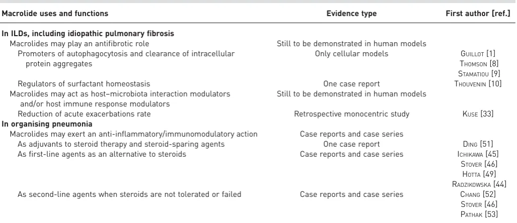
TABLE 1Summary of the main areas of application of low-dose, long-term macrolides in diffuse interstitial lung diseases (ILDs)
Macrolide uses and functions Evidence type First author [ref.]
In ILDs, including idiopathic pulmonary fibrosis
Macrolides may play an antifibrotic role Still to be demonstrated in human models Promoters of autophagocytosis and clearance of intracellular
protein aggregates
Only cellular models GUILLOT[1] THOMSON[8] STAMATIOU[9]
Regulators of surfactant homeostasis One case report THOUVENIN[10]
Macrolides may act as host–microbiota interaction modulators and/or host immune response modulators
Still to be demonstrated in human models
Reduction of acute exacerbations rate Retrospective monocentric study KUSE[33] In organising pneumonia
Macrolides may exert an anti-inflammatory/immunomodulatory action Case reports and case series
As adjuvants to steroid therapy and steroid-sparing agents One case report DING[51] As first-line agents as an alternative to steroids Case reports and case series ICHIKAWA[45]
STOVER[46] HOTTA[49] RADZIKOWSKA[44] As second-line agents when steroids are not tolerated or failed Case reports and case series CHANG[52]
bronchiolar COP in whom steroids (1 mg·kg−1·day−1) and azathioprine had proven to be ineffective and related to side-effects. In the cases available so far, side-effects associated with chronic use of macrolides have been rare and not severe (one case of skin rash associated with clarithromycin) [46].
It is noticeable how the evidence available so far regarding the use of low-dose, long-term macrolides in COP and secondary OP is, in reality, limited to case series. Moreover, in many published cases, macrolides were not used alone but in association with steroids or other immunosuppressive agents, making it impossible to evaluate their actual efficacy [51]. In conclusion, given the lack of large-scale prospective studies, at present there is no definitive data on the efficacy of macrolide therapy either as a first-line or as an adjuvant agent.
Conclusions and future perspectives
The experience gained so far has allowed the identification of many intracellular pathways on which macrolides act, but much remains to be investigated, especially with regard to the role of the lung microbiota and host–microbiota interaction in the genesis and evolution of ILDs and the action that macrolides can play on the latter. The main areas of application known to date are summarised in table 1. In addition, although macrolide use has been assessed in OP and IPF patients, other ILDs might potentially benefit from it. In this regard, the use of macrolides has been tested in animal models or anecdotally in case reports of patients with extrinsic allergic alveolitis and desquamative interstitial pneumonia [54, 55]. It is important to remember that the limited evidence available derives almost exclusively from studies in animal models, case series and retrospective studies. Before being actually able to support the utility of low-dose, long-term macrolides in the treatment of ILD patients, RCTs are certainly needed.
Acknowledgements
The authors thank Giovanna Minelli and Sharon Carlson for their help in the translation process.
References
1 Guillot L, Tabary O, Nathan N,et al.Macrolides: new therapeutic perspectives in lung diseases.Int J Biochem Cell Biol2011; 43: 1241–1246.
2 Maher TM, Wells AU, Laurent GJ. Idiopathic pulmonary fibrosis: multiple causes and multiple mechanisms?
Eur Respir J2007; 30: 835–839.
3 Korfei M, Ruppert C, Mahavadi P, et al. Epithelial endoplasmic reticulum stress and apoptosis in sporadic idiopathic pulmonary fibrosis.Am J Respir Crit Care Med2008; 178: 838–846.
4 Lawson WE, Crossno PF, Polosukhin VV, et al. Endoplasmic reticulum stress in alveolar epithelial cells is prominent in IPF: association with altered surfactant protein processing and herpesvirus infection.Am J Physiol Lung Cell Mol Physiol2008; 294: L1119–L1126.
5 Monick MM, Powers LS, Walters K,et al.Identification of an autophagy defect in smokers’alveolar macrophages.
J Immunol2010; 185: 5425–5435.
6 Wang L, Lyerla T. Histochemical and cellular changes accompanying the appearance of lung fibrosis in an experimental mouse model for Hermansky Pudlak syndrome.Histochem Cell Biol2010; 134: 205–213.
7 Osanai K, Higuchi J, Oikawa R, et al. Altered lung surfactant system in a Rab38-deficient rat model of Hermansky–Pudlak syndrome.Am J Physiol Lung Cell Mol Physiol2010; 298: L243–L251.
8 Thomson AW, Turnquist HR, Raimondi G. Immunoregulatory functions of mTOR inhibition.Nat Rev Immunol
2009; 9: 324–337.
9 Stamatiou R, Paraskeva E, Boukas K,et al.Azithromycin has an antiproliferative and autophagic effect on airway smooth muscle cells.Eur Respir J2009; 34: 721–730.
10 Thouvenin G, Nathan N, Epaud R, et al. Diffuse parenchymal lung disease caused by surfactant deficiency: dramatic improvement by azithromycin.BMJ Case Rep2013; 2013: bcr2013009988.
11 Cui L, Morris A, Huang L, et al. The microbiome and the lung. Ann Am Thorac Soc 2014; 11: Suppl. 4, S227–S232.
12 Rogers GB, Shaw D, Marsh RL,et al.Respiratory microbiota: addressing clinical questions, informing clinical practice.Thorax2015; 70: 74–81.
13 Monsó E, Ruiz J, Rosell A,et al.Bacterial infection in chronic obstructive pulmonary disease. A study of stable and exacerbated outpatients using the protected specimen brush. Am J Respir Crit Care Med 1995; 152: 1316–1320.
14 Goldstein EJC, Citron DM, Goldman PJ,et al.National hospital survey of anaerobic culture and susceptibility methods: III.Anaerobe2008; 14: 68–72.
15 Murphy TF, Brauer AL, Schiffmacher AT,et al. Persistent colonization by Haemophilus influenzaein chronic obstructive pulmonary disease.Am J Respir Crit Care Med2004; 170: 266–272.
16 Murphy TF, Brauer AL, Eschberger K,et al. Pseudomonas aeruginosain chronic obstructive pulmonary disease.
Am J Respir Crit Care Med2008; 177: 853–860.
17 Charlson ES, Bittinger K, Haas AR,et al.Topographical continuity of bacterial populations in the healthy human respiratory tract.Am J Respir Crit Care Med2011; 184: 957–963.
18 Segal LN, Alekseyenko AV, Clemente JC,et al.Enrichment of lung microbiome with supraglottic taxa is associated with increased pulmonary inflammation.Microbiome2013; 1: 19.
20 Ege MJ, Mayer M, Normand AC,et al.Exposure to environmental microorganisms and childhood asthma.N Engl J Med2011; 364: 701–709.
21 Molyneaux PL, Mallia P, Cox MJ, et al. Outgrowth of the bacterial airway microbiome after rhinovirus exacerbation of chronic obstructive pulmonary disease.Am J Respir Crit Care Med2013; 188: 1224–1231. 22 Sethi S, Murphy TF. Infection in the pathogenesis and course of chronic obstructive pulmonary disease.N Engl J
Med2008; 359: 2355–2365.
23 Garzoni C, Brugger SD, Qi W,et al.Microbial communities in the respiratory tract of patients with interstitial lung disease.Thorax2013; 68: 1150–1156.
24 Molyneaux PL, Maher TM. The role of infection in the pathogenesis of idiopathic pulmonary fibrosis.Eur Respir Rev2013; 22: 376–381.
25 Molyneaux PL, Cox MJ, Willis-Owen SAG,et al.The role of bacteria in the pathogenesis and progression of idiopathic pulmonary fibrosis.Am J Respir Crit Care Med2014; 190: 906–913.
26 Seibold MA, Wise AL, Speer MC,et al.A common MUC5B promoter polymorphism and pulmonary fibrosis.
N Engl J Med2011; 364: 1503–1512.
27 Peljto AL, Zhang Y, Fingerlin TE,et al.Association between the MUC5B promoter polymorphism and survival in patients with idiopathic pulmonary fibrosis.JAMA2013; 309: 2232–2239.
28 Han MK, Zhou Y, Murray S,et al.Lung microbiome and disease progression in idiopathic pulmonary fibrosis: an analysis of the COMET study.Lancet Respir Med2014; 2: 548–556.
29 Molyneaux PL, Willis-Owen SAG, Cox MJ,et al.Host–microbial interactions in idiopathic pulmonary fibrosis.
Am J Respir Crit Care Med2017; 195: 1640–1650.
30 Huang Y, Ma SF, Espindola MS,et al.Microbes are associated with host innate immune response in idiopathic pulmonary fibrosis.Am J Respir Crit Care Med2017; 196: 208–219.
31 Molyneaux PL, Cox MJ, Wells AU,et al.Changes in the respiratory microbiome during acute exacerbations of idiopathic pulmonary fibrosis.Respir Res2017; 18: 29.
32 Shulgina L, Cahn AP, Chilvers ER, et al. Treating idiopathic pulmonary fibrosis with the addition of co-trimoxazole: a randomised controlled trial.Thorax2013; 68: 155–162.
33 Kuse N, Abe S, Hayashi H,et al. Long-term efficacy of macrolide treatment in idiopathic pulmonary fibrosis: a retrospective analysis.Sarcoidosis Vasc Diffuse Lung Dis2016; 33: 242–246.
34 Mishra A, Bhattacharya P, Paul S,et al.An alternative therapy for idiopathic pulmonary fibrosis by doxycycline through matrix metalloproteinase inhibition.Lung India2011; 28: 174–179.
35 Fujita H, Sakamoto N, Ishimatsu Y,et al. Effects of doxycycline on production of growth factors and matrix metalloproteinases in pulmonary fibrosis.Respir Int Rev Thorac Dis2011; 81: 420–430.
36 Craig VJ, Zhang L, Hagood JS,et al.Matrix metalloproteinases as therapeutic targets for idiopathic pulmonary fibrosis.Am J Respir Cell Mol Biol2015; 53: 585–600.
37 Raghu G, Rochwerg B, Zhang Y,et al.An official ATS/ERS/JRS/ALAT clinical practice guideline: treatment of idiopathic pulmonary fibrosis. An update of the 2011 clinical practice guideline.Am J Respir Crit Care Med2015; 192: e3–e19.
38 Baque-Juston M, Pellegrin A, Leroy S,et al. Organizing pneumonia: what is it? A conceptual approach and pictorial review.Diagn Interv Imaging2014; 95: 771–777.
39 Cordier JF. Cryptogenic organising pneumonia.Eur Respir J2006; 28: 422–446.
40 Schlesinger C, Koss MN. The organizing pneumonias: an update and review.Curr Opin Pulm Med 2005; 11: 422–430.
41 Cai M, Bonella F, Dai H,et al. Macrolides inhibit cytokine production by alveolar macrophages in bronchiolitis obliterans organizing pneumonia.Immunobiology2013; 218: 930–937.
42 Asano T, Ogushi F, Tani K, et al. Increased macrophage inflammatory protein-1α and -1β in BAL fluid of bronchiolitis obliterans organizing pneumonia.Respirology2003; 8: 461–466.
43 Carré PC, King TE, Mortensen R,et al.Cryptogenic organizing pneumonia: increased expression of interleukin-8 and fibronectin genes by alveolar macrophages.Am J Respir Cell Mol Biol1994; 10: 100–105.
44 Radzikowska E, Roży A, Jagus P,et al.Clarithromycin decreases IL-6 concentration in serum and BAL fluid in patients with cryptogenic organizing pneumonia.Adv Clin Exp Med2016; 25: 871–878.
45 Ichikawa Y, Ninomiya H, Katsuki M, et al. Low-dose/long-term erythromycin for treatment of bronchiolitis obliterans organizing pneumonia (BOOP).Kurume Med J1993; 40: 65–67.
46 Stover DE, Mangino D. Macrolides: a treatment alternative for bronchiolitis obliterans organizing pneumonia?
Chest2005; 128: 3611–3617.
47 Nagai S, Aung H, Tanaka S,et al.Bronchoalveolar lavage cell findings in patients with BOOP and related diseases.
Chest1992; 102: Suppl. 1, 32S–37S.
48 Mukae H, Kadota J, Kohno S, et al. Increase of activated T-cells in BAL fluid of Japanese patients with bronchiolitis obliterans organizing pneumonia and chronic eosinophilic pneumonia.Chest1995; 108: 123–128. 49 Hotta M. Neutrophil chemotactic activity in cryptogenic organizing pneumonia and the response to erythromycin.
Kurume Med J1996; 43: 207–217.
50 Aoki Y, Kao PN. Erythromycin inhibits transcriptional activation of NF-κB, but not NFAT, through calcineurin-independent signaling in T cells.Antimicrob Agents Chemother1999; 43: 2678–2684.
51 Ding QL, Lv D, Wang BJ,et al.Macrolide therapy in cryptogenic organizing pneumonia: a case report and literature review.Exp Ther Med2015; 9: 829–834.
52 Chang WJ, Lee EJ, Lee SY,et al.Successful salvage treatment of steroid-refractory bronchiolar COP with low-dose macrolides.Pathol Int2012; 62: 144–148.
53 Pathak V, Kuhn JM, Durham C,et al.Macrolide use leads to clinical and radiological improvement in patients with cryptogenic organizing pneumonia.Ann Am Thorac Soc2014; 11: 87–91.
54 Knyazhitskiy A, Masson RG, Corkey R, et al. Beneficial response to macrolide antibiotic in a patient with desquamative interstitial pneumonia refractory to corticosteroid therapy.Chest2008; 134: 185–187.