Reverse order method for teaching
cataract surgery to residents
Gustavo Ricci Malavazzi, 1 Jonathan Clive Lake,2 Eduardo Sone Soriano,1 Walton Nose1
To cite: Malavazzi GR, Lake JC, Soriano ES, et al. Reverse order method for teaching cataract surgery to residents.
BMJ Open Ophthalmology
2019;4:e000190. doi:10.1136/ bmjophth-2018-000190
Received 6 September 2018 Revised 14 June 2019 Accepted 14 July 2019
1Ophthalmology, Universidade
Federal de Sao Paulo Escola Paulista de Medicina, Sao Paulo, Brazil
2Ophthalmology, Brasilia Vision
Hospital, Brasilia, Brazil
Correspondence to Dr Gustavo Ricci Malavazzi; gustavo. malavazzi@ gmail. com © Author(s) (or their employer(s)) 2019. Re-use permitted under CC BY-NC. No commercial re-use. See rights and permissions. Published by BMJ.
AbsTrACT
Objective To implement a method to train residents in the performance of phacoemulsification surgery, with the steps completed in reverse chronological order and with the easiest step being undertaken first.
Methods and analysis We created a method for training ophthalmology residents in which we taught phacoemulsification surgery in a series of steps learnt in reverse order. Each resident advanced through the teaching modules only after being approved in the final step and then progressed to the complete performance of surgeries. We analysed the rates of complications in the 2 years after introducing the new method.
results The new method allowed for a standardised approach that enabled replicated teaching of
phacoemulsification regardless of instructor or student. After implementing the new method, residents performed 1817 phacoemulsification surgeries in the first year and 1860 in the second year, with posterior capsule rupture rates of 8.42% and 7.9%, respectively.
Conclusions Teaching residents to perform the steps of phacoemulsification in a standardised reverse order resulted in low rates of complications.
InTrOduCTIOn
Cataract constitutes 51% of the 39 million
cases of blindness per year worldwide.1 The
only effective treatment is surgery, involving removal of the opaque lens and replacement
with an artificial intraocular lens (IOL),2–5
to re-establish the transparency of the media
and promote improved visual acuity.1 5 6 The
WHO set a goal in 2011 of reducing avoidable causes of blindness, among which cataract is
the main curable cause,1 7 by 25% by 2019.3
In well-organised and well-equipped facili-ties, a single surgeon can perform 1000–2000
surgeries yearly,1 so the training of new
surgeons in this technique is essential.
Training for phacoemulsification is
‘step-dependent’,8 9 that is, acquiring surgical
skill depends on the mastery of previous steps. However, the training method for this technique has traditionally been based on teaching young physicians the surgical steps in the temporal order applied during real
surgery; that is, from the first to the last step.9
The complication rate of phacoemulsifica-tion surgery decreases with the learning curve; two studies reported a reduction of 50% after a
few dozen surgeries had been performed,10 11
while another reported a similar reduction
after hundreds of surgeries.12 Acquiring
profi-ciency is challenging because surgery involves people in need of treatment who require
safe procedures.12 Thus, complications must
always be considered when incorporating new pedagogical techniques in a training centre.
The complication rate of ultrasonic phacoemulsification surgery varies among
reports.11–13 This may be due to intrinsic
factors, such as differences in patient age and
comorbidities,14 the technique used and the
level of experience of the surgeon or resident. This, in turn, affects the duration of surgery, which should decrease with the learning
curve.13 15 The posterior capsule rupture
(PCR) rate in phacoemulsification can reach
14.7%16 when performed by trainees and can
remain high even when performed by
experi-enced surgeons.17
The purpose of this study was to investi-gate the complication rate of a new method of training residents in phacoemulsification based not on the order in which the steps are performed during surgery, but rather on the difficulty of each step. In this manner, the final
Key messages
What is already known about this subject? ► Cataract surgery presents several challenges
re-garding the learning process, resulting in varying complication rates.
What are the new findings?
► A standardised approach towards cataract teaching may reduce complication rates among beginning resident surgeons.
How might these results change the focus of research or clinical practice?
► A system of checkpoints and logbooks allows for replication of this teaching method in other teaching facilities and clinical practices.
on September 12, 2020 by guest. Protected by copyright.
step of the surgery (ie, the step that is the least depen-dent on the steps, and thus also the easiest) is taught first. The other steps are subsequently taught in reverse order until the resident has learnt and can perform the entire surgery.
MeTHOds
study design and ethics
This was an observational study in which we evaluated the outcomes of an educational intervention on ophthal-mology residents. Participants were second-year and third-year residents in a university hospital providing free services to the general population.
Participants
The participants in this study were second-year and third-year residents in ophthalmology (R2 and R3). During both years of the study period, the number of residents in training was the same: 12 R2 and 12 R3 in the first year, and 12 R2 and 12 R3 in the second year. All resi-dents participated in the study. R3 resiresi-dents in the first year of the study had been trained previously using the same method of this study. Within the unit, there were two surgical preceptors in each study year; the volume of operations of this type was approximately 2000 surgeries per year. All preceptors adhered to the study protocol during the study period and trained the second-year resi-dents according to the methods described herein.
We evaluated all patients referred to the surgical centre with an indication for phacoemulsification surgery in terms of their suitability for inclusion in the study. On the scheduled day of surgery, the preceptor evaluated the patient to confirm the surgical treatment indication and that they were in suitable condition for surgery. We included only patients with good mydriasis (>6 mm), a transparent cornea, a lens with cataract and an inter-mediate nuclear opacity intensity of 2 on a rating scale ranging from 1 to 4, or a potentially related posterior
subcapsular opacity.15
We excluded patients with ophthalmic comorbidities (ie, pseudoexfoliation syndrome, zonular laxity and shallow anterior chamber (<2 mm)) and those whose clinical condition had certain characteristics that could have made the surgical procedures more difficult (ie, obstructive pulmonary disease, neurological disorders, spinal disorders and morbid obesity). In such cases, expe-rienced surgeons conducted the surgery, and the patients were not entered into the residents’ surgical records.
Patients and public involvement
Patients were not directly involved in the recruitment or conduct of this study. Data obtained from this study were made available on direct request to the corresponding author.
Phacoemulsification teaching methods
We adopted the ‘stop and chop’ technique18
which includes all of the fundamental elements of
phacoemulsification surgery, such as sculpting a central sulcus, performing fracture of the two heminuclei, applying chopping techniques in the heminucleus and emulsification of the quadrants. Thus, after the minimum number of surgeries, the student was able to perform the main phacoemulsification steps.
We used the following parameters throughout the study: for central sulcus sculpturing, we set the vacuum at 80 mm Hg and aspiration at 25 mL/min, and used a continuous ultrasonic energy of 60%. All cases used the Infiniti phacoemulsification system (Alcon Labs, Fort Worth, California, USA). For chopping, emulsifi-cation and aspiration of the lens quadrants, we set the vacuum at 250 mm Hg and aspiration at 40 mL/min, and used a pulsed ultrasonic energy of 60%. All cases were performed under peribulbar anaesthesia.
Since phacoemulsification surgery is step-dependent, we created a method structured according to the degree of difficulty of the steps. The residents began by learning the less step-dependent procedures, and progressed to the next step only after the instructor had approved their performance during the previous one; evaluation check-points were used. Approval was obtained subjectively by the instructor based on manual dexterity, identification of ocular structures (ie, anterior capsule, corneal endo-thelium, lens quadrants and cortex) and time to perform the procedure. We established that residents should perform at least four surgeries before progressing to the next checkpoint (total of 20 surgeries).
We created a logbook, based on that of the Royal
College of Anaesthetists,19 detailing the progress of each
resident in terms of the evaluation checkpoints. A signa-ture in the logbook from the preceptor was mandatory for progression. After each surgery, the resident pasted the patient’s tag (containing their name, registry number and surgery) into the logbook.
Chronological sequence of the surgery and teaching sequence
We broke down the stop and chop technique into a chronological sequence of 10 steps. Adherence to this sequence was mandatory for both instructors and resi-dents. Steps 1–10 denote the order of procedures during actual surgery, but for the purposes of teaching, this order was reversed; thus, the resident began training by watching the complete surgery, but initially, he/she performed only steps 7, 9 and 10 without assistance. Within this system of progressive difficulty, the resident could proceed to the next step only after mastering the previous ones. The sequence of the phacoemulsification steps is presented below.
surgical steps 1. Incisions
1.1. Paracentesis, 15°
1.2. Limbic corneal incision, 2.75 mm
2. Ophthalmic viscosurgical device (OVD) injection 3. Capsulorhexis
on September 12, 2020 by guest. Protected by copyright.
3.1. Opening
3.2. Intermediate capsulorhexis 3.3. Finalisation
4. Hydrodissection and hydrodelineation 5. Nucleus ‘stop and chop’
5.1. Sulcus
5.1.1. Sculpturing of half of the sulcus 5.1.2. Nucleus rotation
5.1.3. Finishing the sulcus sculpture 5.2. Fractures
5.2.1. Sulcus fracture
5.2.2. Fracture (chop) of the first heminucleus 5.2.3. Fracture (chop) of the second heminucleus 5.3. Emulsification
5.3.1. Emulsification of the first quadrant 5.3.2. Emulsification of the second quadrant 5.3.3. Emulsification of the third quadrant
5.3.4. Emulsification of the last quadrant and epinu-cleus
6. Irrigation and aspiration of cortical remnants
7. Injection of OVD into the capsular bag and anterior chamber
8. Insertion of IOL 9. OVD aspiration
10. Suture with nylon, 10.0
Checkpoints
We established checkpoints at which learning could be evaluated to check on the progress of the residents and to decide whether more responsibilities should be allo-cated. At each checkpoint, the instructor assessed the residents’ ability to perform a procedure without assis-tance.
Checkpoint 1
Instructor: Steps 1–6 and 8
Resident: Steps 7, 9 and 10 (OVD injection and aspira-tion, suture and no IOL implantation)
Checkpoint 2
Instructor: Steps 1–3.2 (intermediate capsulorhexis) Resident: Step 3.3
Instructor: Steps 4–5.3.1 (emulsification of quadrant 1) Resident: Steps 5.3.2 and 5.3.3 (quadrants 2 and 3) Instructor: Step 5.3.4 (quadrant 4 and epinucleus) Resident: Steps 6–10
Checkpoint 3
Instructor: Steps 1–3.1 (initial capsulorhexis) Resident: Steps 3.2 and 3.3
Instructor: Step 4
Resident: Steps 5.1–5.2.1 (sulcus fracture) Instructor: Step 5.2.2 (chop of heminucleus 1) Resident: Steps 5.2.3–5.3.3 (quadrant 3)
Instructor: Step 5.3.4 (quadrant 4 and epinucleus) Resident: Steps 6–10
Checkpoint 4
Instructor: Steps 1–3.1 (initial capsulorhexis)
Resident: Steps 3.2 and 3.3
Resident: Steps 4–5.3.3 (quadrant 3)
Instructor: Step 5.3.4 (quadrant 4 and epinucleus) Resident: Steps 6–10
Checkpoint 5
Instructor: Intersperse 1.1, 1.2 and 2 with the resident Resident: Steps 3–10
Outcomes and variables
We collected data from logbooks and medical records to assess complications (frequency and type) during and after surgery. The main outcomes were the absolute and relative rates of PCR during the phacoemulsification surgeries performed by the residents (individually and at the group level). Secondary outcomes included anterior capsule rupture (ACR), IOL rupture, zonular rupture or dialysis, iris injury (IRI), Descemet’s membrane detach-ment and fragdetach-mentation of the nucleus in the vitreous cavity.
statistical analysis
We present the data descriptively, as rates and averages. We compared the PCR rates among the years using the
χ2 test. We estimated ORs of the PCR rates and CIs using
a generalised linear model with binomial distribution. We set the level of significance at p<0.05 and used SPSS V.18.0 for Windows statistical software for the analyses.
resulTs
A total of 3677 procedures were included in this study. In the first year after adopting the new method (year 1), 1817 (49.4%) phacoemulsification surgeries were performed. In the second year (year 2), 1860 (50.6%)
procedures were performed. Table 1 presents that PCR
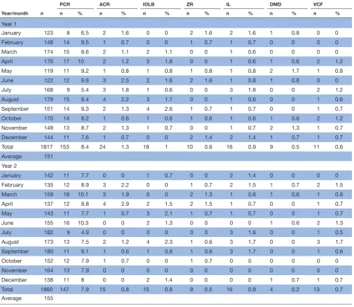
was the most frequent complication. Other compli-cations included ACR, IOL damage, zonular dialysis, IRI, Descemet’s detachment and nuclear fragments in the vitreous chamber. However, these complications affected <3% of all cases. The total numbers of PCR events were 153 in year 1 and 147 in year 2. The rate of PCR complications was not significantly different in years 1 and 2 of the study, with an OR of 0.37 and 0.35,
respectively (table 2).
dIsCussIOn
In this study, we obtained good clinical results using a new training method for cataract surgery designed for resi-dents. This new method features well-defined steps that are performed in the reverse order to that of actual surgery, such that residents learn the easiest step first. We believe that any educational programme for surgery should follow a systematic methodology that is reproducible and can be
compared with other methods in terms of clinical results,8
with the aim of standardising the teaching procedure. The current method should be easily reproducible. The clin-ical outcomes obtained with this method were comparable
on September 12, 2020 by guest. Protected by copyright.
Table 1 Absolute and relative rates of complications of phacoemulsification surgeries performed by second-year and third-year residents
Year/month n
PCR ACR IOLB ZR IL DMD VCF
n % n % n % n % n % n % n %
Year 1
January 123 8 6.5 2 1.6 0 0 2 1.6 2 1.6 1 0.8 0 0
February 148 14 9.5 1 0.7 0 0 1 0.7 1 0.7 0 0 0 0
March 174 15 8.6 2 1.1 2 1.1 0 0 1 0.6 0 0 0 0
April 170 17 10 2 1.2 3 1.8 0 0 1 0.6 1 0.6 2 1.2
May 119 11 9.2 1 0.8 1 0.8 1 0.8 1 0.8 2 1.7 1 0.8
June 122 12 9.8 3 2.5 2 1.6 2 1.6 1 0.8 1 0.8 0 0
July 168 9 5.4 3 1.8 1 0.6 0 0 3 1.8 0 0 2 1.2
August 179 15 8.4 4 2.2 3 1.7 0 0 1 0.6 0 0 1 0.6
September 151 14 9.3 2 1.3 4 2.6 1 0.7 1 0.7 0 0 1 0.7
October 170 14 8.2 1 0.6 1 0.6 1 0.6 1 0.6 1 0.6 2 1.2
November 149 13 8.7 2 1.3 1 0.7 0 0 1 0.7 2 1.3 1 0.7
December 144 11 7.6 1 0.7 0 0 2 1.4 2 1.4 1 0.7 1 0.7
Total 1817 153 8.4 24 1.3 18 1 10 0.6 16 0.9 9 0.5 11 0.6
Average 151
Year 2
January 142 11 7.7 0 0 1 0.7 0 0 2 1.4 0 0 0 0
February 135 12 8.9 3 2.2 0 0 1 0.7 2 1.5 1 0.7 2 1.5
March 159 16 10.1 3 1.9 0 0 2 1.3 1 0.6 1 0.6 1 0.6
April 137 12 8.8 4 2.9 2 1.5 2 1.5 1 0.7 0 0 1 0.7
May 143 11 7.7 1 0.7 3 2.1 1 0.7 1 0.7 0 0 1 0.7
June 155 16 10.3 0 0 2 1.3 0 0 0 0 1 0.6 2 1.3
July 182 9 4.9 0 0 0 0 0 0 3 1.6 0 0 1 0.5
August 173 13 7.5 2 1.2 4 2.3 1 0.6 3 1.7 0 0 3 1.7
September 180 11 6.1 1 0.6 1 0.6 1 0.6 3 1.7 0 0 1 0.6
October 152 12 7.9 1 0.7 0 0 1 0.7 0 0 0 0 0 0
November 164 13 7.9 0 0 0 0 0 0 0 0 0 0 0 0
December 138 11 8 0 0 2 1.4 0 0 0 0 1 0.7 1 0.7
Total 1860 147 7.9 15 0.8 15 0.8 9 0.5 16 0.9 4 0.2 13 0.7
Average 155
ACR, anterior capsule rupture; DMD, Descemet’s membrane detachment; IL, iris lesion; IOLB, intraocular lens breakage; PCR, posterior capsule rupture; VCF, core fragment in the vitreous cavity; ZR, rupture of the zonula.
Table 2 Generalised linear model with a binomial distribution of the posterior capsule rupture (PCR) rate in cataract surgeries performed in years 1 and 2 after introducing a new training programme
Year
PCR, n (%)
OR
95% CI
No Yes Inferior Superior
1 1664 (91.6) 153 (8.4) 0.37 0.28 0.50 2 1713 (92.1) 147 (7.9) 0.35 0.26 0.47
Total 3377 (91.8) 300 (8.2%)
to those reported internationally, and superior to those
recorded previously in our hospital.20
In our study, we excluded patients with comorbidities. These patients might have acted as confounders in the
analyses of complications. We are aware that this might present a risk of bias since patients without comorbid-ities tend to have fewer complications; however, this ensures better homogeneity of the sample. Patients with comorbidities were also excluded in a study by
Carricondo.13
Moreover, we excluded complex patients because previous studies failed to report the criteria used for selecting patients who could be operated on by residents. The complication rates of phacoemulsification surgeries
performed by residents are high.12 16 21 22 In one study,16
the majority of cases of vitreous loss occurred in situa-tions in which the technical difficulty of the surgery could be predicted, or in eyes with a small pupil or a nuclear sclerosis grade of 4+; this emphasises the importance of patient selection for surgical training.
on September 12, 2020 by guest. Protected by copyright.
Evaluation checkpoints are an important component of our training method. The learning curve of a surgical technique sometimes entails the performance of a large number of actual surgeries. However, during the learning curve period, patients may be at higher risk of suffering complications. By requiring the students to pass through the five checkpoints four times, the educational objectives could be met after fewer surgeries. Therefore, surgeries were considered to be performed independently after the twentieth procedure. In this study, the students learnt faster and the complication rates were comparable to
previous reports in the literature.13 23–27
By the end of the 1990s, the overall complica-tion rates of surgeries performed by residents have
decreased.12 28–30 In seven studies published at that time,
the rate of PCR among resident-performed surgeries was 2%–10%, while the vitreous loss rate ranged from 1.8%
to 10.4%.23–27 In 2010, Carricondo13, in Brazil, reported
PCR rates of 11.49% for uncomplicated cataracts.
Subse-quent studies28 29 31–33 reduced these rates even further
(2%–5%), although bias was present and it was unclear which complications were included in each analysis. These large variations in reportage must be reduced to allow comparison of complication rates among studies. National electronic data sets, such as used by the Royal
College of Ophthalmologists,19 have allowed for closer
monitoring of complications. PCR rates for less
experi-enced trainees were as low as 2.3%.30 We chose to ascribe
greater importance to PCR in our study because it was the most frequently encountered complication and directly
affects the visual prognosis.28 In a recently published
study,34 a reverse order teaching method was also assessed.
However, although the rate of PCR was lower after the implementation of the educational programme, the difference was not significant. This may have been due to the small sample size; that study involved 32 surgeons and 609 patients.
Teaching surgery in a step-by-step manner requires
detailed records of each student’s improvement.9 At
our institution, prior to the implementation of the new teaching method, patient medical records were irregular and there was no standardised method for documenting complications (except for PCR); this led to heteroge-neous information regarding the various complications. Furthermore, there was no facility for recording difficul-ties, that is, the steps that students found most difficult to master. Hence, there was a clear need to develop and systematise a logbook system. In a preliminary review of charts before the implementation of this new method, a PCR rate of 19.89% was detected among R2 and R3
procedures.20 This data, while used as a benchmark for
our study, was not included in the study due to statistical and methodological bias. The high rate of complications present in the service did not allow for the comparison of previous and current teaching methods.
We based our logbook on that of the Royal College of
Anaesthetists.19 In this individual record book,
check-points were used to monitor the progress of the residents.
Other fields were used to describe complications and any challenging aspects of the surgery. A resident could move on to the next checkpoint only after approval and with the signature of the supervising surgical preceptor. Thus, the requirement for a signature from the preceptor forced the student to perform all of the required steps and surgeries. This enabled us to obtain more reliable data on complication rates, and also allowed us to implement a teaching method that could be reproduced regardless of student or teaching staff. By standardising the patients, techniques and participating residents (in terms of their proficiency), it should be possible to compare our results with those of future studies.
This study presents limitations that are inherent to observational series. First, the lack of adequate chart data previous to the implementation of this method did not allow for statistical comparison with other teaching methods. Second, complications were registered as occurrences; however, the stage of complication was not registered, this could further help better orientation regarding critical surgical steps. Third, clinical data, such as surgical time, endothelial cell count and corneal central thickness, are not registered, at our service, in routine cases that are used for surgical teaching. Further studies should address these issues, as well as evaluate the implementation of this method for other surgical techniques or evaluation of the learning curve within different stages of learning.
Most studies in the literature focus on the data produced by surgeons in training or residents, not on the training itself. To the best of our knowledge, a fully stan-dardised teaching procedure has not yet been previously reported. This technique should allow for adequate eval-uation of residents’ technique within services or across different locations. This should also allow for evaluation of different stages during learning curves.
This study showed that instructing residents in the steps necessary for phacoemulsification in reverse chronolog-ical order presents low intraoperative complication rates. The creation of a logbook system was useful for instilling the discipline required for progression through the steps, and the logbook also served as a reference tool for evaluating the progress of individual students. Further reduction of residents’ phacoemulsification complica-tion rates over time should be the goal of future studies.
Acknowledgements The authors wish to acknowledge the participation of the residents of the Department of Ophthalmology of the Santa Casa of São Paulo, Brazil.
Contributors GRM was responsible for concept and design, data acquisition and analysis, manuscript drafting, overall content and final approval of the manuscript. JCL was responsible for concept and design, manuscript revision and final approval of the manuscript. ESS was responsible for concept and design, manuscript critical revision and final approval of the manuscript. WN was responsible for concept and design, manuscript supervision and final approval of the manuscript.
Funding CAPES (Coordenação de Aperfeiçoamento de Pessoal de Nível Superior) research support grant to the main investigator(Gustavo Malavazzi, MD). Competing interests None declared.
Patient consent for publication Not required.
on September 12, 2020 by guest. Protected by copyright.
ethics approval The institutional ethics committee approved the study protocol. Provenance and peer review Not commissioned; externally peer reviewed. Open access This is an open access article distributed in accordance with the Creative Commons Attribution Non Commercial (CC BY-NC 4.0) license, which permits others to distribute, remix, adapt, build upon this work non-commercially, and license their derivative works on different terms, provided the original work is properly cited, appropriate credit is given, any changes made indicated, and the use is non-commercial. See: http:// creativecommons. org/ licenses/ by- nc/ 4. 0/.
reFerenCes
1. World Health Organization. Global data on visual impairments 2010. Geneva: World Health organization, 2012. Available: http://www. who. int/ blindness/ GLOB ALDA TAFI NALf orweb. pdf? ua=1 [Accessed 13 Feb 2018].
2. Leske MC, Wu SY, Wu SY, ChylackLTJr. The lens opacities case-control study. risk factors for cataract. Arch Ophthalmol
1991;109:244–51.
3. World Health Organization. Universal eye health. A global action plan 2014-2019. Geneva: World Health organization, 2013. Available: http://www. who. int/ blindness/ AP2014_ 19_ English. pdf? ua=1 [Accessed 20 Dec 2016].
4. Taylor A, Jacques PF, Epstein EM. Relations among aging, antioxidant status, and cataract. Am J Clin Nutr 1995;62(Suppl 6):1439S–47.
5. Brian G, Taylor H. Cataract blindness--challenges for the 21st century. Bull World Health Organ 2001;79:249–56.
6. World Health Organization. Prevention of blindness and visual impairment. causes of blindness and visual impairment. Available: http://www. who. int/ blindness/ causes/ en/ [Accessed 20 Dec 2016]. 7. Ramke J, Welch V, Blignault I, et al. Interventions to improve access
to cataract surgical services and their impact on equity in low- and middle-income countries. Cochrane Database Syst Rev 2014;19. 8. Soriano ES. Cataract surgery teaching. Arq Bras Oftalmol
2015;78:V–VI.
9. Kloek CE, Borboli-Gerogiannis S, Chang K, et al. A broadly applicable surgical teaching method: evaluation of a stepwise introduction to cataract surgery. J Surg Educ 2014;71:169–75. 10. Ament CS, Henderson BA. Optimizing resident education in cataract
surgery. Curr Opin Ophthalmol 2011;22:64–7.
11. Corey RP, Olson RJ. Surgical outcomes of cataract extractions performed by residents using phacoemulsification. J Cataract Refract Surg 1998;24:66–72.
12. Ghanem VC, Mannis MJ. O Professor E O estudante Na facoemulsificação: OS dez princípios para O sucesso. Arq Bras Oftalmol 2003;66:93–9.
13. Carricondo PC. Análise DOS custos E complicações dA cirurgia de catarata realizada POR residentes [thesis]. São Paulo, Faculdade de Medicina da Universidade de São Paulo, 2010.
14. Blomquist PH, Morales ME, Tong L, et al. Risk factors for vitreous complications in resident-performed phacoemulsification surgery. J Cataract Refract Surg 2012;38:208–14.
15. Taravella MJ, Davidson R, Erlanger M, et al. Characterizing the learning curve in phacoemulsification. J Cataract Refract Surg
2011;37:1069–75.
16. Allinson RW, Metrikin DC, Fante RG. Incidence of vitreous loss among third-year residents performing phacoemulsification.
Ophthalmology 1992;99:726–30.
17. Lee JY, Kim K-H, Shin KH, et al. Comparison of intraoperative complications of phacoemulsification between sequential and combined procedures of pars plana vitrectomy and cataract surgery.
Retina 2012;32:2026–33.
18. Koch PS, Katzen LE. Stop and CHOP phacoemulsification. J Cataract Refract Surg 1994;20:566–70.
19. Hammond EJ, Sweeney BP. Electronic data collection by trainee anaesthetists using palm top computers. Eur J Anaesthesiol
2000;17:91–8.
20. Costa JBG, Lake JC. Surgical and visual results of cataract surgeries performed by residents of the Department of ophthalmology of the SANTA Casa of São Paulo. paper presented at: annual scientific meeting of the Department of ophthalmology of the SANTA Casa de Misericórdia de São Paulo. Brazil: São Paulo, 2008.
21. Cruz OA, Wallace GW, Gay CA, et al. Visual results and complications of phacoemulsification with intraocular lens implantation performed by ophthalmology residents. Ophthalmology
1992;99:448–52.
22. Barreto Junior J, Primiano Junior H, Espíndola RF, et al. Cirurgia de catarata realizada por residentes: avaliação dos riscos [Cataract surgery performed by residents: risk analysis]. Rev Bras Oftalmol
2010;69:301–5.
23. Lambert LC, Occhiutto ML, Paparelli CM, et al. Resultados visuais e incidência de complicações em facoemulsificação com LIO por residentes [Visual results and complications of phacoemulsifications with intraocular lens implantation performed by residents]. Rev Bras Oftalmol 1997;56:953–6.
24. Smith JH, Seiff SR. Outcomes of cataract surgery by residents at a public County Hospital. Am J Ophthalmol 1997;123:448–54. 25. Prasad S. Phacoemulsification learning curve: experience of
two junior trainee ophthalmologists. J Cataract Refract Surg
1998;24:73–7.
26. Badoza DA, Jure T, Zunino LA, et al. State-Of-The-Art phacoemulsification performed by residents in Buenos Aires, Argentina. J Cataract Refract Surg 1999;25:1651–5.
27. Bhan A, Squirrel D, Longstaff S. Teaching junior ophthalmologists phacoemulsification under topical anaesthesia. Eye 2000;14 Pt 5:810–1.
28. Ionides A, Minassian D, Tuft S. Visual outcome following posterior capsule rupture during cataract surgery. Br J Ophthalmol
2001;85:222–4.
29. Rutar T, Porco TC, Naseri A. Risk factors for intraoperative complications in resident-performed phacoemulsification surgery.
Ophthalmology 2009;116:431–6.
30. Royal College of Ophthalmologists. United Kingdom. National ophthalmology database audit, 2018. Available: https://www. nodaudit. org. uk/ u/ docs/ 20/ avusuryktz/ NOD% 20Audit% 20Annual% 20Report% 202018. pdf [Accessed 29 Dec 2018].
31. Blomquist PH, Rugwani RM. Visual outcomes after vitreous loss during cataract surgery performed by residents. J Cataract Refract Surg 2002;28:847–52.
32. Pingree MF, Crandall AS, Olson RJ. Cataract surgery complications in 1 year at an academic institution. J Cataract Refract Surg
1999;25:705–8.
33. Powe NR, Schein OD, Gieser SC, et al. Synthesis of the literature on visual acuity and complications following cataract extraction with intraocular lens implantation. cataract patient outcome research team. Arch Ophthalmol 1994;112:239–52.
34. Suryawanshi M, Gogate P, Kulkarni AN, et al. Comparison of the posterior capsule rupture rates associated with conventional (start to finish) versus reverse methods of teaching phacoemulsification.
Middle East Afr J Ophthalmol 2016;23:163–7.
on September 12, 2020 by guest. Protected by copyright.