A
A
V
V
E
E
R
R
A
A
G
G
E
E
A
A
N
N
D
D
L
L
O
O
N
N
G
G
T
T
E
E
R
R
M
M
S
S
U
U
R
R
V
V
I
I
V
V
A
A
L
L
I
I
N
N
P
P
R
R
I
I
M
M
I
I
T
T
I
I
V
V
E
E
C
C
A
A
R
R
C
C
I
I
N
N
O
O
M
M
A
A
O
O
F
F
T
T
H
H
E
E
P
P
A
A
R
R
O
O
T
T
I
I
D
D
G
G
L
L
A
A
N
N
D
D
O
O
c
c
t
t
a
a
v
v
i
i
a
a
n
n
C
C
h
h
i
i
ş
ş
11,
,
S
S
i
i
l
l
v
v
i
i
u
u
A
A
l
l
b
b
u
u
22,
,
A
A
m
m
a
a
l
l
i
i
a
a
A
A
n
n
d
d
r
r
e
e
e
e
a
a
C
C
h
h
i
i
ş
ş
33,
,
M
M
a
a
r
r
i
i
l
l
e
e
n
n
a
a
C
C
h
h
e
e
p
p
t
t
e
e
a
a
11,
,
C
C
o
o
r
r
i
i
n
n
a
a
V
V
e
e
r
r
n
n
i
i
c
c
441 “Prof. Dr. I. Chiricuţă” Institute of Oncology, Cluj-Napoca 2 “Iuliu Haţieganu” Medicine and Pharmacy University OF Cluj-Napoca
3 Private medical, dental, orthodontics, Cluj-Napoca
4 “Victor Babeş” Medicine and Pharmacy University OF Timişoara
ABSTRACT: The long term analysis of the general survival
rate and of the disease-free interval on 54 patients with
primary parotid carcinoma and the description of the involved prognosis factors.
Material and Methods. The paper analyses data recorded for 54 patients with primary parotid carcinoma, selected according to the criteria of admission into the study. The applied treatment was surgery or surgery with post surgical radiotherapy between 1995 and 2008 at the “Prof. Dr. I. Chiricuţă” Institute of Oncology in Cluj-Napoca. Univariate and multivariate methods of statistical analysis have been employed.
Results. We reached a 27.8% disease-free interval at 10 years, with a general survival rate of 48.8%.
Conclusion. The results indicate that post-therapeutic interval at 10 years in the disease-free survival was significantly influenced by the patients’ age (p=0.037) and in general survival by patients’ age (p=0.015), pT
(p=0.026), perineural invasion (p=0.043) and the
histopathologic subtype (p=0.024).
KEYWORDS: primary parotid cancer, post-therapy survival, significance factors.
1. INTRODUCTION
Through the formulation of diagnosis therapeutic and prognosis conclusions after the analysis of long-term survival, namely the observation for 10 years of patients with primary parotid cancer, the present study aims at continuing our previous research that focused on the analysis of survival and of the disease-free interval at 3 and 5 years, post-therapeutically (results presented in another study [C+13]).
Parotid cancer is considered a rare entity, with a percentage of 1.2-3% of all tumoral afflictions of head and neck; nevertheless, through its etiopathogenic and histopathologic polymorphism, it is an affliction that has always been and still is researched intensely, with an extremely serious prognosis and development if one also takes into consideration the mortality involved in this localization.
The parotid gland, one of the major salivary glands, with mix serous-mucous type or predominantly serous secretion, a pair gland with the largest volume, is, from a tumoral malign perspective, involved in ca.
80% of all tumors of the salivary glands (as previously mentioned [SB02]).
It is believed that 20-25% of all tumors of the parotid gland are carcinoma; the latter are the topic of the present analysis, besides the stressing of long term survival of these patients following treatment, namely for 10 years, post therapeutically.
The diagnostic identification of malign parotid tumors starts with the clinical observation of a tumoral formation in the upper laterocervical region, supported imagistically through tomographic examination and magnetic resonance, confirmed through fine-needle aspiration punction, extemporaneous biopsy, and the histopathologic examination of the surgical specimens, mandatory stages in subsequent treatment and follow-up.
The malign nature of a swelling, identified through palpation in the sack of the parotid gland and stressed imagistically, is supported by the rapid global increase in volume, followed in some cases by motion deficit in the territory of the facial nerve with the presence of local pain and some segmental or total facial paralyses.
2. MATERIAL AND METHOD
Data of patients included in the study were gathered from the database of the “Prof. Dr. I. Chiricuţă” Institute of Oncology Cluj-Napoca, center of diagnosis and treatment of cancer and other tumoral afflictions, on all anatomic localizations, including the cancer of head and neck.
The study is analytical, of the observational type, retrospective, and includes data on 54 patients with malign primary parotid tumors, with the approval of the Ethics Commission no. 5.691 of July 8th 2009, part of the “Prof. Dr. I. Chiricuţă” Institute of Oncology Cluj-Napoca.
Between 1995 and 2003, 54 patients with malign primary tumors of the parotid gland were recorded at the “Prof. Dr. I. Chiricuţă” Institute of Oncology Cluj-Napoca for diagnosis and/or treatment, who benefited from surgical interventions on the level of the parotid gland to various degrees: partial parotidectomy, total parotidectomy with/without facial nerve resection and neck dissection. These cases subsequently followed another therapeutic sequence, associated to or independent from surgery at the institute, mainly post-surgical radiotherapy.
For the 54 patients with the diagnosis of parotid gland cancer we analyzed their development over a ten-year period, with regard to the treatment followed, the main factors of prognosis and survival, considered as a long-term analysis of this localization.
The clinical examination, diagnostic biopsy/surgical intervention and stadialization of these cases were performed at the “Prof. Dr. I. Chiricuţă” Institute of Oncology in Cluj-Napoca.
The following inclusion criteria of patients in the “second stage” of the research were employed:
- age between 14 and 90 at the time the diagnosis was established;
- both genders;
- paraclinical imagistic investigations (ultrasonography, thoracic Rx., computer tomography) and laboratory investigations;
- identical presurgical balance;
- fine-needle aspiration punction;
- surgical intervention: partial or total parotidectomy, with or without the preservation of the facial nerve with modified radical laterocervical lymphanedectomy;
- with or without postsurgical radiotherapy;
- histopathologic result of surgical items, with the confirmation of the malignity and pTNM rendering;
- histopathologic diagnosis of primary malign tumor of the parotid gland;
- stadialization according to the AJCC [***02];
- absence of distant metastasis at the time of the surgical sequence;
- absence of other, previous specialized treatments;
- data on patient control land status recorded at intervals of 3, 6, 12, 36, 60, and 120 months.
The interpretation of results was also performed by comparison with data published in the specialized literature.
The descriptive and analytical statistical analysis was performed with the aid of frequency indicators, tests, and specific methods.
In order to estimate the possibility of disease-free survival and the general survival rate we employed the Kaplan-Meier method (multivariate analysis). We also analyzed the average and median survival time (disease-free interval).
The comparison of the “disease-free interval” rate between groups was performed with the aid of the log rank test and considering statistically significant values 0.05.
The Cox regression analysis was employed for the hazard rate in the multivariate analysis. A degree of probability of 0.05 was considered statistically significant.
The survival / disease-free interval at 3 and 5 years were described through survival and hazard curves. Statistic processing (descriptive and analytical) was performed with the SPSS 13 software.
3. RESULTS AND DISCUSION
The study includes 54 patients with primary carcinoma of the parotid gland.
Tumors of the salivary glands, including those of the parotid gland, are entities eminently treatable through surgery according to the clinical stage at presentation and are naturally classified starting from their histopathologic character, which is in fact the main prognosis characteristic [SS92].
The analyzed parameters are prognosis factors in cancers of the parotid gland: age (2 groups, namely 50 years and 50 years respectively), patient gender, degree of malignity (low, high), sub-degree of malignity, stage (grouped into incipient and advanced stages) (Table 1), perineural invasion, lymphatic vascular emboli, venous vascular emboli (Table 2), type of surgical intervention, and therapeutic sequence (surgeryradiotherapy).
The average age (in years) of the 54 patients was of 5316 (95%IC: 49-58) with interval 14-86 years. The proportion of male and female patients was almost equal, namely 40.7% men and 59.3% women, with average age values for the 22 men of 5316 (95%IC: 47-61) in an interval between 14 and 80 years, respectively 5316 (95%IC: 47-59) in an interval between 19 and 86 years.
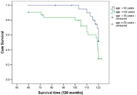
In the analysis of the prognosis factor age and its involvement in survival and the disease-free interval we included the patients in two age groups: 50 years (22 patients, 40.7%) and 50 years (32 patients, 59.3%).
treated in a combined manner, through surgery followed by radiotherapy.
The significance of the surgical sequence in the lot of 54 patients with parotid cancer was represented in almost equal proportion by total parotidectomy, parotidectomy with facial nerve preservation (25 cases, 46.3%) total parotidectomy, and parotidectomy with facial nerve resection (21 cases, 38.9%). Partial parotidectomy was performed in 14.8% of cases. All 54 cases of primary parotid tumors presented under clinical examination laterocervical adenopathy in various stages and required lymphanedectomy. As part of the surgical treatment, lymphanedectomy of various proportions was also performed, besides parotidectomy, in the case of all 54 patients (Table 3).
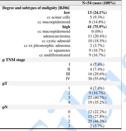
Table 1. Postsurgical histopathologic survey of the 54 patients with malign parotid tumor
N=54 cases (100%)
Degree and subtypes of malignity [BJ06]
low 13 (24.1%)
cc acinar cells 5 (9.3%) cc mucoepidermoid 8 (14.8%)
high 41 (75.9%)
cc mucoepidermoid 0 (0%) adenocarcinoma 11 (20.4%) cc cystic adenoid 10 (18.5%) cc ex pleomorphic adenoma 2 (3.7%)
cc squamous 9 (16.7%) cc undifferentiated 9 (16.7%)
p TNM stage
I 4 (7.4%)
II 4 (7.4%)
III 16 (29.6%)
IV 30 (55.6%)
pT
1 4 (7.4%)
2 9 (16.7%)
3 22 (40.7%)
4 19 (35.2%)
pN
0 12 (22.2%)
1 15 (27.8%)
2 25 (46.3%)
3 2 (3.7%)
Table 2. Frequency of prognosis factors according to gender, on 54 patients with malign primary parotid tumor (database of the “Prof. Dr. I. Chiricuţă” Institute of Oncology Cluj-Napoca 1995-2008, surgical protocols,
institutional registry) prognosis factors gender total 54 (100%)
female male
N=32 (59.3 %)
N=22 (40.7 %)
lymphatic
vascular emboli 18 (33.3 %) 13 (24.1 %)
31 (57.4 %) venous
vascular emboli 8 (14.8 %) 5 (9.3 %)
13 (24.1 %) perineural invasion
11 (20.4 %) 10 (18.5 %) 21 (38.9 %) facial nerve
paralysis 9 (16.7 %) 7 (13 %)
16 (29.6 %) skin invasion
5 (9.3 %) 4 (7.4 %) 9 (16.7 %) pain
8 (14.8 %) 6 (11.1 %) 14 (25.9 %)
Table 3. Frequency of laterocervical ganglionary resection types on 54 patients with malign primary parotid tumors (database of the “Prof. Dr. I. Chiricuţă”
Institute of Oncology Cluj-Napoca 1995-2008, surgical protocols, institutional registry)
type of lymphanedectomy cases parotid cancer
N=54 (100%)
modified radical
laterocervical lymphanedectomy
35 (64.8 %) radical
laterocervical lymphanedectomy
10 (18.5 %) selective
laterocervical lymphanedectomy
9 (16.7 %)
633 ganglia have been resected, that, according to the degree of ganglionary metastases identified through histopathologic examination, determined the creation of two groups of patients, namely:
- in 8 patients (14.81%), out of the ganglia resected and examined histopathologically 72 (12.16%) turned out negative;
- 46 patients (85.18%) with ganglia resected and examined histopathologically 226 such ganglia (35.71%) turned out positive while 330 (52.15%) were negative (Table 4).
The average duration for the disease-free interval at 10 years, calculated from the date of the surgical intervention, was in months 95.758.82 (95%IC: 78.45-113.04) with the median also expressed in months of 9725.27 (95%IC: 47.45-146.52).
The average period for survival at 10 years, calculated from the date of the surgical intervention, was in months 145.139.24 (95%IC: 127-163.25) with the median also expressed in months of 123.2713.07 (95%IC: 97.64-148.89).
Table 4. Characteristics of the number of laterocervical ganglia extracted through lymphanedectomy, in 54 patients with malign primary parotid tumors (database
of the “Prof. Dr. I. Chiricuţă” Institute of Oncology Cluj-Napoca 1995-2008, surgical protocols, institutional
registry)
status / ganglio N (%) N patients (100%) average SD (95%IC) interval minimum -
maximum N. (median,
mode)
number of extracted ganglia: 633 (100%)
54 (100%)
11.724.15 (10.59-12.85) 3-24 (12; 8) number of negative extracted ganglia: 407 (64.29%) 8 (14.81%)
7.413.74 (6.77-8.05) 0-21 (7; 5) number of positive extracted ganglia: 226 (35.71%) 46 (85.18%)
3.673.40 (3,09-4.25)
0-14 (3; 0)
Table 5. Prognosis factors in the evaluation of the disease-free interval and general survival in patients
monitored for a period of 10 years (54 patients with malign primary parotid tumors from the database of the “Prof. Dr. I. Chiricuţă” Institute of Oncology
Cluj-Napoca 1995-2008)
variable disease-free
interval
general survival
10 years rate(%)
value
“p” 10 years rate(%) value
“p”
27.8% 48.1%
age
50 years
50 years
40.9% 18.8%
0.037 59.1% 40.6% 0.015 gender male female 22.7% 31.3%
0.055 31.8% 59.4% 0.276 therapeutic sequence surgery surgery+radiotherapy 18.8% 31.6%
0.165 56.3% 44.7% 0.486 p stage I-II (early) III-IV (advanced) 42.9% 25.5%
0.670 85.7% 42.6% 0.317 pT 1-2 3-4 46.2% 22%
0.410 92.3% 34.1% 0.026 p N „N0” „N+” (1-2-3) 41.7% 23.8%
0.066 75% 40.5% 0.223 perineural invasion no yes 39.4% 9..5%
0.138 66% 19% 0.043 lymphatic vascular emboli no yes 43.5% 16.1%
0.219 69.6% 32.3% 0.204 venous vascular emboli no yes 31.7% 15.4%
0.537 56.1% 23.1%
0.110
degree of malignity
low high
23.1% 29.3%
0.432 76.9% 39%
0.534
histopathologic subtype
cc with acinar cells 40% 0.223 80% 0.024 cc mucoepidermoid
with a low degree of malignity
12.5% 75%
cc mucoepidermoid with a high degree of malignity
- -
adenocarcinoma 36.4% 45.5%
cc cystic adenoid 50% 60%
cc expleomorfic adenoma
50% 50%
cc squamous 22.2% 33.3%
cc undifferentiated 0% 11.1%
In both analyses, a higher rate was calculated for: the group of female patients, patients aged 50 years, incipient stages (stage I and II), pT1 and pT2, absence of ganglia (pN”0”), absence of perineural invasion, absence of lymphatic vascular emboli and absence of venous vascular emboli.
The authors of an American study [TF86] that analyzed malign parotid tumors, have reported a proportion of T1-T2 and T3-T4 cases of 1.7:1 with a 62% disease-free survival at 10 years for patients who underwent combined treatment, surgery+radiotherapy, as compared to only 22% for patients who were only
treated through surgery. Our results indicate that the proportion of T1-T2 cases as compared to T3-T4 cases is of 1:3.15, while for the disease-free survival rate at 10 years for the group of patients with combined treatment (surgery+radiotherapy) we calculated an almost double absolute value as compared to the group of patients who only underwent surgery (31.6% and 18.8% respectively). The situation changes in the case of the survival rate that is higher for the group of patients with surgery, namely 56.3% as compared to the survival rate for the group of patients with radiotherapy associated to surgery, 44.7%.
As for the degree of malignity, the analysis of the disease-free survival has indicated that the rate is higher among the group of patients suffering from tumors with a high degree of malignity (29.3%) than it is among patients with tumors malign to a low degree (23.1%).
Analyzing the rate of general survival, at the same parameter, we noted a higher rate of survival in case of patients with tumors with a low degree of malignity (76.9%) as compared to patients with tumors with a high degree of malignity (39%).
At 10 years, the rate of disease-free survival was significantly influenced by the age of the patients (p=0.037), while the rate of general survival was influenced significantly by: age (p=0.015), pT (p=0.026), perineural invasion (p=0.043) and the histopathologic subtype (p=0.024) (Fig. 1, Fig. 2, Fig. 3, Fig. 4, Fig. 5).
The obtained results indicate that age is a major prognosis factor for both the rate of disease-free survival and the general survival rate, as both reached higher values for the group of patients aged 50 years (40.9% and 59.1%).
As for the rate of general survival at 10 years, we have identified the following factors with an influence on prognosis: age, pT category, perineural invasion, and the histopathologic subtype (Table 5). For the group of patients with histopathologic pT1, the general survival rate recorded was of 100%. For the histopathologic subtype, the best long-term survival at 10 years was calculated for the group of patients with carcinoma containing acinar cells of the parotid gland (80%), followed by those with mucoepidermoids with a low degree of malignity (75%).
For the disease-free survival at 10 years, the rate was of 27.8%, while for the general survival, the calculated rate was of 48.1%.
In another study with a similar pathology [N+09], performed on a lot of 104 patients, the authors have reported a diseases-specific survival at 10 years of 71% and a rate of local control of 82%.
A group of authors [M+05] who analyzed 224 patients with cancer of the salivary glands have reported a general survival rate at 10 years of 44%, with the T category and the type of treatment as having a major impact on prognosis.
The authors of a study performed at the Christie Hospital, Manchester [R+99], on 103 patients with parotid cancer and similar treatment, have reported a slightly higher rate of survival at 10 years (65%), with the following prognosis factors in the univariate analysis: tumoral volume T, patients’ age, clinical N, histological type, perineural invasion and micrometastases in the periparotid ganglia.
Fig. 1. Disease-free interval at 10 years according to age groups for the 54 patients with parotid gland cancer
Fig. 2. General survival at 10 years according to age groups for the 54 patients with parotid gland cancer
Fig. 3. General survival at 10 years according to pT for the 54 patients with parotid gland cancer
Fig. 4. General survival at 10 years according to the perineural invasion for the 54 patients with parotid
gland cancer
Fig. 5. General survival at 10 years according to the histopathologic subtype for the 54 patients with parotid
4. CONCLUSIONS
The results of the study performed on the lot of 54 patients with primary carcinoma of the parotid gland indicate that at 10 years the disease-free survival rate was significantly influenced by the patients’ age and the rate of general survival was also influenced significantly by the same factor ( 50 years).
In the average and long-term monitoring, the following major factors of prognosis have been identified: pT, perineural invasion, and the histopathologic subtype.
For the disease-free survival at 10 years, the rate was of 27.8% while for the global survival the calculated rate was of 48.1%.
REFERENCES
[BJ06] Byron J. Bailey, Johans T. Johnson -
Head and Neck Surgery 2006, Lippincott
Williams and Wilkins, V2, pp.:1515-16;
[C+13] O. Chis, S. Albu, G. Cirebea, M. Cheptea, C. Vernic - Data on the treatment and survival of patients with parotid cancer at the „Prof. dr. I. Chiricuta” Institute of oncology in
Cluj-Napoca. Annals. Computer Science
Series. 11th Tome 2nd Fasc. – 2013 pp: 34-40;
[L+05] R. A. Lima, M. R. Tavares, F. L. Dias, J. Kligerman, M. E. Nascimento, M. M. Barbossa, C. R. Cernea, J. R. Soares, I.C. Santos, S. Salviano -
Clinical prognostics factors in malignant
parotid gland tumors. Otolaryngology
Head and Neck Surg. 2005, nov.133(5):702-8;
[M+05] W. M. Mendenhall, C. G. Morris, R. J. Amdur, J. W. Werning, D. B. Villaret -
Radiotherapy alone or combined with surgery for salivary gland carcinoma.
Cancer, 2005, june 15, 103-(12): 2544-2550;
[N+09] M. Nagliati, A. Bolner, V. Vanoni, L. Tomio, G. Lay, R. Murtas, M. A. Deidda, A. Madeddu, E. Delmastro, R. Verna, P. Gabriele, M. Amichetti -
Surgery and radiotherapy in treatment of malignant parotid tumors: a retrospectiv
multicenter study. Tumori, 2009, jul-aug,
95(4): 442-448;
[R+99] A. G. Renehan, E. N. Gleave, N. J. Slevin, M. McGurk - Clinicopatho-logical and treatment – related factors influencing survival in parotid cancer.
British Journal of Cancer, (1999) 80 (8): 1296-1300;
[SB02] P. M. Speight, A. W. Barred - Salivary
gland tumours. Oral Dis. 8(5): 230-242,
2002;
[SS92] G. Seifert, L. H. Sobin - The World
Health Organisation's histological
classification of salivary gland tumors.
Cancer 1992, N70, pp.: 379-85;
[TF86] C. Theriault, P. J. Fitzpatrick -
Malignant parotid tumors. Prognostics
factors and optimum treatment.
Am.J.Clin.Oncology, 1986, dec; 9(6): 510-517;
[***02] *** - AJCC – Cancer staging handbook,
Sixth Edition, 2002, pp: 81-87;