Volume 2012, Article ID 362610,4pages doi:10.1155/2012/362610
Case Report
Primary Hydatid Cyst of the Axillary Region: A Case Report
Mehrangiz Zangeneh,
1Mahmood Amerion,
2S. Davar Siadat,
3and Mohsen Alijani
11Department of Infectious Diseases, Islamic Azad University, Tehran Medical Branch, Tehran, Iran 2Department of General Surgery, Islamic Azad University, Tehran Medical Branch, Tehran, Iran 3Department of Microbiology, Pasteur Institute, Tehran, Iran
Correspondence should be addressed to Mehrangiz Zangeneh,zangeneh4@yahoo.com
Received 8 July 2012; Revised 30 September 2012; Accepted 30 September 2012
Academic Editor: Nima Rezaei
Copyright © 2012 Mehrangiz Zangeneh et al. This is an open access article distributed under the Creative Commons Attribution License, which permits unrestricted use, distribution, and reproduction in any medium, provided the original work is properly cited.
Introduction. Hydatid disease is a disease caused by the cestode Echinococcus. Echinococcus granulosus is the most common Echinococcus species affecting human. It may affect any organ and tissue in the body, most in the liver and lung. Disease is endemic in some regions of the world, and is common in Iran. Primary hydatid cyst of the axillary region is an unusual and rare localization of hydatid disease. So far, only sixteen cases have been published in the all medical literature. Case Report. Herein, we present a 33-year-old woman because of a mass in the axillary region of four months duration. Axillary ultrasonography showed a thick wall cystic lesion. No abnormality was found in mammographic examination of either breast, or in abdominal ultrasonography and chest X-ray. The mass was excised for pathological examination that showed a typical laminated membrane of hydatid cyst. Postoperative IgG- ELISA serology in this case was negative. Based on pathology an axillary hydatid cyst was diagnosed. Conclusion. Hydatid cyst should be considered in endemic areas in patients presenting with a soft tissue mass in the axillary region.
1. Introduction
In human, three forms of echinococcosis are known to occur: cystic echinococcosis, caused by Echinococcus granulosus, alveolar echinococcosis, caused by Echinococcus multiloc-ularis, and polycystic Echinococcosis due to Echinococcus vogeli or Echinococcus oligarthrus. Hydatid disease is a parasitic disease usually caused by the larval stage of a small zoonotic tapeworm primarily found in dogs Echinococcus granulosus [1]. Echinococcus granulosus (cystic echinococco-sis) is the most common species, E. granulosus has worldwide distribution and is endemic in many countries, especially the Mediterranean region, Australia, South America, the Middle East, South Africa, and Eastern Europe [1–3]. Iran is also an endemic area for hydatid diseases, the prevalence of cystic echinococcosis in humans detected by ultrasound ranges from less than 0.5% to 1.5%. Human seropositivity was greater than 5% in the west and southwest of the Islamic Republic of Iran, with 2–18% seroprevalence in nomadic groups [4, 5]. The hydatid cysts of E. granulosus tend to form in the liver (50% to 70% of patients) or lung (20% to 30%) but may through the capillary systems
reaches the general circulation and passes to all viscera and soft tissues. For this reason, hydatid cysts may arise in atypical sites such as the brain, heart, orbit, urinary bladder, chest wall, subcutaneous tissue, tibia, parotid gland, breast, cervicofacial region, thyroid, and in any organ of the body (10%) [1, 3, 6, 7]. The diagnostic methods include imaging techniques, mainly X-ray for lung echinococcosis, ultrasound and computed tomography examination for abdominal echinococcosis and other affected organs, and immunodiagnostic tests (enzyme-linked immunosorbent assay (ELISA), IFAT, and immunoblot) for confirmation. IgG-ELISA is about 90% sensitive for liver cyst infection but less sensitive for lung (80%) or other organ involvementand (90%), and its specificity is 90% [1].
2 Case Reports in Medicine
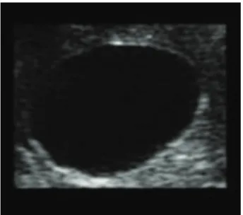
Figure 1: Ultrasound image of axillary cyst.
case of a primary hydatid disease which originated in the subcutaneous tissue in the right axillary region in a 36-year-old woman.
2. Case Report
A 33-year-old woman in 2010 was presented to our hospital with painless right axillar swelling of four-month duration. She had no history of breast mass, fever, other symptoms, or other mass, and also had no history of hydatid cyst. On physical examination, a firm to hard 5×5 cm mobile mass was noted in the right axilla. Laboratory investigations were done which revealed normal blood counts and biochemical result. Ultrasound showed a 5 × 5 cm thick wall cystic lesion comparable with type CE1 of WHO classification
(Figure 1). No abnormality was found in mammographic
examination of either breast. In this case it was difficult to differentiate a cyst with uncharacteristic imaging findings (type CE1 of WHO Classification based on ultrasound) from simple subcutaneous cyst, haematoma, necrotic tumor, or lymph node based on ultrasound. Under general anesthesia, the entire cyst was excised without rupture and sent for histopathological examination. On macroscopic examina-tion, a unilocular cyst with a hard fibrotic wall was seen. Microscopic examination revealed the presence of a typical laminated membrane of hydatid cyst (Figure 2). The hydatid IgG-ELISA test in our case was negative. Radiographs of chest, spine, and long bones were done for evaluation of hydatid in organs and all were normal. Abdominal ultra-sound examination was normal. The patient was discharged from the hospital with albendazole 400 mg twice a day for four weeks. During one-year followup, examinations since her surgery have shown no relapse of hydatidosis.
3. Discussion
Hydatid disease is a common clinical pathology in many parts of the world. The main species pathogenic for humans in Mediterranean and Southern European countries is Echinococcus granulosus [1, 2]. Hydatid cysts most often develop in the liver and lung. primary axillary hydatid disease
is rare, as shown in Table 1, we could find only sixteen previous case reports in the medical literature [3,6,8–21]
(Table 1).
Hydatid cysts grow 5 to 10 cm in size within the first year and can survive for years or even decades. The cyst may be present for many years in the organ in which it is located with no clinical symptoms or signs, and sometimes it may exhibit clinical symptoms depending on the size and location of the cyst and the pressure of the growing cyst [1,6]. The sonographic and tomographic appearances of subcutaneous hydatid disease such as axillary hydatid cyst are similar to those in other organs. Hydatid cyst may be unilocular at earlier stages, whereas older cysts are usually multilocular. They may either be made up of daughter cysts or have a solid appearance made up of multiple septated cysts. However, hyperintense hydatid cysts in the axillary region can be misdiagnosed as a soft tissue tumor or lymphadenopathy. As Iran is an endemic area for hydatid disease, this should be in the differential diagnosis for patients presenting with tissue masses including haematoma, abscess, sarcoma, lymphadenopathy, breast cancer, or metastatic lesions [1,
3, 18]. Infection is suspected based on imaging studies (ultrasonography, CT, and MRI), and it may be confirmed by a specific enzyme-linked immunosorbent assay (ELISA) and Western blot serology. Ultrasonography is an easy available, and affordable with high diagnostic sensitivity imaging test for screening of hydatid cyst in endemic areas and in family members based on WHO classification. The sonographic imaging in our case showed a simple cystic lesion comparable with type CE1 of WHO Classification and did not confirm a diagnosis of hydatid cyst; it is difficult to differentiate a cyst with uncharacteristic imaging findings from simple subcutaneous cyst, haematoma, necrotic tumor, or lymph node based on ultrasound. Immunodiagnostic tests (enzyme-linked immunosorbent assay (ELISA), IFAT, and immunoblot) are used for confirmation. IgG-ELISA is about 90% sensitive for liver cyst infection but less sensitive for lung (80%) and other organ involvement (90%), and its specificity is 90% [1]. Postoperative IgG-ELISA test in this case was negative. Abdominal ultrasonography and a plain chest radiography are mandatory to detect liver and lung involvement. Chest X-rays and imaging studies showed no other involvement in our patient. In routine practice, the accurate diagnosis in patient with soft tissue hydatidosis is frequently delayed until histopathologic examination after surgery. In this case, for accurate diagnosis, the entire cyst was excised.
Figure 2: Section of cyst wall showing germinal epithelium and underlying laminated membrane (haematoxylin-eosin stain, magnification
×10).
Table 1: Reported cases of hydatid cysts of the axilla.
Author Year Age/gender Origin of cysts Daughter cysts Organ involvement Screening Follow
Thomson [8] 1899 NA Axillary NA NA NA NA
Lamotte et al. [9] 1967 32/M Right axillary vein Multiple None CXR NA Zamfir et al. [10] 1997 11/F Left neurovascular None pulmonary CXR, US NA Navarro Mart´ın et al. [11] 1998 84/M Subcutaneous Multiple None CXR, US NA Mayol Mart´ınez et al. [12] 1994 67/F Muscle Multiple None CXR,US NA Ekrem Unal et al. [13] 2001 53/F Right pectoral M None None MRI, CT 9 months Dilege et al. [14] 2003 15/F Right axillary region NA Pulmonary CXR, CT NA Losanoffet al. [15] 2004 38/M Subcutaneous tissue NA None NA NA Sain Guven et al. [16] 2004
Borovik et al. [17] 2006 31/F Left axillary Multiple None CXR, CT 6 months Singh et al. [18] 2009 28/M Left lateral chest wall NA None CXR, CT 8 years
¨
Unalp et al. [3] 2011 48/F Left axillary fossa Multiple None CXR, US, CT 6 months Ozsoy et al. [6] 2011 45/F The axillary region None None CXR, US, CT 1 year Ruso et al. [19] 2011 84/F Cervicoaxillary region Multiple None CXR, US, CT 5 years Arsalane et al. [20] 2012 43/M Left axilla Multiple None CXR, US, CT NA Saylam et al. [21] 2012 36/F Right axillar None None CXR, US 17 months
F: female, M: male, CT: computed tomography, CXR: chest X-ray, LP: laparoscopy, RS: radionuclide scintigraphy, US: ultrasonography, NA: data not available, MRI: magnetic resonance Imaging.
medication is widely used postoperatively and preoperatively for the purpose of cyst size reduction and to limit the risk of intraoperative dissemination of daughter cysts. All patients are treated with albendazole 10 mg/kg/day for at least two weeks preoperatively, and this is continued postoperatively for four weeks [22]. However, the experience with scolicidal agents such as albendazole (400 mg/kg) and praziquantel (50 mg/kg) or a combination of these drugs in the treatment of soft tissue hydatid disease is very limited and results of the medical treatment in the hydatidosis are unclear [23,24]. We gave the patient albendazole 400 mg twice a day for four weeks postoperatively.
In conclusion, hydatid disease is a widespread public health problem in developing countries especially in endemic regions; therefore, it should be considered in the differential diagnosis of a palpable mass in the axillary region.
Conflict of Interests
The authors declare that they have no conflict of interests.
Consent
Written informed consent was obtained from the patient for publication of this case report and accompanying images.
References
4 Case Reports in Medicine
[2] P. S. Craig, D. P. McManus, M. W. Lightowlers et al., “Prevention and control of cystic echinococcosis,” The Lancet
Infectious Diseases, vol. 7, no. 6, pp. 385–394, 2007.
[3] H. R. ¨Unalp, E. Kamer, T. Rezanko, ¨O. Kılıc¸, M. Tunakan, and M. A. ¨Onal, “Primary hydatid cyst of the axillary region: a case report,” Balkan Medical Journal, vol. 28, no. 2, pp. 209–211, 2011.
[4] First WHO report on neglected tropical diseases 2010: working to overcome the global impact of neglected tropical diseases. WHO/HTM/NTD/2010. 1.
[5] A. Zamani and S. Kalikas, “Hydatid cyst of parotid gland: a case report,” Iranian Journal of Pediatrics, vol. 16, no. 1, 2006. [6] M. Ozsoy, C. Keles, M. Kahy, and G. Keles, “Primary
echinococcal cyst in the axillary region,” Journal of Infection
in Developing Countries, vol. 5, no. 11, pp. 825–827, 2011.
[7] I. Iynen, O. Sogut, M. E. Guldur, R. Kose, H. Kaya, and F. Bozkus, “Primary hydatid cyst: an unusual cause of a mass in the supraclavicular region of the neck,” Journal of Clinical
Medicine Research, vol. 3, no. 1, pp. 52–54, 2011.
[8] P. J. Thomson, M. R. C. S. ENG., and L. R. C. P. Lond, “Hydatid cyst of the axilla in a child,” The Lancet, vol. 153, no. 3932, pp. 25–26, 1899.
[9] M. Lamotte, J. Perrotin, A. Julliard, and G. Timsit, “Hydatid cysts of the soft tissues. Apropos of a case of hydatid cyst of the axilla,” Annales de Chirurgie, vol. 21, no. 23, pp. 1463–1467, 1967 (French).
[10] T. Zamfir, R. B˘al˘anescu, T. P˘atr˘acus¸ et al., “The diagnosis and surgical treatment in the case of 2 rare sites of hydatid cyst in children,” Chirurgia, vol. 92, no. 6, pp. 413–415, 1997 (Romanian).
[11] L. M. Navarro Mart´ın, J. Pardo Lled`ıas, I. Galindo P´erez, and R. Querol Prieto, “Medicine in images. Incidental diagnosis of a mass subcutaneously located in the axillary region,”
Revista Cl´ınica Espa˜nola, vol. 198, no. 10, pp. 703–704, 1998
(Spanish).
[12] J. Mayol Mart´ınez, P. J. Gonz´alez Noguera, R. Peromingo Fresneda, and J. Alvarez Fern´andez-Represa, “Mass localized in the axilla,” Revista Cl´ınica Espa˜nola, vol. 194, no. 3, pp. 199– 200, 1994 (Spanish).
[13] A. Ekrem Unal, S. Can Ulukent, S. Bayar, A. Demirkan, and H. Akg¨ul, “Primary hydatid cyst of the axillary region: report of a case,” Surgery Today, vol. 31, no. 9, pp. 803–805, 2001. [14] S. Dilege, M. Aksoy, I. Okan, A. Toker, G. Kalayci, and M.
Demiryont, “Hydatid cystic disease of the soft tissues with pulmonary and hepatic involvement: report of a case,” Surgery
Today, vol. 33, no. 1, pp. 69–71, 2003.
[15] J. E. Losanoff, B. W. Richman, and J. W. Jones, “Primary hydatid cyst of the axilla,” ANZ Journal of Surgery, vol. 74, no. 5, pp. 393–394, 2004.
[16] G. Sain Guven, H. Simsek, B. Cakir, O. Akhan, and O. Abbasoglu, “A hydatid cyst presenting as an axillary mass,”
The American Journal of Medicine, vol. 117, no. 5, pp. 363–364,
2004.
[17] A. Borovik, D. Massasso, and K. Gibson, “Axillary hydatid disease,” The Medical Journal of Australia, vol. 184, no. 11, p. 585, 2006.
[18] S. Singh, S. Khichy, M. Singh, and J. Singh Gill, “Recurrent solitary hydatid cyst of the subcutaneous tissue,” Indian
Journal of Surgery, vol. 71, no. 3, pp. 162–164, 2009.
[19] L. Ruso, G. Rodriguez, A. Gatti et al., “Primary cervico— axillary hydatid disease,” Cirugia y Cirujanos, vol. 79, pp. 306– 312, 2011.
[20] A. Arsalane, M. El Hammoumi, F. El Oueriachi, A. Traibi, A. Darbi, and E. H. Kabiri, “Primary axillary hydatid cyst,”
General Thoracic and Cardiovascular Surgery, vol. 60, no. 6, pp.
359–362, 2012.
[21] B. Saylam, V. Vural, A. P. Duzgun, M. V. Ozer, and F. Coskun, “Primary hydatid cyst of the axilla: report of a case,” Medical
Principles and Practice, vol. 21, no. 1, pp. 79–81, 2012.
[22] O. Akhan, B. Gumus, D. Akinci, M. Karcaaltincaba, and M. Ozmen, “Diagnosis and percutaneous treatment of soft-tissue hydatid cysts,” CardioVascular and Interventional Radiology, vol. 30, no. 3, pp. 419–425, 2007.
[23] A. E. Mohamed, M. I. Yasawy, and M. A. Al Karawi, “Com-bined albendazole and praziquantel versus albendazole alone in the treatment of hydatid disease,” Hepato-Gastroenterology, vol. 45, no. 23, pp. 1690–1694, 1998.
Submit your manuscripts at
http://www.hindawi.com
Stem Cells
International
Hindawi Publishing Corporationhttp://www.hindawi.com Volume 2014
Hindawi Publishing Corporation
http://www.hindawi.com Volume 2014
INFLAMMATION
Hindawi Publishing Corporation
http://www.hindawi.com Volume 2014
Behavioural
Neurology
Endocrinology
International Journal ofHindawi Publishing Corporation
http://www.hindawi.com Volume 2014
Hindawi Publishing Corporation
http://www.hindawi.com Volume 2014
Disease Markers
Hindawi Publishing Corporation
http://www.hindawi.com Volume 2014
BioMed
Research International
Oncology
Journal ofHindawi Publishing Corporation
http://www.hindawi.com Volume 2014
Hindawi Publishing Corporation
http://www.hindawi.com Volume 2014
Oxidative Medicine and Cellular Longevity
Hindawi Publishing Corporation
http://www.hindawi.com Volume 2014
PPAR Research
The Scientific
World Journal
Hindawi Publishing Corporation
http://www.hindawi.com Volume 2014
Immunology Research
Hindawi Publishing Corporation
http://www.hindawi.com Volume 2014
Journal of
Obesity
Journal ofHindawi Publishing Corporation
http://www.hindawi.com Volume 2014
Hindawi Publishing Corporation
http://www.hindawi.com Volume 2014 Computational and Mathematical Methods in Medicine
Ophthalmology
Journal ofHindawi Publishing Corporation
http://www.hindawi.com Volume 2014
Diabetes Research
Journal ofHindawi Publishing Corporation
http://www.hindawi.com Volume 2014
Hindawi Publishing Corporation
http://www.hindawi.com Volume 2014
Research and Treatment
AIDS
Hindawi Publishing Corporation
http://www.hindawi.com Volume 2014
Gastroenterology Research and Practice
Hindawi Publishing Corporation
http://www.hindawi.com Volume 2014
Parkinson’s
Disease
Evidence-Based Complementary and Alternative Medicine
Volume 2014 Hindawi Publishing Corporation