http://www.sciencepublishinggroup.com/j/ijnfs doi: 10.11648/j.ijnfs.20190803.12
ISSN: 2327-2694 (Print); ISSN: 2327-2716 (Online)
Preparation of α-Amylase Inhibitor from Seeds of White
Kidney Bean Using a Novel and Scalable Process Based on
Enzymatic Hydrolysis
Yifeng Rang
1, Wei Zhao
2, * 1Department of Research and Development, New Industry Health Technology (Zhuhai) Co., Ltd., Zhuhai, China 2School of Food Science and Technology, Jiangnan University, Wuxi, China
Email address:
*
Corresponding author
To cite this article:
Yifeng Rang, Wei Zhao. Preparation of α-Amylase Inhibitor from Seeds of White Kidney Bean Using a Novel and Scalable Process Based on Enzymatic Hydrolysis. International Journal of Nutrition and Food Sciences. Special Issue: Natural Active Ingredients for the Management of Diabetes and Obesity. Vol. 8, No. 3, 2019, pp. 52-58. doi: 10.11648/j.ijnfs.20190803.12
Received: July 13, 2019; Accepted: August 13, 2019; Published: September 6, 2019
Abstract:
This study proposed and evaluated a novel and scalable process based on enzymatic hydrolysis for the preparation of α-amylase inhibitor (α-AI) in seeds of white kidney bean (Phaseolus vulgaris). The process mainly involved heat treatment (70°C/30 min), enzymatic hydrolysis, isoelectric precipitation and 70% ethanol precipitation. The optimal preparation parameters for enzymatic hydrolysis and isoelectric precipitation were as follows: Flavourzyme 500MG was used for enzymatic hydrolysis and the ultimate hydrolysate was obtained at 180 min, followed by isoelectric precipitation at pH 3.6. The loss of miscellaneous proteins and purification fold in the novel process were relatively low (85.84% and 4.74, respectively), while the α-AI activity yield (67.12%) was much higher than the values obtained by chromatography. Combined with the SDS-PAGE analysis, enzymatic hydrolysis proved to have modified the pI and alcohol-solubility of the miscellaneous proteins, which has a favorable influence on isoelectric precipitation and ethanol precipitation for the preparation of the α-AI.Keywords:
α-Amylase Inhibitor (α-AI), White Kidney Bean, Enzymatic Hydrolysis, Isoelectric Precipitation1. Introduction
White kidney bean (Phaseolus vulgaris) is adapted to different ecological environments and has been widely planted in the world for centuries [1]. It is popular for the excellent nutrition in its seeds as well [2]. Since Bowman reported the presence of α-amylase inhibitor (α-AI) in seeds of white kidney bean, the α-AI has been comprehensively studied and its physico-chemical properties have also been elucidated [3-4]. Nowadays, the α-AI has been regarded as a potential remedy for the treatment of diabetes and obesity because of its higher inhibition activity on the α-amylase within human intestine, as well as its physiological safety [5-6]. Besides, the α-AI makes up about 9-11% of the total seed proteins, which could provide a substantial yield of the α-AI [7]. Consequently, there is a realistic meaning to prepare α-AI from seeds of white kidney bean especially in such a world where the incidence of diabetes and obesity is on a rise [8-9].
the α-AI based on enzymatic hydrolysis is put forward. The novel process mainly consists of heat treatment (70°C/30 min), enzymatic hydrolysis, isoelectric precipitation and 70% ethanol precipitation, so its procedure is simple and easily scalable. In this case, it is worthwhile to use the novel process to prepare the α-AI.
Although some digestive enzymes would not affect the α-AI, the pH of the hydrolysis will inevitably cause adverse influences on the α-AI activity [18]. The α-AI tends to retain its activity in the condition with pH between 3 and 7, which is suitable for some commercial proteases [16]. Consequently, the proteases used in the novel process are picked out from five commercial proteases. The objective of the present study was to propose and evaluate a novel process based on enzymatic hydrolysis for the preparation of the α-AI in seeds of white kidney bean. The novel process took advantage of the chemical properties of the α-AI and thus enriched the methods for the preparation of the α-AI in seeds of white kidney bean.
2. Materials and Methods
2.1. Materials and Reagents
Seeds of white kidney bean were produced from Lijiang, Yunnan Province, China. Porcine pancreatic α-amylase was obtained from Sigma Chemical Company (St. Louis, MO, USA). Alcalase 2.4L, Flavourzyme 500MG and Neutrase 1.5MG were purchased from Novozymes. Prote AX and Protease N “Amano” G were purchased from Amano Enzyme Inc.. Among the five proteases, except that Alcalase 2.4L was liquid protease, the others were solid proteases. All other chemicals were of analytical grade.
2.2. Process for the Preparation of the α-AI
Figure 1. The novel process for the preparation of the α-AI.
The process to obtain the α-AI was shown in Figure 1. The seeds were mashed with deionized water (1:4 w/v) for 10 min in a waring blender. The suspension was then passed through a 150-mesh screen to separate the fiber solids [19]. This filtered suspension was left to stand for 1 h to eliminate the starch from the initial supernatant [20]. The initial supernatant was then heated at 70°C for 30 min and then the supernatant called α-AI extract (protein concentration 25 mg/mL, pH 6.5) was collected by centrifugation (10,000 r/min, 20 min) [21]. α-AI extract was thoroughly hydrolyzed by a moderate protease. The ultimate hydrolysate was adjusted to moderate pH by using 1 mol/L HCl and stayed for 1 h. The supernatant obtained by centrifugation (10,000 r/min, 20 min) was adjusted to pH 6.9 with 1 mol/L NaOH and then to 70% ethanol at 4°C. After 2 h of stirring at 4°C, the α-AI was obtained by volatilizing the remnant ethanol in the precipitate collected by centrifugation (10,000 r/min,20 min) [21].
2.3. Enzymatic Hydrolysis
100 mL α-AI extract was hydrolyzed by proteases and hydrolysis curves were obtained using the hydrolysis parameters as follows: protein concentration 25 mg/mL; enzyme/substrate ratio 1/10 (w/w) for solid proteases and 1/300 (v/v) for liquid protease; pH 6.9; temperature 50°C. The pH was maintained by 1 mol/L NaOH. The hydrolysis was stopped by heating to 80°C for 20 min [22]. The supernants obtained by centrifugation (10,000 r/min, 20 min) were used for degree of hydrolysis measurements.
2.4. Selection of Proteases Used for Enzymatic Hydrolysis
100 mL α-AI extract was thoroughly hydrolyzed by Alcalase 2.4L, Flavourzyme 500MG, Neutrase 1.5MG, Prote AX and Protease N “Amano” G respectively. The proteases in the hydrolysates were inactivated by extremely acidic treatment, followed by adjusting the pH of the hydrolysates to 6.9 with 1 mol/L NaOH. The supernatants collected by centrifugation (10,000 r/min, 20 min) were then adjusted to 70% ethanol at 4°C. After 2 h of stirring at 4°C, the precipitates collected by centrifugation (10,000 r/min, 20 min) were dissolved in 100 mL 0.2 mol/L PBS buffer (pH 6.9) after volatilizing the remnant ethanol. α-AI extract which hadn’t been hydrolyzed was served as a control. The α-AI activities of the precipitates were measured after having been diluted 8 times. All dilutions of the α-AI solutions obtained in the research were performed with 0.2 mol/L PBS (pH 6.9).
2.5. Optimization for Proteases Used for the Process
remnant ethanol. α-AI extract was served as a control without enzymatic hydrolysis. The protein contents of both the supernatants and the precipitates were determined. Meanwhile, the α-AI activities of the supernatants obtained in the control were measured after having been diluted 35 times.
2.6. Determination for α-AI Activity Losses
In order to determine the influence of heat treatment (70°C/30 min) and 70% ethanol precipitation on the α-AI activity, 100 mL α-AI extract obtained by heat treatment (70°C/30 min) from the initial supernatant (Method 2.2) was adjusted to pH 6.9 and then to 70% ethanol at 4°C. After 2 h of stirring at 4°C, the resulting precipitate collected by centrifugation (10,000 r/min, 20 min) were dissolved in 100 mL 0.2 mol/L PBS buffer (pH 6.9) after volatilizing the remnant ethanol. The initial supernatant, α-AI extract and the resulting precipitate were used for the α-AI activity measurement after having been diluted 35 times.
2.7. Determination for Degree of Hydrolysis
Degree of hydrolysis (DH) was calculated by determining free amino groups by reaction with TNBS [23]. For seeds of white kidney bean, the total number of peptide bonds per protein equivalent was 7.66 mmol/g of protein [19].
2.8. Assay for α-AI Activity
The α-AI activity was measured according to Yang with some modifications [21]. In this reaction system, the concentration of soluble starch solution added was 1% (w/v) and the volume of 3, 5-ditrosalicylic acid used was 1 mL. The α-AI activity was expressed as the inhibitory rate which was the ratio of the inhibited α-amylase activity and the total α-amylase activity. According to Marshall and Lauda, the α-AI activity was proportional to the amount of the α-AI when no more than approximately 50% inhibition of the α-amylase activity took place [14]. In order to ensure the inhibitory rates below 50%, samples had been moderately diluted before measurements. In the present study, the dilution times of the α-AI solutions in a consistent procedure were kept same, so the changes of the inhibitory rates were equal to those of the α-AI contents. Protein content was measured by the Lowry method, using bovine serum albumin as the standard [24].
2.9. Extremely Acidic Treatment on Proteases
50 mg solid proteases and 0.25 mL liquid protease were respectively dissolved in 50 mL 0.2 mol/L PBS buffer (pH 6.9), followed by adjusting the pH of the enzyme solutions to 2.0 with 1 mol/L HCl at room temperature. Keep this treatment for 30 min with stirring and then adjust the pH back to 6.9 with 1 mol/L NaOH. Determinate the protease activities before and after the treatment according to Maria [25].
2.10. Polyacrylamide Gel Electrophoresis and Statistical Analysis
SDS–PAGE of solutions with 12% (w/v) gel strength was
performed at a constant voltage (80 V) in a MiniProtean Tetra system with PowerPac Basic Power Supply (Bio-Rad Laboratories, Inc., Richmond, CA) [26]. Protein markers, α-lactalbumin (14.4 kDa); trypsin inhibitor (20.1 kDa); carbonic anhydrase (31.0 kDa); ovalbumin (43.0 kDa); bovine serum albumin (66.2 kDa); phosphorylase B (97.4 kDa) were used as standard molecular weight markers. Following electrophoresis, the gels were stained by the Coomassie blue G-250 method.
Values were given as mean of three determinations ± standard deviation (SD). Statistical calculation employed a one-way analysis of variance (ANOVA) test by using SPSS version 17 and means were separated using a least significant difference (LSD) test at p<0.05.
3. Results and Discussion
3.1. Selection of Proteases Used for Enzymatic Hydrolysis
3.1.1. Duration for Enzymatic Hydrolysis
Figure 2 showed the hydrolysis curves of the five proteases. For Flavourzyme 500MG, the rate of hydrolysis was nearly constant and its degree of hydrolysis (6.46%) was the lowest of all. For the others, the rates of hydrolysis were much higher at the beginning and tended to decrease with the duration of time. Prote AX had the highest initial rate of hydrolysis and the highest degree of hydrolysis (25.55%). What’s in common was that the degree of hydrolysis reached a maximum at 180 min, which meant the proteases had interacted thoroughly with the proteins including the α-AI and the ultimate hydrolysates had also been obtained at this time.
Figure 2. Hydrolysis curves of the five proteases.
3.1.2. Inactivation Effect of Extremely Acidic Treatment on Proteases
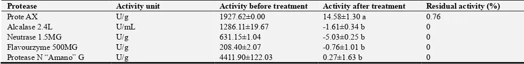
present research, which might attribute to the differences of the varieties [6]. Thus, an extremely acidic treatment was exerted to inactivate the proteases instead of heat treatment. Table 1 showed the losses of protease activities caused by such a treatment. Except that Prote AX remained subtle activity (0.76%), the other four proteases were all irreversibly inactivated. Although Prote AX wouldn’t be thoroughly inactivated, the residual activity was so low and the preincubation period (10 min) was so short that Prote AX could hardly perform its function and might have no influence
on the determination of the α-AI activity [21]. Consequently, the extremely acidic treatment was applicable to inactivate the proteases. Because the α-AI concentration in α-AI extract was pretty high, the extremely acidic treatment wouldn’t lead a low α-AI activity which was difficult to determine exactly. It was the reason why only high concentration of the α-AI was effective in vivo [27]. Besides, the α-AI would get into gastric environment, so it was not necessary to inactivate the proteases during the process.
Table 1. Activity changes of proteases when treated by extremely acidic treatment.
Protease Activity unit Activity before treatment Activity after treatment Residual activity (%)
Prote AX U/g 1927.62±0.00 14.58±1.30 a 0.76
Alcalase 2.4L U/mL 1286.11±19.67 -1.61±0.34 b 0
Neutrase 1.5MG U/g 631.15±1.04 -5.03±0.25 b 0
Flavourzyme 500MG U/g 208.40±2.07 -0.76±1.01 b 0
Protease N “Amano” G U/g 4411.90±122.03 0.27±1.63 b 0
Values with same letters within one column are not significantly different at p﹤0.05.
3.1.3. Suitable Proteases for Enzymatic Hydrolysis
During enzymatic hydrolysis, the short peptides produced might possess α-AI activity and should be separated from the α-AI in case of interference [28]. Because of the excellent solubility of short peptides in high concentration of ethanol, the ultimate hydrolysates with the proteases inactivated were precipitated with 70% ethanol to separate the short peptides from the α-AI. Table 2 showed the loss of the α-AI activity caused by enzymatic hydrolysis with the five proteases. Compared with the control, Flavourzyme 500MG and Neutrase 1.5MG had no significant effect on the α-AI activity (p<0.05) and the loss of the α-AI activity caused by them was 3.54% and 2.67% respectively. The other proteases significantly reduced the α-AI activity (p>0.05) and the influence of Alcalase 2.4L on the α-AI activity was the greatest of all (88.53%). Consequently, Flavourzyme 500MG and Neutrase 1.5MG were suitable for enzymatic hydrolysis.
Table 2. The loss of the α-AI activity caused by enzymatic hydrolysis with the five proteases.
Protease Inhibitory rate (%) Activity loss (%)
Flavourzyme 500MG 41.12±0.91 a 3.54
Protease N “Amano” G 31.52±0.12 b 26.06
Alcalase 2.4L 4.89±0.66 d 88.53
Neutrase 1.5MG 41.49±0.36 a 2.67
Prote AX 28.45±0.36 c 33.26
Control 42.63±0.36 a 0
Values with same letters within one column are not significantly different at p
﹤0.05.
3.2. Optimization of Proteases Used for the Novel Process
3.2.1. Effects of Isoelectric Precipitation on the α-AI
Table 3 showed the α-AI activities in the supernatants obtained from the α-AI extract by isoelectric precipitation at pH between 3.6 and 6.0. Compared with α-AI extract, the α-AI activities in the supernatants reduced with the decrease of the pH while the differences were not significant (p>0.05). The loss of the α-AI activity at pH 6.0 and 3.6 was 0.64% and 5.34%
respectively (Table 3). This result demonstrated few of the α-AI precipitated in acidic solutions. The stability of the α-AI activity in acidic solutions was accordance with the conclusion reported by Golparvar & Naseri [16]. Although the pI of the α-AI ranged from 4.0 to 6.0, the α-AI near pI did’t sediment, which might attribute to its carbohydrate contents which ranged from 8.6% to 15% and accordingly improved its solubility [4, 15]. Consequently, during isoelctric precipitation, the α-AI nearly had no loss and even distributed in one phase, which made it easy to recover all of the α-AI. Thus, isoelectric precipitation was an ideal method for the preparation of the α-AI.
Table 3. The α-AI activities in the supernatants obtained by isoelectric precipitation.
Isoelectric precipitation Inhibitory rate (%) Activity loss (%)
α-AI extract 24.92±0.72 a
Supernatant
pH 6.0 24.76±1.37 a 0.64
pH 5.2 24.30±0.80 a 2.49
pH 4.4 23.79±0.72 a 4.53
pH 3.6 23.59±0.64 a 5.34
Values with same letters within one column are not significantly different at p
﹤0.05.
3.2.2. Optimal Operation Parameters for the Process
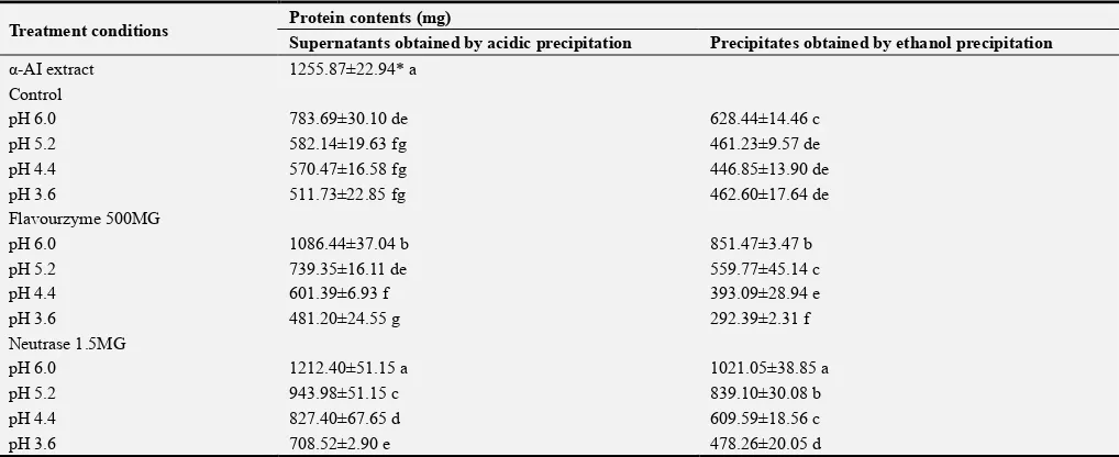
the α-AI were always less than those of the corresponding supernatants. The pH used for electric precipitation had a significant influence on protein contents in the α-AI and the protein contents significantly reduced with the decrease of the pH (p<0.05). Consequently, isoelectric precipitation at pH 3.6 eliminated the most of miscellaneous proteins. At the same pH, the protein contents of the α-AI deriving from hydrolysate F were significantly lower than the ones deriving from hydrolysate N (p<0.05), so Flavourzyme 500MG was more suitable for the preparation of the α-AI considering the purity of the α-AI.
So far, the optimal operation parameters of the novel process described in 2.2 had been obtained as follows: the protease used for enzymatic hydrolysis was Flavourzyme 500MG; the hydrolysis time was 180 min; the pH used for isoelectric precipitation was 3.6.
3.3. Evaluation for the Novel Process
The novel process was evaluated by the α-AI activity yield and the loss of miscellaneous proteins. As Flavourzyme 500MG was introduced into the process, it was hard to directly determine the changes of the α-AI activity during the process because of the interference. Thus, the α-AI activity yield was determined indirectly by calculating the losses of the α-AI activity caused by influential factors which included heat
treatment (70°C/30 min), enzymatic hydrolysis, isoelectric precipitation and 70% ethanol precipitation. The α-AI activities of the initial supernatant, α-AI extract and the resultant precipitate were 31.64%, 24.92% and 23.26% respectively, which indicated the loss of the α-AI activity caused by heat treatment (70°C/30 min) and ethanol precipitation was 21.24% and 6.66% respectively. Besides, enzymatic hydrolysis and isoelectric precipitation caused 3.54% and 5.34% loss of the α-AI activity respectively as mentioned in 3.1.3 and 3.2.1. Consequently, the α-AI activity yield was 67.12% in total (Table 5). The value was much higher than the result (37%) presented by Marshall and Lauda who had used CM-cellulose chromatography [14]. The protein contents in different steps of the process were presented in Table 5. Every procedure could significantly reduce the protein contents (p<0.05) except for enzymatic hydrolysis. 85.84% of the miscellaneous proteins were eliminated by the novel process. The loss of miscellaneous proteins was much lower than the result (94.96%) reported by Mridu et al. who had used ion exchange chromatography, which lead to a relatively low purification fold (4.74) [13]. However, according to Jay et al., the α-AI was only effective at high doses in vivo, thus the α-AI activity yield might be the most important indicator considering the practical application of the α-AI [29]. Consequently, the novel process could better ensure the efficacy of the α-AI.
Table 5. The loss of miscellaneous proteins, the α-AI activity yield and purification fold during the novel process.
Step Protein content (mg) Protein loss (%) Activity yield (%) Purification fold
Initial supernatant 2064.30±55.76 a 0 100 1
α-AI extract 1255.87±22.94 b 39.16 78.76 1.29
Hydrolysate 1227.23±4.63 b 40.55 75.97 1.28
Supernatant 481.20±24.55 c 76.69 71.91 3.09
α-AI 292.39±2.31 d 85.84 67.12 4.74
Values with same letters within one column are not significantly different at p﹤0.05.
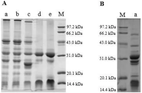
3.4. SDS-PAGE Analysis
SDS-PAGE analyses of the proteins in different steps of the novel process were shown in Figure 3 (A). The α-AI obtained by the process contained two peptide fractions with the molecular weight between 14.4 and 20.1 kDa, together with another two peptide fractions around 28 to 32 kDa (Figure 3 (A), Lane e). This profile was similar to the α-AI profile for a common bean mutant obtained by three phase partition [12]. The α-AI purified from seeds of white kidney bean should have ranged between 36 and 56 kDa, which might be because the α-AI was dissolved into relatively smaller peptides during electrophoresis [30]. Although heat treatment (70°C/30 min) reduced 39.16% of the protein contents in the initial supernatant, the protein profile of the initial supernatant and α-AI extract were the same (Figure 3 (A), Lane a and Lane b), which might be because the heat-sensitive proteins were in a relatively high amount and couldn’t be effectively eliminated by the heat treatment (70°C/30 min). Compared with α-AI extract (Figure 3 (A) Lane b), the bands of peptide fractions with molecular weight above 97.2 kDa and near 29 kDa (Figure 3 (A) Lane b) slightly moved downward and two peptide fractions with lower molecular weight above 97.2 kDa
and near 27 kDa appeared in the hydrolysate (Figure 3 (A) Lane c), which indicated some new proteins with smaller molecular weight might be produced by enzymatic hydrolysis.
Although two chief peptide fractions with molecular weight near 32 and 16 kDa were nearly close to each other in the supernatant deriving from the hydrolysate and α-AI extract (Figure 3 (A), Lane d; Figure 3 (B), Lane a), the peptide fractions in the supernatant deriving from the hydrolysate were less than those in the supernatant deriving from α-AI extract, which was accordance with the significant differences (p<0.05) in protein contents of the two supernatants (Table 4). On the other hand, in the supernatant deriving from the hydrolysate, the third chief peptide fraction near 29 kDa in the supernatant deriving from α-AI extract turned much weaker and the newly produced peptide fraction near 27 kDa in the hydrolysate even disappeared. This result demonstrated that enzymatic hydrolysis was in favor of isoelectric precipitation by altering the pI of more miscellaneous proteins to 3.6.
During the novel process, although the peptide fractions of the supernatant and the α-AI were the same (Figure 3 (A), Lane d and Lane e), 39.24% of the miscellaneous proteins in the supernatant were eliminated by ethanol precipitation. As to the supernatants obtained in the control, the highest miscellaneous protein loss caused by ethanol precipitation was 21.67% (Table 4). This might attribute to the existence of the newly produced proteins with smaller molecular weight. Because of the relatively low molecular weight, these proteins weren’t presented in the SDS-PAGE and were able to disperse in higher concentration of ethanol. Consequently, enzymatic hydrolysis was in favor of ethanol precipitation to eliminate the miscellaneous proteins by elevating the amount of alcohol-soluble proteins.
Table 4. Protein contents in the supernatants and the precipitates (the α-AI) when the ultimate hydrolysates obtained from the α-AI extract were subjected to isoelectric precipitation, followed by ethanol precipitation. Meanwhile, the α-AI extract was served as a control without enzymatic hydrolysis.
Treatment conditions Protein contents (mg)
Supernatants obtained by acidic precipitation Precipitates obtained by ethanol precipitation
α-AI extract 1255.87±22.94* a
Control
pH 6.0 783.69±30.10 de 628.44±14.46 c
pH 5.2 582.14±19.63 fg 461.23±9.57 de
pH 4.4 570.47±16.58 fg 446.85±13.90 de
pH 3.6 511.73±22.85 fg 462.60±17.64 de
Flavourzyme 500MG
pH 6.0 1086.44±37.04 b 851.47±3.47 b
pH 5.2 739.35±16.11 de 559.77±45.14 c
pH 4.4 601.39±6.93 f 393.09±28.94 e
pH 3.6 481.20±24.55 g 292.39±2.31 f
Neutrase 1.5MG
pH 6.0 1212.40±51.15 a 1021.05±38.85 a
pH 5.2 943.98±51.15 c 839.10±30.08 b
pH 4.4 827.40±67.65 d 609.59±18.56 c
pH 3.6 708.52±2.90 e 478.26±20.05 d
* This value presented here referred to the protein content of the α-AI extract. Values with same letters within columns are not significantly different at p<0.05.
4. Conclusions
Flavourzyme 500MG and Neutrase 1.5MG had no significant influence on the α-AI activity (p<0.05), which indicated the α-AI was highly resistant to the hydrolysis of the two proteases. Besides, the α-AI had a quite good solubility in acidic solutions with pH between 3.6 and 6.0, where the α-AI activity had no significant decrease (p<0.05). On the other hand, the novel process based on enzymatic hydrolysis proved feasible for the preparation of the α-AI in seeds of white kidney bean. Although the loss of miscellaneous proteins and purification fold in the novel process were relatively low, the α-AI activity yield was higher than the methods which used chromatography, which was crucial for the practical application of the α-AI. Combined with the SDS-PAGE analysis, enzymatic hydrolysis proved to have modified the pI and alcohol-solubility of the miscellaneous proteins, which had a favorable influence on isoelectric precipitation and ethanol precipitation.
Acknowledgements
The authors thank National Science Council of China for supporting the research by National Natural Science Fund (31271977).
Author Contributions
Wei Zhao designed the study and interpreted the results. Yifeng Rang further adjusted the study design, collected test data and drafted the manuscript.
References
[2] Du SK, Jiang H, Ai Y, et al. 2014. Physicochemical properties and digestibility of common bean (Phaseolus vulgaris L.) starches. Carbohydrate Polymers, 108, 200-205.
[3] Bowman DE. 1945. Amylase inhibitor of navy beans. Science, 102, 358-359.
[4] Obiro WC, Zhang T & Jiang B. 2008. The nutraceutical role of the Phaseolus vulgaris α-amylase inhibitor. British Journal of Nutrition, 100, 1–12.
[5] Wu CT, Chiu CY, Huang CF, et al. 2018. Genotoxicity and 28-day oral toxicity studies of a functional food mixture containing maltodextrin, white kidney bean extract, mulberry leaf extract, and niacin-bound chromium complex. Regul Toxicol Pharmacol, 92, 67-74.
[6] Kusaba NM, Ki M, Iwamoto M, et al. 2000. CM3, one of the wheat α-amylase inhibitor subunits, and binding of IgE in Sera from Japanese with atopic dermatitis related to wheat. Food and chemical toxicology, 38, 179–185.
[7] Ho MF & Whitaker JR. 2010. Subunit structures and essential amino acid residues of white kidney bean (Phaseolus vulgaris) alpha-amylase inhibitors. Journal of Food Biochemistry, 17 (1), 35-52.
[8] Chen XP & Yang WY. 2014. Epidemic trend of diabetes in China. Journal of Diabetes Investigation, 5, 478-481.
[9] Mokdad AH, Bowman BA, Ford ES, et al. 2001. The continuing epidemics of obesity and diabetes in the United States. Jama, 286, 1195-1200.
[10] None. 2016. Fabenol? max, a standardised aqueous extract from phaseolus vulgaris l. and ‘reduces the absorption of carbohydrates’: evaluation of a health claim pursuant to article 13 (5) of regulation (ec) no 1924/2006. EFSA Journal, 14 (2).
[11] Skop M & Chokshi D. 2006. Purified amylase inhibitor and novel process for obtaining the same. http://www.freepatentsonline.com/20060147565.html (accessed 10 April 2007).
[12] Wang H, Chen C, Jeng T, et al. 2011. Comparisons of α-amylase inhibitors from seeds of common bean mutants extracted through three phase partitioning. Food Chemistry, 128, 1066–1071.
[13] Gupta M, Sharma P & Nath AK. 2014. Purification of a novel α-amylase inhibitor from local himalayan bean (Phaseolus vulgaris) seeds with activity towards bruchid pests and human salivary amylase. Journal of food science and technology, 51,
1286–1293.
[14] Marshall JJ & Lauda CM. 1975. Purification and properties of phaseolamin, an inhibitor of α-amylase, from the kidney bean,
Phaseolus vulgaris. Journal of Biological Chemistry, 250,
8030–8037.
[15] Kumar S, Verma AK, Das M, et al. 2013. Clinical complications of kidney bean (Phaseolus vulgaris L.) consumption. Nutrition, 29, 821-827.
[16] Golparvar Z & Naseri B. 2016. Comparative reproductive performance and digestive enzymatic activity of helicoverpa armigera (noctuidae) on seven bean cultivars. Journal of the Lepidopterists Society, 70 (2), 121-129.
[17] Yanli M, Yifeng R, Ruijin Y, et al. 2018. Effect of white kidney bean extracts on estimated glycemic index of different kinds of porridge. LWT, 96, 576-582.
[18] Nciri N & Cho N. 2018. New research highlights: impact of chronic ingestion of white kidney beans (Phaseolus vulgarisl. var. beldia) on small-intestinal disaccharidase activity in wistar rats. Toxicology Reports, 5, 46-55.
[19] Betancur AD, Gallegos TS & Chel GL. 2004. Wet-fractionation of Phaseolus lunatus seeds: partial characterization of starch and protein. Journal of the Science of Food and Agriculture, 84, 1193–1201.
[20] Singh N, Kaur M, Sandhu KS, et al. 2004. Physicochemical, thermal, morphological and pasting properties of starches from some Indian black gram (Phaseolus mungo L.) cultivars.
Starch-Stärke, 56, 535-544.
[21] Yang M, Zhang X, Ma Y, et al. 2008. Purification and partial characterization of a glycoprotein alpha-amylase inhibitor from white kidney bean (Phaseolus vulgaris L.). Journal of Food Biochemistry, 32, 72–84.
[22] Betancur AD, Sosa ET, Ruiz RJ, et al. 2014. Enzymatic hydrolysis of hard-to-cook bean (Phaseolus vulgaris L.) protein concentrates and its effects on biological and functional properties. International Journal of Food Science & Technology, 49, 2–8.
[23] Adler NJ. 1979. Determination of the degree of hydrolysis of food protein hydrolysates by trinitrobenzenesulfonic acid.
Journal of Agricultural and Food Chemistry, 27, 1256-1262.
[24] Lowry OH, Rosebrough NJ, Farr AL, et al. 1951. Protein measurement with the Folin phenol reagent. J biol Chem, 193,
265–275.
[25] Maria HCC, Arcy LA, Roy MH, et al. 2001. Aspartic protease in leaves of common bean (Phaseolus vulgaris L.) and cowpea (Vigna unguiculata L. Walp): enzymatic activity, gene expression and relation to drought susceptibility. FEBS Letters, 492, 242-246.
[26] Zhao W, Yang R, Tang Y, et al. 2009. Investigation of the protein–protein aggregation of egg white proteins under pulsed electric fields. Journal of agricultural and food chemistry, 57,
3571–3577.
[27] Mahomoodally MF & Ramalingum N. 2015. An investigation into the consumption patterns, attitude, and perception of mauritians towards common medicinal food plants. Journal of Herbal Medicine, 5 (2), 99-112.
[28] Yu Z, Yin Y, Zhao W, et al. 2011. Novel peptides derived from egg white protein inhibiting alpha-glucosidase. Food Chemistry, 129, 1376–1382.
[29] Jay U, Ollie T & Jhanna M. 2018. Systematic review and meta-analysis of a proprietary alpha-amylase inhibitor from white bean (phaseolus vulgaris l.) on weight and fat loss in humans. Foods, 7 (4), 63-72.
[30] Bellincampi D, Camardella L, Delcour JA, et al. 2004. Potential physiological role of plant glycosidase inhibitors.