R E S E A R C H A R T I C L E
Open Access
Altered microRNA expression profile during
epithelial wound repair in bronchial epithelial cells
Aleksandra Szczepankiewicz
1*, Peter M Lackie
2and John W Holloway
2,3Abstract
Background:Airway epithelial cells provide a protective barrier against environmental particles including potential pathogens. Epithelial repair in response to tissue damage is abnormal in asthmatic airway epithelium in comparison to the repair of normal epithelium after damage. The complex mechanisms coordinating the regulation of the processes involved in wound repair requires the phased expression of networks of genes. Small non-coding RNA molecules termed microRNAs (miRNAs) play a critical role in such coordinated regulation of gene expression. We aimed to establish if the phased expression of specific miRNAs is correlated with the repair of mechanically induced damage to the epithelium.
Methods:To investigate the possible involvement of miRNA in epithelial repair, we analyzed miRNA expression profiles during epithelial repair in a cell culture model using TaqMan-based quantitative real-time PCR in a TaqMan Low Density Array format. The expression of 754 miRNA genes at seven time points in a 48-hour period during the wound repair process was profiled using the bronchial epithelial cell line 16HBE14o-growing in monolayer. Results:The expression levels of numerous miRNAs were found to be altered during the wound repair process. These miRNA genes were clustered into 3 different patterns of expression that correlate with the further regulation of several biological pathways involved in wound repair. Moreover, it was observed that expression of some miRNA genes were significantly altered only at one time point, indicating their involvement in a specific stage of the epithelial wound repair.
Conclusions:In summary, miRNA expression is modulated during the normal repair processes in airway epithelium in vitrosuggesting a potential role in regulation of wound repair.
Keywords:Epithelial cells, Wound repair, miRNA, Profiling, Cluster analysis, Pathway analysis
Background
The airway epithelium has been recognized to play a cen-tral role in the integration of innate and adaptive immune responses [1-4]. The airway epithelium is also crucial to the origin and progression of respiratory disorders such as asthma, chronic obstructive pulmonary disease, cystic fi-brosis and pulmonary fifi-brosis. In asthma, chronic airway inflammation underlies aberrant repair of the airway that subsequently leads to structural and functional changes in the airway wall. This remodeling is responsible for a num-ber of the clinical characteristics of asthmatic patients.
Normal epithelial repair occurs in a series of overlapping stages. Damage to the epithelium or challenge associated with damage can result in loss of structural integrity or barrier function and local mucosal activation [5]. Studies in animals have shown that the repair of normal airway epithelium after minor damage involves the migration of the remaining epithelial cells to cover the damaged area. This is a rapid process, suggesting an autonomous re-sponse by cells in the vicinity of the damage [6]. It in-cludes an acute inflammatory response, with recruitment of immune cells as well as epithelial spreading and migra-tion stimulated by secreted provisional matrix. Once the barrier is reformed, the differentiated characteristics are then restored. The regulation of these processes require complex sequential changes in the epithelial cell biology driven by the phased expression of networks of genes [7]. * Correspondence:alszczep@ump.edu.pl
1Laboratory of Molecular and Cell Biology, Department of Pediatric
Pulmonology, Allergy and Clinical Immunology, Poznan University of Medical Sciences, 27/33 Szpitalna St., 60-572 Poznan, Poland
Full list of author information is available at the end of the article
One biological mechanism that plays a critical role in the coordinate regulation of gene expression such as that re-quired during epithelial wound repair is the expression of small non-coding RNA molecules termed microRNAs (miRNAs) [8]. To date, more than 1000 human miRNAs have been identified [http://microrna.sanger.ac.uk], with documented tissue-specific expression of some of these miRNAs in lung and involvement in the development of lung diseases including lung cancer, asthma and fibrosis [9-15]. MiRNAs have been demonstrated to play a crucial role in epithelial cell proliferation and differentiation [16-18]. The expression in lung epithelium of Dicer, the en-zyme responsible for processing of miRNA precursors, is essential for lung morphogenesis [16] and there is differen-tial expression of miRNAs during lung development [17]. Furthermore, transgenic over-expression of miR-17-92 (shown to be over-expressed in lung cancer) in the lung epithelium promotes proliferation and inhibits differenti-ation of lung epithelial progenitor cells [18]. Recently, it has been reported that miRNA-146a modulates survival of bronchial epithelial cells in response to cytokine-induced apoptosis [19]. In experimental studies, mice lacking miR-155 demonstrated autoimmune phenotypes in the lungs with increased airway remodeling and leukocyte invasion, phenotypes similar to those observed in asthma [20,21].
While a number of studies have examined the role of miRNA in lung development and in disease [9-15], their influence on the regulation of gene expression involved in epithelial wound repair remains unresolved and com-prehensive studies on miRNA involvement in epithelial repair and the pathogenesis of airway remodeling are lacking. However in the skin, miRNAs were found to play a crucial role in wound closure by controlling mi-gration and proliferation of keratinocytes in an in vitro model of wound repair [22].
Thus the hypothesis of the study was that the stages of wound repair in respiratory epithelium are regulated by the phased expression of specific miRNA species. The aim was to investigate the possible involvement of miRNAs by examining their expression profile in epithelial repair in a cell culture model. Understanding the effect of altered miRNA activity on protein expression during repair pro-cesses can be further used to identify pathways targeted by miRNAs that regulate epithelial wound repair, potentially providing a novel therapeutic strategy for asthma and other respiratory diseases with underlying aberrant epithe-lial wound repair.
Methods
Cell culture and wounding assays
The 16HBE14o- bronchial epithelial cell line was cultured under standard conditions [23]. For the wounding assay, cells were seeded on 6-well plates at the initial density of 3x10⁵cells and cultured until confluent. Forty eight hours
after reaching full confluence cells were damaged by scrap-ing off the monolayer with a hatch-cross woundscrap-ing pattern using a P200 Gilson pipette tip. After that, the medium and cell debris were removed by pipetting off the medium and 2 ml of fresh serum-containing medium was added to the remaining cells. For all experiments, at least two points of reference per well of a 6-well plate were used for post-injury analyses. Several time-lapse experiments were per-formed to establish consistent experimental conditions and the timing of the stages of wound repair.
Time lapse microscopy
Time lapse images were captured at 15 minute intervals on a Leica DM IRB phase-contrast inverted microscope (Leica; Milton Keynes, UK) in a chamber maintained at 36 ± 1°C and 5% CO2atmosphere. The images were collected with a
cooled Hamamatsu ORCA digital camera (Hamamatsu Photonics, Welwyn Garden City, UK) connected to a com-puter running Cell^P software (Olympus, London, UK) for 30 hours (ensuring complete wound closure is included in the time course). For quantitative analysis of the area of damage and hence ongoing repair in time lapse serial im-ages ImageJ software [24] was used.
RNA isolation
RNA isolation was performed with the use of an Exiqon RNA isolation kit. Samples were collected in triplicate for each of the following time points: baseline, 2, 4, 8, 16, 24 and 48 hours after wounding. RNA isolation was performed according to the manufacturer’s protocol from 6-well plates (9.5 cm2of growth area) and the amount of starting mater-ial was 1×10⁶cells per well. Samples were frozen at−70°C for subsequent use in microarray experiments.
RNA quality control
The concentration of total RNA in each sample was deter-mined using a NanoDrop 1000 spectrophotometer. The integrity of total RNA extracted was assessed by a Lab901 Gene Tools System. The passing criteria for use in micro-arrays was: sample volume of 10–30μl, RNA concentra-tion > 50 ng/μl, SDV≤3.1 (ScreenTape Degradation Value), which corresponds to RIN≥9.0, purity: OD 260/ 280 > 1.7, OD 260/230 > 1.4.
Micro RNA profiling
A and B). The resulting cDNA combined with TaqMan Universal PCR Master Mix, No AmpErase UNG was loaded into the arrays and TaqMan real-time PCR was per-formed using the 7900HT Fast Real-Time PCR System (Applied Biosystems) according to the manufacturer’s protocol.
Raw data were obtained using SDS 2.3 software (Applied Biosystems). All SDS files were analyzed utilizing the RQ Manager 1.2 software (Applied Biosystems). The compara-tive analysis of miRNA expression datasets between base-line and each time point following the wounding assay was performed using DataAssist software v.3.01 (Applied Bio-systems). The comparative CT method [25] was used for calculating relative quantitation of gene expression after re-moving outliers with use of Grubbs’ outlier test together with a heuristic rule to remove the outlier among technical replicates and data normalization was based on the en-dogenous control gene expression method (U6 snRNA-001973) where the mean of the selected endogenous control was used to normalize the Ct value of each sample. The data from miRNA profiling have been deposited in ArrayExpress database (accession no. E-MEXP-3986).
Cluster analysis
To identify the clusters of miRNAs following the same ex-pression profile over time, we performed cluster analysis using STEM (Short Time series Expression Miner) software available at: http://www.cs.cmu.edu/~jernst/stem [26].
Target genes and pathways prediction
To identify potential common biological pathways for miRNAs showing similar expression profiles in cluster analysis, we performed pathway enrichment analysis. The best predicted candidate mRNA genes for each differentially expressed miRNA were identified using the miRNA BodyMap tool available at: http://www. mirnabodymap.org. The tool enables the selection of target genes based on the use of several prediction al-gorithms at a time: DIANA, PITA, TargetScan, RNA22 (3UTR), RNA22 (5UTR), TargetScan_cons, Micro-Cosm, miRDB, RNA22 (5UTR), TarBase and miRe-cords. To minimize the target prediction noise, only target genes predicted by five or more of the prediction algorithms mentioned above were included.
The lists with predicted target genes were then analysed with use of The Database for Annotation, Visualization and Integrated Discovery (DAVID) v.6.7 [27,28] to identify BioCarta & KEGG pathways [29,30] enriched functional-related gene groups and biological themes, particularly gene ontology (GO) terms [31] in which the analysed sets of target genes were statistically the most overrepresented (enriched).
Statistics
The statistics applied by Data Assist software for each sample included calculation of the relative quantification (RQ) = 2 (−ΔCt)/2(−ΔCt reference). The standard devi-ation (SD) was calculated for CT values of each of the three technical replicates and was used to calculate the RQ Min and RQ Max [RQ Min = 2(−ΔCt– SD)/2(−ΔCt reference), RQ Max = 2 (−ΔCt + SD)/2(−ΔCt reference)]. Pearson's product moment correlation coefficient (r) was calculated for CT orΔCT values of sample pairs as below and plotted on the Signal Correlation Plot and Scatter Plot respectively.
r¼ ffiffiffiffiffiffiffiffiffiffiffiffiffiffiffiffiffiffiffiffiffiffiffiffiffiffiffiffiNΣXY−ðΣXÞðΣYÞ
NΣX2−ðΣXÞ2
q ffiffiffiffiffiffiffiffiffiffiffiffiffiffiffiffiffiffiffiffiffiffiffiffiffiffiffi
NΣY2−ðΣYÞ2 q
Distances between samples and assays were calculated for hierarchical clustering based on the ΔCT values using Pearson’s correlation or the Eucidian distance calculated as follows [https://products.appliedbiosystems.com]. For a sample pair, the Pearson's product moment correlation co-efficient (r) was calculated considering allΔCT values from all assays, and the distance defined as 1–r. For an assay pair, r was calculated considering all ΔCT values from all samples and the distance defined as 1– r. Euclidean
Dis-tance calculated as
ffiffiffiffiffiffiffiffiffiffiffiffiffiffiffiffiffiffiffiffiffiffiffiffiffiffiffiffiffiffiffiffiffiffiffiffiffiffiffiffiffiffiffi
Σ Δð CT ið Þ−ΔCT jð ÞÞ2 q
where, for a sample pair, the calculation is done across all assays for sample i and sample j while for an assay pair, the calcula-tion is done across all samples for assay i and assay j.
Results
Characterisation of epithelial wound repair model
Global miRNA expression profile altered during epithelial wound repair
Expression profiling analysis revealed a large number of mature miRNAs that were modulated at different time points during epithelial repair with a fold change above 2.0 (Table 1). Numerous miRNAs showed significantly increased or decreased expression (>10-fold) at differ-ent time points as compared to baseline (presdiffer-ented in Additional file 1). Based on the high fold change values at different time points, ten miRNAs were found to undergo a significant modulation (both up- or down-regulation) at five or more of the seven time points analysed (Additional file 2). We also observed that the alterations in expression of some miRNA genes were limited to a single time point of wound repair, whereas at the other time points the ex-pression levels did not differ much from the baseline, sug-gesting their involvement at a particular stage of repair (marked in red in Additional file 1).
Cluster analysis
We then hypothesized that, given the number of miRNA genes undergoing significant changes during the epithelial
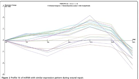
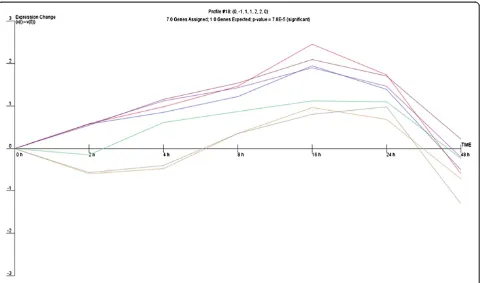
repair process, a common expression profile might be shared by miRNAs whose expression is regulated by par-ticular transcriptional activation pathways. Therefore, we analysed the expression of miRNA genes with use of the clustering algorithm STEM [26], assigning each gene pass-ing the filterpass-ing criteria to the model profile that most closely matches the gene's expression profile as determined by the correlation coefficient. Since the model profiles are selected by the software by random allocation, independent of the data, the algorithm then determines which profiles have a statistically significant higher number of genes assigned using a permutation test. It then uses standard hy-pothesis testing to determine which model profiles have significantly more genes assigned as compared to the aver-age number of genes assigned to the model profile in the permutation runs. Our cluster analysis revealed three sig-nificant miRNA expression profiles (16, 1 and 18) over 48 hours of wound repair (Figures 2, 3 and 4).
Profile 16 included genes that gradually increase between 2 and 16 hours and then display a sudden drop in expres-sion 16 hours post-wounding, which corresponded to the completion of cell proliferation and the restoration of the monolayer after wounding in time lapse observations. Pro-file 1 was characterized by significant decrease of miRNA expression 4 hours after wounding followed by a significant increase with a maximum 16 hours post-wounding, sug-gesting induction of transcription of genes involved in the early response to stress due to mechanical cell damage which are subsequently switched off. Profile 18 shared some similarities with profile 1, although it showed a more
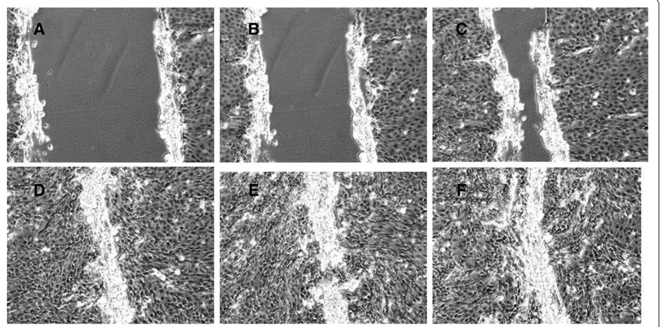
Figure 1Stages of wound repair at different time points (A–2 hrs, B–4 hrs, C–8 hrs, D–16 hrs, E–24 hrs, F–48 hrs post wounding), n=3 wells for each time point.
Table 1 Number of miRNAs with >2.0-fold change in expression at different time points after wounding
Time point (post wounding)
Mode of miRNA alteration 2 hrs 4 hrs 8 hrs 16 hrs 24 hrs 48 hrs
Upregulated 70 128 85 37 35 252
Figure 2Profile 16 of miRNA with similar expression pattern during wound repair.
gradual decrease in miRNA expression 4 hours post-wounding, that then increased steadily to reach a max-imum at 16 hours and afterwards gradually decreased. The different profiles of miRNA expression are shown in Figures 2, 3 and 4. The miRNA genes sharing the common expression pattern during epithelial wound repair are listed in Additional file 3.
Identification of biological processes regulated by miRNAs Pathways in clusters of miRNAs
The next question to be addressed was if miRNA clusters of characteristic expression profile during epithelial wound repair identified using STEM were regulating target genes from the same biological pathways or processes. To analyse this, we used highly predicted miRNA targets (mRNAs confirmed to be a target of specific miRNA by at least five different algorithms) to create a list of potential miRNA target genes, which were then analysed utilising the DA-VID online database for annotation and visualization [27,28]. The use of DAVID enabled the integration of the miRNA target genes into common pathways or GO pro-cesses. Analysis of targets predicted for each miRNA ex-pression cluster generated by STEM enabled us to predict four significantly enriched pathways for profile 16, includ-ing the neurotrophin signallinclud-ing pathway, ERBB signallinclud-ing pathway, MAPK signalling pathway and the RIG-I-like re-ceptor signalling pathway. Six pathways were predicted for the targets of miRNAs demonstrating expression in profile
1: adherence junction, acute myeloid leukaemia, small lung cancer, cell cycle, pathways in cancer and the chemo-kine signalling pathway. No common pathways were pre-dicted for profile 18. The prepre-dicted pathways are shown in Additional files 4 and 5.
For all the profiles, DAVID also predicted numerous bio-logical processes where miRNAs targets play a significant role (see Additional file 6). In general, predicted biological processes and pathways were mainly associated with cell cycle regulation and induction of mitotic divisions, switch-ing on anti-apoptotic genes (ECM, PKB/Akt and IKK) and genes stimulating proliferation (such as MEK, PPARγ) that are of known importance in epithelial wound repair. Apart from well documented biological processes, we also ob-served that, surprisingly, the most significantly overrepre-sented were target genes involved in the neurotrophin signalling pathway which suggests its importance in epithe-lial wound repair process (Additional file 7).
Target pathways at different stages of wound repair
To identify the most important pathways involved at different stages of epithelial wound repair in vitro we also performed pathway enrichment of miRNAs signifi-cantly altered only at one time point of wound repair (see Additional file 1, genes in red). For those genes, tar-gets were predicted as above and DAVID was used to identify potential pathways and biological processes. The main observation for epithelial cells in the early
phase of repair (2 hours post-wounding) were miRNAs being up-regulated, suggesting switching off target genes and processes associated with response to cellular stress (MAP kinase pathway), regulation of actin cyto-skeleton, cell proliferation and migration. The main pathways targeted by up-regulated miRNAs identified for the repair 4 hours after cell damage included genes involved in negative regulation of transcription, RNA metabolism, regulation of cell motion and the cytoskel-eton. The most important processes at 8 hours after wounding involved a number of up-regulated miRNAs at this time point and indicating the switching off of genes involved in negative regulation of gene expression and negative regulation of cell communication. At 16 hours following epithelial cell wounding we observed a number of miRNA genes that were down regulated and, therefore, switching on genes involved in mitotic cell cycle, negative regulation of cell death, cell prolifer-ation, ERBB signalling pathway (cell proliferprolifer-ation, sur-vival, migration). This may suggest the predominance of a proliferating phenotype of cells after the damaged area was closed by spreading and migrating cells. After 24 hours post-wounding we observed further down regulation of miRNA genes. Two were of particular interest as they are responsible for switching on genes involved in p53 signal-ling pathway (cell cycle arrest), IL-10 (anti-inflammatory response), regulation of apoptosis, cell death, RNA trans-port and localization. This indicates that at this time point cells have proliferated sufficiently and are beginning to dif-ferentiate. At 48 hours after wounding, we observed mainly up regulation of miRNA genes responsible for silencing genes involved in protein catabolic processes, alternative splicing, spectrins, mRNA splicing and processing as well as methylation indicating that cells are undergoing physio-logical processes and restoring a normal phenotype.
Discussion
The main finding of this study is the involvement of mul-tiple miRNA genes in the process of epithelial wound re-pair in vitro. We found three distinct expression patterns of miRNA genes clusters that are predicted to further regulate numerous pathways and biological processes in-volved in wound repair. We have applied here for the first time the cluster analysis of time-series miRNA expression data (using STEM) to identify basic patterns and predict pathways (using DAVID) involved in repair processes of airway epithelium.
Such an approach has enabled us to identify common miRNA expression profiles during wound repair giving comprehensive information about activated miRNA genes. The relationships amongst these genes, their regulation and coordination during wound repair over time can also be explored. Further validation of individual protein, gene or miRNA changes will be required in subsequent studies,
but it seems clear that specific expression profiles of clus-ters of miRNAs correlates with repair of mechanically in-duced damage to the epithelium. For expression profile 16 we demonstrated that, among other plausible signalling pathways, the neurotrophin signalling pathway may be in-volved in wound repair in epithelial cells, in addition to the inflammatory response in airway epithelium in allergy and asthma as reported previously [35-38]. The involve-ment in wound repair may further suggest that this path-way is important in the regulation of airpath-way remodelling in asthma. Indeed, in the study by Kilic et al. [39] it was observed that blocking one of the neurotrophins, nerve growth factor (NGF), prevented subepithelial fibrosis in a mouse model of asthma and that NGF overexpression exerted a direct effect on collagen expression in murine lung fibroblasts. The involvement of neurotrophins in re-pair processes has been also confirmed recently by Palazzo et al. [40] in wound healing in dermal fibroblasts. Moreover, miRNAs involved in this pathway such as the miR-200 fam-ily were reported to control epithelial-mesenchymal transi-tion (EMT) [41], the process that is suggested to underlie airway remodelling in asthma. In the recent study of Ogawa et al. [42] utilising a mouse model of asthma, it was ob-served that mice challenged with house dust mite allergen exhibited an increase in NGF that was primarily expressed in bronchial epithelium and was positively correlated with airway hyperresponsiveness and substance P-positive nerve fibers. However in this model siRNA targeted NGF inhib-ited hyperresponsiveness and modulation of innervation but not subepithelial fibrosis and allergic inflammation.
For expression profile 1 we observed a significant down-regulation at the beginning of wound repair followed by sharp increase in miRNA expression with a maximum at 16 hours after cell damage. This may indicate the induc-tion of the six pathways predicted by enrichment analysis in the early phase of wound repair, which are then being switched off by the miRNAs with increased expression up to 16 hours post-wounding.
The process of wound repair in vivo in the airways in-volves cell spreading and migration as the primary mecha-nisms in the first 12–24 hours after injury, while proliferation begins by 15–24 h and continues for days to weeks. Similarly in our study we have confirmed that epi-thelial wound repair in vitromimics thein vivo situation but in a shorter time frame, and that in its early stage this involves spreading and migration of neighbouring epithe-lial cells to cover the damaged area (2 and 4 hours after wounding). This is followed by migration and proliferation of progenitor cells to restore cell numbers (8 and 16 hours after cell damage) and differentiation to restore function (24 and 48 hours post-wounding) (Figure 1) [43-48].
switching off pathways regulating cell proliferation and dif-ferentiation and activating cellular stress responses (chemo-kine signalling pathway) as well as cell migration and cell death (corresponding to time points at 2, 4 and 8 hours after injury). Furthermore, at later time points cells are undergo-ing intensive proliferation and secretundergo-ing extracellular matrix which is supported by the involvement of ERBB signalling pathway and NFAT pathway stimulating cell proliferation and the regulation of transcription of immune genes (that corresponds to 16 hours after injury). Once confluent, cells restore their phenotype so that the cell cycle is arrested (in-hibition of cell division) and differentiation processes are switched on. In parallel to this, the IL-10 anti-inflammatory signalling pathway is induced to deactivate immune cells stimulated during the early stages of wound repair.
Conclusions
In summary, we report here for the first time that ex-pression of multiple miRNAs is significantly altered during airway epithelium wound repair processes. Dif-ferent patterns of expression have been observed and the target genes of those miRNA clusters coordinate several biological pathways involved in the repair of in-jury. Our work provides a starting point for a systematic analysis of mRNA targets specific for wound repair. This will help to identify regulatory networks control-ling these processes in airway epithelium to better understand their involvement in respiratory diseases.
Additional files
Additional file 1:List of significantly modulated mature miRNAs (>10.0-fold) and their respective fold induction at each time point. * miRNAs with significant change in expression at one time point only (marked in red).
Additional file 2:Fold change of the top ten miRNAs undergoing significant modulation (>10-fold) during wound repair process at, at least, five time points.
Additional file 3:MiRNA genes assigned to each expression profile during wound repair (values given for each time point represent expression change after normalization in STEM software).
Additional file 4:The significantly overrepresented pathways (enriched) in the analysed sets of target genes of miRNAs included in the profile 16.
Additional file 5:The significantly overrepresented pathways (enriched) in the analysed sets of target genes of miRNAs included in the profile 1.
Additional file 6:The most significant biological processes predicted using DAVID tool undergoing regulation of miRNA target genes from the same expression profile (processes were ranked based on their Fisher Exact Probability value from the gene enrichment analysis to identify those showing significant overrepresentation).
Additional file 7:Neurotrophin signaling pathway with miRNA genes and their predicted targets.
Competing interests
The authors declare that they have no competing interests.
Authors’contributions
AS performed in vitro cell experiments and wounding assays, miRNA profiling, data analysis, cluster and pathway analysis, drafted the paper and approved its final version. PL contributed to the study design and
methodology regarding cell experiments drafted the paper and approved its final version. JWH contributed to the study design and methodology regarding miRNA analysis, drafted the paper and approved its final version.
Acknowledgements
This study was supported by the Polish National Science Centre grant no. 2011/01/M/NZ3/02906 and the AAIR Charity (Asthma, Allergy and Inflammation Research).
AS was a recipient of an EAACI Exchange Research Fellowship 2010. We thank the personnel from the Biomedical Imaging Unit, University of Southampton for the assistance and technical support.
Author details
1Laboratory of Molecular and Cell Biology, Department of Pediatric
Pulmonology, Allergy and Clinical Immunology, Poznan University of Medical Sciences, 27/33 Szpitalna St., 60-572 Poznan, Poland.2Clinical and
Experimental Sciences, Faculty of Medicine, University of Southampton, Southampton, UK.3Human Development and Health, Faculty of Medicine, University of Southampton, Southampton, UK.
Received: 21 December 2012 Accepted: 31 October 2013 Published: 5 November 2013
References
1. Hammad H, Lambrecht BN:Dendritic cells and epithelial cells: linking innate and adaptive immunity in asthma.Nat Rev Immunol2008,8:193–204. 2. Holgate ST:Epithelium dysfunction in asthma.J Allergy Clin Immunol2007,
120:1233–1244. quiz 1245–1236.
3. Holgate ST:The epithelium takes centre stage in asthma and atopic dermatitis.Trends Immunol2007,28:248–251.
4. Kato A, Schleimer RP:Beyond inflammation: airway epithelial cells are at the interface of innate and adaptive immunity.Curr Opin Immunol2007, 19:711–720.
5. Sha Q, Truong-Tran AQ, Plitt JR, Beck LA, Schleimer RP:Activation of airway epithelial cells by toll-like receptor agonists.Am J Respir Cell Mol Biol2004, 31:358–364.
6. Erjefalt JS, Erjefalt I, Sundler F, Persson CG:In vivo restitution of airway epithelium.Cell Tissue Res1995,281:305–316.
7. Heguy A, Harvey BG, Leopold PL, Dolgalev I, Raman T, Crystal RG: Responses of the human airway epithelium transcriptome to in vivo injury.Physiol Genomics2007,29:139–148.
8. He L, Hannon GJ:MicroRNAs: small RNAs with a big role in gene regulation.Nat Rev Genet2004,5:522–531.
9. Eder M, Scherr M:MicroRNA and lung cancer.N Engl J Med2005, 352:2446–2448.
10. Hurteau GJ, Carlson JA, Spivack SD, Brock GJ:Overexpression of the microRNA hsa-miR-200c leads to reduced expression of transcription factor 8 and increased expression of E-cadherin.Cancer Res2007,67:7972–7976. 11. Jay C, Nemunaitis J, Chen P, Fulgham P, Tong AW:miRNA profiling for
diagnosis and prognosis of human cancer.DNA Cell Biol2007,26:293–300. 12. Miller RL, Ho SM:Environmental epigenetics and asthma: current
concepts and call for studies.Am J Respir Crit Care Med2008,177:567–573. 13. Nana-Sinkam SP, Karsies T, Riscili B, Ezzie M, Piper M:Lung microRNA: from
development to disease.Expert Rev Respir Med2009,3:373–385. 14. Takamizawa J, Konishi H, Yanagisawa K, Tomida S, Osada H, Endoh H,
Harano T, Yatabe Y, Nagino M, Nimura Y,et al:Reduced expression of the let-7 microRNAs in human lung cancers in association with shortened postoperative survival.Cancer Res2004,64:3753–3756.
15. Solberg OD, Ostrin EJ, Love MI, Peng JC, Bhakta NR, Hou L, Nguyen C, Solon M, Nguyen C, Barczak AJ,et al:Airway epithelial miRNA expression is altered in asthma.Am J Respir Crit Care Med2012,186:965–974. 16. Harris KS, Zhang Z, McManus MT, Harfe BD, Sun X:Dicer function is
essential for lung epithelium morphogenesis.Proc Natl Acad Sci USA2006, 103:2208–2213.
18. Lu Y, Thomson JM, Wong HY, Hammond SM, Hogan BL:Transgenic over-expression of the microRNA miR-17-92 cluster promotes proliferation and inhibits differentiation of lung epithelial progenitor cells.Dev Biol
2007,310:442–453.
19. Liu X, Nelson A, Wang X, Kanaji N, Kim M, Sato T, Nakanishi M, Li Y, Sun J, Michalski J,et al:MicroRNA-146a modulates human bronchial epithelial cell survival in response to the cytokine-induced apoptosis. Biochem Biophys Res Commun2009,380:177–182.
20. Rodriguez A, Vigorito E, Clare S, Warren MV, Couttet P, Soond DR, van Dongen S, Grocock RJ, Das PP, Miska EA,et al:Requirement of bic/ microRNA-155 for normal immune function.Science2007,316:608–611. 21. Thai TH, Calado DP, Casola S, Ansel KM, Xiao C, Xue Y, Murphy A,
Frendewey D, Valenzuela D, Kutok JL,et al:Regulation of the germinal center response by microRNA-155.Science2007,316:604–608. 22. Bertero T, Gastaldi C, Bourget-Ponzio I, Imbert V, Loubat A, Selva E, Busca R,
Mari B, Hofman P, Barbry P,et al:miR-483-3p controls proliferation in wounded epithelial cells.FASEB J2011,25:3092–3105.
23. Adam EC, Holgate ST, Lackie PM:Epithelial repair is inhibited by an alpha (1,6)-fucose binding lectin.Am J Physiol Lung Cell Mol Physiol2007, 292:L462–L468.
24. Schneider CA, Rasband WS, Eliceiri KW:NIH Image to ImageJ: 25 years of image analysis.Nat Methods2012,9:671–675.
25. Schmittgen TD, Livak KJ:Analyzing real-time PCR data by the comparative C(T) method.Nat Protoc2008,3:1101–1108.
26. Ernst J, Bar-Joseph Z:STEM: a tool for the analysis of short time series gene expression data.BMC Bioinformatics2006,7:191.
27. da Huang W, Sherman BT, Lempicki RA:Systematic and integrative analysis of large gene lists using DAVID bioinformatics resources. Nat Protoc2009,4:44–57.
28. da Huang W, Sherman BT, Zheng X, Yang J, Imamichi T, Stephens R, Lempicki RA:Extracting biological meaning from large gene lists with DAVID.Curr Protoc Bioinformatics2009. Chapter 13:Unit 13 11. 29. D. N:BioCarta.Biotech Softw Internet Rep2001,2:117–120.
30. Kanehisa M, Goto S, Kawashima S, Okuno Y, Hattori M:The KEGG resource for deciphering the genome.Nucleic Acids Res2004,32:D277–D280. 31. Ashburner M, Ball CA, Blake JA, Botstein D, Butler H, Cherry JM, Davis AP,
Dolinski K, Dwight SS, Eppig JT,et al:Gene ontology: tool for the unification of biology. The gene ontology consortium.Nat Genet2000,25:25–29. 32. Adam EC, Holgate ST, Fildew CJ, Lackie PM:Role of carbohydrates in
repair of human respiratory epithelium using an in vitro model.Clin Exp Allergy2003,33:1398–1404.
33. Howat WJ, Holgate ST, Lackie PM:TGF-beta isoform release and activation during in vitro bronchial epithelial wound repair.Am J Physiol Lung Cell Mol Physiol2002,282:L115–L123.
34. Leir SH, Holgate ST, Lackie PM:Inflammatory cytokines can enhance CD44-mediated airway epithelial cell adhesion independently of CD44 expression.Am J Physiol Lung Cell Mol Physiol2003,285:L1305–L1311. 35. Hahn C, Islamian AP, Renz H, Nockher WA:Airway epithelial cells produce
neurotrophins and promote the survival of eosinophils during allergic airway inflammation.J Allergy Clin Immunol2006,117:787–794. 36. Lommatzsch M, Braun A, Renz H:Neurotrophins in allergic airway
dysfunction: what the mouse model is teaching us.Ann N Y Acad Sci
2003,992:241–249.
37. Olgart Hoglund C, Frossard N:Nerve growth factor and asthma. Pulm Pharmacol Ther2002,15:51–60.
38. Prakash Y, Thompson MA, Meuchel L, Pabelick CM, Mantilla CB, Zaidi S, Martin RJ:Neurotrophins in lung health and disease.Expert Rev Respir Med
2010,4:395–411.
39. Kilic A, Sonar SS, Yildirim AO, Fehrenbach H, Nockher WA, Renz H:Nerve growth factor induces type III collagen production in chronic allergic airway inflammation.J Allergy Clin Immunol2011,128:1058–1066. e1051-1054. 40. Palazzo E, Marconi A, Truzzi F, Dallaglio K, Petrachi T, Humbert P, Schnebert
S, Perrier E, Dumas M, Pincelli C:Role of neurotrophins on dermal fibroblast survival and differentiation.J Cell Physiol2012,227:1017–1025. 41. Adam L, Zhong M, Choi W, Qi W, Nicoloso M, Arora A, Calin G, Wang H,
Siefker-Radtke A, McConkey D,et al:miR-200 expression regulates epithelial-to-mesenchymal transition in bladder cancer cells and reverses resistance to epidermal growth factor receptor therapy.Clin Cancer Res
2009,15:5060–5072.
42. Ogawa H, Azuma M, Uehara H, Takahashi T, Nishioka Y, Sone S, Izumi K: Nerve growth factor derived from bronchial epithelium after chronic
mite antigen exposure contributes to airway hyperresponsiveness by inducing hyperinnervation, and is inhibited by in vivo siRNA.Clin Exp Allergy2012,42:460–470.
43. Puchelle E, Zahm JM, Tournier JM, Coraux C:Airway epithelial repair, regeneration, and remodeling after injury in chronic obstructive pulmonary disease.Proc Am Thorac Soc2006,3:726–733. 44. Stripp BR, Reynolds SD:Maintenance and repair of the bronchiolar
epithelium.Proc Am Thorac Soc2008,5:328–333.
45. Ware LB, Matthay MA:The acute respiratory distress syndrome.N Engl J Med2000,342:1334–1349.
46. Zahm JM, Chevillard M, Puchelle E:Wound repair of human surface respiratory epithelium.Am J Respir Cell Mol Biol1991,5:242–248. 47. Zahm JM, Kaplan H, Herard AL, Doriot F, Pierrot D, Somelette P, Puchelle E:
Cell migration and proliferation during the in vitro wound repair of the respiratory epithelium.Cell Motil Cytoskeleton1997,37:33–43.
48. Zahm JM, Pierrot D, Chevillard M, Puchelle E:Dynamics of cell movement during the wound repair of human surface respiratory epithelium. Biorheology1992,29:459–465.
doi:10.1186/1471-2466-13-63
Cite this article as:Szczepankiewiczet al.:Altered microRNA expression profile during epithelial wound repair in bronchial epithelial cells.BMC Pulmonary Medicine201313:63.
Submit your next manuscript to BioMed Central and take full advantage of:
• Convenient online submission
• Thorough peer review
• No space constraints or color figure charges
• Immediate publication on acceptance
• Inclusion in PubMed, CAS, Scopus and Google Scholar
• Research which is freely available for redistribution