ÜONNEXIN EXPRESSION AND FUNCTIONAL: GAP JUNCTIONAL COMMUNICATION IN ECTODERMALLY DERIVED EMBRYONte
ùç
TISSUES OF THE RATA
KARINA NICOLE POTTER
Thesis submitted for the degree of PhD
DEPARTMENT OF ANATOMY AND DEVELOPMENTAL BIOLOGY UNIVERSITY COLLEGE LONDON
GOWER STREET LONDON
ProQuest Number: 10042839
All rights reserved
INFORMATION TO ALL USERS
The quality of this reproduction is dependent upon the quality of the copy submitted.
In the unlikely event that the author did not send a complete manuscript and there are missing pages, these will be noted. Also, if material had to be removed,
a note will indicate the deletion.
uest.
ProQuest 10042839
Published by ProQuest LLC(2016). Copyright of the Dissertation is held by the Author.
All rights reserved.
This work is protected against unauthorized copying under Title 17, United States Code. Microform Edition © ProQuest LLC.
ProQuest LLC
789 East Eisenhower Parkway P.O. Box 1346
ACKNOWLEDGEMENTS
First and foremost I would like to thank my supervisor Dr. Barbara Fulton for all her advice, help and encouragement throughout my thesis. I would also like to greatly thank my husband, Nimesh Patel for his constant moral support throughout the four years of study. I would like to thank Dr. David Becker, Dr. Colin Green, Dr. Paul Martin and Dr. Helen Makarenko va for technical help and advice throughout my thesis and Dr. Christina Danevic, Janie McClusky, Jovita Er and Dr. Yvonne Lawrence for their friendship and support at UCL. I would like to thank everyone from
ABSTRACT
The aim of my PhD project was to examine the developmental expression and functional significance of gap junctional communication in ectodermally derived embryonic tissues (nervous system and epidermis) of the rat, with particular emphasis on elucidating the role of gap junctions and putative mechanisms of gap junctional regulation in the development of dorsal root ganglion (DRG) neurons.
In this study, immunohistochemistry in combination with the technique of the laser scanning confocal microscopy (LSCM) with its PC-IMAGE image analysis programme was used to determine the developmental expression of gap junction proteins
(connexins). Antisera specific to certain peptide sequences of the three major connexin (Cx) types (Cx26, Cx32 and Cx43) were used. Gap junction-mediated intercellular communication was assessed by observing the transfer o f the low molecular weight fluorescent dye, Lucifer Yellow CH (LY), from an intracellularly microinjected cell to neighbouring cells under epifluorescence optics. In thick tissue, the extent of dye transfer was visualized subsequently on the LSCM.
These immunohistochemical and dye-injection techniques were used to study connexin expression and functional gap junctional communication in developing non-neural ectoderm (predominantly flank epidermis) and in neural derivatives of ectoderm (predominantly DRG neurons). A developmental study of flank epidermis revealed a temporal increase in Cx26 and Cx43 from embryonic days 10 to 14 (E l0 -E l4). LY dye-coupling assays carried out in parallel were consistent with the
epidermis is transformed from a simple unilayer of ectoderm into an epidermal and peridermal bilayer.
DRG neurons had previously been shown to be dye-coupled in small groups from E 13- El 5, but during subsequent embryonic development, both the number of cells that transfer dye and the mean number of cells in a coupled group decreased steadily (Fulton, B P J. Physiol. 426: 122P, 1990). Immunohistochemical studies of these developing neurons established that this decrease in dye-coupling was accompanied by a decrease in expression of Cx26 and Cx43.
TABLE OF CONTENTS
PAGE
ACKNOWLEDGEMENTS 2
ABSTRACT 3
1 INTRODUCTION
Formation of ectodermally derived tissues 15
Rationale for studying connexin expression in specific types 21 of ectodermally derived tissues
The gap junction 22
Ultrastructural analysis 22
Members of the gap junction polypeptide family
Nomenclature o f the gap junction polypeptide family 25
Topology of the connexin protein 27
Connexin distribution 29
Gap junction genes 30
Gene structure 30
Chromosomal location of gap junction genes 30
Gap junction assembly and disassembly 32
Gap junction assembly 32
Gap junction disassembly 33
Biophysical properties of the gap junction channel 34
Gap junction conductance 34
Gap junction permeability 34
Gating mechanisms of gap junctions 35
Cation gating of gap junctions 36
M odulation of gap junctional communication 37
Modulation of gap junctional communication by regulating the
amount or type of connexin expressed 37
Changes in the functional state of gap junction channels 39
Roles of gap Junctions in adult tissue 40
General roles o f gap junctions 40
Roles of gap junctions in non-neural excitable cells 42
Role of gap junctions in the adult nervous system 42
The role of gap junctions in development 47
Role of gap junctions in mediating global signals 47
Role of gap junctions in mediating local interactions 50
Correlatons between gap junction communication & cell fate
restriction 51
Role of gap junctions in the developing nervous system 53
Role of gap junctions in patterning the developing nervous system 53
Role of gap junctions in inductive interactions in the developing
nervous system 54
Roles of gap junctions which are specific to the needs of the
developing nervous system 55
Examples of transient interactions mediated by gap junctions 55
Examples of long-term interactions mediated by gap junctions 56
2 CHARACTERIZATION OF CONNEXINS LABELLED BY ANTIPEPTIDE ANTIBODIES
2.1 Introduction 60
2.2 M aterials and M ethods 65
Fixation and histological procedures 65
Laser scanning confocal microscopy (LSCM) 67
2.3 Results 69
Establishment of an optimal immunohistochemical protocol 69
Immunolabelling of Zamboni's fixed tissue 69
Immunolabelling of tissue fixed by methanol freeze substitution 69
Immunolabelling of frozen sections post fixed in methanol 71
Controls 71
2.4 Discussion 76
3 CONNEXIN EXPRESSION AND DYE COUPLING IN TWO REGIONS OF NON-NEURAL ECTODERM OF RAT EMBRYOS: FLANK EPIDERMIS AND THE APICAL ECTODERMAL RIDGE OF THE RAT
3.1 Introduction 80
3.2 Materials and Methods 91
Peptide synthesis and antibody production 91
Animals and tissue collection 91
Immumohistochemistry 92
Intracellular dye-injection 94
3.3 Results 97
Immunohistochemistry 97
Intracellular dye-injection 115
3.4 Discussion 128
Immunolabelling of the flank epidermis, AER and underlying
mesenchymal tissue 128
Dye-coupling studies of the flank epidermis and AER in
embryonic rats 132
Putative roles of gap junctions in the flank epidermis, periderm,
AER & limb bud mesenchyme 139
4 GAP JUNCTIONS BETWEEN DRG NEURONS (CONNEXIN
EXPRESSION, DYE COUPLING AND PUTATIVE MECHANISMS OF REGULATORY CONTROL
4.1 Introduction 145
General introduction to dorsal root ganglia 145
Histogenesis of the dorsal root ganglia 146
Timing of events during sensory neuron development 147
Innervation patterns of developing DRG neurons 149
Possible factors which are thought to influence innervation patterns 151
Gap junctions in the developing somatosensory system 153
4.2 M aterials and Methods 155
Immunohistochemistry 155
Dye-coupling between DRG neurons and effects of exogenously
applied NGF 157
4.3 Results 161
Immunohistochemistry 161
Effects of NGF on dye-coupling between DRG neurons 174
4.4 Discussion 187
Connexin expression in developing DRG neurons 187
Regulation of gap junction expression in developing DRG neurons 192
Dye-injection of freshly prepared DRG neurons 194
Dye-injection of DRG explants exposed to NGF 195
Dye-injection of DRGs in organotypic slice cultures 196
5 CONNEXIN EXPRESSION AND DYE COUPLING IN THE DEVELOPING SPINAL CORD
5.1 Introduction
Organization of the spinal cord 204
Histogenesis of the rat spinal cord 204
Gap junctions in the developing spinal cord 204
Patterning of the neural tube 205
5.2 M aterials and Methods 209
Immunohistochemistry 209
Dye-injection 209
5.3 Results 210
Immunolabelling of lumbar neural tube 210
Dye-coupling in lumbar neural tube 216
5.4 Discussion 219
6 CONCLUSIONS AND FUTURE STUDIES
Summary of finding 225
Prospective research arising from the thesis 233
FIGURES AND TABLES
CHAPTER I
Fig. 1.1. Neural plate and neural tube stage 17
Fig. 1.2. Vertical and planar signals involved in neural tube patterning 18
Fig. 1.3. Migratory pathways of the neural crest 19
Fig. 1.4. Structure of gap junctions derived from X-ray diffraction 23
Fig. 1.5. Phylogenetic tree of connexins 26
Fig. 1.6. Illustration of connexin topography 28
Table 1.1. Expression of connexins in mammalian tissues 30
CHAPTER 2
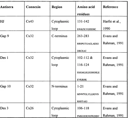
Fig. 2.1. Diagrammatic representation of site specific antisera 62 Fig. 2.2. Immunolabelling of Zamboni's fixed mouse heart wax sections
with HJ 70
Fig. 2.3. Immunolabelling of MFS fixed mouse heart & liver wax sections
with HJ & Gap 10 72
Fig. 2.4. Immunolabelling of cryosectioned rat & mouse liver with
Gap 10, Des 1 & Gap 9 73
Fig. 2.5. Immunolabelling of cryosectioned mouse heart & liver
with Des 3 & HJ 74
Table 2.1. Summary of site specific antisera to the three main connexins 61 Table 2.2. Summary of IR detected in mouse heart & mouse & rat liver 75
CHAPTER 3
Fig. 3.2. Different stages in rat limb bud development 84
Fig. 3.3. Structure of the limb bud 87
Fig. 3.4. Immunolabelling of E13 rat epidermal tissue with Des 3 98 Fig. 3.5. Tmmunolabelling of E10-E14 rat epidermal tissue with
HJ 99
Fig. 3.6. Immunolabelling of E10-E14 rat epidermal tissue with
Des 3 100
Fig. 3.7. Immunolabelling of E13 rat epidermal tissue with
Des 1 102
Fig. 3.8. Immunolabelling of E14 rat epidermis & mesenchyme with
Des 3 103
Fig. 3.9. Graph illustrating temporal changes in the frequency &
size of Cx43 & Cx26 IR plaques in the epidermis 104 Fig. 3.10. Immunolocalization of Des 3 & HJ in different epidermal
cell types in the developing rat 106
Fig. 3.11. Immunolabelling of newborn rat skin with Des 3 109 Fig. 3.12. Immunolabelling of the AER with Des 3 & HJ in rat hindlimb 112 Fig. 3.13. Graph illustrating temporal changes in the frequency & size of IR
plaques in the AER 113
Fig. 3.14. Immunolabelling of hindlimb bud mesenchyme with
Des 3, HJ & Des 1 114
Fig. 3.15. LY-transfer in E10-EI4 rat flank epidermis after
10 & 30 min 117
Fig. 3.16. Number of dye-filled cells in E10-E14 rat flank epidermis 118 Fig. 3.17. Asymmetric dye-transfer in E11-E12 rat flank epidermis 119 Fig. 3.18. Graph illustrating the mean number of epidermal cells in
Fig. 3.21. Graph illustrating the mean number of epidermal cells in a
dye-coupled group (estimated using LSCM) 124
Fig. 3.22. Dye transfer in the AER at different stages of rat hindlimb
development. 127
CHAPTER 4
Fig. 4.1. Different preparations used for DRGs experiments 156 Fig. 4.2. Immunolabelling of EI3 lumbar DRGs with HJ,
Des 3 & Des I 164
Fig. 4.3. Immunolabelling of E14 lumbar DRGs with HJ,
Des 3 & Des 1 165
Fig. 4.4. Survey of Des 3 immunolabelling in an EI3 lumbar slice 166 Fig. 4.5. Des 3 immunolabelling in E14 dorsal roots 167 Fig. 4.6. Des 3 & HJ immunolabelled DRG mini-explants and
dissociated DRG neurons 168
Fig. 4.7. Immunolabelled DRGs at PO 170
Fig. 4.8. Des 3 immunolabelled DRG at E13 & PO 171
Fig. 4.9. Des 3 immunolabelled hair follicle in the newborn rat 173 Fig. 4.10. Graph illustrating dye-coupling in E12 DRG explants
cultured with various concentrations of NGF 176
Fig. 4.11. Graph illustrating dye-coupling in EI3 DRG explants
cultured with various concentrations of NGF 177
Fig. 4.12. Graph illustrating dye-coupling in E14 DRG explants
cultured with various concentrations of NGF 178
Fig. 4.13. Examples of dye-coupling in E12 DRG explants
cultured with various concentrations of NGF 180
Fig. 4.14b. Axo-somatic / axo axonic coupling in EI3 DRG explants cultured
with 0.25ng/ml NGF 181
Fig. 4.14c. Large dye-coupled unit in E14 DRG explants cultured
with 0.25ng/ml NGF 181
Fig. 4.15. Absence of dye-coupling in migrating DRG neurons 182 Fig. 4.16. Graph illustrating dye-coupling in E12 DRG organotypic
slices cultured with & without NGF 184
Fig. 4.17. Graph illustrating dye-coupling in E13 DRG organotypic slices
cultured with & without NGF 185
Fig. 4.18. Examples of dye-coupling in E12 DRG organotypic slice cultures 186
CHAPTER 5
Fig. 5.1. Immunolabelling of E13 lumbar neural tube with Des 3 211 Fig. 5.2. Immunolabelling of E13 lumbar DRGs & neural tube
with Des 3 212
Fig. 5.3. Immunolabelling of E13 lumbar neural tube with HJ 213 Fig. 5.4. Absence of Des 1 immunolabelling in E13 lumbar neural tube 214 Fig. 5.5. Immunolabelling of Des 3 at the dorsal surface of
E13 neural tube 215
Fig. 5.6. Immunolabelling of Des 3 in ventro lateral regions
of the neural tube & ventral roots 217
CHAPTER 1
The aim of my PhD project has been to examine connexin expression and functional gap junctional communication in cells of four ectodermally derived tissues; flank epidermis, apical ectoderm ridge (AER), dorsal root ganglia and neural tube. Thus, firstly, I will outline how these tissues are formed from the ectodermal germ layer giving approximate times for each event in the rat. Secondly, I will briefly discuss the rationale for examining connexin expression and gap junctional communication in the four ectodermally derived tissues mentioned above. Finally, I will give an account of the structure, biochemistry, physiological properties, modulation and roles of gap junctions.
FORMATION OF ECTODERMALLY DERIVED TISSUES:- EPIDERMIS, NEURAL TUBE AND DORSAL ROOT GANGLIA , WITH APPROXIMATE TIMES GIVEN FOR THE RAT.
In mammals the epiblast of the bilaminar disc, through the process of gastrulation, forms the three germ layers of the embryo proper (endoderm, mesoderm and ectoderm) (E5.5-E6). Neural induction is generally thought to begin during
hepatoctye growth factor / scatter factor (HGF/SF) which is thought to promote neural differentiation by evoking competence to respond to neural inducing signals or by direct action (Kelly and Melton, 1995).
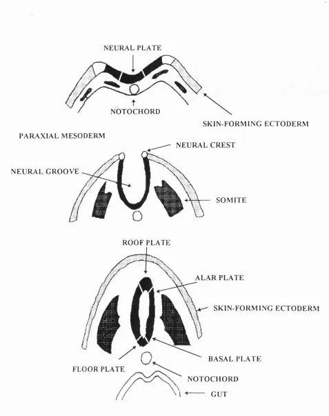
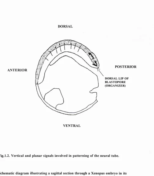
The neural plate forms as a result of these inductive events. It becomes elevated laterally, forming the neural folds, and depressed centrally, forming the neural groove (E6-8). These folds fuse at the midline, in a rostro-caudal sequence (neurulation ) to form the neural tube (E8-9) (Fig. 1.1). Cephalic regions o f the neural tube dilate to form the primary brain vesicles, whereas regions caudal to this form the future spinal cord. Regional patterning of the neural tube is thought to be established by the combined effects of vertical and planar signalling throughout gastrulation and neurulation (see Fig. 1.2 and Chapter 5 for more detail).
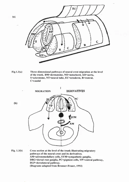
As the neural folds fuse, cells at the lateral edge of the folds delaminate from the developing neural tube, and migrate through the surrounding mesenchyme (E l0-E l 1). These are known as neural crest cells (Fig. 1.1) and give rise to diverse and numerous derivatives, ranging from melanocytes and cranial cartilage to adrenal chromaffin cells and the neurons and glial cells of the peripheral nervous system.
Neural crest cells are regionalized so that cells derived from different axial levels follow distinct migratory pathways. At the level of the trunk, two migratory pathways are present (see Fig. 1.3a). Neural crest cells migrate either ventrally through the anterior half of the somites, along which precursors to the sensory and
sympathoadrenal precursors pass, or dorsally underneath the ectoderm, as is the case for melanocyte precursors (Weston, 1963; Serbedzija et al., 1989) (E10-E13)
N E U R A L P L A T E
N O T O C H O R D P A R A X I A L M E S O D E R M
N E U R A L G R O O V E
S K I N - F O R M I N G E C T O D E R M N E U R A L C R E S T
S O M I T E
R O O F P L A T E
F L O O R P L A T E
A L A R P L A T E
S K I N - F O R M I N G E C T O D E R M
B A S A L P L A T E N O T O C H O R D I --- G U T
DORSAL
ANTERIOR
POSTERIOR
DORSAL LIP OF BLASTOPORE (ORGANIZER)
VENTRAL
Fig. 1.2. Vertical and planar signals involved in patterning of the neural tube.
EC
AO
Fig. 1.3(a) Three-dimensional pathways of neural crest migration at the level of the trunk. DM =dermatome, NO=notochord, AO =aorta,
S=sclerotome, NT=neural tube, EC=ectoderm , R=rostral, C=caudal
MIGRATION DERIVATIVES
(b)
PC
RG
VP
;VM
AM
Fig. 1.3(b) Cross section at the level of the trunk illustrating migratory pathways of the neural crest and its derivatives.
AM =adrenomedullary cells, SYM =sym pathetic ganglia,
DRG=dorsal root ganglia, PC=pigment cells, VP=ventral pathway, DLP=dorsolateral pathway.
Dorsal root ganglia (DRGs) form next to the neural tube (Fig. 1.3b) in a metameric manner, determined by the differential properties of rostral and caudal sclerotome (El 1-El2). The rostral sclerotome is not only permissive for neural crest migration (Keynes and Stern, 1984; Rickman et al., 1985; Bronner-Fraser, 1986) but also mitogenic for DRG precursor cells (Goldstein et al., 1990). It has recently been discovered in chick that the neural crest is not the only source of cells which comprise the sensory ganglia. A second wave of cells migrate away from the dorsal region of the spinal cord to the spinal ganglia after neural crest migration is complete (E l4) (Shama et al., 1995). This emigration occurs when cells of the dorsal spinal cord are still undifferentiated, leaving the spinal cord at the level o f the dorsal root entry zone (DREZ) and migrating through the dorsal roots to the DRGs.
Clonal analysis has clearly demonstrated that early migrating neural crest cells are either multipotential / bipotential progenitors or fully committed unipotential cells. Newly formed DRGs may also contain some multi / bipotential progenitor cells either o f the glial lineage [Schwann and satellite cell precursors (Le Douarin et al., 1991)], neuronal lineage [sensory and autonomic precursors (see Le Douarin and Smith,
1988)] or both.
At about the same time that neural crest cells take part in gangliogenesis, the
plates. An intermediate area will form between the alar and basal plates and give rise to pre-ganglionic sympathetic neurons.
Initially the non-neural, surface ectoderm is a simple monolayer o f cells (E8-E11), which differentiates into a bilayer (E l2 -E l4). However, as embryogenesis progresses, it forms a highly differentiated, stratified epithelium called the epidermis. During epidermal histogenesis some regions of epidermis become specialized instructive epithelia which control the outgrowth and morphogenesis of underlying mesenchyme e.g. the AER, epidermal facial primordia and odontogenic placode epithelia, whereas other regions o f epidermis, in the head region, form placode-derived components of the eye, ear and nose and neurons of several cranial sensory ganglia, e g vestibular and nodose ganglia.
RATIONALE FOR CHOOSING THE FOLLOWING ECTODERMALLY DERIVED TISSUES: FLANK EPIDERMIS, AER, DRGS AND NEURAL TUBE
THE GAP JUNCTION
Ultrastructural analysis of the gap junction
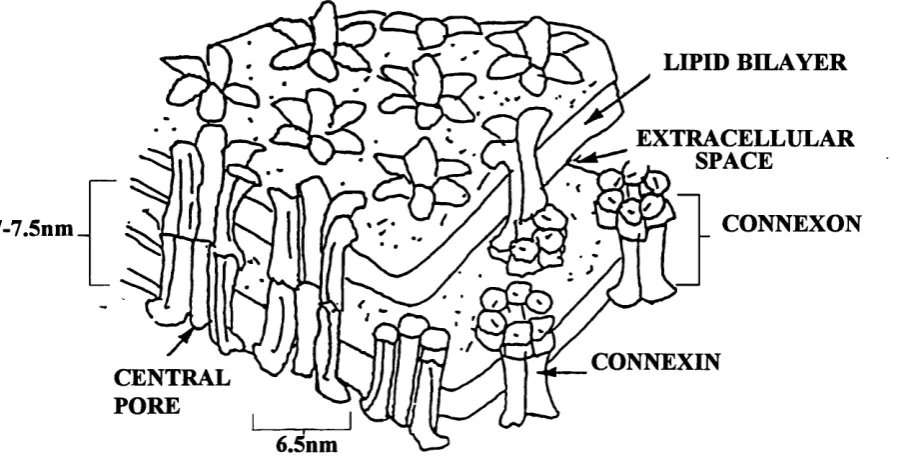
Gap junctions were first visualized ultrastructurally in electron micrographs, as sites where the plasma membranes of two apposed cells closely approach each other but are separated by a small "gap" of l-2nm (Robertson, 1963; Revel and Kamovsky, 1967). Freeze fracture replicas showed this structure to be a plaque-shaped, differentiated region of the plasma membrane consisting of intramembrane particles (connexons) on the P-fracture face and a complementary array o f pits or depressions on the E-ffacture face. Each particle, or connexon, appeared to have a pore in its centre which was especially well detected in deep etched preparations (Hirokawa and Heuser, 1982).
Further structural analysis of the gap junction has been made possible due to the development of gap junction isolation procedures; this has permitted the use of such techniques as negative stain (Casper et al., 1977; Unwin and Zampighi, 1980) or frozen-hydrated (Unwin and Ennis, 1984) electron microscopy. X-ray diffraction analysis (Makowski et al , 1977, 1984; Makowski, 1988) and atomic force microscopy (Hoh et al , 1991). They all support a model in which the gap junction plaque is composed of thousands of channels and a single gap junction channel is formed by the joining of two connexons, analogous to two cylinders, in the plasma membranes of two
adjacent cells. Each of these protein cylinders has been estimated to be 7-7.5 nm in length and 6.5 nm in diameter with an axial water filled channel of 1.5-2 nm in diameter which provides a direct aqueous route between the cytoplasm of the two coupled cells. It was deduced that each connexon is composed of six integral
LIPID BILAYER
EXTRACELLULAR ^ SPACE
CONNEXON 7-7.5nm_
CONNEXIN CENTRAL
PORE
6.5nm
Fig. 1.4. Schematic representation of the supramolecular structure of gap junctions as derived from X-ray diffraction (adapted from Makowski et al.,
Members of the gap junction polypeptide family: biochemical and molecular approaches
Sodium dodecylsulphate-polyacrylamide gel electrophoresis (SDS-PAGE) of gap junction preparations from various tissues has assisted in the isolation and
characterization of gap junction polypeptides. Gap junctions isolated from rat liver are composed of a 27-kD polypeptide and a 21-kD polypeptide (Hertzberg and Gilula,
1979; Traub et al., 1989), whereas gap junctions isolated from rat heart contain a 43- 47 kD polypeptide (Kensler and Goodenough, 1980). Edman's degradation sequencing has shown that liver (Nicholson et al , 1987) and heart (Gros et al., 1981; Nicholson et al., 1985) gap junction polypeptides show some homology in their amino acid sequence and on Western blots, some antisera raised to liver gap junctions were specific,
whereas others cross reacted with other gap junction polypeptides (Goodenough et al., 1988). These studies gave the first indication that gap junction polypeptides might be homologous enough to be encoded by genes of the same family, but divergent enough not to arise through alternative RNA splicing or post-translational modification (Nicholson et al , 1985).
cloning and polymerase chain reaction (PCR) amplification of genomic DNA has led to identification of further gap junction polypeptide types, giving a total, to date, of 12 in mammals (Kumar and Gilula, 1986; Paul, 1986; Beyer et al., 1987; Zhang and
Nicholson, 1989; Hoh et al., 1991b; Paul et al., 1991; Willecke et al., 1991a & b; Haeflinger et a l , 1992; Kanter et al., 1992), three in chick (Beyer 1990a; Musil et al.,
1990a) and three mXenopus (Gimlich et al , 1988; Gimlich et al , 1990; Ebihara et al., 1989).
Nomenclature of the gap juction polypeptide family
Due to discrepancies in the molecular weights of these polypeptides on SDS-PAGE (Green et al., 1988) a new nomenclature for these gap junction polypeptides has been proposed. The term "connexin" (Cx) was assigned for the gap junction polypeptide family, followed by its predicted molecular mass in kilodaltons deduced from its cDNA and an indication of the animal species from which the cDNA originates (Beyer et al., 1987, 1988, 1990). Thus, the rat liver polypeptides of 27-kDa and 21-kDa and the rat heart polypeptide of 43-kDa are termed rat Cx32, rat Cx26 and rat Cx43
respectively.
Recently, due to advances in the gap junction molecular genetics field, a further nomenclature has been proposed, which subdivides the gap junction family into two major classes: alpha (a) and beta (P). This categorization is based primarily upon differences of a motif in the M3 channel forming domain (see below), and the overall sequence of the connexin (Kumar and Gilula, 1992). This novel approach of gap junction classification offers an immediate source of information about the overall
Rat CX31
Class I
(Beta Group)
Class II
(Alpha Group)
Rat CX31.1
Rat CX26
Xenopus CX30
Rat CX32
Human CX32
Human CX43
Rat CX43
Chick CX43
Xenopus CX43
Rat CX33
Rat CX37
Rat CX46
Rat CX40
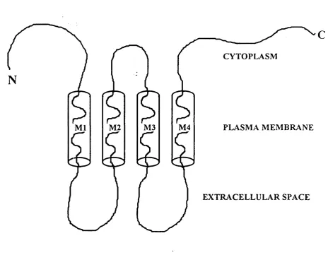
Topology of the connexin protein
The topology of the connexin has been postulated on the basis of hydrophobicity plots of the complete amino acid sequence deduced from its cDNA sequence (Unwin, 1989), and the validity of these results tested using site directed antisera in combination with specific proteases to amino acid residues (Zimmer et al., 1987; Milks et al., 1988).
CYTOPLASM
PLASMA MEMBRANE M4
M2 M3
M l
EXTRACELLULAR SPACE
c
Connexin distribution
Anatomical region Type of connexin (Cx)
Brain Cx26, Cx32, Cx43, Cx37, Cx31, Cx45
Neurons Cx32
Oligodendrocytes Cx32
Astrocytes Cx43
Pinealocytes Cx26
Ependyma Cx26, Cx43
Leptomeninges Cx26, Cx43
Skin Cx43, Cx26, Cx31, CX45, Cx31.1,Cx37
Lens Cx43, Cx46, Cx50
Epithelial cells Cx43
Fiber cells Cx46, Cx50
Cardiovascular system Cx43, Cx37, Cx40, Cx45, Cx46
Heart Cx43, Cx40, Cx45, Cx46
Smooth muscle Cx43, 0x37
Endothelium Cx43, Cx40
Lung Cx37, Cx40, Cx31, Cx43,
Smooth muscle Cx43
Testis Cx33, Cx43, Cx26, Cx32, Cx43
Leydig cells Cx43
Sertolli cells Cx33, Cx43
Pancreas Cx26, Cx32, Cx43,
Exocrine Cx26, Cx32
Endocrine Cx43
Placenta Cx31
Uterus Cx32, Cx26, Cx43
Endometrium Cx32, Cx26
Myometrium Cx43, Cx26
GAP JUNCTION GENES
Gene structure
The genomic organization of connexin genes has not yet been established for the entire polypeptide family. With the aid of Southern blots, PCR experiments and genomic DNA sequencing some characterization and genomic sequences have been published concerning the genes for rat Cx32 (Miller et al., 1988), human Cx43 (Fishman et al., 1991a), rat Cx31 (Hoh et al., 1991b), mouse Cx31 (Hennemann et al., 1992), mouse Cx37 (Willecke et al., 1991a), mouse Cx40 (Hennemann et al., 1992), mouse Cx26 and Cx32 (Hennemann et al., 1992). These genes all have a similar structure
consisting of two exons; the first is small, containing the 5'- untranslated sequence; the second contains the entire coding region which is uninterrupted by introns (Zhang and Nicholson, 1989; Beyer, 1990 a & b; Kanter et al , 1992).
Chromosomal location of gap junction genes
Chromosome mapping of human connexin genes places Cx46 and Cx26 on
GAP JUNCTION ASSEMBLY AND DISASSEMBLY
G ap junction assembly
Oligomerization of the connexin monomers to form a connexon hexamer is an important process after translation. Recent studies provide evidence that
oligomerization of Cx43 into connexons occurs in the Golgi apparatus, after leaving the E.R (Musil and Goodenough, 1993). This is a different process from that o f other major integral membrane proteins for which assembly in the HR is a prerequisite for transport through the secretory pathway. The reason for this disparity may be to prevent intracellular pairing of connexons and subsequent gap junction formation between membrane bilayers of the ER or Golgi ci sterna (Musil and Goodenough,
1993). Further studies have shown that the intramolecular disulphide bonds which link the extracellular loops in gap junctions are formed in the E.R. (Rahman et al., 1993) and that the presence of the extracellular loops is crucial for connexin trafficking (Evans, 1994).
The short latency with which cells can become coupled to one another, and the lack of effect of protein synthesis inhibitors on coupling, is indicative that connexons may exist in pools (Epstein et al., 1977), in a form (maybe vesicles) which permits rapid
is the "docking" of the extracellular loop of apposing connexins that promotes functional channel formation by the two connexons (Meyer et al., 1990).
Plaque formation occurs when gap junction channels located at cell-cell interfaces form very high-density clusters (Loewenstein, 1981; Yamasaki, 1990). Phosphorylation of the connexin protein appears to be an important step in the establishment of gap junctional plaques following connexon assembly, and may be induced by interactions
with the cell adhesion molecule E-cadherin (Musil et al., 1990b; Musil and Goodenough, 1991).
Gap junction disassembly
Gap junctions appear to be removed from the plasma membrane by an endocytotic mechanism, in which the entire gap junction is taken up into one of the coupled cells (Larsen, 1983; Mazet et al., 1985). Fusion of the plasma membranes gives rise to a double-walled vesicle which encapsulates the "annulated" gap junction, ultimately to be degraded within a lysosome.
It should be taken into account that the formation / removal of functional gap junctions depends on the rate of connexin turnover. The turnover of Cx43, Cx32 and Cx26 is very rapid (2-5hr) (Traub et al., 1989; Laird et al., 1991) and the extent of connexin expression will ultimately be determined by the rate of each step in connexin
BIOPHYSICAL PROPERTIES OF THE GAP JUNCTIONAL CHANNEL
Gap-junction-mediated communication between cells, at a gross level, is quantitatively assessed by measuring macroscopic junctional conductance or junctional permeability, which, in turn, is determined by the number of channels present in the gap junctional plaque, the unitary conductance of each channel and the open probability of channels.
Gap junction conductance
Using standard voltage clamp procedures, junctional conductance at a macroscopic level can be studied (see Bennett and Verselis, 1992; Saez et al., 1993) and single channel conductances can be measured using patch-clamp techniques. The unitary conductance for Cx32 is about 120-150pS (Eghbali et al., 1990; Neyton and
Trautmann, 1985), and for Cx43 the value is about SOpS (Veenstra and DeHaan, 1988; Burt and Spray, 1988; Rook et al., 1988). Cx26 has not been analysed to date.
Gap junction permeability
Although gap junction unitary conductance is modest, gap junctions are permeable to large ions and molecules. Gap junction permeability has been measured either directly by monitoring the intercellular transfer of intracellularly injected fluorescent dyes or indirectly by examining metabolic cooperation between cultured cells. Mammalian cells pass molecules up to 900 daltons and have channels that are able to discriminate between charged and neutral molecules, giving a functional channel diameter of 1.6- 2nm for neutral molecules (Loewenstein, 1966, 1978; see Peracchia and Girsch,
propidium iodide and ethidium bromide penetrate poorly between Cx31 and Cx32 transfectants, 4,6b Diamidino-2-phenylindole (DAPI) dihydrochloride showed less transfer through Cx31 and Cx43 and Neurobiotin was poorly transferred among Cx31 transfectants (Elfang et al., 1995).
Gating mechanisms of gap junctions
Gap junctions appear to undergo conformational changes which alter their
conductance. These changes are rapid and reversible and leave the fine structure of gap junctions, as seen by electron microscopy, virtually unchanged (Hanna et al., 1985).
Image reconstruction of isolated junctions using low-dose electron microscopy suggests closure of the channel is caused by tilting of connexin monomers (Unwin,
1989). The amphipathic alpha helix (M3), which is thought to line the channel, may tilt or rotate to move phenylalanine residues in such a way which would cause channel closure (Catterall, 1988).
Voltage dependency of gap junctions
Voltage can cause changes of conductance in many gap junctions, and single channel recordings reveal that gating is "all-or-none". Although the first example of voltage sensitivity was of the asymmetric type, in the rectifying (presumably heterotypic) gap junction of the crayfish giant synapse (Furshpan and Potter, 1959), subsequent studies
suggested that most gap junctions are electrically linear. However, in the early 1980's a symmetrical voltage dependency of a homotypic gap junction comprising Cx38, was described in amphibian blastomeres (Harris et al.,1981) and subsequently in ascidian embryos (Harris et al., 1983); in both, transjunctional voltage of either sign decreased junctional conductance. Cx37 acts in a similar way to Cx38, and both exhibit a voltage
role in mediating gap junctional communication during embryonic development arises from studies first carried out on amphibian embryos where gap junctions between blastomeres are higldy sensitive to transjunctional voltage ( Harris et al., 1981). Symmetrical voltage dependencies have also been exhibited by Cx32 in oocytes, transfected cells (Spray et al., 1991a) and hepatocytes (Spray et al., 1991b), and by Cx26 in oocytes; the degree of sensitivity, however, is very low and does not imply a physiological function. Cx43 in myocytes appears to be insensitive to weak voltage (Spray et al, 1985).
Cation gating of gap junctions
Turin and Warner (1977) were the first to demonstrate, by exposing early amphibian embryos to CO2 that an increase in cytoplasmic levels decreased junctional
conductance. This decrease in gap junction conductance was found to be totally reversible even after complete closure of junctional channels. pH sensitivity was subsequently shown to vary depending on the type of tissue involved (Turin and Warner, 1980; Spray et al., 1982) and to affect voltage dependency of gap junctions (see Spray et al., 1985; Peracchia and Girsch, 1985; Bennett et al., 1988).
An increase in the concentration of intracellular free calcium ions has also been shown to decrease junctional conductance in a variety of different species and tissues
(DeMello, 1975), although whether this effect occurs at physiological concentrations o f Ca^'*' remains controversial. The sensitivity of Ca^"*" ions appears to depend on the ambient pH in such a manner that as pH falls, the gap junction becomes less sensitive to rises in calcium.
extent of their intercellular spread. If physiological concentrations of Ca^'*' are not sufficient to regulate gap junctional conductance, Ca^'^dependent uncoupling may be important during pathological conditions, allowing a healthy cell to uncouple from a damaged cell.
MODULATION OF GAP JUNCTIONAL COMMUNICATION
The plasticity of gap junctional communication appears to be a result of a multitude of factors, potentially capable of modulating gap junctional communication at many stages. These include the transcription of connexin genes, the stability and translation o f connexin transcripts, connexin trafficking and insertion in to the plasma membrane, formation o f assembled plaques, regulation of channel function, and the removal and degradation o f junctions.
Modulation of gap junctional communication by regulating the amount or type of connexin expressed
This may involve alterations in the transcription rate, mRNA stability, translation and post-translational modifications (trafficking of connexins to the plasma membrane). These changes are slow compared to direct actions on the channel, which would change unitary conductance or gating, and are also likely to involve connexin
expressed is a feasible regulatory mechanism for modulating gap junction communication.
It would be beneficial if there was a system where a common signal could be used to produce a variety of effects depending on the tissue and connexin type involved. A candidate signalling molecule, shown to exert its effects after a few hours, is the second messenger cAMP. It increases gap junctional communication in various cell types (Saez et al., 1986; Flagg-Newton et al., 1981; Azamia et al., 1981; Weiner and Loewenstein, 1983; Kessler et al., 1984; DeMaziere and Scheuermann, 1988; Saez et al., 1990a) and decreases gap junctional communication in uterine smooth muscle cells (Cole and Garfield, 1986). Recent experiments demonstrate that the rate of connexin mRNA transcription (Stagg et al., 1990) and the stability of connexin transcripts (Saez et al., 1989; Stagg et al., 1990) are both important regulatory stages in the modulation o f macroscopic junctional conductance by cAMP. Consistent with there being a cAMP control mechanism for transcription, is the presence of a sequence, upstream to the encoding region for Cx32, corresponding to cAMP response elements (Miller et al., 1988).
Antibody blocking experiments (Keane et al., 1988) and cDNA transfection experiments (Mege et al., 1988) have given evidence that cell adhesion molecules (C AMs) promote the insertion of gap junctions into the plasma membrane. This has been further confirmed by the finding that in CAM deficient cell lines gap junction protein remained intracellularly compartmentalized (Musil et al., 1990b; Jongen et al.,
1991). Conversely, connexin expression has been shown to affect the type of cell adhesion molecule expressed (Eghbali et al., 1991), which further suggests that gap junctions and CAMs share very intimate and highly specific reciprocal relationships
However phospho-connexins are not an absolute requirement for connexon insertion into the plasma membrane (Lash et al., 1990; Werner et al., 1991) as, in the same way, CAMs are not an absolute requirement for gap junction phosphorylation (Berthoud et al., 1992).
Changes in the functional state of gap junction channels
Gating of gap junctions from open to closed states and vice-versa, as discussed earlier, can occur, as a result of changes in voltage, pH, calcium. In these cases the
macroscopic junctional conductance is ascribable to the open probability of each channel in the gap junction.
Subtle changes in junctional conductance, which involve alterations in unitary conductance are usually characterized by gap junctional phoshorylation [although an example of phosphorylation altering the open probability of channels has been
described (McMahon et al., 1989)]. These changes can occur in minutes and may be induced by growth factors, hormones and neurotransmitters (Loewenstein et al., 1981; Saez et al., 1990 a & b; Stagg and Fletcher, 1990; see Saez et al., 1993), often
triggering intracellular signalling cascades involving kinases. The effects of
phosphorylation on gap junctional conductance depend not only on the connexin and cell type but also on which kinase is activated. This accounts for the unpredictable effects of kinase activating agents on junctional conductance (see Saez et al., 1993)
Experiments examining the effects of phosphorylation on Cx3 2 junctional conductance reveal that stoichiometrical differences of the phospho-connexin depend on whether cAMP-dependent kinase (PKA) and protein kinase C (PKC) are involved (Saez et al.,
et al., 1990) its phosphorylation sites are at serine, tyrosine and threonine residues on the C-terminus (Swenson et al.,1990; Laird et a!., 1991; Lau et al., 1991). The single
channel conductance of Cx43 alters depending on its phosphorylation state (Moreno et al., 1992).
ROLES OF GAP JUNCTIONS IN ADULT TISSUES
General roles of gap junctions
Tissue homeostasis
Gap-junction-mediated intercellular communication appears to play a fundamental role in coordinating tissue activity. Tissue homeostasis and integrity are established by allowing the exchange of small molecules between cells. This phenomenon, called " metabolic cooperation" (Subak-Sharpe, Burke and Pitts, 1969), occurs between cells and can be put to use in a variety of ways. For example, local homogeneity amongst cells is important when cells differ in their accessibility to a blood supply [an extreme example of this occurs in the lens, where gap junctions account for as much as 25% of the cell surface, compensating for the lack of blood supply to the lens (Benedetti et al.,
1976)].
obvious candidate which could act as an intercellular communicator in response to such extracellular signals (Flagg-Newton et al., 1981).
Growth control
In 1968, Furshpan and Potter were the first to postulate that a loss in gap-junction- mediated intercellular communication contributes to the formation of uncontrolled growth. By 1979, Loewenstein had reviewed this issue, presenting much evidence in favour of this hypothesis. Further, recent authors have confirm the viability of this hypothesis (Trosko et al.,1988; Yamasaki, 1988; Neveu et al., 1990), and many results seem to fit into the general inverse relationship between rate of growth and the extent of gap junction communication (e.g. Jursnich et al., 1990; Naus et al., 1992).
However, the simplicity of this statement should be treated with caution as it has been demonstrated that several kinds of tumour cells are coupled by gap junctions
(Sheridan, 1970) and many malignant cell lines are coupled, although to a reduced extent to that normally expected (Mege et al., 1988; Eghbali et al., 1990). Recent studies comparing connexin expression in malignant mouse hepatocytes (Stutenkemper et al., 1992) and malignant human hepatocytes and epidermal cells (Wilgenbus et al.,
Role of gap junctions in non-neural excitable cells
Impulse propagation
In non-neural excitable cells, the main function of gap junctions appears to change from one of metabolic cooperation to that of intercellular flow of current, by the maintenance of low resistance pathways between cells. An example exists in the / mammalian myocardium where these low resistance pathways allow current to flow from the sino-atrial node, through the Purkinje conductance system, ultimately leading to rhythmic contractions of the ventricles (Weidmann, 1952). The ability of intestinal smooth muscle to generate peristaltic waves (Barr et al., 1968) and of uterine
myometrium to synchronize contractile activity at the time o f parturition (Dahl and Berger, 1978; Garfield et al., 1978), are also dependent on the formation of low resistance pathways for impulse propagation.
Ro!e of gap junctions in the adult nervous system.
In neurons
The first unequivocal experiments suggesting the existence of electrotonic coupling in phylogenetically higher regions of the mammalian CNS came from electrophysiological studies on the mesencephalic root of the Vth cranial nerve in rat (Baker and Llinas,
1971), the vestibular nucleus of the rat (Korn et al., 1973) and inferior olive of the cat (Llinas et al., 1974; 1981). The strength of coupling detected in the mammalian CNS was o f a much lower order than that experienced from the initial studies in
invertebrates and lower vertebrates. Gap junction plaques, electrotonic coupling and / or dye coupling have subsequently been demonstated in the paraventricular nucleus (Andrew et al., 1981), hippocampal pyramidal neurons and dentate granule cells (Andrew et al., 1982; Nunez et al., 1990).
Recent immunohistochemical localization studies have detected gap junction protein like immunoreactivity in the CNS, to a greater extent than predicted from physiological or ultrastructural studies (Nagy et al., 1988; Dermietzel et al., 1989; Shiosaka et al.,
1989; Yamamoto et al., 1989). Connexin mRNA has been mapped using in situ hybridization and results suggests a widespread, complex regional organization of gap junctions throughout the CNS, Cx32 being the main neuronal connexin (Micevych and
Abelson, 1991). In addition to mediating synchronous activity of active cells, as in the hippocampus (Dudek and Snow, 1985; Llinas, 1985), inferior olive (Llinas, 1974,
1981) and spinal nucleus of the bulbocavemosus of males (Matsumoto et al., 1988), the characteristic weak electrotonic coupling of neurons in the mammalian nervous system appears to be better suited in mediating more subtle interactions. For example, they may be required to ensure that the activity of no individual neuron prevails (Peinado et al., 1993a & b).
The high abundance of connexin expression in defined neuronal subcompartments (Dermietzel et al., 1989; Nagy et al., 1988; Micevych and Abelson, 1991) further suggests a possible role for gap junctions in the adult brain, of setting up
hippocampus (Christie et al., 1989) and is well illustrated in the retina In the latter case, neuronal types form highly organized a rrays of tracer-coupled, cell specific mosaics in the plane of the retina (Vaney et al., 1991; Vaney, 1994). A functional significance of cell type specific coupling in the retina may be to regulate common cell phenotype. However, their main function appears to be that of regulating receptive field size (Lasater and Dowling, 1985; McMahon et al., 1989, 1992a). The finding that gap junctions possess different properties and respond differently to intracellular and extracellular control mechanisms, depending on which cells they couple (Cook and Becker, 1995) gives them the potential to influence every stage of the visual
processing sequence in the retina with great flexibility.
It has recently been demonstrated that second messenger molecules can be elevated in the postsynaptic cell as a consequence of direct diffusion through gap junctions. This suggests a further role of gap junctions at mixed (chemical and electrical) synapses, in modulating neurotransmitter release, and they may even play a role in the generation of long term potentiation (Yang et al., 1990).
The finding that gap junctional communication in the CNS can be altered in response to various functional factors, such as neurotransmitters [e.g. catecholamines effect Cx32 expression in the striatum (Fisher et al., 1990), coupling strength in the retina (Lasater and Dowling, 1985; McMahon et al., 1989, 1992a) and neostriatum (Cepeda,
1992)] and hormones [e.g. steroids affect dye-coupling between hypothalamic
paraventricular nucleus magnocellular neurons (Cobbett et al., 1987), and the size and number of junctional plaques between lumbar motoneurons in the spinal nucleus of the bulbocavemosus (Matsumoto et al.,1988)], suggests the functional nature of gap junctions in the CNS is one of great dynamicity, highly responsive to a variety of
In macroglia
It is now known that macroglial cells play a large number of roles in the CNS (see Barres, 1991) and are the most prominently coupled elements in the CNS (Mooren and Nelson, 1978; Gutnick et al., 1981). They utilize gap-junction-mediated
intercellular communication to ensure that extensive communication can occur. Astrocytic tissue o f the CNS examined by immunolabelling (Nagy et al., 1988; Dermietzel et al., 1989; Yamamoto et al.,1990; Batter et al., 1992), in situ
hybridization (Micevych and Abelson, 1991) and dye-coupling (Batter et al., 1992) studies have revealed abundant, widespread, but heterogeneous Cx43 expression and coupling patterns. This region-specific presence of gap junctions may subserve functional specializations within the CNS and lead to the establishment of largely separated syncytial compartments (Yamamoto et al., 1990; Batter et al., 1992).
Astrocytic gap junctions are thought to play an important role in the generation of slow electrical fields associated with neural activity (Castellucci and Goldring, 1970; Ransom and Goldring, 1973; Ransom, 1974). In tissue homeostasis they may buffer activity surrounding active neurons by providing intercellular pathways either to assist in the disposal o f into the perivascular compartments (Gardner-Medwin et al., 1983; Newman, 1986) or to establish an adjustable buffer sink for excess K""" from particularly active neurons (Kettenmann and Ransom, 1988).
in response to neuronally released substances (Dani et a ! , 1992), further suggesting that gap-junction-mediated interactions may play an active role in neuromodulation (Giaume et al., 1991).
Gap junctions are also present in oligodendrocytes (Massa et al., 1984; Kettenmann and Ransom, 1988) and consist of Cx32 protein (Dermietzel et al., 1989; Micevych and Abelson, 1991). Electrophysiological analysis o f gap junctions in cultured
oligodendrocytes suggests that oligodendrocytes are widely coupled by gap junctions, but display only weak electrical interactions; such junctions would be more selective to smaller metabolically active metabolites (Kettenmann and Ransom, 1988), and
suggests that K""" buffering may be more suited to the highly coupled syncytium present in astrocytes (Kettenmann and Ransom, 1988). Oligodendrocytes have also been shown to be functionally coupled to astrocytes in vivo, their gap junctions being heterotypic (Robinson et al., 1993).
TH E ROLE OF GAP JUNCTIONS IN DEVELOPMENT
The development of a multicellular adult organism from a single cell is an intricate process, wliich involves prodigious organisation and the implementation of a precisely regulated series o f events. During early embryonic development, events are
remarkably similar despite the variety of forms that exist across the animal phyla. Extensive cell-cell interactions are essential during development and much correlative evidence suggests gap junctions may play a vital role in this process.
The hypothesis that direct intercellular communication between embryonic cells might provide a channel for the exchange of information used to direct development was originally postulated as early as 1966 by Potter, Furshpan and Lennox when they detected electrical coupling between cells of the squid embryo and correctly attributed this phenomenon to gap junctions. They found ubiquitous communication between cells during early development, but, as development progressed, cell populations within an embryo became increasingly more specialized and uncoupled from one another. Since these pioneering experiments, electrical and / or dye-coupling between cells in the early embryo has been confirmed in a number of animals at a variety of
developmental stages, including invertebrates, such as the starfish Asterias (Tupper and Saunders, 1972) and the mollusc f ( D o r r e s t e i j n et al., 1982), amphibians such as the newt Triturus (Ito and Hori, 1966) and the frog Xenopus (Slack and Parmer, 1969), chick (Sheridan, 1968) and mammals such as the mouse (Lo and Gilula, 1979a& b) and the human (Dale et al., 1991).
Role of gap junctions in mediating global signals: embryonic patterning
to acquire its own positional identity and ultimately to differentiate according to its identity (Wolpert, 1971).
Gap junctions have been suggested to play a role in this global control of embryonic patterning, by providing a direct, intercellular route for the passage of these
morphogens. The pathway provided by gap junctions has the potential to transfer such morphogens if they are small enough, but unfortunately little is known about the nature of these chemicals. So far in only one species, the invertebrate Hydra, have these elusive substances been partially identified as a hydrophilic nonpeptide with a molecular size of around 500 daltons with head inhibitor properties (Schaller and Bodenmuller, 1981). Retinoic acid (RA) may be a naturally occurring morphogen (Tickle et al., 1982; Thaller and Eichele, 1987), however, since it is a hydrophobic molecule it would not need to move through gap junctions. There is evidence, however, that it can affect gap junction permeability (Mehta et al., 1989), and it has recently been demonstrated that RA also affects Cx43 expression at the
posttranscriptional level (Bex et al., 1995). Thus RA may have an indirect effect on embryonic patterning by gap junctions.
been used extensively to examine the role of gap junctions in pattern formation (Lo, 1985). The organization of the wing imaginai disk into a number of discrete
multicellular domains has been confirmed using lineage analysis techniques (Garcia- Bellido et al., 1973). The finding that these lineage compartments are polyclonal in origin raises the interesting question of whether restrictive communication
compartments play a role in insect pattern formation by providing the basis for the formation of these lineage compartments. Dye-coupling studies demonstrated that, as in Oncopeltus and Calliphora, communication restrictive boundaries coincided with lineage restriction boundaries (Lawrence, 1973). However, in the engrailed homeotic mutant of Drosophila, although the normal A / P lineage border was not observed (Morata and Lawrence, 1975), communication restrictive compartments remained the same as in the wild type (Lo, 1985), implying that lineage compartments are not responsible for the formation of communication compartments but that communication compartments may be responsible for establishing lineage compartments
In the early mouse embryo, communication compartments exist between the inner cell mass (ICM) and trophectoderm (Lo and Gilula, 1979b), and at a later stage, cells in the embryo proper and yolk sac endoderm form separate communication restrictive
compartments from that of the ectoplacental cone and extraembyonic ectoderm (Kamili and Lo, 1988). Whether these communication compartments play a role in setting up morphogenic fields, or are simply a consequence of progressive restrictions in gap junction communication resulting from the determination of cell fate (see below) is not clear.
by the limb morphogen, RA (Tickle, 1991). Thus gap junctions, RA, and Hex genes may act in concert, to establish correct tissue patterning of the limb. Consistent with the graded expression of Cx43 are scrape loading experiments which reveal that dye transfer is greater in posterior compared to anterior limb bud mesenchyme (Coelho and Kosher, 1991).
Despite the large body of information suggesting that gap junctions play a role in embryonic patterning much of the evidence, until recently, has been merely correlative or circumstantial. However, the first conclusive evidence was produced by Warner and colleagues in 1984. A polyclonal antibody raised against Cx32, was injected into a single cell, on the dorsal side of 8 cell stage Xenopus embryos. When these embryos were allowed to develop and examined at stage 36, specific developmental defects were observed and structures which fate maps indicate would normally form from the injected cell, for example the eye and part of the brain, were misplaced or even totally absent. Further antibody blocking experiments in preimplantion mouse embryos (Lee et al., 1987), in developing chick limb (Allen et al , 1990), and regenerating hydra (Fraser et al., 1987) also resulted in patterning defects.
Role of gap junctions in mediating local interaction.
During embryogenesis local tissue interactions are a vital part of development. One example is that of induction, whereby one tissue induces the specific differentiation of another tissue in close proximity, either by releasing an extracellular inducing signal or through direct cell-cell contact.
other. Conjugates left to develop until the late neurula stage accumulate a muscle- specific gene transcript diagnostic of their mesoderm fate in animal pole cells. It was shown that if gap junction antibodies were loaded into vegetal pole cells before making conjugates, electrical coupling between these two tissues was prevented, but,
mesodermal cells were still induced, suggesting gap junctions are not involved in passing the mesoderm inducing signal between conjugates.
Another example of this phenomenon, is found in the limb bud where the apical ectodermal ridge (AER) signals to the underlying mesoderm (secondary induction). Both of these tissues are dye-coupled, but not to each other (Laird et al., 1992), suggesting that the signal probably does not pass between tissue types by gap junctions, but that gap junctions might play a role in relaying the signal transduction
cascades triggered by these inductive signalling molecules.
Correlations between gap junction communication and cell fate restriction
Gap junction communication might parallel the progressive determination of cell fate, thus, cells differentiating in a similar direction might be able to communicate freely, where communication might become restricted between cell populations with different fates. This phenonemom, first postulated by Potter et al., 1966 has since been found to occur in various developing systems. For example, in Xenopus development,
uncoupling of the myotome from undifferentiated somite mesenchyme parallels progressive differentiation (Blackshaw and Warner, 1976). In molluscan embryos, Lymnea and Patella, dye-transfer is initially very extensive, but becomes progressively restricted between cells of different presumptive lineages (Dorresteijn et al., 1983; Serras and Biggelaar, 1987).
first appear in the mouse at the 8-cell stage just before the time of compaction (Ducibella et al., 1975). It is at this time that blastomeres lose their totipotency and become determined to differentiate into two cell types; the trophectoderm and inner cell mass (ICM). Initially, electrical and dye-coupling is extensive between the trophectoderm and ICM (Lo and Gilula, 1979a), but as time progresses, dye-transfer becomes restricted to cells of the same tissue (Lo and Gilula, 1979b), and later still, confined to small groups of cells within either the trophectoderm or ICM (Lo and Gilula, 1979b). During gastrulation, the ICM differentiates into three layers:
presumptive ectoderm, mesoderm and endoderm. Although electrical coupling persists between germ layers, dye-coupling studies reveal that not only is dye-transfer restricted between germ layers but it is also compartmentalized into box-like domains within the ectoderm and mesoderm (Lo and Gilula, 1979b).
Correlations between cell differentiation and gap junction expression are also found in the developing nervous system, for example, in the developing spinal cord neuronal differentiation occurs in a rostro-caudal and ventro-dorsal direction; this correlates temporally and spatially with the expression pattern of Cx43 (Ruangvoravat and Lo,
1992). Also in the developing amphibian retina gap junctions disappear from the central portion of the retina at the exact time of specification (Dixon and Cronly- Dillon, 1972).
RO LE OF GAP JUNCTIONS IN THE DEVELOPING NERVOUS SYSTEM
Gap junctions appear to play similar roles in the developing nervous system to that in development in general, however, there are cases in which gap junction communication takes on a unique role, specific to the complex needs of the developing nervous system
Role of gap junctions in patterning the developing nervous system.
There are many observations which suggest that gap junctions play a role in patterning in the developing nervous system, as mentioned above, by mediating morphogenic gradients within a developmentally significant domain. Dye-transfer studies reveal that communication compartments coincide with the segmental units of the developing hindbrain, the rhombencephalic neuromeres (rhombomeres), of the neural tube (Martinez et al., 1992). It has been shown that rhombomeric cells transfer LY or biocytin between each other, but that dye does not transfer from rhombomere cells to boundary cells or between boundary cells, although electrical coupling between rhombomeric cells was not affected by their position. The communicational isolation of interrhombomeric boundaries relative to the transfer of small molecules may represent an early developmental mechanism of patterning specification within the developing hindbrain. However, this type of communication restriction does not appear to respect cell lineage compartments [lineage studies have shown that after boundary formation, clonal restriction occurs at the midline of the inter-rhombomeric boundary not the rhombomere-boundary interface (Fraser et al., 1990)] as
Further evidence suggesting that gap junctions may play a role in patterning of the CNS, comes from studies where connexin mRNA expression has been mapped
throughout the developing brain by in situ hybridization (Ruangvoravat and Lo, 1992). Using this sensitive method of detection it was found that Cx43 mRNA is expressed in large amounts within discrete domains. Interestingly, at the midbrain / hindbrain border, Cx43 coincides with wnt-1 expression (Wilkinson et al., 1987). Wnt-1 belongs to the Wnt gene family of cell signalling molecules and is the mammalian homologue of the Drosophila segment polarity gene wingless (see McMahon, 1992b). It has been found not only to be important in patterning of the brain (Thomas and Capecchi,
1990), but also to increase gap junction communication (Olson et al., 1991), indicating that these molecules may be linked. Gradients of Cx43 mRNA coincide with
expression patterns of Hox genes in certain regions of the brain (Gaunt, 1991), again suggesting that these molecules may be functionally related.
Role of gap junctions in inductive interactions in the developing nervous system
To date, there is no direct evidence to suggest that gap junctions provide a passage for inducing signals from the notocord to the neural tube, and although electrical coupling between these two tissues exist (Sheridan, 1968; Warner, 1973), direct contact
Roles of gap junctions which are specific to the needs of the developing nervous system
In the nervous system at later stages of embryonic development and early postnatal life, there is an additional developmental requirement: the complex establishment of neuronal connectivity. Much evidence suggests that gap junctions play a major role in resolving this developmental difficulty. I will consider ways in which gap junctions may participate in the establishment of neuronal circuitry, firstly by considering their function at a local level which involves discrete, transient cell-cell interactions, and then turning to their possible functioning at a more global level, where their effects are longer lasting, and may play a role in the organisation and refinement of functional units as a whole.
Examples of transient interactions mediated by gap junctions
During axogenesis a variety of guidance cues are used by the axon to establish correct connectivity with its target. In the grasshopper embryo, the growth cones of pioneer sensory neurons are guided to their CNS targets by interacting with a succession of special "guidepost cells" en route (Bate, 1976; Bentley and Keshishian, 1982).
Transient gap-junction-mediated dye-transfer has been detected between the guidepost cells and pioneer neurons (Bentley and Keshishian, 1982: Goodman et al., 1982), and between later growing axons and pioneer axons (Goodman et al., 1982). In the leech embryo, Hirudo medicinalis, oppositely directed axons of homologous anterior pagoda (AP) neurons overlap extensively with each other, inhibiting one another’s growth (Gao and Macagno, 1987a &b). The presence of tracer and electrical coupling between these neurons suggests that gap junctional communication may mediate the exchange of signals which are involved in this récognition-inhibition process (Wolszon et al., 1994).
revealed that the gap junctional contacts between guidepost cells and growing axons mediate the transfer of Ca^"^ (Bently et al., 1991). However, during pathfinding of primary motoneurons in zebrafish embryos, where transient coupling exists between pioneer and later axons, ablation of pioneer neurons suggest that coupling is not essential during this aspect of axon guidance (Eisen et al., 1989). Further, in a study involving Xenopus retinal ganglion cells during the period of axogenesis and target recognition, dye-coupling was not observed between the growth cones of growing axons and putative guidance cells in their pathway (Holt, 1989). However, in this study, gap junctional communication was examined only using the relatively large molecular weight dye LY; studies involving axogenesis of the AP neurons in leech embryos demonstrated that whilst a small tracer [5-HT (5-hydroxytryptamine) visualized using anti-5-HT immunohistochemistry] passed through gap junctions LY did not (Wolszon et al., 1994), leaving open the possibility that this could also be the case in Xenopus retinal ganglion cells.
Another form of transient coupling appears to occur in cultures consisting o f myocytes and neurons from Xenopus embryos. It was found that electrical and dye-coupling was present between these two tissues during stages of early synaptogenesis (Allen, et al.,
1986). This may allow cells to "test" each other for the purpose of synaptic specificity.
Examples of long-term interactions mediated by gap junctions