THESIS SUBMITTED FOR THE DEGREE OF
MASTER OF PHILOSOPHY IN BIOPHYSICS
OF THE UNIVERSITY OF LONDON
CHRISTINE LESLEY KATHERINE ASTIN BSc, FBCO, DCLP, FAAQ
DEPT. OF VISUAL SCIENCE
INSTITUTE OF OPHTHALMOLOGY
UNIVERSITY OF LONDON
BATH STREET, LONDON EClV
All rights reserved
INFORMATION TO ALL USERS
The quality of this reproduction is dependent upon the quality of the copy submitted.
In the unlikely event that the author did not send a complete manuscript
and there are missing pages, these will be noted. Also, if material had to be removed,
a note will indicate the deletion.
uest.
ProQuest U076010
Published by ProQuest LLC(2016). Copyright of the Dissertation is held by the Author.
All rights reserved.
This work is protected against unauthorized copying under Title 17, United States Code.
Microform Edition © ProQuest LLC.
ProQuest LLC
789 East Eisenhower Parkway
P.O. Box 1346
National health services are receiving increased pressure to
improve cost efficiency and quality of patient treatment outcome.
Hence it is useful to re-evaluate corneal changes following
cataract extraction, with a view to minimising astigmatism and
corneal contour stabilisation time, such that patients can more
rapidly obtain their best corrected vision and return to
Independent life.
In a prospective study of 140 patients undergoing routine
extracapsular cataract extraction, the changes in refraction, in
corneal curvature as determined by keratometry and keratoscopy,
and in regional corneal thickness were closely monitored
preoperatively and for 6 months postoperatlvely. Two groups were
compared, one (C) receiving a corneal incision and nylon sutures,
the other (L> receiving a limbal incision and silk sutures.
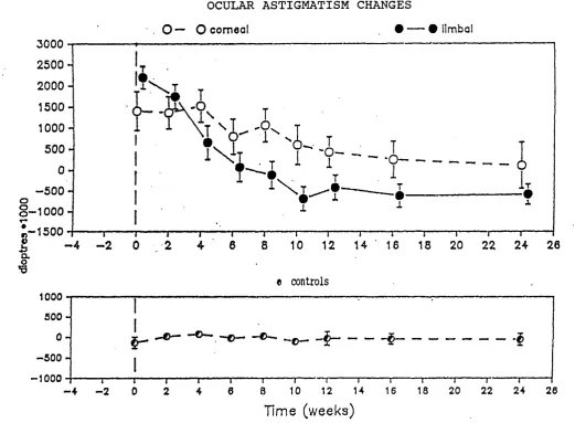
Both groups demonstrated a large 'with the rule' (steep
meridian vertically) astigmatic shift immediately
postoperatlvely. The (L) group showed faster stabilisation of
refractive (8 to 10 weeks) and corneal astigmatism (8 weeks), and
a moderate final mean 'against the rule' astigmatism value. The
<C) group remained as 'with the rule' and stabilised later for
refractive <12 weeks) and corneal astigmatism <16 weeks).
variability, which was also shown by the two control groups.
Although greater corneal thickness change had been anticipated
for the (C) group than the vL) group, no significant difference
was found between the two groups even by their stabilisation time
of about 10 weeks postoperatively. Only when comparing overall
changes was C O group found to show the ,greater amount.
Using the results, a flow chart guide was created for
consideration by surgeons to help produce the most desirable
optical outcome. A predictive second study of 45 patients
demonstrated the valuable advantage of using this chart in
conjunction with preoperative measurements, causing the majority
of the patients to obtain minimal or appropriately balanced
ocular astigmatism by 6 to Ô weeks and therefore a more
satisfactory optical result.
KEY WORDS: cataract extraction, corneal astigmatism, corneal
Page
TITLE PAGE 1
ABSTRACT 2
CONTENTS 4
LIST OF FIGURES 10
LIST OF TABLES ^ 13
ACKNOWLEDGMENTS 15
ABBREVIATIONS 16
CHAPTER 1 INTRODUCTION
1.1 Reasons for this study of corneal parameters 17
1.2 Basic corneal anatomy and physiology 21
1.2.1 Corneal anatomy 21
1.2.2 Corneal transparency 23
1.3 Corneal astigmatism 23
1.3.1 Types and description of astigmatism 24
1.3.2 Changes in astigmatism 29
1.4 Models of the cornea 31
CHAPTER 2 MANAGEMENT OF APHAKIA
2.1 Visual outcome in aphakia 33
2.1.1 Assessment of visual acuity and refraction 33
2.2 Corneal response to cataract surgery 36
CHAPTER 3 ASTIGMATISM AFTER CATARACT EXTRACTION SURGERY
3.1 Review of literature 40
3.2 Surgical techniques ot cataract extraction 42
3.2.1 Intracapsular cataract extraction (ICCE) 42
3.2.2 Extracapsular cataract extraction ΠC C E ) 42
3.2.3 Phacoemulsification 43
3.2.4 Aspiration 43
3.3 Surgically Induced astigmatism 44
3.3.1 IncIsion 44
3.3.2 Surgical technique 45
3.3.3 Tissue healing 48
3.3.4 Sutures 49
3.4 Preoperative astigmatism 52
3.5 Current methods to reduce astigmatism 53
3.5.1 During surgery 53
3.5.2 Postoperatively 53
CHAPTER 4 KERATOMETRY
4, 1 History 55
4.2 Keratometer design 56
4.3 Sources of error in keratometry 63
5.1 Methods of assessing corneal topography 68
5.2 Photokeratoscopy 69
5.2.1 Introduction 69
5.2.2 Advantages and disadvantages of photokeratoscopes 70
5.2.3 Prerequisites for reliable photo^eratography 71
5.2.4 Autocollimation 72
5.3 Photoelectronic keratoscopy PEK 73
5.3.1 PEK instrument 73
5.3.2 Corneal analysis of PEK photograph 78
CHAPTER 6 PACHOMETRY
6.1 Pachometry description and history 84
6.1.1 Introduction 84
6.1.2 History 84
6.2 Factors affecting reliability of measurements 87
6.2.1 Reliability factors 87
6.2.2 Ultrasonic versus optical pachometer 90
6.2.3 Angle Kappa 91
6.3 Variations in normal corneal thickness 92
6.3.1 Age and gender 92
6.3.2 Diurnal variation 93
6.3.3 Menstrual related variation 93
6.3.4 Osmotic effect 94
6.4 Use ot pachometry on abnormal eyes and after surgery 95
6.4.1 To judge treatment effect 95
6.4.2 Diagnostic aid 97
CHAPTER 7 EXPERIMENTAL PROCEDURE
7.1 Patient sample and measurement frequency 98
7.2 Refraction and corrected visual acuity 101
7.3 Keratometry procedure 102
7.4 Keratoscopy procedure 104
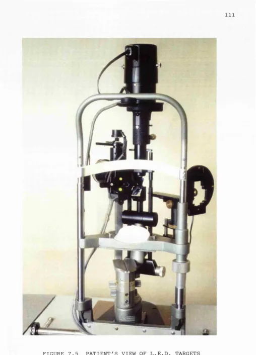
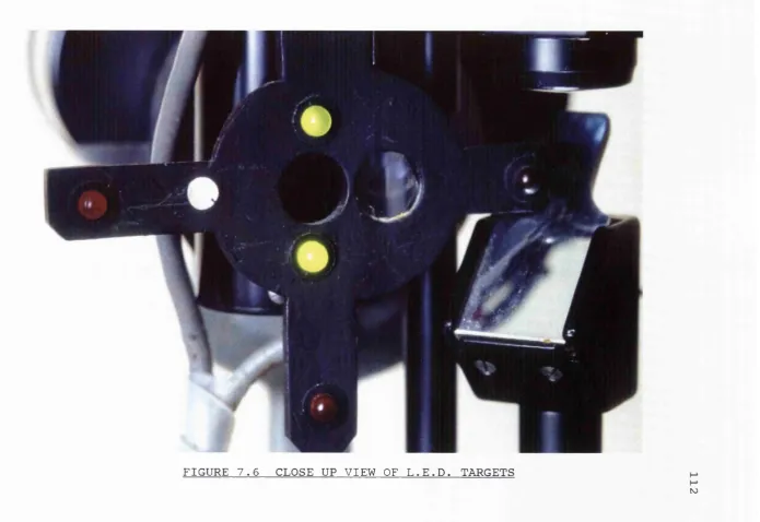
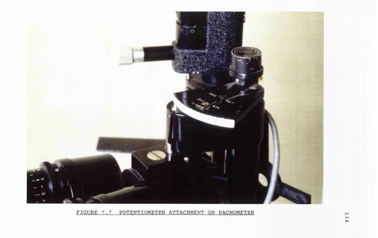
7.5 Regional pachometry procedure 106
7.5.1 Description 106
7.5.2 Calibration 113
7.5.3 Regional pachometry 115
CHAPTER 8 PART I CORNEAL TOPOGRAPHY RESULTS
8.1 Calibration results 117
8.1.1 Calibration of keratometer 117
8.1.2 Calibration of PEK 117
8.2 Description of patient groups and data analysis 118
8.2.1 Patient groups 118
8.2.2 Data analysis 120
8.3 Ocular astigmatism shown by refraction 123
8.4 Corneal astigmatism shown by keratometry 126
8.6 Horizontal shape factor 145
8.7 Vertical shape factor 150
CHAPTER 8 PART II CORNEAL THICKNESS RESULTS
8.8 Corneal thickness 156
8.8.1 Calibration of pachometer ^ 156
8.8.2 Control and Non-operated groups corneal thickness 157
8.9 Patient groups corneal thickness 159
8.9.1 Superior region 159
8.9.2 Central region 162
8.9.3 Inferior region 164
8.9.4 Nasal region 167
8.9.5 Temporal region 170
CHAPTER 8 PART III PARAMETER RELATIONSHIPS
8.10 Parameter relationships 174
8.10.1 Parameter comparison 174
9.1.1 Flow chart design 178
9.1.2 Patient groups and experimental procedure 180
9.2 Results 183
9.2.1 Astigmatism by retraction and keratometry 183
9.2.2 Corneal thickness ^ 192
9.3 Discussion 200
CHAPTER 10 DISCUSSION
10.1 Introduction 205
10.2 Considerations in performing measurements 207
10.2.1 Problems in monitoring 207
10.2.2 Comparative usefulness of parameter measurement 208
10.3 Response of the control groups 209
10.4 Response of the patients 210
10.4.1 Corneal topography 210
10.4.2 Corneal thickness 215
10.5 Recommendations for clinical management and research 219
10.5.1 Clinical management 219
10.5.2 Suggestions for further research 221
REFERENCES 223
LIST OF FIGURES
1.1 Cross section ot basic optical apparatus of the eye 22
1.2 Diagrammatic meridional section of the human cornea 22
1.3 Corneal astigmatism 25
1.4 Astigmatism foci 27
1.5 Letter 'O' distorted as by astigmatism 28
2.1 Calculation of effective lens power 35
2.2 Optical correction of aphakia 37
3.1 Incision site 46
3.2 Suture techniques 51
4.1 Optical principle of the keratometer 57
4.2 Image doubling by prism 58
4.3 American Optical keratometer 60
4.4 Mire image reflected by the cornea 61
4.5 Telescope apertures 62
4.6 Observers view of doubled mire images 62
5.1 Wesley Jessen photoelectronic keratoscope PEK 75
5.2 Corneal photograph using the PEK 76
5.3 Problems with curved image plane 77
5.4 Diagram of the Wesley Jessen PEK 77
5.5 Computer analysis of PEK photograph 80
5.6 Central curvature and shape factor 82
6.1 Jaegers pachometry method 86
6.2 Microscope with Haag Streit pachometer 88
LIST OF FIGURES (continued)
7.1 Keratometer in position 103
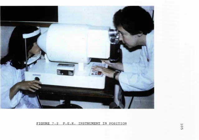
7.2 PEK instrument in position 105
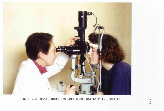
7.3 Haag Streit pachometer and slitlanip in position 107
7.4 Observers view ot' split image in pachometry 109
7.5 Patients view of LED targets ' 111
7.6 Close up view of LED targets 112
7.7 Potentiometer attachment on pachometer 114
8.1a Ocular astigmatism changes related to time 125
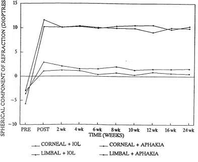
8.1b Spherical component of refraction related to time 127
8.2.1 Corneal astigmatism changes related to time 129
8.2.2(a-j) Distribution of astigmatism axis 131-140
8.3 Corneal astigmatism (PEK) changes related to time 142
8.4 Horizontal shape factor changes related to time 146
8.5 Vertical shape factor changes related to time 151
8.6 Superior corneal thickness changes related to time 160
8.7 Central corneal thickness changes related to time 163
8.8 Inferior corneal thickness changes related to time 165
8.9 Nasal corneal thickness changes related to time 168
8.10 Temporal corneal thickness changes related to time 171
9.1 Ocular astigmatism and corneal astigmatism
related to time 188
9.2 Ocular astigmatism and corneal astigmatism
LIST OF FIGURES (continued)
9.3 Superior corneal thickness related to time 193
9.4 Central and Inferior corneal thickness related 194
to time
9.5 Nasal and temporal corneal thickness related to time 195
9.6 Superior corneal thickness changes related to time 196
9.7 Central and inferior corneal thickness changes
related to time 197
9.8 Nasal and temporal corneal thickness changes
LIST OF TABLES IN APPENDIX
3. 1 Studies on astigmatism following cataract extraction 252
3.2 Clinical merits ot various cataract extraction methods257
3.3 Postoperative complications with various methods 258
4. 1 Diameter ot corneal reflection areas for a single rnire259
5. 1 Keratoscopy investigations 260
6. 1 Pachometry reliability factors 261
5. 2 Normal corneal thickness by optical methods 265
7. 1 Form of consent record 266
8. 1 Keratometer calibration readings 267
6. 2a PEK calibration readings 267
8. 2b PEK repeatability readings 267
8.3a Patient age distribution and attendance 268
8.3b Patients who failed to complete the study 269
8. 4 Ocular astigmatism 270
8. 5 Corneal astigmatism 271
8. 6 Corneal astigmatism (PEK) 272
8. 7 Horizontal shape factor 273
8. 8 Vertical shape factor 275
8.9 Pachometer calibration readings 276
8. 10 Corneal thickness (controls and non operated) 277
8. 11 Superior region corneal thickness 277
8. 12 Central region corneal thickness 278
8. 13 Inferior region corneal thickness 278
LIST OF TABLES (continued)
8.15 Temporal region corneal thickness 279
8.16 Parameter comparisons 280
8.17 Parameters related to lOL 280
8.18 Corneal thickness changes related to preop. value 281
8.19 Corneal thickness changes related'to preop. value
and lOL 282
9.1 Guide for choice of cataract extraction method
to adjust astigmatism 283
9.2 Analysis of parameters 284
ACKNOWLEDGEMENTS
I wish to express my appreciation, to my two supervisors
Dr.V.M.Reading and Prof.E.G.Woodward for their careful guidance
and patience during the course of this work.
I would like to thank the Director of the Contact Lens Dept.,
M r .R . J .Buck ley and also the other consultant ophthalmologists at
Moorfields Eye Hospital for their permission to carry out
measurements on their patients.
I am grateful for the contribution given by consultant
ophthalmologist, Mr.S.N.Cox, in the early stages of this work and
ABBREVIATIONS
ECCE = extracapsular cataract extraction
ICCE = intracapsular cataract extraction
lOL = intraocular lens
PEK = photoelectronic keratoscope
HSF = horizontal shape factor
VSF = vertical shape factor
VA = visual acuity
WTR = with the rule
ATR = against the rule
mm = millImetres
SD = standard deviation
5EM = standard error of the mean
ANOVA = analysis of variance
(C) = corneal incision group
(L) = limbal incision group
N = number in sample
D = dioptres of power
IGF = intraocular pressure
LED = light emitting diode
PMMA = polymethylmethacrylate
CHAPTER 1 INTRODUCTION
1.1 REASONS FOR THIS STUDY OF CORNEAL PARAMETERS
Cataract is probably the largest single correctable cause ot
blindness in the Western world. It is 'currently not preventable
but can be corrected by the surgical procedure of crystalline
lens extraction. The increasing frequency of surgery for cataract
has been analysed by Jay and Devlin (1990). Many thousands of
cataract extractions are performed in the United Kingdom each
year, the yearly average at Moorfields Eye Hospital being about
5000. The College of Ophthalmologists Cataract Audit aims to
assess this increasing surgery demand (Courtney 1992).
Complete lens extraction renders the patient aphakic, where
an optical correction is necessary to help obtain sharply focused
retinal images. This correction can be given by thick positive
power spectacles, a contact lens, intraocular lens implantation
or, rarely, an epikeratophakia procedure (Swinger and Troutman
1980, Kaufman and McDonald 1964, Lass et al 1987). The currently
most popular method is by an intraocular lens (lOL) which is
usually inserted into the eye at the time of surgery. Most lOL
patients, termed pseudophakes, still require spectacles for
residual correction due to inherent errors in lOL power
calculation. At this time of financial stringency in the National
spectacle or contact lens changes needed during the postoperative
management can be reduced, this will benefit both the patient and
the Health Service. Modern methods of performance Indicators and
audit necessitate greater patient throughput per annum. At the
same time, the patients visual outcome relating to astigmatism
reduction, stabilisation factors etc. can be enhanced so improved
quantity and quality of patient care, as also sought by the OCTET
study (1986), can be achieved.
The aim of this study of the preoperative corneal parameters
and cataract extraction methods was to determine which methods
enabled patients to obtain more rapidly stabilised corneal
contour and refraction, enabling them to reach their optical
correction and best visual status In a shorter time. It Is often
an advantage to attain minimal astigmatism once the eye settles
postoperatively, since this reduces the complexity and supply
time of optical correction. Contact lens fitting can more easily
be performed with stock lenses with less instability of lens
movement. lOL patients can obtain Improved unaided vision and
rapid spectacle correction, without the disorientation of
adapting to high degrees of astigmatic correction. Consideration
of the corneal parameters at certain management stages can assist
the surgeon In decisions regarding surgical method and suture
removal. If necessary. These alter the astigmatism accordingly,
minimising the amount or balancing with that of the fellow eye
to avoid problems of aniseikonia (Image Imbalance between the
Changes In surgically produced corneal astigmatism have been
extensively documented, especially with relation to suture
technique, materials, size and type of section employed. Some
studies (see Ch.3) suggest possible methods of minimising final
corneal astigmatism. The rate of corneal stabilisation Is an
Important factor when determining the time of Initiating the
final optical correction. A number of papers give details of
methods of Incision and wound closure, but few have followed the
corneal changes sufficiently frequently to give a satisfactory
guide as to when stabilisation occurs. The study described In
this thesis involved corneal parameter measurements performed
preoperat1vely and postoperatively every two weeks until 12
weeks, then at 16 and 24 weeks. Few previous studies have
included peripheral corneal topography and regional corneal
thickness measurements as does this study, yet these are
particularly Important both In indicating wound healing progress
and In contact lens fitting; they can reveal continuation of
corneal changes even when the cornea appears clear by general
clinical assessment and the central corneal astigmatism (shown by
keratometer or by refraction) appears to be stabilised.
THE RESEARCH AIMS WERE :
1) To quantify the changes in corneal curvature of the central
region (2-3 mm diameter centred on the visual axis) and of
the peripheral region (4-5 mm beyond the visual axis) after
To relate these changes to the time interval postoperatively,
thereby determining the period elapsed until stabilisation.
3) To measure the pattern ot fluctuation of refractive astigmatism with time postoperatively.
4) To quantify the changes in central and peripheral corneal
thickness following surgery.
The aim was to compare two groups of patients; one having
corneal incision and nylon sutures (C), the other having limbal
incision and virgin silk sutures (L), so that conclusions might
be drawn between differences and similarities. Measurements were
carried out over six p o s t o perative months. All 140 patients
underwent extra capsular cataract extraction (ECCE) and were
monitored as regards refraction, corneal curvature and thickness
(central and peripheral) at the following stages: pre-op., 1 day,
2,6,8,10,12,16,24 weeks postoperatively. Certain patient
categories (eg. diabetes, previous anterior segment surgery) were
excluded as they had other factors which could influence the
results. A number of control eyes and a group of non-operated
eyes were measured over 6 m o nths for comparison.
Firstly this thesis describes basic corneal anatomy and
physiology, astigmatism, and the refractive correction and
chapters provide information on the measurement instruments and
techniques used in the study. Chapter 8 describes the results and
the indications of relationships between the parameters and the
surgical methods. After completion of this study, a further
prospective trial was carried out, where the patients
preoperative parameters were analysed to indicate which surgical
method should give the best optical outcome. The surgeon took
this into consideration when planning the surgical method, and
these patients were monitored and described in Chapter 9. The
significance of these is discussed in Chapter 10.
1.2 BASIC CORNEAL ANATOMY AND PHYSIOLOGY
1.2.1. Corneal Anatomy
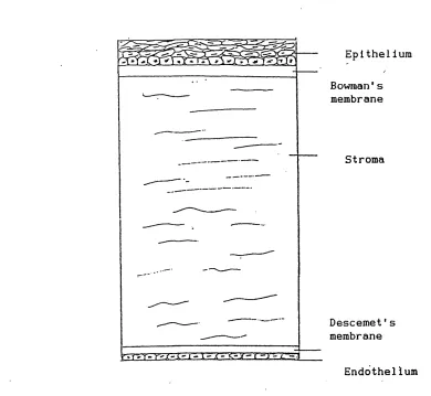
The cornea (Fig.1.1.) consists largely of the stromal tissue
(Fig.1.2.), covered externally by Bowmans membrane and the
epithelium, and internally by Descemets membrane and the
endothelium (Ruskell 1989). The mean human central corneal
thickness is 0.523 mm SD 0.039,, but towards the periphery this
becomes 0.660 mm,SD 0.076 (Martola à Baum 1968). The epithelium
may be considered as the anterior continuation of the
conjunctiva. It is 50-100 microns thick and consists of five or
six layers of squamous epithelial cells. The superficial cells
gradually fragment and are shed, being renewed from the basal
Iris Cornea
AqueDuS" Humour Crystal line Lens
Retina VItreous Humour Fovea
FIGURE 1.1 CROSS SECTION OF BASIC OPTICAL APPARATUS OF THE EYE
Epithelium
Bowman ’ s membrane
Stroma
D e s cemet's membrane
Endothellum
The stroma appears as a set of lamellae, superimposed on each
other and running parallel to the surface. These fibrous layers
consist of mucopolysaccharide and collagen fibrils. Bowmans layer
Is 8 to 14 microns thick, consisting of random but closely
packed collagen fibrils merging with the stroma. Descemets layer
is 5 to 13 microns thick. The endothelium is a layer of flattened
cells, Involved In the active pump of water to maintain the
correct hydration of the stroma.
1.2.2. Corneal Transparency
Increased hydration affects corneal transparency in addition
to Increasing the thickness as described by Maurice (1962), and
Benedek (1971). The corneal tissue power to exclude fluid
relates to the metabolic energy available to resist the Intrusion
of fluid from the aqueous humour. In the normal eye, the active
removal of fluid by the epithelium and endothelium just balances
the Influx from the limbus and tears. Interference by means of
reduction of oxygen supply, trauma or provision of metabolic
toxins leads to Increased thickness with associated mistiness of
the cornea.
1.3 CORNEAL ASTIGMATISM
Corneal astigmatism is defined as that component of the
astigmatism of the eye, beingthe difference in power between the
two principal meridians of the corneal curvature, (at 90 degrees
to each other). Refractive astigmatism is the power difference
total power determination of the eye. Astigmatism is due to the
curvature and power of the cornea and other ocular structures.
When the received spherical image is focused at two different
planes by the astigmatic surface, a distorted image reaches the
retina. The angle between these planes and the horizontal
meridian is the angle ot axis ot the astigmatism. In this thesis,
resultant astigmatism refers to the astigmatism which remains
following treatment or surgery
1.3.1 Types and description of astigmatism
With the Rule astigmatism WTR, is when the more vertical
meridian, ie. that positioned between 60 and 120 degrees, has a
steeper curvature (shorter radius of curvature) than the
horizontal meridian, see Fig. 1.3.1. Against the Rule
astigmatism ATR, is where the horizontal meridian, ie. that
positioned between 0 and 30 or between 150 and 180 degrees, is
more steeply curved than is the vertical meridian, see F i g . 1.3.2.
Oblique astigmatism is when the astigmatic axis is at an angle
of approximately 45 degrees to the horizontal meridian.
F ig.1.3.3. Hirsch (1959) described the percentage of these types
in people over the age of forty, indicating the change with age
e.g. in 5th decade: 57% WTR, 21% ATR, 22% Spherical; in 7th
decade: 32% WTR, 37% ATR, 31% Spherical.
Regular astigmatism.
Parallel rays of light from a distant source, incident to the
1) with the rule (WTR)
2) against the rule (ATR)
3) oblique
two images do not lie at the same plane of focus. The astigmatism
types are shown in F i g . 1.4 (Ruben and Woodward 1982).
Astigmatism affects the retinal image by distortion, an example
is shown in F i g . 1.5. By using a spectacle lens which gives
optical correction corresponding to each of the two principal
meridians, a clear Image can be obtained.
During contact lens fitting, corneal astigmatism affects not
only the optical system, but also the lens fit and centration. A
high degree of corneal astigmatism increases the risk of excess
lens mobility and tolerance problems (Astin 1985). Soft
(hydrophilic) contact lenses rely on the cornea for support and
follow the corneal contours, so that even if lens mobility is
acceptable, most of the visual distortion is revealed through the
lens. Toric lenses, which are more steeply curved in one meridian
than the other, may be fitted but often lead to problems with
lens tolerance and visual image quality.
Irregular astigmatism.
This is when the refractive power of the ocular system cannot
be resolved into two principal meridians. If the corneal surface
is markedly irregular, eg. due to scarring after trauma or in
conditions like keratoconus, the corneal shape is difficult to
measure and spectacle correction is inadequate. The provision of
an improved optical image by trapping tear fluid between the
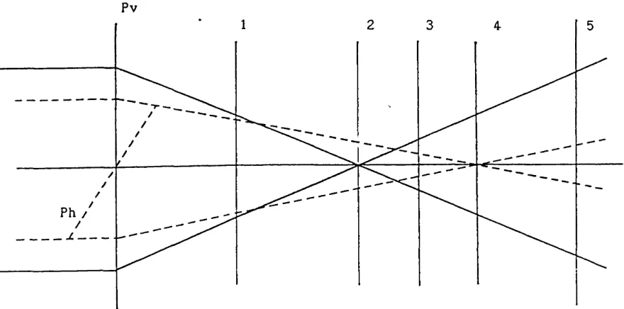
Pv
Ph /
Pv = vertical eye plane Ph = horizontal eye plane
The positions of the retina for the five astigmatism types are shown
1 = compound hyperopic 2 = simple hyperopic 3 = mixed
4 = simple myopic 5 = compound myopic
Target
Image
Few patients can tolerate a full correction of high degrees of
astigmatism (e.g. 4.00D) straight away. Their spatial orientation
is upset by meridional anisometropia, because they previously
compensated for their uncorrected astigmatism, and confusion
exists, eg. a corrected straight image is Interpreted as sloping
and circular images appear oval, until they have cortical ly
adapted to their new spatial images. sMost people have a small
amount of physiological astigmatism between 0 to l.OD at age
forty, not requiring correction (Anstlce 1971).
1.3.2 Changes in astigmatism
a) Due to lid influence
Wilson et al (1982), studying 36 eyes, confirmed that for
corneas of more than 1.GOD WTR astigmatism, lid retraction gave a
decrease In astigmatism. He concluded that lid pressure on the
globe is a factor influencing astigmatism. Vihlen and Wilson
(1983), measuring 195 eyes, determined an elastic coefficient of
lid tension but no correlation to WTR corneal toricity; both of
these showed a statistically significant decrease with age.
Pressure from thickened swollen lids after ocular surgery may
encourage WTR astigmatism in corneas weakened by incisions.
b) Related to gender
Stenstrom (1948) showed that the distribution of total
refracting power and corneal radius may be considered as samples
taken at random from a normal distribution, and suggested that
(1959) found high astigmatism more in men than women. Both
Richards et al (1966) and Bishara et ai (1986) concur that there
is no significant difference between male and female groups,
with regards to corneal astigmatism.
c ) Anterior segment tissue defect
Pterygium and pseudo-pterygium can induce astigmatism as shown
by Bedrossian (1960), and Hansen and Norn (1980). Gridley and
Perlman (1986) studied variable astigmatism for different
positions of gaze caused by a pseudopterygium. Patients with
anterior segment defect were excluded from the current study.
d) Muscle tension
Kushner (1986) demonstrated that the tightening of the
superior oblique muscle produced a long term Incyclo-rotation,
altering the axis of astigmatism. Past hypotheses proposed that
the eye is squeezed by the lids and orbicularis muscle each time
the eye is closed, causing steepest corneal curvature in the
vertical meridian. Lopping and Weale (1965) obtained results
which supported the hypothesis that horizontal corneal curvature
is accentuated by the continual pull of the internal recti
muscles which govern convergence. Elderly corneas showed
negligible change on convergence as stromal cross linkages are
often formed as the tissue ages so increasing its rigidity. No
curvature change was found with accommodation without
convergence. Patients with abnormal ocular muscle tensions, eg.
e; Age
The axis of corneal astigmatism broadly changes with age from
WTR to ATR, Reading (1973) demonstrated that the horizontal
curvature steepened with age. Hirsch (1959) reviewed a number of
investigations and described this shift towards ATR. His study on
1,606 eyes over a 40 year span, found the mean astigmatic error
changed from 0.25D WTR to 0 . 75D ATR. Lyle (1971) did repeated
keratometry over a mean period of 24.1 years to show this shift.
Anstice (1971) measured 621 subjects to reveal that total and
corneal astigmatism have a high correlation, and WTR astigmatism
decreases after the age of 40 years. Baldwin and Mills (1981)
also supported this hypothesis. Kiely, Smith and Carney (1984)
determined the corneal shape of 196 eyes by photokeratoscopy.
Their results showed reduced WTR astigmatism with age, but no
substantial variation in asphericity.
1. 4 MODELS OF THE CORNEA
Various models for the corneal topography have been proposed
e.g. Bibby (1976), Townsley (1970). Gull Ion, Lydon and Wilson
(1986), proposed an ellipsoid corneal model based on a
photokeratoscopic and keratometric study of 220 eyes
representative of a normal population. This study showed a
systematic difference between central keratometric and
photokeratoscopic measurements that was statistically but not
clinically significant; a significant number of corneas steepen
at the periphery; the corneal shape varies greatly within a
are usually similar. The following data were given: a mean radius
of curvature of 7.85 mm <SD +/-0.25) for the flattest meridian of
the central cornea, and a mean difference between that of the
flattest and steepest meridians of 0.15mm (SD +/-0.15); a mean
shape factor (expressed in a different manner from that of Bibby)
for the flattest meridian of the peripheral cornea of 0.85 (SD
+/-0.15), and a mean difference between that of the flattest and
CHAPTER 2
MANAGEMENT OF APHAKIA
2.1 VISUAL OUTCOME IN APHAKIA
Aphakia Is a condition where the crystalline lens is absent
from the correct optical positioning within the eye, due to
trauma, surgery or dislocation. ' After lens extraction
accommodative ability is lost. The main categories of aphakia are
age-related, paediatric and traumatic. Only patients with the
age-related cataract extraction were considered in this study.
The yellow filter eftect of the sclerosed lens is lacking so
they benefit from a ultra violet light filter in their optical
correction. A rapid, success!ul outcome whether correction is by
lOL or contact lens proves cost-effective for the patient, the
clinician and the community iWoodward and Drummond 1964, Davies
et al 1986) and improves the aphake's visual function iBernth-
Petersen (1981, 1982). The aphakic patients are loaned a set of
high spherical power spectacles on their operation day. Those
with lOLs are left temporarily uncorrected. After extracapsuiar
cataract extraction, routine management often involves clinical
checks at 2, 6 and 12 weeks and refraction for spectacle
correction performed about 6 to 12 weeks after the operation.
After small incision surgery, this can be done at 2 weeks.
2.1.1 Assessment of visual acuity and refraction
The target distance, illumination and contrast should be
for VA measurement consists ot test letters viewed at six metres,
subtending standard angles at the nodal point of the eye. Several
factors affect this measurement including the eye's depth of
rocus iTucker and Charman 1975; and whether the trial case lenses
have dirty, misty or scratched surfaces. The traditional
refraction method iRabbetts 1973) using trial frame and lenses
imitates the situation of spectacle wear. Automated instruments,
e.g .Dioptron, iKempster 1975), Autoretractor 6600, Ophthalmetron,
ana Humphrey subjective retractometer are bulky and expensive.
Their accuracy relies on the subject having clear media, relaxed
accommodation, steady eye tixation and no distortion ot
retracting surfaces. Hence they were not used for this study
because preoperatively the patients media were not clear, also
suitable funding was not available.
The distance between the plane of the lens and the principal
plane of the eye is termed the back vertex distance BVD. This is
important when relating retraction astigmatism to keratometry
astigmatism. For example, for a spectacle prescription of +14.GOD
and +16.00D in the two principal meridians, hence refraction
astigmatism ot +2.00D, at the corneal plane these powers are
effectively +16.63D and +19.60D, giving +2.97D of corneal
astigmatism measured by keratometry. For a high positive power
lens vmore than +6.00D; the BVD markedly affects the image
magnification and effective lens power iFe; at the principal
B PL
F = lens power
f = focal length of lens
fsj = second focal point
PL = plane of lens
B = plane to measure effective power
d = distance (m > from lens to effective power plane
Fe = effective lens power = < 1
2.1.2 visual problems in aphakia
Problems associated with external optical correction are:
binocular - diplopia, suppression; - ghost images,
variations in visual acuity and quality; spectacle
magnification, prism effects, restriction or field, distortion,
aberration, lens weight; contact lens - magnification, prism
effects, variation in visual acuity and quality due to lens
movement and condition. Descriptions are given by Bennett U 9 6b,
I960, 1972/3), Stone <1977), Ruben <1975), Ruben and Woodward
<1902), Port <1969). Fig.2.2 indicates the focal planes in
aphakia optical correction. The spectacle lens thickness, lens
form, position and distance from the eye affect the effective
power and aberration <Ruben and Woodward 1962).
2.1.3 Contact Lens and lOL
Many of the optical and physical problems of spectacle
correction are much reduced by contact lens correction <Astin
1964,1966). Lens insertion and removal takes time and correct
disinfection and regular aftercare are vital <Graham and Dart
1966, Stapleton et al 1989). Corneal astigmatism complicates the
fitting, the optical correction, the stability and comfort of the
lens so it is preferable to keep this to a minimum.
An lOL is a rigid plastic imitation lens held in position by
haptic loops or 'legs'. Posterior chamber lOLs are also
supported by the posterior capsule after extracapsuiar cataract
fP = far point of focus of the eye f a = second focal length of the eye
R = position of retina
Fo = ocular refraction at anterior corneal surface Fs = spectacle refraction
d = vertex distance between spectacle plane and anterior cornea
fp = focal length of the eye
EXAMPLE
when f P = 70mm ( approximately equivalent to 15.0 diotres ) Fo = +45.0 dioptres, d = 15mm
spectacle lens focal length = fP + d = 70 + 15 = 85mm spectacle lens power = F® = 1000/85 = +11.75 dioptres.
pupil with the legs resting on the scleral spur. Advantages ot
an lOL Include: similar magnification to that with the original
eye, as the lens is nearer to the optical entrance pupil of the
eye; the patient need not maintain the appliance, and there are
few distortions and visual problems. Generally there are lower
costs to the health service (Davies et al 1986) and fewer visits
needed than with contact lens aftercare. Preoperative corneal
curvature measurements by keratometry are important in the
calculation of the lOL power (Tutton 1985). Maltzman et al
(1986) showed that the cornea and not the lOL was responsible for
the postoperative astigmatism experienced by patients.
Usually the lOL remains in the correct place for satisfactory
vision, and any residual astigmatism following surgery can be
corrected by low power spectacle lenses. If serious problems
occur with lens dislocation or corneal decompensation, the lOL
can be removed. In some cases, refractive keratoplasty provides
an alternative correction method (Waring 1985).
2.2 CORNEAL RESPONSE TO CATARACT SURGERY
The corneal tissues respond to the surgical trauma and to the
new physiological balance In the eye. The future functioning of
the ocular tissues will be influenced by the extent of this
trauma and by any postoperative damage. Corneal rigidity is
reduced in an eye that has undergone a penetrating Incision. This
depends on the size and type of incision and the strength of the
scar tissue. Corneal sensitivity in the superior region is
but may partially recover kGuilion and Morris 1962J. This
facilitates adaptation to contact lens wear, but corneal
sensation and response to epithelial damage is reduced. Partial
sensitivity is regained over two years, depending on the patients
age. Often thin superficial vessels invade the superior
peripheral region of the cornea following a corneal incision to
reach the suture penetration sites. The vessels may increase if
the corneal oxygen supply is diminished as in extended contact
lens wear iHolden et al 19d5>.
Corneal thickness measurement was recoramenaed by Dohlman and
Hyndiuk (19 72) to monitor corneal oedema alter cataract
extraction. Holden et al (19b0) and Guillon and Morris (1961)
showed that under reduced levels of oxygen, the cornea of the
aphakic eye swells less than that ot the fellow eye, although
corneal oxygen uptake was shown to be unchanged. One theory is
that there is a lower oxygen demand because oxygen is no longer
required tor the crystalline lens metabolism. Increases in
corneal oedema and thickness following cataract extraction are
Often attributed to endothelial damage (Liesgang et al 1964,
Buckley 1965). Less endothelial damage is usually caused by the
use of small rather than wide apertures. Studies regarding
endothelial cell damage following lOL Implantation have been
performed by Bourne and Kautman (1976), Kaufman and Katz (1976),
Katz et ai (19/7), Kirk et ai (19//), Cheng et al (197/), Stark
et ai (1979), Sherrard (1963), Vannas et al (1965) and Jacob
CHAPTER 3
ASTIGMATISM AFTER CATARACT EXTRACTION SURGERY
3. 1 REVIEW OF LI TERATURE
The renewed attention to astigmatism following cataract
extraction (described as resultant astigmatism.» has been rocusea
by the reduced Incidence ot serious postoperative complications,
the revival ot extracapsuiar cataract extraction ΠC C E ) and its
comparison to phacoemulsification, improvements in surgical
techniques and materials, and increased visual expectations ox
the patient. Improvements in lOL insertion and design have
usually minimised astigmatism due to iOL tilt, especially when
the lOL position is near to the nodal point of the eye iKosaki et
al 1991^). Maltzman et al (1986) on studying 127 posterior lOL
cases and 54 anterior IOL cases tound that the cornea and not the
lOL was most responsible tor postoperative astigmatism.
The amount of postoperative astigmatism is influenced by a
number of surgical factors: operating technique, incision size
and position, suture material and technique, wound profile and
healing. Changes in postoperative astigmatism can be made by
adjustment of any of these, and by manipulation of the sutures
and wound during the postoperative period (Jaffe 1976,Atkins and
Ropei— Hall 1985). A number of papers regarding these factors is
Generally, there is agreement that small Incisions such as
those Often used with phacoemulsification or aspiration
techniques cause the least degree of astigmatism and the shortest
stabilisation time (.Arnott 1973, Hotter 19d4, Reading 1984,
Bambery 1986). Several reports on the influence of the corneal
incision site concur that a greater degree and variability oi
astigmatism are found f ollowing this ^ method but they suggest
stabilisation times ranging from 10 to 24 weeks. Several workers,
eg. Jampel (1986), concluded that limbal incisions caused less
corneal astigmatism than did corneal incisions. However, most of
the studies followed the astigmatic changes at 6, 12 and 26
weeks.
Jaffe and d a y m a n (1975), and Meredith and Maumenee (1979),
who monitored large numbers of patients, described the various
influences of sutures, incisions and other factors on the degree
of astigmatism but gave no definite conclusions regarding
stabilisation time, Baranyovits <.1990) performed regular serial
refraction to determine the rate of stabilisation and found that
after a limbal incision this occurred at three months if silk
sutures were used but up to four to five months if the sutures
were nylon. Swinger (1987) provided a useful review of studies on
postoperative astigmatism and methods of its reduction.
Two other groups of surgeons placed more emphasis on
astigmatism control, the first (Terry 1980, Troutman 1980) by
judging corneal astigmatism during the operation using a
selective suture removal at the clinical tollow-up visits. These
methods still needed experience and care to achieve moderately
predictable results. Most ot these authors monitored only
central corneal astigmatism and at widely varying intervals so
their contribution was limited. There was still the need for the
Investigation of peripheral corneal shape and thickness changes
and tor a closer study of postoperative astigmatism to more
clearly determine the stabilisation time.
3.2 SURGICAL TECHNIQUES OF CATARACT EXTRACTION
The most common methods are described as follows:
3.2.1 Intracapsular Cataract Extraction (ICCE)
A limbal based incision is made into the anterior chamber, of
size between 110"-160" of arc. The lens supporting zonule fibres
are weakened by the application of 5% alphachymotrypsin. The
cataractous lens is bodily removed with minimal pressure and
slight traction with the forceps, or attached to a cryoprobe.
3.2.2 Extracapsuiar Cataract Extraction <ECCE)
Through the corneal or limbal incision, a capsulotomy needle
or equivalent is inserted into the anterior chamber. This is
used to cut a central disc of anterior lens capsule, which is
then removed. The lens nucleus is expressed by pressure against
the inferior lirabus and behind the incision. The posterior lens
capsule remains in situ. Irrigation and aspiration with buffered
3.2.3 Phacoemulslf icat Ion
A small incision (approximately 3.0 ram) is made behind the
superior limbus or in clear cornea, and extracapsuiar extraction
carried out using a phaco-emulsit1er. This uses ultrasonic energy
to fragment the lens tissue, which is removed by simultaneous
irrigation and aspiration. A manipulator is inserted via a
second, smaller incision to guide the lens fragments towards the
phacoemulslfier. A more detailed description is given by Snyder
and Donnenteld (1994).
3.2.4 Aspiration
Aspiration, or lensectoray, is a form of extracapsuiar
extraction where two paracenteses, 1.5mm wide separated by 90-
180' arc, are made in the cornea near the superior limbus. Fine
cannulae, of 0.7 to 0.9 mm external diameter, are inserted so
that after removal of the anterior capsule, one is used for
aspiration of the soft lens material, the other for irrigation of
the anterior chamber.
Advantages and disadvantages of the various techniques are
given in Tables 3.2 and 3.3. Several complications can follow
cataract extraction surgery, as it involves penetration of the
globe and manipulation of tissues. In a few cases, stretching of
the levator aponeurosis by the superior rectus stay suture or by
the lid retractor can result in ptosis. After ECCE or
phacoemulsification, the remaining posterior capsule can thicken,
the capsule to restore vision in a non-lnvaslve procedure. After
ICCE the vitreous may protrude into the anterior chamber
affecting fluid drainage, producing glaucoma, and pulling at weak
retinal areas, producing retinal detachment. High prescription
myopes often have peripheral retinal thinning which is vulnerable
to tearing when a age-related cataractous lens is removed.
Postoperative vitreous floaters are common (Buratto 1991).
3.3 SURGICALLY INDUCED ASTIGMATISM
Astigmatism may be induced during surgery by the incision
method, surgical technique, tissue healing and sutures.
3.3.1 Incision
Size The larger the wound size the greater is the influence
of the sutures and the greater are the variations in corneal
astigmatism (Gills 1974, Reading 1984, Heslin and Guerriero
1984). Small incision techniques such as phacoemulsification
show smaller variations in the induced changes in mean radii of
corneal curvature. A wound of 4.0mm or less rarely tends to gape.
There is a decreased tendency to overtighten sutures, if used.
Martin et al (1993), using keratoscopy, found significantly
less wound related localised corneal flattening with small
incisions compared to large incisions, but no significant
difference in mean keratometric astigmatism. Several others
(Steinert et ai 1991, El-Maghraby et al 1993, Hayashl et al 1993)
as 4.0mm compared to larger Incisions as 6.5mm. LeWagne and
K'allay (1993) found this even with a scleral pocket incision.
Site Incisions distant from the corneal optical zone tend
to induce less astigmatism (Gills 1974, Bambery 1986). Jaffe and
d a y m a n (1975) found WTR astigmatism to be associated with a mid
limbal or a corneal incision, ATR astigmatism with a more scleral
site. Placement of the incision further posteriorly decreases the
iatrogenically induced astigmatism (Hardten and Lindstrom 1993).
Storr-Paulsen (1991) also found significantly less surgically
induced astigmatism after 1, 3, and 6 months postoperative1 y with
a scleral incision compared to a corneoscleral incision. He
concluded that the configuration of the incision appeared to be
more important for the early surgically induced astigmatism than
was the type of wound closure.
Some surgeons prefer the limbal site as this heals faster and
does less damage to the corneal endothelium. Others prefer a
corneal site (Fig.3.1) and recommend deep suturing and a
peripheral corneal incision. These have less effect on limbal
vessels, no haemorrhages, coaptation of wound edges is easier to
control and correct, delayed reformation of the anterior chamber
occurs more rarely and epithelial ingrowth is rare.
3.3.2 Surgical Technique
In ECCE, the surgical knife makes a superior region scleral or
CORNEAL
LIMBAL
SMALL CORNEAL AS IN
PHACOEMULSIFICATION
this trauma effectively increases the tightness ot the sutures
resulting in a large postoperative shift to WTR astigmatism. ATR
astigmatism can result later due to the wound gaping or
vertically stretching, thereby increasing the globe circumference
and flattening the vertical curvature (Ernest 1994;. The corneal
incision technique and its advantages have been described by
Steele (1977). Agapitos (1993) gave a good general review of
surgical techniques. The corneal incision allows a better view
during surgery of the anterior chamber and the lens extraction.
More recently, the scleral tunnel technique is gaining
popularity. This method begins with a scleral groove, followed by
a scleral tunnel and continues into the cornea before entering
the anterior chamber. Studies on this method indicated a
significant reduction in surgically induced astigmatism and
stabilisation time (Stort— Paulsen 1991, Taylor, Solomon à Boyaner 1992, Hall et al 1993).
Example of a Corneoscleral (limbal) technique
This uses a limbal based conjunctival flap, diathermy to the
limbal vessels and an incision just behind and parallel to the
lirabus, but with a 90-130* arc. The corneoscleral wall is opened
to approximately 90% of tissue depth, with initial groove at
right angles to the globe surface then a posteriorly slanted
incision edge made. Five to seven separate nylon 10/0
monofilament sutures are inserted to 75% or greater depth and
silk sutures are more rarely used nowadays, these begin to weaken
by the fifteenth postoperative day.
Example of a Corneal technique
The incision is made free-hand, with a 90-130" arc, anteriorly
to the limbal vessels, so diathermy is rarely needed. The
direction of the initial incision groove through 907. of the
corneal depth is vertical and the second stage of incision slopes
posteriorly, or else a single posterior slanting incision is made
. Closure is with five 10/0 monofilament nylon sutures placed at
approximately 95% or more of the tissue depth, tensioning to give edge to edge closure and with the knots burled beneath the
corneal surface. Mechanical stresses as the anterior chamber
reforms tend to press the inner wound margin against the outer
one to aid secure wound closure.
3.3.3 Tissue Healing
Troutman (1979) proposed that postoperative ATR astigmatism
was due to a weak superficial corneal scar induced by too shallow
a wound closure with sutures which loosen early, allowing scar
tissue thinning and the superior and central corneal regions to
flatten. A corneal incision gave less tissue trauma, hence a
quiet white eye with lower risk of secondary raised intraocular
pressure or uveitis. For aphakic patients Kersley (1985)
recommended early soft contact lens fitting to give a bandage
Several wound healing factors were described by Flaxel and
Swan (1969), tor example:
a) Thickness and rigidity of the sclera and cornea,
b) Raised intraocular pressure which may lead to wound gape and
alteration of suture tension,
c) Steroids which slow wound healing,
d) General health and healing ability of patient,
Excessive eye rubbing causing sutures to loosen.
3.3.4 Sutures
Materials
The major types are: non-absorbable material, eg. nylon,
silk, and absorbable eg. catgut, collagen, some synthetics. The
suture materials used by the co-operating surgeons at the time of
this study were virgin silk and monofilament 10/0 nylon. With the
latter, there was usually less tissue Irritation, less
postoperative hyperaemia and inflammation, and neater smooth
scar formation due to prolonged firm apposition of the wound.
Tissue reaction to nylon was minimised and scar formation delayed
so that the nylon was usually left in the incision for several
months. Nylon sutures could give significant suture induced
astigmatism, especially if they were tight, but this could be
reduced by selected interrupted suture removal, which was easier
if the incision was corneal rather than subconjuctiva 1.
Induced astigmatism with virgin silk sutures was less as
weakening of individual sutures caused them to break, releasing
tension on the incision and reducing the WTR astigmatism. The
irritant sutures encouraged vessel activity and rapid scar
formation; hence the astigmatism could not readily be influenced
after the first two to three weeks since the sutures began to
biodegrade. For corneal incisions, the suture had to remain firm
for approximately eight weeks due to the slower wound healing of
stromal tissue, therefore nylon sutures were used.
Gills (1974) confirmed that for all materials WTR astigmatism
was present for ten to fourteen days postoperatively. Dekkers
and Buijs (1989) found that silk sutures led to ATR while nylon
led to WTR astigmatism. They explained that although silk is
chemically non-absorbable, a special polymer is present in virgin
silk which provokes a tissue reaction. The softening of tissue
diminishes the tensile strength of the virgin silk suture and
hence it behaves in a similar manner to an absorbable suture.
They concluded that with respect to postoperative astigmatism,
the suture material seemed more important than suture method.
Suture Techniques
The loop must be 75% (or preferably more) of the corneal depth
to control wound apposition. The closer the sutures are placed
to each other, or sited in a crossed pattern as in the bootlace
method, the more stable is the incision. Different suturing
patterns have been described and compared by Roper Hall (1982),
and Emery and McIntyre (1983) (Fig. 3.2). Extra sutures or knots
INTERRUPTED
CONTINUOUS
BOOTLACE
sutures affect the wound for up to a year according to Gills
(1974) and Meredith and Maumenee (1979). Continuous and mattress
sutures tend to give a tighter wound closure with a steeper
vertical curvature and more WTR astigmatism than do interrupted
sutures.
3.4 PREOPERATIVE ASTIGMATISM
Preoperative corneal astigmatism may be due to: corneoscleral
lirabus shape, cicatricial modification, ectasia, and local
modification of corneal thickness. Congenital astigmatism is
usually associated with an oval limbal shape where the larger
diameter corresponds to the tlatter meridian. If the preoperative
astigmatism is known, the surgeon can adjust the suturing
method in order to reduce this. Floyd (1951) found that the sixth
month postoperative astigmatism was often of the same type as
that preoperatively. Brown and Sparrow (1988) tried to compensate
for preoperative astigmatism by using additional sutures and
agreed with Bambery (1986) that postoperative astigmatism can be
controlled by selected suture removal.
Jampel et al (1986) recommended using computer analysis of
postoperative astigmatism for each surgeon to determine the
typical surgically induced astigmatism and to provide a basis for
preoperative planning of surgical technique. They concluded that
astigmatism changes continue towards a stable situation at four
to six months postoperat1v e l y . Naeser (1990) attempted to express
astigmatism and its direction in figures for mathematical
3.5 CURRENT METHODS TO REDUCE ASTIGMATISM
3.5.1 During surgery
Both the surgical keratometer and the keratoscope have been
used to monitor astigmatism during surgery. Although there are a
number of apparent advantages ( Troutman, 1974, 1976, 1977, 1976,
1980, Terry 1960, Colvard et al 1981, Amoils 1986); other
studies by Samples and Binder (1984) and Jacobi and Strobel
(1985), found several errors remain with a surgical keratometer
even with an experienced user. It was an asset in guarding
against high degrees of astigmatism during the operation, but did
not give as fine a control as its proponents suggested.
Troutman mentioned difficulties in using this keratometer
effectively when using the interrupted suture technique. Roper—
Hall (1982) emphasised that even if the desired corneal
curvature is obtained with the aid of a surgical keratometer by
the end of the operation, the final postoperative astigmatism is
still not fully predictable. Conditions at the end of surgery are
abnormal, healing processes are subject to many variables
including intraocular pressure, patient health and treatment,
which lead to changing corneal curvature and refraction. Tissue
reaction to suture tension and material varies.
3.5.2 Postoperat ivelv
Selected Interrupted suture removal within the early weeks of
healing has been recommended by several surgeons (Ropei— Hall
1982,1985, Atkins and Ropei— Hall 1985, Bambery 1986, Stanford et
meridian of steepest curvature (the axis of the positive power
cylinder in refraction), so It could be removed under topical
anaesthesia at the slit lamp to give a rapid reduction of corneal
astigmatism. The retraction and keratometry was reassessed and
the process repeated until optimal correction was obtained
without wound gaping. Continuous sutures were gripped by
mlcrosurgical suture forceps and eased towards the tight steepest
curvature meridian. For cases without lOL, Buxton (1975,1976)
advised the surgeon to aim for postoperative WTR astigmatism to
facilitate the fitting of aphakia contact lenses three to four
CHAPTER 4 KERATOMETRY
4.1 HISTORY
The main function ot the keratometer is tor the measurement ot
the radius of curvature of the central portion of the anterior
surface of the cornea, usually referred to as the optic cap.
Helmholtz (1909, cited in 19241 is usually credited with the
invention of the keratometer, but Levene (1965,1977) suggested
that Scheiner attempted this in 1619, and that Hare and Ramsden
in 1795 were the first to measure corneal radii. According to
Helmholtz (1909 translated 1924), Kohlrausch in 1640 and Senff in
1846 performed corneal radius measurements on a living eye.
Woodward (1980) noted that Valk in 189/, Tscherning in 1904, and
Sorsby in 1961 used keratometry to measure the ocular refracting
elements. As the cornea is aspheric, assumptions in central
sphericity led to errors.
Woodward described L a n d o l t ’s use in 1878 of a prism for
doubling the keratometer image to assist measurement, and the
use by Javal and Schiotz (1881) of a Wollaston prism with
specially shaped 'mires' as test objects. The advantage of this
method over Helmholtz's glass plates doubling method was that the
operator could obtain a direct reading without further
calculations. The work of Helmholtz and others in the development
4.2 KERATOMETER DESIGN
Keratometrlc measurements are useful in contact lens fitting
and aftercare; to aid calculations of the refracting power of
optical elements and lOLs; to aid diagnosis of a corneal
condition; to guide and monitor the progress of ocular surgery
«.Ruben 1975). Measurements are obtained indirectly from the
angular size of the reflected image formed by the cornea of an
object of known linear size (h> at a predetermined distance from
the image plane <.d). The derivation of the radius of curvature is
shown in Fig. 4.1. If the eye was stationary, the image size
could be measured directly using a graticule in the eyepiece, but
the eye is continually moving with small saccadic movements, so a
doubling principle must be Incorporated, The two images can then
be juxtaposed because even it moving slightly, they do so with
the same speed and direction (Fig 4.2).
In practice, the object limits are represented by a pair of
Internally illuminated mires whose corneal images are seen
magnified through a short focus telescope as described by
Sheridan (1989). This incorporates the doubling device which
produces the four images seen in the telescope field, the two
central ones being brought into contact or superimposed. To
obtain adjacent or superimposed images, either h' may be varied
by altering the mire separation h, while the power and position
of the doubling device are fixed; or the image size h* and the
mire separation h may be fixed while the power of the doubling
T T
The optical principle of the Keratometer is that r is directly proportional to the image
size, h * .
Q and R are the limits of an object of size h Q' and R' are the limits of the Image of size h'
formed by the reflection at the anterior corneal surface
F = principal focus RQ = object plane
d = distance between image plane R'Q* and object plane RQ
X = distance between F and RQ A = pole of the cornea
C = centre of curvature of cornea surface
.. r = AC and r/2 = AF
h'/h = ( r/2)/x but d is approximately equal to x then
h'/h = ( r/2)/d .. r = 2dh'/h
JL
L
For a prism of power P dioptres fixed halfway in an observation aperture
h' = image size - seen doubled
a = distance between plane of the prism and the position at which the doubled image are exactly adjacent to each other
h'/a = P/100 h' = aP/100 mm