SCREENING BIOACTIVES FROM VEGETAL SOURCES AS POTENTIAL SKIN LIGHTENING AGENTS USING AN ENZYMATIC MODEL OF TYROSINASE INHIBITION, CORRELATIONS AMONGST ACTIVITY,
PHENOLIC COMPOUNDS CONTENT AND CYTOTOXICITY
Muñoz K1*, Londoño J1, Arango G1, Arenas J1, Mira L1, Ochoa J1, Sierra J2
1
Grupo de Investigación en Sustancias Bioactivas –GISB- Corporación Académica para el Estudio y Control de Patologías Tropicales CAEPT, Facultad
de Química Farmacéutica. 2 Grupo de Inmunodeficiencias Primarias, Corporación Biogénesis. Universidad de Antioquia, Colombia.
Summary
For many people, irregularities in skin pigmentation had become an aesthetic problem, mainly when they are affecting the face. For this reason, there is a big interest in products with skin lightening properties for treating irregularly pigmented skin areas. Skin pigmentation, is the consequence of production and dispersion of melanin, a pigment sinthesized in epidermal melanocytes were the precursor amino acid tyrosine is sequentially oxidized into L-dihydroxyphenylalanine and dopaquinone by the copper-containing enzyme tyrosinase (EC 1.14.18.1), the key and rate limiting enzyme in melanin biosynthesis (1). In our search for tyrosinase inhibitors from plants, we study Ruta graveolens and citrus sinensis extracted by different means, for inhibition of the oxidation of L-3,4-dihydroxyphenylalanine (L-DOPA) catalyzed by mushroom tyrosinase and correlated activity with the content of phenolic compounds and its cytotoxicity. In our case, although phenolics could be the main compounds responsible for activity, no clear correlations exist between cytotoxicity, tyrosinase inhibition and the content of phenolic compounds, however the contents of this compounds increased as a more astringent method of extraction was used.
Materials and methods
Reagents: Cell Culture media and reagents, fetal bovine serum (FBS), trypsin, antibiotics, were purchased from Gibco BRL. Reagents used in the tyrosinase inhibition, cytotoxicity and Folin Ciocalteau assays were of the highest purity (Sigma), including mushroom tyrosinase, L-DOPA, Sodium cyanide, dimethylsulfoxide, and MTT.
Plant material: All material was obtained from nearby cultivars. 170g. of Ruta graveolens were extracted with water:EtOH 1:1 using sohxlet (2h), percolation (24h), and ultrasound bath (2h) for evaluating effects of the extraction method, and Citrus sinensis flavonoid fraction was precipitated from fruits according to Montoya et al method (2).
Tyrosinase assay: The spectrophotometric assay for tyrosinase was performed as described previously (3). Briefly, all the compounds were dissolved in DMSO to a concentration of 2.5%. L-DOPA was used as the substrate to measure the DOPA oxidase activity of tyrosinase. The activity and the absorption were measured with a Cary Bio 50 spectrophotometer (Varian). The inhibition percentage value used in the present study was defined as the product concentration that was indicated by the change in absorbance at 480 nm per min.
All the studies have been carried out at least in triplicates and the results here represents means and standard deviation of the mean. Here in these experiments Sodium cyanide was used as standard tyrosinase inhibitor.
Mammalian cell culture and harvesting
For the toxicity assays we selected the widely used cell line for cytotoxicity and reactivity tests L-929 (ATCC CCl-1TM), a line firstly isolated from mouse subcutaneous connective tissue; cells were cultured under standard conditions in RPMI 1640, supplemented with 10% fetal bovine serum (FBS). Cells were subcultured each two days, prior to each experiment cultures were trypsinized and at least 2 x10⁶ cells were seeded into 75 cm2 flasks and cultured for 24 h.
Cell treatments
Cytotoxicity assay: The MTT assay is based on the protocol described for the first time by Mosmann (4). Cells undergoing exponential growth were trypsinized and seeded in treated 96-well microplates at a density of 1 x 10⁴ cells/well. The plates were incubated for 12 h to allow adherence, then, fresh medium containing the fractions was added to give the final concentrations described above. Cultures containing test compounds were incubated 72 h, then fresh medium containing MTT 0.83 mg/ml was added and cells incubated at 37°C for 4 h. The formazan was dissolved by adding DMSO. Optical densities were read at 550 nm with background subtraction at 630 nm in a Power Wave X microplate reader.
MTT assays were performed in triplicated wells at least three times each. Results of IC50 were interpolated of a toxicity regression curve and are presented as mean of triplicates and standard deviation.
Phenolic compounds content: Phenolic compounds were measured using the Folin-Ciocalteau method as is described by Sigleton et al (5), Briefly, a reaction mix was prepared as follows: to 750 µL of distillated water was added, extract 100 µl (prepared in DMSO:MetOH 1:1);100 µL of 10% sodium carbonate solution, and 50 µL Folin-Ciocalteau’s reagent, the mix were incubated in the dark for 1h and then absorbance was measured at 760 nm.
Results and discussion
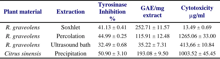
In the present paper, tyrosinase inhibition studies of Ruta graveolens extracts and the flavonoid fraction from Citrus sinensis have been discussed; as is noted in Table 1, there are statistically significant differences in the inhibition of oxidation of L-DOPA by R. graveolens extracts generated by all methods used here, although this screening results cannot be explained directly by the content of phenolic compounds as we initially expected, since many phenolics i.e kaempherol, quercetin, rutin, ferulic acid, apigenin, azelaic acid, cathecol, morin, chrysin and other flavonoids (6) has been shown to be good inhibitors of tyrosinase, and some are even used in cosmetic formulas. However to clarify this point, is necessary to estimate IC50 values and comparing this data normalized to contents of phenolics.
Until now, various tyrosinase inhibiting plant extracts has been included in cosmetic formulas, either alone, i.e. Lilium candidum, Arctostaphylos uva-ursi (L.) Sprengel, Polygomun bistorta, Coix lacrymajobi y Sophora angustifolia (7), or in combination Rumex occidentalis S. Wats, R. maritimus L., R. Pseudonatronatus, R. Stenophyllus (Tysrostat®) (8)
Table 1 Results of tyrosinase inhibition, phenolic compounds content and cytotoxicity according to the extraction method used
Plant material Extraction
Tyrosinase Inhibition
%
GAE/mg extract
Cytotoxicity µg/ml
R. graveolens Soxhlet 41.13 ± 0.41 252.71 ± 11.57 13.49 ± 0.69
R. graveolens Percolation 44.99 ± 0.25 115.91 ± 12.48 1265.06 ± 33.00
R. graveolens Ultrasound bath 32.49 ± 0.68 35.22 ± 7.31 413,66 ± 10.84
Citrus sinensis Precipitation 50.90 ± 3.10 193.08 ± 9.50 1003.52 ± 45.45
Data of basal cytotoxicity is a good starting point in an integrated assessment of potential in vivo toxicity for predicting the acute effects of compounds in vivo. As is well accepted that, if a compound is acutely toxic, in most cases, this reflects an insult to the intrinsic functions of cells and this approach has been successfully applied in a validated in vitro method to assess phototoxicity (9), Moreover in a large study of a diverse range of chemicals, it was found a reasonably good correlation between basal cytotoxicity and acute toxicity in animals and humans (10).
Our data shows how extraction method strongly affects the cytotoxicity data, and as in tyrosinase inhibition there is not a clear correlation with the content of phenolics, so we think that cytotoxicity could be determined by the quality of the extracted compounds rather than the quantity; we firstly thought that the most astringent methods, soxhlet and ultrasound, could generate either extraction artefacts due to temperature (60° C) or just extract compounds that could not be extracted using percolation, in table 1 could be noted that, only R graveolens percolation extract and C. sinensis flavonoid fraction, have IC50 values higher than 1000 mg/ml; and according to Gad Shayne classification of toxicity for natural products (11) ranked as potentially non toxic compounds.
References
1. Del Marmol V, Beermann F. Tyrosinase and related proteins in mammalian pigmentation, FEBS Lett 1996; 381: 165–168.
2. Montoya GL, Londoño JA, Arango GJ. Obtención de la fracción flavonoide a partir de residuos de la industria citrícola colombiana: Evaluación de la actividad inhibidora de la oxidación de LDL. In: 2005 14 Congreso Científico Internacional – CNIC, La Habana.
3. Chen QX, Kubo I. Kinetics of mushroom tyrosinase inhibition by quercetin, J. Agric. Food Chem 2002; 50: 4108–4112.
4. Mosmann T. Rapid colorimetric assay for cellular growth and survival: Application to proliferation and cytotoxicity assays. J Immunol Meth 1983; 65:55-63.
5. Singleton VL, Orthofer R, Lamuela-Raventos RM. Analysis of total phenols and other oxidation substrates and antioxidants by means of Folin-Ciocalteu reagent. Methods Enzymol 1999; 299: 152-178
6. Kubo I., Kinst-hori I, Chaudhuri S. et al; Flavonols from Heterotheca inuloides:Tyrosinase inhibitory activity and structural criteria. Bioorg Med Chem 2000; 8: 1749-1755.
7. Curto E, Kwong C, Hermersdörfer H et al. Inhibitors of mammalian melanocyte tyrosinase: In vitro comparisons of alyl esters of gentisic acid whit other putative inhibitors. Biochem Pharmacol 1999; 57: 663-672.
8. Fox C. Technically Speaking. Kosey KK. Patents skin lightening formula. Cosmetics & Toiletries 1998 Dec p 23
9. Simonot D, McColl J, Thome D. Tyrosinase inhibitors: Activity of a Rumex extract in combination with Kojic acid and Arbutin. Cosmetics & Toiletries 2002; 117: 51-56.
10.Eisenbrand G, Pool-Zobel B, Baker V, Balls M, Blaauboer BJ , A. Boobis A, Carere A, Kevekordes S, Lhuguenot J.-C., R. Pieters R., Kleiner J. Methods of in vitro toxicology; Food Chem Toxicol 2002; 40:193–236