R E S E A R C H
Open Access
Salinity tolerance mechanisms in glycophytes: An
overview with the central focus on rice plants
Tomoaki Horie
1*, Ichirou Karahara
2and Maki Katsuhara
3Abstract
Elevated Na+levels in agricultural lands are increasingly becoming a serious threat to the world agriculture. Plants suffer osmotic and ionic stress under high salinity due to the salts accumulated at the outside of roots and those accumulated at the inside of the plant cells, respectively. Mechanisms of salinity tolerance in plants have been extensively studied and in the recent years these studies focus on the function of key enzymes and plant morphological traits. Here, we provide an updated overview of salt tolerant mechanisms in glycophytes with a particular interest in rice (Oryza sativa) plants. Protective mechanisms that prevent water loss due to the increased osmotic pressure, the development of Na+toxicity on essential cellular metabolisms, and the movement of ions via the apoplastic pathway (i.e. apoplastic barriers) are described here in detail.
Background
The global climate change is feared to promote rapid soil degradations in agricultural lands worldwide. Soil salinization is one of the serious soil degradations, which can arise from natural causes and human-mediated ac-tivity such as irrigation in arid and semi-arid regions. Approximately 20% of the irrigated lands in the world are presumably affected by soil salinization (Yeo 1999). Salinity stress significantly reduces growth and product-ivity of glycophytes, which are the majority of agricul-tural products. The term “salinity” represents all the problems of the soil accumulating excessive salts, which can be categorized into sodic (or alkaline) and saline soils (IRRI 2011). Sodic soils having a poor soil structure generally spread over arid and semi-arid regions, retain-ing high concentrations of Na+ at the exchangeable site of clay particles in the soil, which shows high pH (greater than 8.5) with a high exchangeable sodium per-centage (ESP>15) (IRRI 2011). Saline soils can be gen-erally found in arid regions, estuaries, and coastal fringes, which are dominated by Na+ ions with electrical conductivity (EC) more than 4 dS/m that corresponds to approximately 40 mM NaCl (IRRI 2011; Munns and Tester 2008). Moreover, saline soils exhibit ESP of<15
and much lower pH values than the sodic soils (IRRI 2011).
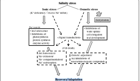
Plants have to cope with two major stresses under high salinity, osmotic stress and ionic stress (Figure 1). The former stress immediately comes over plants in accord-ance with a rise in salt levels outside the roots, which leads to inhibitions of water uptake, cell expansion and lateral bud development (Figure 1) (Munns and Tester 2008). The latter stress phase develops later when toxic ions such as Na+ accumulate in excess in plants particu-larly in leaves over the threshold, which leads to an in-crease in leaf mortality with chlorosis and necrosis, and a decrease in the activity of essential cellular metabolisms including photosynthesis (Figure 1) (Yeo and Flowers 1986; Glenn et al. 1999). Recent molecular physiological and molecular genetic studies have increasingly gained knowledge for the protection mechanisms that plants use to cope with detrimental effects of salinity stress (Blumwald 2000; Zhu 2002; Pardo et al. 2006; Munns and Tester 2008; Horie et al. 2009; Hauser and Horie 2010). Many studies also highlight the significance and relevancy of the functions/regulations of important membrane proteins such as water channels and Na+ transporters (Horie and Schroeder 2004; Maurel et al. 2008; Ward et al. 2009) and also signaling molecules (Zhu 2002) to plant salt tolerance.
In this review, we summarize the problems caused by soil salinity and molecular mechanisms that protect plants from salinity stress, combining knowledge from * Correspondence:horie@shinshu-u.ac.jp
1
Division of Applied Biology, Faculty of Textile Science and Technology, Shinshu University, 3-15-1, Tokida, Ueda, Nagano 386-8567, Japan Full list of author information is available at the end of the article
classic physiology with the recent findings. Rice is the most salt sensitive among cereals (Munns and Tester 2008). In rice, it has been observed that the rate of Na+ uptake into shoots mediated by the intrusive apoplastic ion transport is considerably high under salinity stress (Yeo et al. 1987; Yadav et al. 1996; Ochiai and Matoh 2002). Therefore, in addition to the first two sections where responses of plants to osmotic stress and ionic stress are mentioned, we particularly highlight morpho-logical traits/barriers of plant roots under salinity stress in the last section of this review. Current achievements of investigators and future prospects are discussed.
1. Responses to osmotic stress caused by high salinity
Salinity-induced osmotic stress reduces water uptake into plant roots. Plants regulate water transport under salinity stress because a sufficient amount of water is in-dispensable for the cells to maintain their growth and vital cellular functions such as photosynthesis and meta-bolisms. In the long distance water transport from roots to shoots, evaporation is one of the main motive forces for the water movement, especially in the apoplastic pathway. Salinity/osmotic stress directly (Yeo et al. 1985) or indirectly via hormonal regulation (Jia et al. 2002) induces a stomatal closure, which leads to a re-duction in the evaporation and overall water transport.
Along with the apoplastic pathway, symplastic and transcellular pathways are also important in water trans-port in plants. In these pathways, where the water is transported across the membrane, the water potential (Ψ) plays a central role in the driving force for the water movement. Although some theoretical issue regarding a biological cause of the water flux remains to be dis-cussed (Kramer and Boyer 1985), water flux is positively correlated with the product of water potential difference (ΔΨ) and hydraulic permeability (Lp). In case of water uptake in root cells, ΔΨ is the difference between Ψ of extracellular solution and intracellular sap solution. Under non-stress condition, intracellular Ψ is generally more negative than that of the soil solution, resulting in water influx into roots according to the water potential gradient. The water potential (Ψ) is approximately con-sistent with the sum of the pressure potential (Ψp) and
the osmotic potential (Ψosm, equivalent to the osmotic
pressure of salt solution but with minus sign because they work in opposite direction).
Because of dissolved ions that decrease extracellular
Ψosm, salinity stress immediately reducesΔΨthus water
influx. If the water potential gradient is reversed due to severe salinity/osmotic stress (that is, an excessive amount of dissolved ions decreases extracellular Ψosm
remarkably), water efflux from roots (dehydration) can Salinity stress
Osmotic stress Ionic stress
(K+deficiency excess Na+influx)
• Inhibitions of: water uptake cell elongation leaf development
Dehydration
Recovery/Adaptation Ion homeostasis
Na+ extrusion/ Na+compartmentation/ Na+reabsorption
Osmotic adjustment
Accumulations of
ions/solutes/organic compounds <Signal transduction>
(Cell death) • leaf senescence
• Inhibitions of: photosynthesis protein synthesis enzyme activity
Na+toxicity
occur. To minimize the influence of a reduction in water influx or dehydration upon the growth under salinity/os-motic stress, plants set independent strategies in motion by regulating the rootLp (Lpr) and attempting to restore
ΔΨ (Figure 2). Active regulation of intracellular Ψosm,
which re-establishes ΔΨ, can be achieved by accumulat-ing solutes includaccumulat-ing organic compounds (for details, see below). However, time is required (> several hours or days) to accumulate enough solutes inside the cell to get a decrease in intracellular Ψosm (osmotic adjustments).
Signal transduction and changes of related-gene expres-sion, in contrast, are a relatively quick response (Figure 2). Fine-tuning ofLprthat occurs within hours is important
for adaptive regulation under salinity/osmotic stress. Sev-eral days after the salinity/osmotic stress, the whole-root water conductance can also be regulated by increasing the total root surface area via changes in the root morphology because the whole-root water conductance is the product ofLprby the total root surface area. For example, a
stimu-lation of the lateral root formation was observed in Arabi-dopsis thaliana under mild salt stress (Zolla et al. 2010). In rice, increased growth of roots in depth was found under drought stress (Asch et al. 2005), but the detailed response of rice roots to drought or salinity stress is yet to be elucidated.
Regulations ofLprand aquaporin water channels
Intracellular Ψ of cells with full turgor of maize (Zea mays) or wheat (Triticum aestivum) was reported to be in a range of 0.5-0.7 MPa (Munns, 2002). Consistently, the corresponding Ψ of rice root cells was measured to be -0.5 MPa (Katsuhara, unpublished data). Osmotic Ψ of -0.5 MPa is equivalent to a 220 mOsm solution or a 124 mM NaCl solution, which means ΔΨis reduced but not eliminated by salinity stress of 100 mM NaCl or less. Under such circumstances, an enhancement ofLprmight
compensate the reduction of water influx. As a matter of fact, however, reductions in Lpr were recorded in several
plant species including Arabidopsis and maize under
salinity stress of less than 100 mM NaCl (Azaizeh and Steudle 1991; Peyrano et al. 1997; Carvajal et al. 1999; Martínez-Ballesta et al. 2000; Martínez-Ballesta et al. 2003; Boursiac et al. 2005). In roots of barley (Hordeum vulgare) seedlings, no significant change in the Lpr has
been reported when plants were treated with 100 mM NaCl for 4 hrs in a sharp contrast to the severeLpr
repres-sion by 200 mM NaCl treatments for 4 hrs (Horie et al. 2011b). However, more detailed time courseLpranalyses
using barley seedlings imposed by 100 mM NaCl stress demonstrated that severe Lpr repression occurs within
1 hr and the repression status lasts at least for 24 hrs with a complex temporal Lpr retrieval (Kaneko, Horie and
Katsuhara, unpublished). Such Lpr reductions should be
effective to prevent dehydration under stress conditions more severe than -0.5 MPa (i.e. equivalent to 124 mM or more of NaCl). Severe salinity stress markedly decreases
Ψof the soil solution, which can reverse the osmotic gradi-ent between the inside and the outside of root cell, which generates water efflux (dehydration). In these conditions, shutdown of the water transport attributed to theLpr
re-duction should be essential to minimize water loss at the initial phase of severe salt stress for survival (Kjellbom et al. 1999; Hachez et al. 2006; Horie et al. 2011b). How-ever, the reason why NaCl treatments with the concentra-tion of less than 100 mM evokes significant reducconcentra-tions in
Lprin many plant species is yet to be clarified. Possible
sig-nificances of the phenomenon would be: (i) Plants shut downLpreven upon moderate salinity stress conditions to
get ready for more severe stress in advance because such a sequence occurs in nature (that is, moderate stress grad-ually succeeds to more severe one); and (ii)Lprreductions
could be a sign of conversion of the growth status of plant cells from the rapid growth mode with high water absorp-tion to the protect/tolerant one with less water uptake as a strategy for the survival under salinity stress (Horie et al. 2011b).
Interestingly, no significant change inLprwas observed
in the plants of japonica rice cultivar Nipponbare under
Salt -stress-induced osmotic change
hours days weeks Time after stress
Post-translational regulation of aquaporins
Transcriptional regulation of aquaporins
Structural changes of roots
Signal transduction and metabolic changes Accumulation of ions and compatible solutes
Figure 2Timeline of regulations and changes in symplastic water-related functions of a plant cell after salt-induced osmotic changes.
salinity stress of 100 mM NaCl within 24 hrs by the pres-sure chamber method (Kaneko and Katsuhara, unpub-lished data). The result suggests that at least Nipponbare rice plants might not be able to promote an immediate re-pression of the Lpr in response to the osmotic stress
phase. Similar exceptions of no influence of salinity stress onLprhave been reported using tobacco (Tyerman et al.
1989) and relatively mature barley plants (Munns and Pas-sioura 1984). Whether the salinity/osmotic-induced Lpr
down-regulation is an essential component of salt tolerant mechanisms in plants and how much influence the exist-ence of the down-regulation has on plant salt tolerance are important questions to be addressed. Moreover, whether the pattern of regulation ofLprupon
salinity/os-motic stress could be changed depending on the growth stage of the plant species as has been seen in two inde-pendent studies using mature barley plants (Munns and Passioura 1984) and barley seedlings (Horie et al. 2011b) is also an interesting question to pursue.
Water transport across cellular membranes is mediated by water channel activity of proteins that belong to the major intrinsic protein (MIP) family called aquaporins (Tyerman et al. 1999; Javot and Maurel 2002; Chaumont et al. 2005; Maurel et al. 2008). Aquaporins are known to be a pore-forming membrane protein, which transport water and low-molecular weight neutral compounds (Tyerman et al. 2002; Maurel et al. 2008). The plasma membrane intrinsic proteins (PIPs) being divided into two phylogenic subgroups PIP1 and PIP2 are one of the four major subfamilies of plant aquaporins and the most abun-dant aquaporins in the plasma membrane (Maurel et al. 2008). Genetic evidence using tobacco and Arabidopsis
plants have indicated that PIP aquaporins mediate water uptake by roots and are a predominant component of the
Lpr (Martre et al. 2002; Siefritz et al. 2002; Javot et al.
2003). Furthermore, the residualLprof salt-stressed Ara-bidopsisand paprika plants have been shown to be resist-ant to mercury that is a potent inhibitor of aquaporin-mediated water transport, indicating that the down-regulatedLprcomponent consists of most likely PIP
aqua-porin activity in these conditions (Carvajal et al. 1999; Martínez-Ballesta et al. 2003). These earlier studies to-gether point that uncovering regulatory mechanisms on aquaporins at the molecular level, including transcrip-tional and post-translatranscrip-tional modifications, are indispens-able to understand the physiological significance of Lpr
down-regulation upon salinity/osmotic stress in plants. Aquaporin genes were found to form a large gene family in several plant species including Arabidopsis and maize (Chaumont et al. 2005). Salinity-induced down-regulation of most aquaporin transcripts has been observed in roots of salt-stressed Arabidopsis and maize plants (Maurel et al. 2008), suggesting that a transcriptional regulation on aquaporin genes contributes to the Lpr down-regulation
under salinity stress. In roots of barley seedlings, the accu-mulation of ten HvPIP transcripts from 100 mM NaCl-treated plants is similar to those from non-stress condition. However, 200 mM NaCl stress significantly down-regulated the level of six out of ten HvPIP transcripts (Horie et al. 2011b). The importance of post-translational mechanisms such as protein phosphorylation/dephosphorylation and dy-namic changes in subcellular localization via membrane internalization on aquaporin-mediated water transport in roots is being focused (Boursiac et al. 2005; Boursiac et al. 2008; Maurel et al. 2008; Horie et al. 2011b). In Arabidop-sis, 100 mM NaCl treatments induced redistribution of PIPs from the plasma membrane to internal compartments, which could account for the rapidLpr down-regulation in
100 mM NaCl-treated Arabidopsis roots (Boursiac et al. 2005). More recent findings further demonstrated that salinity-induced rapidLprdown-regulation and
redistribu-tion of PIPs to internal compartments are controlled by the salicylic acid-mediated accumulation of reactive oxygen species (ROS), which is stimulated by salinity stress (Boursiac et al. 2008). Together, these findings suggest that mechanisms to regulate PIP aquaporins are one of key mechanisms used by plants to maintain properLpr,
there-fore water homeostasis during salinity/osmotic stress. In rice plants, 33 aquaporin genes including 11 PIPs
(Kitagawa, personal communication). These complicated results might be attributed to the different functions of introduced aquaporin genes. Further investigations are needed to reveal the exact physiological roles of aquapor-ins in salinity/osmotic tolerance mechanismsin planta.
Osmotic adjustments and compatible solutes
Osmotic adjustments by means of solute accumulations inside the cell are essential to reduce the cellular Ψosm
against an osmotic gradient between root cells and out-side saline solution, which eventually restore the water uptake into roots during salinity stress (Greenway and Munns 1980). Ion accumulations in the cytosol (mainly K+) and in the vacuole (Na+, especially in salt tolerant cultivars/species) are also found to be important for the osmotic adjustment of plant cells (Gorham et al. 1985). In addition to the accumulation of ions for cellular os-motic adjustment, certain organic compounds are known to accumulate in the cytosol under salinity/os-motic stress conditions. Such compounds are called compatible solutes (Bohnert and Shen 1999). Compatible solutes were initially determined as compounds that are non-toxic even when they are highly accumulated in the cytosol and contribute to decrease the cytoplasmic water potential. In addition to the role in osmotic adjustments, compatible solutes seem to function as a chaperone pro-tecting enzymes and membrane structures, and as a scavenger reducing radical oxygen species under stress conditions including salinity stress (Bohnert and Shen 1999). Rice has two genes encoding the betaine aldehyde dehydrogenase, which catalyzes betaine aldehyde to gly-cine betaine (GB), a compatible solute. However, rice cannot synthesize GB because of the lack of an upstream enzyme, the choline monooxidase (CMO), which con-vert a choline to a betaine aldehyde. Introductions of spinach CMO genes or theArthrobacter pascenscholine oxidase into rice plants promoted the synthesis of GB in the transgenic rice plants (Sakamoto et al. 1998; Shirasawa et al. 2006). However, only relatively small amount of GB accumulation and slight enhancement of salt
tolerance of transgenic rice plants were observed in some conditions tested, probably because of low activ-ities and/or miss-localization of the introduced enzymes (Shirasawa et al. 2006).
2. Na+over-accumulation and components for the protection from Na+toxicity under salinity stress
Over-accumulated Na+ in the cytoplasm during salinity stress develops toxicity and disturbs essential cellular metabolisms such as protein synthesis, enzyme activity and, in the case of cells that compose the source organ, photosynthesis (Yeo and Flowers 1986; Glenn et al. 1999; Tsugane et al. 1999; Blaha et al. 2000). At the whole plant level, salinity stress leads to Na+ over-accumulation in shoots particularly in old leaves, and many reports have suggested that restricting Na+ accu-mulation in shoots under salinity stress is associated with salt tolerance of wheat and barley (Jeschke 1984; Gorham et al. 1990; Munns and James 2003; Garthwaite et al. 2005). Moreover, it has been also reported that Na
+
accumulation in shoots is relatively well correlated with the survival of rice plants under salinity stress (Yeo et al. 1990). Ionic stress eventually triggers premature senescence of older leaves with stress symptoms such as chlorosis and necrosis (Munns 2002; Munns et al. 2006), which in turn significantly reduces growth and product-ivity of cereals. Therefore, effective strategies for glyco-phytes to cope with salinity stress are to keep cytosolic Na+ levels low at the cellular level and to keep shoot Na+ concentrations low at the whole plant level. In addition to these factors, acquisition and maintenance of K+ were found to have a considerable impact on plant salt tolerance (Wu et al. 1996; Zhu et al. 1998). Mainten-ance of high cytosolic K+/Na+ ratios especially in shoots have been strongly suggested to be crucial for salt toler-ance of glycophyte plants (Gorham et al. 1987; Gorham et al. 1990; Blumwald 2000; Ren et al. 2005; Sunarpi et al. 2005; Yamaguchi and Blumwald 2005; Hauser and Horie 2010). In fact, rice cultured cells overexpressing
OsKAT1 cDNA, which encodes a Shaker-type K+
Figure 3Localizations of OsPIP2;1 in rice root tissues.Rice plants were grown in a hydroponic culture solution and are placed in a growth chamber under a 12 h dark (20 C)/12 h light (25 C) photoperiod (370μmol s-1m-2) for 38 d. Tap root samples were fixed at 3 h after the onset of the light period. Root sections at around 4 mm from the root tip were subjected to immunocytochemistry using the anti-OsPIP2;1 antibody.a,
channel, had enhanced cell growth in the presence of 100 mM and 200 mM NaCl (Obata et al. 2007). OsKAT1-expressing cells accumulated more K+ during salinity stress, which resulted in higher K+/Na+ ratios of OsKAT1-expressing cells than control cells (Obata et al. 2007). Furthermore, a more recent study demonstrated that tobacco cultured cells expressing the rice OsHAK5 transporter that exhibits relatively Na+insensitive K+ up-take activity showed an enhancement of growth under salinity stress due to increases in K+ accumulations ac-companied with decreases in Na+ accumulations as proved by the high K+/Na+ ratios of the cells (Horie et al. 2011a). These findings further supported a posi-tive impact of a stable K+ acquisition on the cellular salt tolerance.
The underlying mechanisms of Na+ entries into plant roots via both symplastic and apoplastic pathways are largely unknown. Based on the proposition by Yeo et al. 1987, at least four different entry mechanisms can be assumed: (i) ion channels/transporters that mediate Na+ selective transport at the plasma membrane of root epi-dermal/cortical cells, (ii) ion channels/transporters that mediate non-selective cation transport at the plasma membrane of root epidermal/cortical cells, (iii) Na+ in-trusion into the root symplastic pathway due to a direct leakage through membrane bilayers or an injury in membrane bilayers, and (iv) a direct apoplastic intrusion into the xylem from the outside environment without biological selectivity.
During intrusive Na+ entries into the root, plants can exert “selectivity” at three independent biological mem-branes: the plasma membrane of epidermal/cortical cells, the tonoplast of cells in roots and shoots, and the plasma membrane of the xylem parenchyma cell. In this section, we focus on the physiological functions of sev-eral essential components that have been demonstrated to protect plants from Na+ toxicity on each biological membrane, with the special attention to the apoplastic Na+flow, which is remarkably considerable in rice plants in the presence of high concentrations of Na+.
A contribution of the apoplastic Na+flow to the shoot Na+accumulation in rice
A significance of the apoplastic space for the nutrition of higher plants in addition to the symplastic and transcel-lular solute transports, mediated by plasma membrane-localized channels/transporters and plasmodesmata, has been suggested (Sattelmacher et al. 1998). It has been shown that a significant amount of Na+ transported to the shoots during salinity stress is through the apoplastic pathway (the so-called “bypass flow”) in the case of the rice plant, which is the most salt sensitive species among the cereals (Yeo et al. 1987; Yadav et al. 1996; Ochiai and Matoh 2002; Anil et al. 2005; Krishnamurthy et al.
2009). Interestingly, in the presence of 100 mM NaCl, the upward Na+ transport rate in barley, which is the most salt tolerant cereal, is much lower (only 20%) when compared to that in rice plants (Munns 1985), suggest-ing a significant contribution of Na+ bypass flow in salinity-induced shoot Na+ accumulation in rice plants. In roots, there are morphological components to prevent non-selective apoplastic flow of water and ions into the stele. These morphological components are Casparian bands and suberin lamellae at the root exo- and endo-dermis (Enstone et al. 2003). Casparian bands and su-berin lamellae are deposited in anticlinal walls and on the inner face of the primary cell walls, respectively. Though the mechanism of bypass flow has not been completely understood, bypass flow-mediated Na+ over-accumulation in shoots of rice plants is believed to be the outcome of a passive leakage of Na+into the xylem over the morphological barriers. Since the apoplastic space of the leaf is relatively small, the effect of a large quantity of Na+reaching the xylem in saline conditions is significant. In other words, the accumulation of even only a small portion of Na+in the leaf apoplastic space causes large changes in ion concentrations of the space. According to the estimate of Yeo and Flowers (1986), even if 99% of arriving Na+ is successfully sequestered into the expanded rice leaves during salinity stress, the apoplastic Na+ concentration could reach 500 mM within 7 days, which would lead to severe cell dehydra-tion and stomatal closure. Furthermore, shoot apoplas-tic Na+ accumulations were found to be negatively correlated with the survival of rice varieties including a highly salt tolerant cultivar Pokkali (Krishnamurthy et al. 2009; Krishnamurthy et al. 2011). Therefore, reducing Na+ transport to the shoots via apoplastic bypass flow is one of the primary subjects to solve in order to enhance salinity tolerance of rice plants.
Despite respectable efforts, the precise entry site for Na+ bypass flow remains to be determined. Due to the nature of apoplastic bypass flow, locations in roots with injuries and weak barrier areas were expected to be the potential entry sites. These include lateral root emerging sites and cell walls near the root apices (Yeo et al. 1987) (Figure 4a). In monocot plants, lateral roots initiate to emerge at the pericycle near the phloem, disrupt the endodermal Casparian bands, and eventually break through the barrier in the exodermis as they develop (Ranathunge et al. 2005). It was observed that Casparian bands and suberin lamella in both exo- and endodermis are undetectable at the root tip region (Ranathunge et al. 2003; Schreiber et al. 2005), suggesting an immature bar-rier status of the rice root tip. Recently, Faiyue et al. (2010) have reported that bypass flow was significantly increased in two independent lateral rootless mutants,
an apoplastic tracer dye, trisodium-8-hydroxy-1,3,6-pyr-enetrisulphonic acid (PTS). Based on their results, they concluded that the lateral root emergence does not con-tribute to be the entry site for the Na+ bypass flow (Faiyue et al. 2010). In contrast, however, a more recent study has indicated the leakage of the tracer PTS into the primary root through the breaks created by lateral root emergences in both a salt sensitive cultivar IR20 and Pokkali plants (Krishnamurthy et al. 2011). On the other hand, Ochiai and Matoh (2002) indicated that the tracer dye Fluostain I (also known as Calcoflour White M2R New) was intruded into the xylem with the strong fluorescence around the rice root tip region as well in the presence of 100 mM NaCl, suggesting a significant role of the root apical region in triggering bypass flow. It
appears that the issue of the Na+ entry site for bypass flow still remains to be an open question. Further studies are needed to elucidate the locations for the Na+ entry in the apoplastic flow and the cause of shoot Na+ accu-mulation during salinity stress in rice plants. For more details of the apoplastic water and solute flow in plants, see the section below.
Components for the Na+extrusion from saline roots Electrochemical analyses using barley and corn have pre-dicted that active Na+extrusion across the plasma mem-brane occurs under high salinity (Jeschke 1984). Interestingly, the external K+-dependent net Na+ extru-sion from Na+-loaded roots (K+-Na+ exchange) were found in cereals such as barley, wheat and rye, but not
b
PM of root surface cells
PM of xylem-parenchyma
cells
Tonoplast in root and shoot cells
HKT1
In
Out In Out CytosolOut: In
SOS1
SOS1 VIC/
NSCC
NHX1
a
Xylem Lateral root
Xylem Parenchyma
cell
Na+
Na
+?
Na
+?
Na+
Na+
Figure 4Schematic summaries of Na+influx pathways into saline roots and primary protective mechanisms mediated by Na
+transporters on important biological membranes. (a)Schematic representations of several entries for Na+influx into roots including cell-to-cell and apoplastic pathways. Thick red arrows represent hypothetical Na+entry sites for the apoplastic bypass flow (see text).
in other plant species such as onion, saltbush and buck-wheat (Jeschke 1984). This Na+ extrusion system, how-ever, seems not to depend on the plasma membrane Na+/K+-ATPase that mediates the efflux 3Na+ and the influx 2 K+, which is ubiquitous in animal cells (Jeschke 1984). Rather, the whole system were suggested to be composed of several independent activities in the plasma membrane, H+-pump ATPases, H+-Na+antiport and the high-affinity K+ uptake (Jeschke 1984), suggesting that the H+-driven Na+ exclusion is coupled to the high-affinity K+ uptake with the unknown mechanism in cereals (Figure 4b).
Three independent salt overly sensitive SOS mutant loci have been identified in the model plantArabidopsis thali-ana by a genetic screen, which render plants hypersensi-tive to high concentrations of Na+and Li+but not to the general osmotic stress (Zhu et al. 1998). The initial genetic and physiological analyses revealed growth deficiency of
sos mutants under low K+ conditions, which leads to an assumption thatsosmutant loci are essential components for K+ acquisition and signal transduction during salinity stress (Wu et al. 1996; Zhu et al. 1998). In particular,sos1
plants were demonstrated to exhibit deficiency in the high-affinity K+ uptake in roots, which suggested a pri-mary role of theSOS1locus in mediating high-affinity K+ absorption into roots (Wu et al. 1996). Interestingly, how-ever, the SOS1 gene was found to encode the plasma membrane-localized Na+/H+ antiporter, which extrudes Na+out of the cell (Shi et al. 2000).SOS2andSOS3genes were found to encode a protein kinase and a Ca2+binding protein, respectively (Liu and Zhu 1997; 1998; Halfter et al. 2000). They are later grouped into large protein fam-ilies of calcineurin B-like proteins (CBL) and CBL-interacting protein kinases (CIPK), and therefore SOS2 and SOS3 are also known as CIPK24 and CBL4, respect-ively (Kolukisaoglu et al. 2004). The working model of es-sential SOS components were proposed in which the SOS2/SOS3 complex targeted to the plasma membrane via N-myristoylation of SOS3 phosphorylates the SOS1 transporter to exclude Na+ (Qiu et al. 2002; Quintero et al. 2002) (Figure 4b). Further physiological analyses to-gether with the SOS1 promoter-GUS analyses revealed that sos1 plants over-accumulate Na+ in both xylem sap and shoots, and that the physiological function of the SOS1 transporter is in Na+ extrusion particularly in the root apex and also in the long distant Na+ transport through the xylem vessel during salinity stress (Shi et al. 2002) (Figure 4b). Note that an electrophysiological study later showed evidence that SOS1-dependent Na+ extru-sion is in action in more matured epidermal cells as well (Shabala et al. 2005). For more details, see a review (Zhu 2002). Taken together, a significant reduction in the high-affinity K+ transport activity due to dysfunctional muta-tions in theSOS1gene inArabidopsisplants implicates a
complex interaction between high-affinity K+ uptake and Na+ extrusion in roots as has been observed in roots of cereals (see above), which remains to be elucidated.
A recent genetic study using barley on Zn2+ accumula-tion identified a candidate locus named HvNax4, which happened to have a considerable influence on the shoot Na+ accumulation (Lonergan et al. 2009). TheHvNax4
locus was narrowed down to an approximately 200 kb region of the long arm of barley chromosome 1 H, and theSOS3gene homolog in barley,HvCBL4, in the region was found to co-segregate with the HvNax4 locus (Rivandi et al. 2011). It has been shown that rice plants also maintain large CBL and CIPK protein families simi-lar to Arabidopsisplants (Kolukisaoglu et al. 2004). Fur-thermore, the cDNAs encoding OsSOS1, OsSOS2 (OsCIPK24) and OsSOS3 (OsCBL4) have been isolated and the functions of those OsSOS proteins were investi-gated in yeast cells and inArabidopsisplants (Martínez-Atienza et al. 2007). The results demonstrated that all OsSOS proteins could coordinately function with AtSOS proteins in yeast cells and nicely complemented muta-tions in the corresponding sos mutant of Arabidopsis
plants (Martínez-Atienza et al. 2007). Together, these results suggest that the SOS-like salinity tolerance mech-anism seems also to be conserved in barley and rice plants. However, important questions remain to be investigated: (i) why the high-affinity K+ uptake activity is significantly reduced insos1 Arabidopsismutants? (ii) whether the SOS1-type Na+/H+ exchanger exists and plays a primary function in salt tolerant mechanism in barley as inArabidopsis?, (iii) if so, whether the SOS-like mechanism is a component constituting the high-affinity K+ uptake-coupled Na+ extrusion system in barley roots?, and (iv) whether rice roots exhibit activity of the high-affinity K+ uptake-coupled Na+ extrusion as found in other cereals, in which the OsSOS1-3 protein are involved?
helps in maintaining the osmotic driving force by pro-moting water uptake in saline environments.
Biochemical evidence of Na+/H+ antiport activity at the tonoplast membrane was reported in sugar beet (Blumwald and Poole 1985; Blumwald and Poole 1987). The molecular identity of the vacuolar Na+/H+ exchan-ger ofA. thaliana plants, AtNHX1, was identified based on the similarity search for theNHX1gene of Saccharo-myces cerevisiaeonA. thaliana genome sequence (Apse et al. 1999; Gaxiola et al. 1999) (Figure 4b). Apse et al. (1999) indicated thatAtNHX1-overexpression conferred increased salinity tolerance to the transgenicArabidopsis
plants, which were able to grown in the presence of 200 mM NaCl. This result demonstrates that vacuolar Na+/H+ antiport activity is indeed crucial for plant salt tolerance.NHXgene families have been identified in dif-ferent plant species including cereals such as wheat, bar-ley and rice since AtNHX1 was isolated (Pardo et al. 2006; Rodríguez-Rosales et al. 2009; Yamaguchi et al. 2999). Transgenic plants expressing various NHX1 transporters showed increased salt tolerance (For details, see a review: Yamaguchi and Blumwald 2005), support-ing the essentiality of Na+ sequestration in salt tolerance (Apse et al. 1999).
In Arabidopsis, six AtNHX genes were identified, whose gene products can be divided into two classes, class I (AtNHX1-4) and class II (AtNHX5 and 6) (Pardo et al. 2006). All class I transporters characterized to date are targeted to the tonoplast (Pardo et al. 2006). The SOS2/CIPK24 protein kinase has been found to involve the regulation of class I NHX-mediated tonoplast Na+/H+ exchange inArabidopsis(Qiu et al. 2004). Tonoplast vesi-cles fromsos2 mutant plants but not from eithersos1 or
sos3mutant plants exhibited a significant reduction in the Na+/H+exchange activity (Qiu et al. 2004), suggesting that SOS2/CIPK24 also functions in vacuolar Na+ sequestra-tion presumably with some CBL(s) other than SOS3/ CBL4. Unlike vacuolar-localized class I transporters, AtNHX5 and 6 class II transporters were postulated to function in endosomal vesicles (Pardo et al. 2006). A re-cent study demonstrated that AtNHX5 and AtNHX6 in-deed localize at endosomal compartments associated with Golgi and the trans-Golgi network (TGN) and loss of functions of both transporters rendered the atnhx5 atnhx6 double mutant plants more salt sensitive (Bassil et al. 2011). Moreover, theatnhx5 atnhx6plants exhibited reduced growth phenotypes even under normal growth condition. These phenotypes include decreases in leaf cell size and number in addition to the impairment of vesicular trafficking to the vacuole (Bassil et al. 2011). These findings suggest that class II NHX-mediated ion homeostasis in endosomal compartments affects protein trafficking from the Golgi/TGN to the vacuole, which could cause increased sensitivity to salinity due to the
deficiency in supply of essential components such as AtNHX1 into vacuoles when plants face salinity stress (Bassil et al. 2011). For more details of NHX proteins, see reviews: (Pardo et al. 2006; Rodríguez-Rosales et al. 2009; Yamaguchi et al. 2999.
The vacuolar Na+/H+ exchange activity is driven by the vacuolar proton gradient established by two inde-pendent proton pumps, vacuolar H+-ATPase (V-ATPase) and vacuolar H+-translocating pyrophosphatase (V-PPase) (Blumwald 1987) (Figure 4b). Constitutive over-expression of a V-PPase ofArabidopsis, AVP1, conferred increased resistance to high concentrations of Na+ and drought stress by enhancing cation uptake into vacuoles (Gaxiola et al. 1999; Gaxiola et al. 2001). These findings indicate that enhancement of the driving force for vacu-olar Na+/H+ exchange activity is an efficient strategy to increase salinity tolerance in plants, and that vacuolar Na+sequestration is crucial for plant salt tolerance.
Earlier biochemical analyses provided evidence that salt-tolerant Plantago maritima plants maintains a greater salt-induced Na+/H+ antiport activity on the tonoplast than that of salt-sensitive Plantago media
plants, suggesting that the innate difference in the ability for the Na+ sequestration into vacuoles could be the cause of the difference in salt sensitivity (Staal et al. 1991). Therefore, important questions to be addressed are whether salt tolerant cultivars of glycophytes or halo-phytes retain better systems such as superior enzyme ac-tivity including more preferable acac-tivity/ion selecac-tivity of transporting proteins and more efficient regulations on the genes/proteins involved.
Components for the Na+reabsorption from the xylem vessel
Na+ions that reach the xylem by passing through barrier mechanisms in roots under salinity stress are trans-ported to shoots. In addition to independent barrier components introduced above, plants retain a different protection mechanism at the cell-xylem apoplast inter-face. It has been shown that Na+ reabsorption occurs from the xylem stream by surrounding tissues, and as a result, reduces the net Na+ flow into shoots (Läuchli 1984; Lacan and Durand 1996).
shoots especially in leaves by controlling the net Na+ flow in the long distant Na+ transport via the stele (Berthomieu et al. 2003; Sunarpi et al. 2005; Davenport et al. 2007; Horie et al. 2006; Møller et al. 2009). Particu-larly, Sunarpi et al. (2005) indicated using the anti-AtHKT1;1 peptide antibody that anti-AtHKT1;1 localizes at the plasma membrane of xylem parenchyma cells known as a key cell layer controlling the flux of water and solutes in the xylem stream, demonstrating that AtHKT1;1 is a crucial factor in Na+ reabsorption from the xylem vessel (Figure 4b). Supporting this, the enhan-cer trap-mediated targeting expression of AtHKT1;1 in root stellar cells increased an efficiency of the shoot Na+ exclusion and salt tolerance in Arabidopsis plants (Møller et al. 2009). Recently, AtHKT1;1-mediated ion currents were characterized by patch clamp analyses using GFP-labeled root stelar cells from wild-type and
athkt1;1mutant plants and the result provided biophys-ical evidence that AtHKT1;1 mediates passive Na+ chan-nel transport (Xue et al. 2011). Furthermore, independent
athkt1;1mutant alleles caused a reduction in the K+ level in xylem vessels and shoots, which were inverted to the increased Na+ levels in the same tissues (Sunarpi et al. 2005). Together, these results led to a hypothesis that Na+ exclusion mediated by Na+ channel activity of AtHKT1;1 from xylem vessels into xylem parenchyma cells indirectly stimulates K+ loading to xylem vessels (Sunarpi et al. 2005) (Figure 4b). This hypothesis is consistent with the earlier physiological findings that Na+ reabsorption at the xylem occurs in exchange for K+(Läuchli 1984). This mechanism is ideal since plants can manage to not only prevent Na+over-accumulation but also enhance K+ accumulation in shoots (Figure 4b), which increases a K+/Na+ ratio in shoots and most im-portantly in leaves during salinity stress (Horie et al. 2009; Hauser and Horie 2010).
Similar Na+ reabsorption mechanisms have been found in cereals such as rice and wheat based on genetic QTL analyses. In rice, theOsHKT1;5 gene has been identified, based on the influence of the shoot K+ content (SKC1) locus on the K+ accumulation in xylem sap and shoots during salinity stress, as one of the primary genes causing a difference in salt tolerance between a tolerant indica cul-tivar Nona Bokra and a susceptible japonica culcul-tivar (Ren et al. 2005). The locus was also found to cause a signifi-cant increase in the Na+ accumulation in xylem vessels and shoots while K+ in the same tissues accumulated in the opposite manners (Ren et al. 2005). Given that the product of theOsHKT1;5gene from the salt tolerant culti-var encoded a Na+ transporter that appears to exhibit a higher Na+transport activity than that from sensitive cul-tivar, the importance of Na+reabsorption from the xylem and the accompanied K+homeostasis for salt tolerance of rice plants were proposed (Ren et al. 2005). The
OsHKT1;5-mediated salt tolerant mechanism in rice was later found to be similar to the AtHKT1;1-mediated Na+ reabsorption mechanism inArabidopsis(Horie et al. 2009; Hauser and Horie 2010). In wheat,T. aestivum, an essen-tial salt tolerant locus, Kna1, which maintains a high K+/Na+ ratio in shoots during salinity stress, had been identified (Gorham et al. 1987). A more recent QTL ana-lyses using durum wheat revealed that theNax2locus that reduces Na+ transport from roots to leaf blades by restricting Na+ contents of xylem sap is homoeologous to the Kna1 locus, which were eventually suggested to be theHKT1;5gene in wheat (Munns et al. 2003; James et al. 2006; Byrt et al. 2007). Interestingly, wheat QTL analyses also highlighted the Nax1 locus, which is an-other salt tolerant locus that shows very similar features to the Nax2 locus (Munns et al. 2003; James et al. 2006). The Nax1 locus has been suggested to encode the HKT1;4 transporter (Huang et al. 2006), which is a close homolog of AtHKT1;1 and HKT1;5 transporters in rice and wheat, all of which are classified into the class I HKT transporter (Horie et al. 2009; Hauser and Horie 2010). Further details regarding HKT transpor-ters were extensively reviewed elsewhere (Munns and Tester 2008; Horie et al. 2009; Hauser and Horie 2010). Together, these findings strongly suggest that class I HKT transporter-mediated Na+ reabsorption at xylem parenchyma cells is a key component for plants to maintain a high K+/Na+ ratio in leaves, which result in salt tolerance of the plants during salinity stress.
In addition to xylem Na+ loading, the SOS1 trans-porter inArabidopsisplants has been suggested to play a crucial role in Na+ retrieval from xylem vessels depend-ing on the degree of salinity stress (Shi et al. 2002). This function overlaps with the role of class I HKT transpor-ters in the stele. However, after considering the thermo-dynamics of Na+ transport with the estimated pH level of the stele, SOS1-mediated Na+retrieval was deemed to be less likely (Munns and Tester 2008).
sensitive cultivars (Shabala et al. 2010), suggesting that higher xylem K+ loading and Na+/H+antiport activity in leaves could be more predominant mechanisms for bar-ley plants to resist salinity stress. The molecular identity of the protein(s) that mediates K+ loading to the xylem apoplastic space under salinity stress is yet to be deter-mined. However, K+ efflux activity mediated by the K+ outward-rectifying channel (KORC) and/or the nonse-lective outward-rectifying channel (NORC) (Wegner and Raschke 1994; Wegner and De Boer 1997) could be pri-mary candidates for it (Horie et al. 2009; Hauser and Horie 2010).
In comparison with increasing evidence for the es-sentiality of the function of some HKT transporters at the cell-xylem apoplastic surface in salt tolerance, regulation mechanisms controlling their activity and expression are largely unknown. A combination of genetic analyses with high-throughput elemental pro-filing and DNA microarray-based bulk segregant ana-lysis has revealed that the AtHKT1;1 expression was controlled by the tandem repeat region, which is sub-sequently found to be a dense cluster of small RNAs, that localizes at approximately 4 to 5 kb upstream of
AtHKT1;1 (Rus et al. 2006). A more recent molecular genetic approach demonstrated that a plant hormone cytokinin regulates AtHKT1;1-mediated Na+ accumu-lation in shoots through the transcription factors, ARR1 and ARR12, in Arabidopsis plants (Mason et al. 2010). Further research focusing on the regulation mechanisms of xylem-parenchyma localized HKT transporters will be important to elucidate the contri-bution of Na+ reabsorption to plant salt tolerance.
Toxic Na+influx into roots
An increase in the Na+concentration of the outer envir-onment triggers passive Na+ influx into root cortical cells (Blumwald 2000; Tester and Davenport 2003). Net Na+ entry in plant roots is the consequence of passive Na+influx and active Na+efflux. The initial unidirectional Na+ influx into saline roots was found to be of a very high rate (Tester and Davenport 2003). Several electro-physiological studies have indicated a primary role of voltage-independent (or weakly voltage-dependent) nonselective cation channels (VIC/NSCC) in such a flux of Na+ in saline conditions (see reviews: Amtmann and Sanders 1999; Demidchik et al. 2002; Tester and Daven-port 2003). VIC/NSCC currents were found in the cell/ root of several plant species including cereals such as wheat (Tyerman et al. 1997; Davenport and Tester 2000) and barley (Amtmann et al. 1997). Using the fluorescent sodium-sensitive dye, Kader and Lindberg (2005) sug-gested an involvement of NSCCs in Na+ influx into root cells of salt-stressed rice plants. Although the molecular identity of the protein mediating toxic Na+ influx is not
yet known, cyclic nucleotide-gated channels and ionotro-pic glutamate receptor-like channels are the primary can-didates (Demidchik et al. 2002).
Other potential pathways for the toxic Na+ influx into roots are via K+ channels/transporters and HKT trans-porters. A characteristic feature of HKT transporters is the channel-like Na+ transport activity in the presence of a large amount of Na+ (Gassmann et al. 1996; Xue et al. 2011), which could contribute to Na+ over-accumulation in plants. Unlike the case of Arabidopsis
plants, HKT transporters were found to form a gene family in cereals including rice (Garciadeblás et al. 2003; Huang et al. 2008). TaHKT2;1-knockdown wheat plants exhibit low Na+ influx phenotypes under salt stress and have reduced salt sensitivity (Laurie et al. 2002). A Na+ transporter OsHKT2;1, one of the seven OsHKT trans-porters in a japonica rice cultivar Nipponbare, has been demonstrated to mediate Na+ influx into roots under K+-starved conditions (Horie et al. 2001; Garciadeblás et al. 2003; Horie et al. 2007; Yao et al. 2010). However, it was also demonstrated that the Na+transport activity of OsHKT2;1 is rapidly down-regulated in intact rice roots even upon a mild salt stress, suggesting that the contribution of OsHKT2;1 to toxic Na+ influx during salinity stress is little (Horie et al. 2007). Given that the OsHKT2;4 transporter exhibited a broad cation trans-port activity including Ca2+ in Xenopus laevis oocytes, and that immunological detection localized the OsHKT2;4 protein at the plasma membrane of rice root hair cells, a novel physiological role of OsHKT2;4 in Ca2+-linked processes has been proposed (Lan et al. 2010). More recently, OsHKT2;4 was demonstrated to exhibit a strong K+selectivity over divalent cations such as Ca2+and Mg2+with an atypical low Na+transport activity compared to other HKT transporters (Horie et al. 2011c), suggesting that the potential contribution of OsHKT2;4 to the toxic Na+influx is unlikely.
3. Pathways of the radial solute movement and apoplastic transport barriers formed in roots
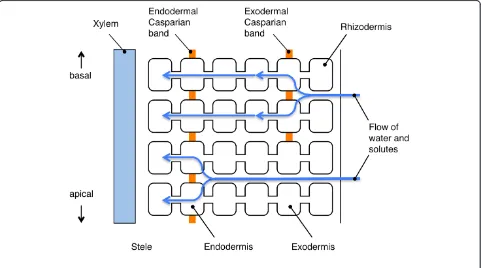
The root is the only organ that is directly exposed to ex-cess salts under salt stress conditions, and at the same time, the root has important function to take up neces-sary solutes from the soil. Therefore, it is important how roots avoid the influx of excess salts. Solutes, once taken up by the roots from root surface, move across the root in the radial direction and enter xylem, where they are transported to the shoot. Potentially speaking, there are three pathways for the radial movement of solutes: apoplastic, symplastic and transcellular pathways (Figure 4a). Among these pathways, the transcellular component may be negligible because of the low mem-brane permeability of most solutes (Steudle and Peterson 1998). A general understanding is that apoplastic trans-port barriers in roots are present in the endodermis and the exodermis (Perumalla and Peterson 1986). The endodermis of roots of all vascular plants (Clarkson and Robards 1975) and the exodermis of roots of many angiosperms (Perumalla and Peterson 1986; Perumalla et al. 1990) develop the Casparian band that is located in the transverse and the radial walls of cells of these tis-sues (Figure 5). Suberin is impregnated in the cell wall (Schreiber et al. 1999; Zeier et al. 1999) and the plasma membrane tightly attaches to the cell wall at the site of the Casparian band (Karahara and Shibaoka 1992; 1994). Suberin lamellae are formed in these tissues on the inner
surface of the primary cell walls as secondary cell wall modifications after the formation of the Casparian band (Perumalla and Peterson 1986). The function of the Casparian band is to avoid non-selective apoplastic ra-dial movements of solutes into the stele, and the func-tion of suberin lamellae is to block the passage of water and ions through the plasma membrane into the root stele (Steudle and Peterson 1998; Schreiber et al. 1999). Based on the earlier physiological and morphological studies, it is generally considered that the initial uptake of solutes could occur at the epidermis, at the exoder-mis, or if soil solution flows apoplastically across the root cortex, it would occur at the endodermis (Enstone et al. 2003).
Bypass flow of solutes and the apoplastic pathway in rice roots
From the general view point mentioned above, it is quite natural to hypothesize that the mechanism of salt exclu-sion could occur at the exodermis or at the endodermis in the radial direction, and from there, salt is transported ra-dially via a symplastic pathway (Storey and Walker 1998; Munns 2002) because flow of solutes through the apoplast is arrested at the site of the Casparian band and finally solutes are selectively transported through the plasma membrane into the symplast (Figure 5). However, as described in the previous section, studies using apoplastic tracer showed that major pathway of uptaking Na+ ions
into the stele is apoplastic pathway in the case of rice (Yeo et al. 1987; Yadav et al. 1996; Ochiai and Matoh 2002).
As discussed by Garcia et al. (1997) and also mentioned in the previous section, the uptake of sodium ions by the bypass flow possibly involves direct apoplastic leakage oc-curring at the root apices (Enstone and Peterson 1992) and the sites of secondary root emergence (Peterson et al. 1981; Ranathunge et al. 2005) (Figure 4a). On the other hand, experiments using pressure probes have indicated a possi-bility of some passage of water and perhaps also of solutes bypassing protoplasts (Steudle and Frensch 1996). With re-gard to the outer part of the root (OPR), Ranathunge et al. (2005) performed experiments introducing precipitates of insoluble inorganic salts to block apoplastic pores and sug-gested existence of predominant apoplastic bypass flow of water as well as high permeability of ions of the OPR, sup-porting at least a possibility of apoplastic bypass flow of solutes in the OPR. Although it may appear somehow con-troversial about the mechanism of the bypass flow of solutes, a fascinating model called "composite transport" of both water and solutes is proposed. This model assumes that a switching between the apoplastic and cell-to-cell pathways occurs depending on given circumstances and explains how plants optimize water uptake according to demands from the shoot (Steudle and Frensch 1996; Steu-dle and Peterson 1998; Miyamoto et al. 2001). The compos-ite transport model may also be applied even to rice roots in which aerenchyma extensively develop (Ranathunge et al. 2005).
A unique point of view accounting for the bypass flow discussed by Munns (2002) is that large gaps created be-tween the plasma membrane and the cell wall may fill with solution and allow an artifactual apoplastic pathway for salts to move radially across the root under salinity stress conditions. However, this view might be unlikely at the sites of the Casparian band because plasma mem-brane tightly attaches to the cell wall at these sites even under osmotic (or salinity) stress conditions that causes severe plasmolysis (Karahara and Shibaoka 1992).
Silicon alleviates abiotic stresses including salinity stress in rice (Matoh et al. 1986; Ma and Yamaji 2006). This function of silicon may be ascribed, in part, to the partial blockage of the transpirational bypass flow (Ma 2004). In the case of alleviation of cadmium toxicity by silicon in maize, possible involvement of a change in the development of endodermal suberin lamellae is sug-gested (Vaculik et al. 2009). Such a possibility could also be suggested in the case of alleviation of salinity stress by silicon in rice.
Responses of roots with regard to the apoplastic transport barriers under salinity stress
To understand the detailed resistance mechanisms to salinity stress as well as of the bypass flow in rice roots,
it is undoubtedly necessary to focus on the development and function of the apoplastic transport barriers in roots. Because an issue about the Casparian band development and its potential functions in salt tolerance has been re-cently reviewed (Chen et al. 2011), here we refer mainly to rice roots. One of the most advantageous features of using rice for plant physiological studies is the availabil-ity of many cultivars (varieties) for comparative studies.
Rice roots develop aerenchyma extensively in the cor-tex (Clark and Harris 1981) and, after its formation, only several cell layers are left intact in the OPR. These cell layers in the OPR are composed of cortical cell layers that appear to be unmodified, sclerenchyma, exodermis, and rhizodermis from the inside to the outside (Clark and Harris 1981) (Figure 6). It is suggested that low overall hydraulic conductivity of rice roots is a result of the existence of apoplastic barriers in the OPR and a strongly developed endodermis (Miyamoto et al. 2001).
Among the structures in the OPR, a unique anatomical feature of rice roots, when compared with other species of cereal plants, is the formation of sclerenchyma of seminal roots and nodal roots (Galamay et al. 1991) (Figure 6). This highly lignified tissue is formed so that it can pre-sumably complement structural weakness caused by the extensive formation of aerenchyma in the cortex of rice roots. This sclerenchyma is composed of single or double cell layers and the number of this cell layers depends on rice varieties and has been considered to have a role in re-ducing water loss from roots (Feldman 1984). However, no suberin could be detected in sclerenchyma by staining with Sudan Red 7B (Ranathunge et al. 2003), indicating in-effectiveness of this tissue as an apoplastic barrier. Never-theless, this sclerenchyma might be related to some unique physiological characteristics of rice roots, which is to be revealed in the future.
From the viewpoint of the issue about the bypass flow of water and ions, function of the OPR as the apoplastic barrier was examined using perfusion techniques (Ranathunge et al. 2003; 2005). Ranathunge et al. (2005) perfused the aerenchyma and the ORP with perfusion media containing different salts. When the salt of the out-side of the root diffused across the apoplast of the OPR, the two different salts formed a coloured precipitate where they met, indicating a considerable permeability of the exodermal Casparian band to salts (Ranathunge et al. 2005). This phenomenon may be attributed to a part of unique variable features of the exodermis (Hose et al. 2001) and testing a permeability of the endodermis using the same technique would be interesting as mentioned by the same authors (Ranathunge et al. 2005).
bands in the cell wall in maize roots (Zimmermann et al. 2000). Suberin is a biopolymer consisting of an aliphatic and an aromatic domain with the aliphatic component being the major contributor to its apoplastic barrier functions (Schreiber et al. 1999). To understand the im-portance of suberin in apoplastic barriers under salinity stress, comparative analyses of apoplastic barrier forma-tion (i.e., morphology and suberin composiforma-tion deter-mined by gas chromatography and mass spectrometry) and Na+ uptake under salinity stress among different rice cultivars have been performed (Krishnamurthy et al. 2009). They have found that suberization of apo-plastic barriers in roots was most extensive in salt-tolerant cultivar, which also has the least sodium accu-mulation in the shoots. They also found that salinity stress induced the strengthening of these barriers in both sensitive and tolerant cultivars by increasing the expression of the genes encoding suberin biosynthetic enzymes. Cai et al. (2011) have also performed a similar comparative study using Fourier transform infrared spectroscopy and have found that development and de-position of suberin at the Casparian band in the endo-and exodermis were earlier, endo-and the amounts of major chemical components in the OPR, such as aliphatic su-berin, lignin and cell wall proteins and carbohydrates, were higher in the most salt-tolerant cultivar examined. These pieces of evidence indicate a correlation between development and suberization of apoplastic barriers and salt tolerance. Furthermore, chemical composition of su-berin in rice roots and the roots hydraulic conductivity were analyzed and compared with those in maize roots by Schreiber et al. (2005). They have detected significantly higher amounts of suberin, i.e., 34 times greater in the
endodermis and six times greater in the exodermis, in the apoplastic barriers of rice corresponded with a substan-tially lower root hydraulic conductivity compared with maize. It is no doubt that this higher amount of suberin content in the endodermis indicates the importance of the endodermis as an apoplastic barrier in rice roots. On the other hand, it is also suggested that this is true only for the endodermis because the OPR of rice root is highly porous and permeable to water and that, therefore, more detailed consideration of both the chemical nature and polymer arrangement of suberins in apoplastic barriers is necessary (Schreiber et al. 2005; Ranathunge et al. 2011). A similar opinion was also mentioned in the case of Ara-bidopsis roots based on a study using suberin mutants (Ranathunge and Schreiber 2011).
depends on cell division rate, cell elongation rate as well as the time required for formation of the band in individ-ual cells. This issue was tested in the case of development of the endodermal Casparian band in maize roots under salinity stress and it was demonstrated that the estimated time required for formation of the endodermal Casparian band in an individual cell did not change under salinity stress even when the distance from the root tip to the low-est position of the Casparian band decreased (Karahara et al. 2004). Therefore, a conclusion that Casparian band development is accelerated by salinity stress cannot be drawn exclusively from the observation that the band forms closer to the root tip under the stress. To solve this problem, a unique integrative analysis was proposed to monitor changes in the developmental processes of a par-ticular cell type in the root, i.e. the rates of cell differenti-ation, production, and elongation (Karahara et al. 2008). As a model case, effects of exogenous ethylene on differ-entiation, i.e. formation of the endodermal Casparian band, division and elongation of endodermal cells were analyzed in maize primary roots. Effects of environmental stresses, including salinity stress, on the development of barrier structures should be examined or re-examined by this approach. A recent finding of a novel protein family mediating Casparian band formation in the endodermis of
Arabidopsis root (Roppolo et al. 2011) has shed a new light on the study of the Casparian band development, which might also be relevant to salt sensitivity of plants.
Conclusion
Soil salinity is a serious problem in the world agriculture. Owing to efforts of investigators and elevated levels of technologies, our knowledge on the mechanisms of plant salinity tolerance is dramatically expanding these days. However, individual plant species exhibits distinct salt sensitivity due to morphological differences and the differ-ence in the ability of protection components that the plant has evolved to depend on. Therefore, challenges to eluci-date the roles/significances of protection mechanisms in plant salt tolerance including morphological barriers at molecular, cellular and whole plant levels in further depth will be crucial to develop high-yielding salt-tolerant cultivars.
Competing interests
The authors declare that they have no competing interests.
Acknowledgements
We thank Dr. Junko Sakura-Ishikawa and Dr. Mari Murai-Hatano (NARO Tohoku Agricultural Research Center, Japan) for providing us with the unpublished data. We would also like to thank Dr. Pulla Kaothien-Nakayama for the comments on the manuscript. This work was supported by the grants from the Ministry of Education, Culture, Sports, Science, and Technology, Japan (Grant-in-Aid for Scientific Research no. 23119507 T.H.) and the Program for Promotion of Basic Research Activities for Innovative Biosciences (PROBRAIN), Japan (to M.K.).
Author details 1
Division of Applied Biology, Faculty of Textile Science and Technology, Shinshu University, 3-15-1, Tokida, Ueda, Nagano 386-8567, Japan. 2
Department of Biology, Graduate School of Science and Engineering, University of Toyama, 3190 Gofuku, Toyama 930-8555, Japan.3Institute of Plant Science and Resources, Okayama University, 20-1, Chuo-2-chome, Kurashiki, Okayama 710-0046, Japan.
Authors’contribution
TH, writing a part of section 1 and the whole part of section2, and coordinating the contents of this review; IK, writing whole part of the section3; MK, writing the section1. All authors read and approved the final manuscript.
Received: 13 June 2012 Accepted: 22 June 2012 Published: 22 June 2012
References
Amtmann A, Sanders D (1999) Mechanisms of Na+uptake by plant cells. Adv Bot Res 29:75–112
Amtmann A, Laurie S, Leigh R, Sanders D (1997) Multiple inward channels provide flexibility in K+/Na+discrimination at the plasma membrane of barley suspension culture cells. J Exp Bot 48:431–440
Anil VS, Krishnamurthy P, Kuruvilla S, Sucharitha K, Thomas G, Mathew MK (2005) Regulation of the uptake and distribution of Na+in shoots of rice (Oryza sativa) variety Pokkali: role of Ca2+in salt tolerance response. Physiol Plant 124:451–464
Apse MP, Aharon GS, Snedden WA, Blumwald E (1999) Salt tolerance conferred by overexpression of a vacuolar Na+/H+antiport inArabidopsis. Science 285:1256–1258
Asch F, Dingkuhn M, Sow A, Audebert A (2005) Drought-induced changes in rooting patterns and assimilate partitioning between root and shoot in upland rice. Field Crop Res 93:223–236
Azaizeh H, Steudle E (1991) Effects of salinity on water transport of excised maize (Zea maysL.) roots. Plant Physiol 97:1136–1145
Bassil E, Ohto MA, Esumi T, Tajima H, Zhu Z, Cagnac O, Belmonte M, Peleg Z, Yamaguchi T, Blumwald E (2011) TheArabidopsisintracellular Na+/H+ antiporters NHX5 and NHX6 are endosome associated and necessary for plant growth and development. Plant Cell 23:224–239
Berthomieu P, Conejero G, Nublat A, Brackenbury WJ, Lambert C, Savio C, Uozumi N, Oiki S, Yamada K, Cellier F, Gosti F, Simonneau T, Essah PA, Tester M, Very AA, Sentenac H, Casse F (2003) Functional analysis of AtHKT1 inArabidopsisshows that Na+recirculation by the phloem is crucial for salt tolerance. EMBO J 22:2004–2014
Blaha G, Stelzl U, Spahn CM, Agrawal RK, Frank J, Nierhaus KH (2000) Preparation of functional ribosomal complexes and effect of buffer conditions on tRNA positions observed by cryoelectron microscopy. Method Enzymol 317:292–309
Blumwald E (1987) Tonoplast vesicles as a tool in the study of ion transport at the plant vacuole. Physiol Plant 69:731–734
Blumwald E (2000) Sodium transport and salt tolerance in plants. Curr Opin Cell Biol 12:431–434
Blumwald E, Poole R (1985) Na+/H+-antiport in isolated tonoplast vesicles from storage tissue ofBeta vulgaris. Plant Physiol 78:163–167
Blumwald E, Poole RJ (1987) Salt tolerance in suspension cultures of sugar beet: induction of Na+/H+antiport activity at the tonoplast by growth in salt. Plant Physiol 83:884–887
Bohnert HJ, Shen B (1999) Transformation and compatible solutes. Sci Hortic 78:237–260
Boursiac Y, Chen S, Luu DT, Sorieul M, van den Dries N, Maurel C (2005) Early effects of salinity on water transport inArabidopsisroots. Molecular and cellular features of aquaporin expression. Plant Physiol 139:790–805
Boursiac Y, Boudet J, Postaire O, Luu DT, Tournaire-Roux C, Maurel C (2008) Stimulus-induced downregulation of root water transport involves reactive oxygen species-activated cell signalling and plasma membrane intrinsic protein internalization. Plant J 56:207–218
Cai X, Chen T, Zhou QY, Xu L, Qu LQ, Hua XJ, Lin JX (2011) Development of Casparian strip in rice cultivars. Plant Signal Behav 6:59–65
Carvajal M, Martinez V, Alcaraz CF (1999) Physiological function of water channels as affected by salinity in roots of paprika pepper. Physiol Plant 105:95–101 Chaumont F, Moshelion M, Daniels MJ (2005) Regulation of plant aquaporin
activity. Biol Cell 97:749–764
Chen T, Cai X, Wu X, Karahara I, Schreiber L, Lin J (2011) Casparian strip development and its potential function in salt tolerance. Plant Signal Behav 6:1499–1502
Clark LH, Harris WH (1981) Observations on the root anatomy of rice (Oryza sativaL.). Am J Bot 68:154–161
Clarkson DT, Robards AW (1975) The endodermis, its structural development and physiological role. In: Torrey JG, Clarkson DT (eds) The development and function of roots. Acedemic Press, London, pp 415–436
Davenport RJ, Tester M (2000) A weakly voltage-dependent, nonselective cation channel mediates toxic sodium influx in wheat. Plant Physiol 122:823–834
Davenport RJ, Munoz-Mayor A, Jha D, Essah PA, Rus A, Tester M (2007) The Na+ transporter AtHKT1;1 controls retrieval of Na+from the xylem inArabidopsis. Plant Cell Environ 30:497–507
Demidchik V, Davenport RJ, Tester M (2002) Nonselective cation channels in plants. Annu Rev Plant Biol 53:67–107
Enstone JE, Peterson CA, Ma FS (2003). Root endodermis and exodermis: structure, function, and responses to the environment. J Plant Growth Regul 21:335–351
Enstone DE, Peterson CA (1992) The apoplastic permeability of root apices. Can J Bot 70:1502–1512
Faiyue B, Vijayalakshmi C, Nawaz S, Nagato Y, Taketa S, Ichii M, Al-Azzawi MJ, Flowers TJ (2010) Studies on sodium bypass flow in lateral rootless mutants lrt1andlrt2, and crown rootless mutantcrl1of rice (Oryza sativaL.). Plant Cell Environ 33:687–701
Feldman LJ (1984) Regulation of root development. Annu Rev Plant Physiol 35:223–242
Galamay TO, Kono Y, Yamauchi A, Shimizu M (1991) Comparative root anatomy of seminal and nodal root axes of summer cereals with special reference to the development of hypodermis and cortical sclerenchyma. Jpn J Crop Sci 60:184–190
Garcia A, Rizzo CA, Ud-Din J, Bartos SL, Senadhira D, Flowers TJ, Yeo AR (1997) Sodium and potassium transport to the xylem are inherited independently in rice, and the mechanism of sodium: potassium selectivity differs between rice and wheat. Plant Cell Environ 20:1167–1174
Garciadeblás B, Senn M, Banuelos M, Rodriguez-Navarro A (2003) Sodium transport and HKT transporters: the rice model. Plant J 34:788–801 Garthwaite AJ, von Bothmer R, Colmer TD (2005) Salt tolerance in wildHordeum
species is associated with restricted entry of Na+and Cl-into the shoots. J Exp Bot 56:2365–2378
Gassmann W, Rubio F, Schroeder JI (1996) Alkali cation selectivity of the wheat root high-affinity potassium transporter HKT1. Plant J 10:869–882 Gaxiola RA, Rao R, Sherman A, Grisafi P, Alper SL, Fink GR (1999) TheArabidopsis
thalianaproton transporters, AtNhx1 and Avp1, can function in cation detoxification. Proc Natl Acad Sci USA 96:1480–1485
Gaxiola RA, Li J, Undurraga S, Dang LM, Allen GJ, Alper SL, Fink GR (2001) Drought- and salt-tolerant plants result from overexpression of the AVP1 H+-pump. Proc Natl Acad Sci USA 98:11444–11449
Gierth M, Mäser P (2007) Potassium transporters in plants-involvement in K+ acquisition, redistribution and homeostasis. FEBS Lett 581:2348–2356 Glenn EP, Brown JJ, Blumwald E (1999) Salt tolerance and crop potential of
halophytes. Crit Rev Plant Sci 18:227–255
Gorham J, Wyn Jones RG, McDonnell E (1985) Some mechanisms of salt tolerance in crop plants. Plant Soil 89:15–40
Gorham J, Hardy C, Wyn Jones RG, Joppa LR, Law CN (1987) Chromosomal location of a K/Na discriminating character in the D genome of wheat. Theor Appl Genet 74:584–588
Gorham J, Wyn Jones RG, Bristol A (1990) Partial characterization of the trait for enhanced K+-Na+discrimination in the D genome of wheat. Planta 180:590–597
Greenway H, Munns R (1980) Mechanisms of salt tolerance in nonhalophytes. Ann Rev Plant Phys 31:149–190
Guo L, Wang ZY, Lin H, Cui WE, Chen J, Liu M, Chen ZL, Qu LJ, Gu H (2006) Expression and functional analysis of the rice plasma membrane intrinsic protein gene family. Cell Res 16:277–286
Hachez C, Zelazny E, Chaumont F (2006) Modulating the expression of aquaporin genes in planta: A key to understand their physiological functions? Biochim Biophys Acta 1758:1142–1156
Halfter U, Ishitani M, Zhu JK (2000) The Arabidopsis SOS2 protein kinase physically interacts with and is activated by the calcium-binding protein SOS3. Proc Natl Acad Sci USA 97:3735–3740
Hauser F, Horie T (2010) A conserved primary salt tolerance mechanism mediated by HKT transporters: a mechanism for sodium exclusion and maintenance of high K/Na ratio in leaves during salinity stress. Plant Cell Environ 33:552–565
Horie T, Schroeder JI (2004) Sodium transporters in plants. Diverse genes and physiological functions. Plant Physiol 136:2457–2462
Horie T, Yoshida K, Nakayama H, Yamada K, Oiki S, Shinmyo A (2001) Two types of HKT transporters with different properties of Na+and K+transport in Oryza sativa. Plant J 27:129–138
Horie T, Horie R, Chan WY, Leung HY, Schroeder JI (2006) Calcium regulation of sodium hypersensitivities ofsos3andathkt1mutants. Plant Cell Physiol 47:622–633
Horie T, Costa A, Kim TH, Han MJ, Horie R, Leung HY, Miyao A, Hirochika H, An G, Schroeder JI (2007) Rice OsHKT2;1 transporter mediates large Na+influx component into K+-starved roots for growth. EMBO J 26:3003–3014 Horie T, Hauser F, Schroeder JI (2009) HKT transporter-mediated salinity resistance
mechanisms inArabidopsisand monocot crop plants. Trends Plant Sci 14:660–668
Horie T, Brodsky DE, Costa A, Kaneko T, Lo Schiavo F, Katsuhara M, Schroeder JI (2011a) K+transport by the OsHKT2;4 transporter from rice with atypical Na+ transport properties and competition in permeation of K+over Mg2+ and Ca2+ions. Plant Physiol 156:1493–1507
Horie T, Kaneko T, Sugimoto G, Sasano S, Panda SK, Shibasaka M, Katsuhara M (2011b) Mechanisms of water transport mediated by PIP aquaporins and their regulation via phosphorylation events under salinity stress in barley roots. Plant Cell Physiol 52:663–675
Horie T, Sugawara M, Okada T, Taira K, Kaothien-Nakayama P, Katsuhara M, Shinmyo A, Nakayama H (2011c) Rice sodium-insensitive potassium transporter, OsHAK5, confers increased salt tolerance in tobacco BY2 cells. J Biosci Bioeng 111:346–356
Hose E, Clarkson DT, Steudle E, Schreiber L, Hartung W (2001) The exodermis: a variable apoplastic barrier. J Exp Bot 52:2245–2264 Huang S, Spielmeyer W, Lagudah ES, James RA, Platten JD, Dennis ES, Munns R
(2006) A sodium transporter (HKT7) is a candidate forNax1, a gene for salt tolerance in durum wheat. Plant Physiol 142:1718–1727
Huang S, Spielmeyer W, Lagudah ES, Munns R (2008) Comparative mapping of HKT genes in wheat, barley, and rice, key determinants of Na+transport, and salt tolerance. J Exp Bot 59:927–937
IRRI (2011) Stress and disease tolerance: 1. Breeding for salt tolerance in rice. http://www.knowledgebank.irri.org/ricebreedingcourse/bodydefault. htm#breeding_for_salt_tolerance.htm. 2006; Accessed 12 October James RA, Davenport RJ, Munns R (2006) Physiological characterization of two
genes for Na+exclusion in durum wheat,Nax1andNax2. Plant Physiol 142:1537–1547
Javot H, Maurel C (2002) The role of aquaporins in root water uptake. Ann Bot 90:301–313
Javot H, Lauvergeat V, Santoni V, Martin-Laurent F, Guclu J, Vinh J, Heyes J, Franck KI, Schaffner AR, Bouchez D, Maurel C (2003) Role of a single aquaporin isoform in root water uptake. Plant Cell 15:509–522 Jeschke WD (1984) K+-Na+Exchange at Cellular Mmbranes, Intracellular
Compartmentation of Cations, and Salt Tolerance. Sanity Tolerance in Plant. Strategies for Crop Improvement. Wiley-Interscience Publication, New York, pp 33–76
Jia W, Wang Y, Zhang S, Zhang J (2002) Salt-stress-induced ABA accumulation is more sensitively triggered in roots than in shoots. J Exp Bot 53:2201–2206
Kader MA, Lindberg S (2005) Uptake of sodium in protoplasts of sensitive and salt-tolerant cultivars of rice,Oryza sativaL. determined by the fluorescent dye SBFI. J Exp Bot 56:3149–3158
Karahara I, Shibaoka H (1992) Isolation of Casparian strips from pea roots. Plant Cell Physiol 33:555–561
Karahara I, Shibaoka H (1994) The Casparian strip in pea epicotyls: effects of light on its development. Planta 192:269–275