O R I G I N A L A R T I C L E
Intramedullary nailing versus proximal plating
in the management of closed extra-articular proximal tibial
fracture: a randomized controlled trial
Ramesh Chand Meena• Umesh Kumar Meena•
Gopal Lal Gupta•Nitesh Gahlot•Sahil Gaba
Received: 26 April 2014 / Accepted: 27 December 2014 / Published online: 15 January 2015 ÓThe Author(s) 2015. This article is published with open access at Springerlink.com
Abstract
Background Extra-articular proximal tibial fractures account for 5–11 % of all tibial shaft fractures. In recent years, closed reduction and minimally invasive plating and multidirectional locked intramedullary nailing have both become widely used treatment modalities for proximal and distal tibial metaphyseal fractures. This study was per-formed to compare plating and nailing options in proximal tibia extra-articular fractures.
Materials and methods This randomized prospective clinical study was conducted on 58 skeletally mature patients with a closed extra-articular fracture of the prox-imal tibia treated with minprox-imally invasive proxprox-imal tibial plating (PTP) or intramedullary nailing (IMN) by trained surgeons at a tertiary trauma center.
Results Postoperative hospital stay (p=0.035), time to full weight-bearing, and union time (p =0.004) were significantly less in the IMN group than in the PTP group, but there was no clear advantage of either technique in terms of operative time (p=0.082), infection rate
(p =0.738), range of motion of the knee (p=0.462), or degrees of malunion and nonunion.
Conclusion Both implants have shown promising results in extra-articular proximal tibial fractures, and provide rigid fixation that prevents secondary fracture collapse. Level of evidence Level 2, randomized controlled trial.
Keywords Intramedullary nailing (IMN)Proximal tibial plate (PTP)Proximal tibial extra-articular fractures Prospective trial
Introduction
Extra-articular proximal tibial fractures account for 5–11 % of all tibial shaft fractures [1,9] and often result from high-velocity trauma. They lead to complex tissue injuries involving bone and surrounding soft tissues [1]. Conservative management of these fractures has often resulted in malunion, nonunion, rotational deformity, or stiffness of adjacent joints [2–4], so there has been a shift towards operative management of these fractures in recent times. However, the optimal method of surgically treating these fractures remains debatable. Options include intra-medullary implant, half-pin external fixation, hybrid or thin-wire external fixation, plate fixation, or a combination of these techniques [5,17]. In recent years, closed reduc-tion with minimally invasive plating and locked intra-medullary nailing have both become widely used treatment modalities for proximal and distal tibial metaphyseal fractures [6–8], despite the absence of any conclusive proof of the superiority of one modality over the other.
Due to the paucity of the relevant literature and the lack of conclusive evidence to guide the selection of treatment options in such cases, we designed this randomized R. C. MeenaU. K. Meena (&)G. L. GuptaS. Gaba
Department of Orthopaedics, SMS Medical College and Hospital, Jaipur 302004, India
e-mail: drumesh_meena@yahoo.co.in
R. C. Meena
e-mail: rc.meena@yahoo.com
G. L. Gupta
e-mail: drglg4u@gmail.com
S. Gaba
e-mail: drsahilgaba@gmail.com
N. Gahlot
Department of Orthopaedics, Postgraduate Institute of Medical Education and Research, Chandigarh, India
controlled study (RCT) in order to compare the plating and nailing options in proximal tibia extra-articular fractures. We intended to compare these options in terms of operative time, duration of hospital stay, period of non-weight-bearing, degree of reduction, union rate, malunion rate, infection rate, and rates of other possible complications which could possibly affect decision-making in relation to such fracture patterns.
Materials and methods
This randomized prospective clinical study was conducted on 58 patients with extra-articular fracture of the proximal tibia (OTA 41-A2/A3) treated with minimally invasive proximal tibial plating (PTP) or intramedullary nailing (IMN) by trained surgeons at a tertiary trauma care center in the Department of Orthopedics, SMS Medical College and Hospital, Jaipur, between January 2009 and December 2012. After excluding 14 patients who were lost to follow-up, a total of 44 patients were included in the final outcome analysis. Ethical committee approval was obtained, and patients were recruited once written informed consent had been provided.
Skeletally mature patients with closed proximal tibial metadiaphyseal fractures were included in this study. The proximal tibia was defined as the region extending from the articular surface up to 1.5 times the medial to lateral width of the articular surface [6]. Patients with metadiaphyseal tibial fractures with an intra-articular extension, tibial shaft fractures, open fractures, pathological fractures, and patients with multiple musculoskeletal injuries to the same or opposite lower limb were excluded from the study.
Patient allocation to groups was randomized by com-puter prospectively through the use of sequentially num-bered opaque envelopes. Envelopes were opened inside the operating theater by a nurse who was blind to the alloca-tion. Group A patients were treated with IMN and group B patients received PTP.
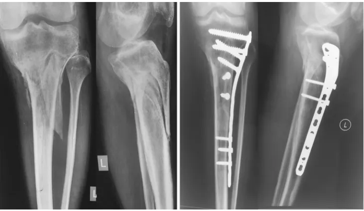
The intramedullary nailing performed in group A was done by creating an entry point just medial to the lateral intercondylar eminence of the tibial plateau through a medial parapatellar approach. Temporary blocking screws, a reduction clamp, a reduction unicortical plate, or a universal fixator was used to achieve reduction and removed after fracture fixation, except for the reduction unicortical plate when used with a reamed intramedullary tibial nail. The intramedullary nail used had a proximal Herzog band and four multilevel, multiplanar, and multidirectional screws (Expert Tibial Nail, Synthes, Zuchwil, Switzerland) (Fig.1). Patients in group B were treated by minimally invasive PTP using curvilinear incision over the lateral aspect of the proximal tibia. Indirect reduction was achieved using axial
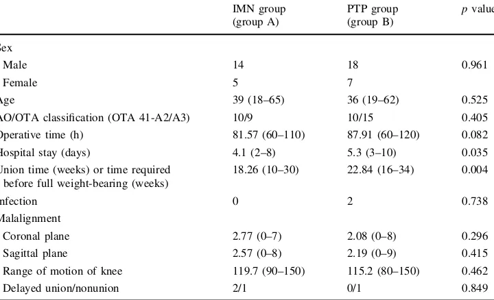
traction and/or the application of a reduction clamp or distractor. Internal fixation was then achieved with a proximal tibial lateral locking compression plate (LCP). A minimum of three screws were used on both sides of the fracture, and plating was done using a minimally invasive technique (Fig.2).
Postoperatively, patients in both groups were given intravenous third-generation cephalosporin antibiotics for 3 days. Ankle pumps and isometric quadriceps strength-ening exercises were started on the first postoperative day, followed by active and assisted knee bending on the second postoperative day. Partial weight-bearing was allowed from the second postoperative day, depending upon the stability of the construct, whereas full weight-bearing was allowed only after complete clinical and radiological union.
All patients were followed up at 2 and 6 weeks, 3 and 6 months, and 1 year postoperatively. Both the immediate postoperative and the final follow-up radiographs were compared to assess the accuracy of reduction and final alignment. Measurements were performed for coronal (varus and valgus) and sagittal (procurvatum and recurva-tum) plane deformities using the measuring technique described by Freedman and Johnson [9]. In AP view, varus/ valgus deformity was evaluated by measuring the angle between the lines drawn perpendicular to the proximal and distal tibial articular surfaces. In lateral view, the pro-curvatum/recurvatum was measured similarly and 8° of posterior slope was subtracted. Malreduction was defined as a deformity of[5°in any plane. Rotational alignment, shortening, and knee ROM were assessed clinically [9,10]. The fracture was considered united if three or more cortices were continuous on two radiographic views. Nonunion was defined as three consecutive months of X-rays that did not show progressive healing.
All data were entered into a pro forma. The statistical analysis was performed by an independent statistician using the Statistical Package for the Social Sciences (SPSS version 22.0; SPSS, Chicago, IL, USA). The chosen level of significance was p\0.05. The two groups were com-pared with respect to age, sex, operating time, hospital stay, infection rate, fracture union time, angulation of the frac-ture, and the knee range of motion. The parameters were compared between the groups. A paired-samplettest was used for the interval data (age, operating time, length of hospital stay, fracture union time, postoperative angulation, and range of motion of the knee).
Results
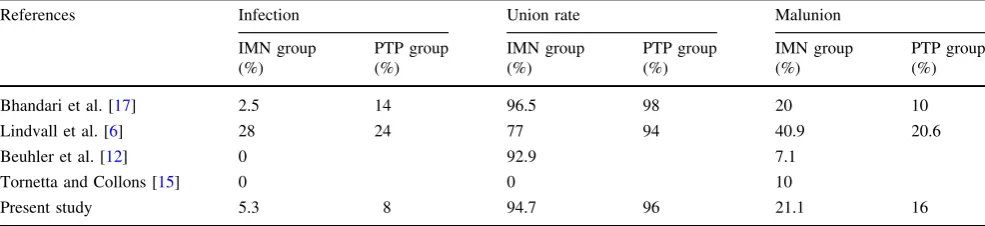
included in the final outcome analysis. Preoperative char-acteristics including age, sex, classification, mode of injury, and time period from injury to operation were comparable in both groups (Table1).
Postoperative hospital stay, time period to full weight-bearing, and union time were significantly less in the IMN
group as compared to the PTP group (Table1). Surgical site infections (SSIs) were seen in two patients in the PTP group, one of which was resolved with debridement while the other necessitated implant removal due to infection.
Delayed union occurred in two patients in the IMN group, for which dynamization was performed by Fig. 1 Patient with a segmental tibial fracture treated with expert tibial nail, showing a good range of motion of the knee postoperatively
removing the distal screw. One case in the nailing group presented nonunion, which ultimately required exchange nailing with bone grafting and fibular osteotomy. There was nonunion in one patient in the PTP group; bone grafting was done in that case, which eventually led to fracture healing.
The alignment of the tibia, as measured by an inde-pendent observer in the immediate postoperative and 1-year follow-up X-rays, did not show any significant difference between the groups, indicating that there was no secondary loss of reduction. The mean postoperative angulation in the coronal plane (varus/valgus) was 2.7°
(range 0–7°, SD=1.98) in the IMN group and 2.1°
(range 0–8°, SD=1.77) in the PTP group; both of these tended towards a varus inclination, but there was no sta-tistically significant difference between the groups (p=0.296). In the sagittal plane, the mean extent of postoperative procurvatum/recurvatum was 2.6° (range 0–8°, SD=1.82) in the IMN group and 2.2°(range 0–9°, SD=1.98) in the PTP group; both of these tended towards procurvatum, but there was no statistically sig-nificant difference between the groups (p=0.415).[5°
of malalignment was seen in four patients (21.1 %) in the IMN group (one patient had varus and three had anterior apex deformity) and in four patients (16 %) in the PTP group (two patients had varus and two patients had pro-curvatum). The average range of motion was 119.7°
(range 90–150°, SD=19.18) in group A and 115.2°
(range 80–150°, SD =17.28) in group B (p =0.462). There were complaints of occasional anterior knee pain and discomfort upon kneeling on the floor from six patients (31.6 %) in group A and two patients (8.0 %) in group B (p =0.097).
Discussion
Data allowing a comparison of tibial nail and minimally invasive plating for extra-articular proximal tibial fractures are scarce. The primary goal of this prospective study was to compare the results of tibial nailing and minimally invasive plating from various aspects.
In the present study, patients in the IMN group had a significantly shorter length of hospital stay compared with those in the PTP group (p\0.05) because of the smaller incision made during closed nailing, meaning that IMN results in less of an economic burden and a lower cost of healthcare to society than PTP.
Although early weight-bearing is inherently associated with a load-sharing device such as an IMN, the literature does not accurately predict an accepted time at which full weight-bearing should be initiated with either procedure. Various studies have often stated that weight-bearing should be initiated when it can be tolerated by the patient [6]. In previous studies of extra-articular proximal tibial fractures treated with IMN, full weight-bearing was initi-ated at various times ranging from 0 to 16 weeks, depending on the fracture location, fracture pattern, and surgeon’s preference [11,19]. Similarly, in extra-articular proximal tibial fractures treated with PLP, time to full weight-bearing has ranged from 6 to 13 weeks for the same reasons [6, 8,18]. In our study, the time required before full weight-bearing, which was done only after complete radiological union, was significantly less in the IMN group (18.26 weeks) as compared to the PTP group (22.84 weeks). Although these times are longer than those stated in previously published reports, we started full weight-bearing only after complete clinical and Table 1 Comparison of the
demographic and postoperative data for both groups
IMN group (group A)
PTP group (group B)
pvalue
Sex
Male 14 18 0.961
Female 5 7
Age 39 (18–65) 36 (19–62) 0.525
AO/OTA classification (OTA 41-A2/A3) 10/9 10/15 0.405
Operative time (h) 81.57 (60–110) 87.91 (60–120) 0.082
Hospital stay (days) 4.1 (2–8) 5.3 (3–10) 0.035
Union time (weeks) or time required before full weight-bearing (weeks)
18.26 (10–30) 22.84 (16–34) 0.004
Infection 0 2 0.738
Malalignment
Coronal plane 2.77 (0–7) 2.08 (0–8) 0.296
Sagittal plane 2.57 (0–8) 2.19 (0–9) 0.415
Range of motion of knee 119.7 (90–150) 115.2 (80–150) 0.462
radiological fracture union. That being said, we started passive and active assisted movements early—from day 2, progressing later to partial weight-bearing. Hence, we found no significant differences in range of motion of the knee between the groups.
Reported infection rates range from 0 to 8 % in nailing patients [5,12,17] and from 0 to 14 % in plating patients [17,18, 20]. But, in the study by Lindvall et al. [6], the authors reported significantly higher infection rates: 28 % in the nailing group and 24 % in the plating group. The most probable reason for this is the higher proportion (42.8 %) of patients with open fractures in their study [6]. In the systemic review by Bhandari et al. [17], the infection rates were 2.5 % in the nailing group and 14 % in the plating group. The infection rates in our series were 5.3 % in the IMN group and 8 % in the PTP group (p =0.738). Malunion is a documented complication of the nailing of proximal tibia fractures and has been reported to occur in 3–100 % of cases in previous studies (Table2) [9,11,17]. In our study, there was a malreduction/malunion rate of [5° in the IMN group (four patients, 21.1 %): varus malalignment in one patient and anterior apex deformity in three patients. Various techniques have been described for preventing malreduction, including the use of blocking screws [5,6], unicortical plating [13], a universal distractor [14], nailing in the semiextended position [15], or the use of a nail with a more proximal Herzog bend [16]. In our study, we used blocking screws in three cases, reduction plating in one case, and a universal distractor in two cases. In the other cases, a reduction clamp was used to prevent proximal fragment extension while inserting the nail. A common technique employed in all of the nailing cases was to make a slightly higher entry point than that normally used for tibial nail insertion. This modification brought our insertion point more in line with the medullary canal of the tibia, hence reducing the extension of the proximal frag-ment. The plating group also had four cases of malunion (16 %), but the difference was not statistically significant. In a systemic review of 17 studies by Bhandari et al. [17], the authors reported a higher malunion rate in the nailing group (20 %) than in the plating group (10 %). Similarly,
Lindvall et al. [6] reported a higher malunion rate in the nailing group—apex anterior malreduction occurred in 36 % of the patients in the IMN group and 15 % of those in the locking plate group—although this difference was not statistically significant.
When union rates after the initial fixation were analyzed in our study, it was found that the union rate in the IMN group was 94.7 and that in the PTP group was 96 % (p =0.849). The high union rates observed in our series are consistent with those stated in various published reports, which range from 91 to 100 % [6,8,11,19]. Our results were, however, higher than seen in a study per-formed by Lindvell et al. [6], where the authors noted union rates of 77 % in the IMN group and 94 % in the PTP group. We believe that this difference in union rates arose because open fractures were excluded from our series, not because of the type of procedure performed. The locked nail technique demonstrated advantages in terms of the operation time, hospital stay, early full weight-bearing, and time required for bony union.
We concluded from our study that intramedullary nail is superior to minimally invasive plating in terms of brevity of hospital stay and speed of union along with early full weight-bearing, but there was no clear advantage of either technique in terms of operative time, infection rate, range of motion of the knee, and rates of malunion and nonunion. Both implants yielded promising results with extra-articu-lar proximal tibial fractures and provided rigid fixation that prevented secondary fracture collapse.
Limitations of this study include the small number of patients, the involvement of multiple surgeons, the absence of long-term follow-up to evaluate the outcome of mala-lignment in terms of the development of osteoarthritis of the knee, and the use of both stainless steel and titanium implants, which may affect infection rates because titanium is more biocompatible than stainless steel, meaning that using titanium reduces the soft-tissue reaction and reduces the chance of infection.
Conflict of interest The authors declare that they have no conflict of interest related to the publication of this manuscript.
Table 2 Comparison of data obtained in the present work with data presented in the literature
References Infection Union rate Malunion
IMN group (%)
PTP group (%)
IMN group (%)
PTP group (%)
IMN group (%)
PTP group (%)
Bhandari et al. [17] 2.5 14 96.5 98 20 10
Lindvall et al. [6] 28 24 77 94 40.9 20.6
Beuhler et al. [12] 0 92.9 7.1
Tornetta and Collons [15] 0 0 10
Ethical standards All of the patients provided informed consent prior to enrollment, ethical clearance was obtained from the institu-tional review board, and the study was performed in accordance with the ethical standards of the 1964 Declaration of Helsinki as revised in 2000.
Open Access This article is distributed under the terms of the Creative Commons Attribution License which permits any use, dis-tribution, and reproduction in any medium, provided the original author(s) and the source are credited.
References
1. Court-Brown CM, McBirnie J (1995) The epidemiology of tibial fractures. J Bone Jt Surg Br 77(3):417–421
2. DeCoster TA, Nepola JV, el-Khoury GY (1988) Cast brace treatment of proximal tibia fractures. A 10-year follow-up study. Clin Orthop Relat Res 231:196–204
3. Jensen DB, Rude C, Duus B, Bjerg-Nielsen A (1990) Tibial plateau fractures. A comparison of conservative and surgical treatment. J Bone Jt Surg Br 72(1):49–52
4. Milner SA, Davis TR, Muir KR, Greenwood DC, Doherty M (2002) Long-term outcome after tibial shaft fracture: is malunion important? J Bone Jt Surg Am 84-A(6):971–980
5. Nork SE, Barei DP, Schildhauer TA, Agel J, Holt SK, Schrick JL et al (2006) Intramedullary nailing of proximal quarter tibial fractures. J Orthop Trauma 20(8):523–528
6. Lindvall E, Sanders R, Dipasquale T, Herscovici D, Haiduke-wych G, Sagi C (2009) Intramedullary nailing versus percuta-neous locked plating of extra-articular proximal tibial fractures: comparison of 56 cases. J Orthop Trauma 23:485–492
7. Hiesterman TG, Shafiq BX, Cole PA (2011) Intramedullary nailing of extra-articular proximal tibia fractures. J Am Acad Orthop Surg 19(11):690–700
8. Naik MA, Arora G, Tripathy SK, Sujir P, Rao SK (2013) Clinical and radiological outcome of percutaneous plating in extra-artic-ular proximal tibia fractures: a prospective study. Injury 44(8):1081–1086
9. Freedman EL, Johnson EE (1995) Radiographic analysis of tibial fracture malalignment following intramedullary nailing. Clin Orthop Relat Res 315:25–33
10. Milner SA (1997) A more accurate method of measurement of angulation after fractures of the tibia. J Bone Jt Surg 79(6):972–974
11. Krettek C, Stephan C, Schandelmaier P et al (1999) The use of Poller screws as blocking screws in stabilizing tibial fractures treated with small diameter intramedullary nails. J Bone Jt Surg Br 81:963–968
12. Beuhler KC, Green J, Woll TS, Duwelius PJ (1997) A technique for intramedullary nailing of proximal third tibia fractures. J Orthop Trauma 11(3):218–223
13. Moed BR, Watson JT (1994) Intramedullary nailing of the tibia without a fracture table: the transfixation pin distractor technique. J Orthop Trauma 8(3):195–202
14. Archdeacon MT, Wyrick JD (2006) Reduction plating for pro-visional fracture fixation. J Orthop Trauma 20(3):206–211 15. Tornetta P, Collons E (1996) Semiextended position of
intra-medullary nailing of the proximal tibia. Clin Orthop Relat Res 328:185–189
16. Henley MB, Meier M, Tencer AF (1993) Influences of some design parameters on the biomechanics of the unreamed tibial intramedullary nail. J Orthop Trauma 7(4):311–319
17. Bhandari M, Audige L, Ellis T (2003) Operative treatment of extraarticular proximal tibial fractures. J Orthop Trauma 17(8):591–595
18. Cole PA, Zlowodzki M, Kregor PJ (2004) Treatment of proximal tibia fractures using the less invasive stabilization system: sur-gical experience and early clinical results in 77 fractures. J Ort-hop Trauma 18(8):528–535
19. Koval K, Clapper M, Brumback R et al (1991) Complications of reamed intramedullary nailing of the tibia. J Orthop Trauma 5(2):184–189
20. Schutz M, Kaab MJ, Haas N (2003) Stabilization of proximal tibia fractures with the LIS-System: early clinical experience in Berlin. Injury 34(Suppl 1):A30–A35