Experience of Using Ultrasonography in the Diagnosis of
Cattle Reproductive Track Diseases in the West Kazakhstan
Region
KAZYBAY KARAYEVICH BOZYMOV , EDIGE NASSAMBAYEV , ALIBI NAUKHANOVICH
BAYAKHOV , BAITLESSOV YERBULAT UPIEVICH and ASSEL KUTTBAEVNA SULTANOVA
Western Kazakhstan Agrarian-Technical University named after Zhangir khan, Republic of Kazakhstan, 090009, Uralsk, Zhangir khan street, 51.
DOI: http://dx.doi.org/10.13005/bpj/577
(Received: February 06, 2015; accepted: March 07, 2015)
ABSTRACT
The study was conducted in the western region of Kazakhstan with beef production livestock. The article presents the results of transrectal ultrasonography at the diacrisis of reproduction tract disease. Ultrasonography made it possible not only to visualize the gynecological organs, but also to provided valuable diagnose information about the size of internal formations, their number, localization and morphotype. The aim of the study was to obtain preliminary information for future research that would allow providing scientific credence for the ways to increase beef cattle production through a fuller implementation of the genetic potential of animals.
Key words: Beef production livestock, Diacrisis of reproductive tract diseases, Transrectal ultrasonography.
INTRODUCTION
According to the report presented by Utitskih and Semenets, 2012, as well as Kostomahina, 2009, the optimal level of cattle reproduction is determined by the competence of cows reproductive and other organs and systems. However, exploiting of breeding stock to a large extent depends on a variety of pathological changes in the body and genitals of an animal that lead to a violation of their reproductive function, since the diseases of reproductive organs can cause temporary and often lengthy disorders of reproduction process.
Shkuratova and Ryaposova, 2011, note that high yielding cows show a rather high level of gynecological diseases regardless of the animals’ welfare technology. At tie-stall housing of cattle it reaches 95.8%, at yard housing – 81.1%, while the more inflammatory diseases of uterus and ovarian
hypofunction are reported in animals, which are limited in free day-night mobility.
According to Kumar et al., 2014, in recent years in the veterinary obstetrics and gynecology of large domestic animals, and, in particular, cows, the increasing application has visual transrectal ultrasonography. This diagnostic technique (Palgrave & Cezon, Saied et al., 2014, Purohit, 2014, Kumar & Purohit, 2009) is one of the most effective in the study of the ovaries and uterus structures. According to Purohit, 2010, ultrasonic technique allows also determining the geometric parameters of reproductive organs and detecting pathological processes, which may be indicated by fluctuations in echogenicity in the formation under visualization.
early pregnancy cows and heifers of Kazakh white-headed breed. The obtained data proved the advantages of using the ultrasonography technique for examination of genital organs of beef cattle breeding in West Kazakhstan region.
METHODOLOGY
The reported studies were carried out from April to November 2014 in the farms of West Kazakhstan region, specializing in the beef production livestock. In total 350 cows and heifers were surveyed.
When carrying out diagnostication, we used portable digital veterinary diagnostic apparatus KH 5200 with rectal linear transducer and disposable plastic gloves for rectal examination.
According to Ernst et al., 2008, Paschenco & Shevtsov, 2008, and Chaudhary & Purohit, 2012, to examine the uterus, a rectal transducer with a frequency of 7.5 MHz (LV2-2/7.5 MHz) was employed. Working surface of the sensor was smeared by sound-conducting gel, and then introduced into the rectum, scanning step by step uterus neck, body and horns, as well as ovaries. To comply with the aseptic and antiseptic rules, the chlorhexidine solution (0.05%) was used. A continuous pattern of the formation located beneath the sensor’s working surface, was observed on the screen.
Ovaries were examined by longitudinal and transverse scanning, while determining their maximum size (length, width, and thickness). Their volume was calculated using the formula for the ellipsoid volume (0.523 multiplied by length, width, and thickness). Special attention was paid to the ovary structure. When examining by ultrasound the infertile cows, thin-walled liquid anechoic formations larger than 20 mm were taken as ovarian cysts, given that at the first and second tests there was no corpus luteum. Cyst morphotype was judged by the presence/absence of the parietal luteal tissue. The pattern images were transferred to and saved in the computer.
Collection and evaluation of vaginal secretions, when diagnosing endometritis, was performed according to procedure described by Saut et al., 2011. The area of the vulva was cleaned and then vaginal secretion was collected by gloved hand. The secret was classified based on its color, proportion of its content, as well as the smell.
RESULTS AND DISCUSSION
The structure of functional disorders of the reproductive organs in beef production cows of West Kazakhstan region is shown in Diagram 1.
As is obvious from the presented diagram, cyst deceases are the major problem of the reproductive organs. This is consistent with the data of Jeengar et al., 2014.
Ovarian cysts are a quite common cause of infertility in cows. According to present-day concepts, they arise because of delayed and/or inadequate secretion of luteinizing hormone during estrus. Anovulation with the transformation of preovulatory follicle into the ovary cyst leads to a lengthening of the calving interval, disturbance of the reproduction rhythm and, consequently, economic losses.
To obtain the pattern images needed for the current work, we conducted an ovaries study of one of the cows with a negative diagnosis for pregnancy. Rectal examination gave an initial diagnosis of “packed follicles” (Fig.1). During subsequent ultrasonographic studies based on cow’s ovarian echogram this diagnosis was updated as “multiple follicular cyst”.
Diagram 1. The structure of functional disorders of the reproductive organs in beef production cows of West Kazakhstan region
During the study of sonographical image of ovarian tissue, voluminous ovarian formations large vesicular cysts were found. Their size, shape, number, location, and especially echo structure of abnormalities were determined visually.
Follicular cysts on echograms were defined as single unilateral or bilateral thin-walled liquid formations of round, oval or irregular shape with a uniform anechoic content and area of signal enhancement on the back surface. In terms of sonography, follicular cysts differed from vesicular follicles only by larger size, an average of 38x31 mm.
During subsequent diacrisis studies, when analyzing sonographic images, we revealed that the pathological process was progressing (Fig. 2).
Later it was visually found that cyst had four approximately identical cystic cavities with nonhomogeneous content, walls thickened up to 7 mm, having a high echolucency. Content of cystic formations was echo-negative and had inclusions of gray color.
Based on obtained images, it was proved that chronic long-lasting process, accompanied by degeneration of the tissues, was characterized by the formation of new similar cystic cavities that were firmly adhered to each other, as well as variation of echo response of liquid contents of cysts, and thickening of the chamber walls that led to polycystic ovary syndrome. Ultrasound diagnostic allowed visual tracing of the polycystic ovary progression and determining echo response of this pathology.
Without proper treatment, the tissue can be significantly compacted, followed by appearance of perifocal cicatrical formation, leading to polycystic ovary syndrome.
The clinical presentation at follicular ovarian cysts was manifested by infertility (100%), nymphomania (72%), or anaphrodisia (25%) involving fibrinous changes in the cyst. Our results are supported by the data of Ryaposova & Sokolova, 2011.
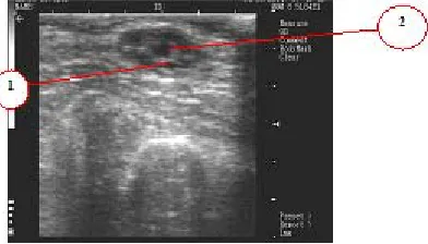
Due to the hypersensibility of the animals’ uterus to the biological, chemical and physical stimuli, inflammatory processes develop more Fig. 1. Ultrasound image of ovary at transrectal
examination of cow’s genitals. Cystic formation in the ovary wall: 1 - echo-positive membrane;
2 - cyst camera
Fig. 2. Ultrasound image of the ovary with cystic formations at transrectal examination of
cow’s genitals: 1 – echo-negative cystic formations; 2 - cysts partitions (echo-positive
membranes)
frequently in the uterus than in other parts of the animal’s reproductive system.
Meira et al., 2012, report that endometriosis is a superficial inflammation of the endometrium and is characterized histologically by a disturbance of the surface epithelium.
According to Studentsov et al., 2005, endometritises are classified depending on their clinical course into acute, subacute and chronic; depending on their symptomatic - into clinical and subclinical (absconded); and depending on inflammation nature - into catarrhal, catarrhal-purulent, catarrhal-purulent, fibrinous, and absconded.
Transrectal examination has shown that uterus, having the shape of an overfull bubble, was increased in volume as in case of 3-3.5 months pregnancy and lowered into the peritoneal cavity. Surrounding the entire uterus and pulling it into the pelvic cavity failed. During palpation, the uterus wall was thickened, boggy, hydropic, dough-like, with no rigidity. Vaginal secretion was opacity, with a strong smell.
Our observations of acute endometritis are consistent with studies of Shabunina et al., 2011.
During cow examination one of the ovarian showed a presence of corpus luteum, which was not resorbed until complete recovery of
the animal. Acute endometritis pattern is shown in Fig. 3.
After treatment, the clinical signs of a benign outcome of endometritis included the disappearance of the strong smell in the exudate and change of its consistency and color; initially the emitted exudate was dominated by mucus, becoming gradually clear and glassy like egg protein.
CONCLUSIONS
At the present stage, the transrectal visual sonography is the most informative and accurate method to diagnose diseases of cattle reproduction tract. By means of ultrasonography it is possible not only to visualize the gynecological organs, but also to obtain valuable diacrisis information about the size of internal formations, their number, localization and morphotypes.
Carrying out comprehensive scientific research, aimed at the elimination of yieldness in cows, allows improving the economic benefit of specialized beef cattle and, consequently, increasing the production of animal husbandry.
ACKNOWLEDGEMENTS
We express our gratitude to V.I.Sorokin, Associate Professor of the Orenburg State Agrarian University, for advices which significantly contributed to the preparation of this article.
REFERENCES
1. Chaudhar y, A., & Purohit, G. Ultrasonographic detection of early pregnancy loss in dairy cows. Journal of Animal Science Advances, 2(8), 707 (2012). 2. Ernst, L., Dzhaparidze, T., & Varnavskiy A. Organization of reproduction of highly productive cows. Dairy and Beef Cattle Breeding, 4: 5-8 (2008).
3. Jeengar, K., Chaudhary, V. & Kumar, A. Ovarian cysts in dairy cows: Old and new concepts for definition, diagnosis and therapy. Animal Reproduction, 11(2): 63
(2014).
4. Kostomakhin, N. Cattle husbandr y. St. Peterburg: Lan (2009).
5. Kumar, V., & Purohit, N. Ultrasonographic diagnosis of the bovine genital tract disorders. Vet Scan, 4(2); 43 (2009). 6. Kumar, Ð. Anestrus in cattle and buffalo:
Indian perspective. Advances in Animal and Veterinary Sciences, 2(3); 130-135 (2014). 7. Meira, E., Henriques, & Gregor y, L.
endometritis in Holstein-Friesian cows. Journal of Dairy Science, 95: 6969 (2012). 8. Paschenco, E. & Shevtsov, F. Pregnancy
diagnosis. Dairy and Beef Cattle Breeding, 5; 28-29 (2008).
9. Palgrave, Ê., & Cezon, N. Improving bovine reproductive management with ultrasound. Veterinary Ireland Journal. 64(1): 45 (2011). 10. Purohit, G. Methods of pregnancy diagnostics in domestic animals: The current status. Webmed Central Reproduction, 1(12): (2010).
11. Purohit, G. Ovarian and oviductal pathologies in the buffalo: Occurrence, diacrisis and therapeutic approaches. Asian Pacific Journal of Reproduction, 3(2): 156 (2014).
12. Ryaposova, M., & Sokolova, O. Sonographical and histomorphological pattern of ovarian with cystic pathology in productive cows. Ural Agrarian Bulletin, 6(85): 23 (2011).
13. Saied, H., Utytskykh, T., & Avrunin, O. Vital diagnostic method for cows’ gonads using ultrasound data and discriminant analysis. International Journal of General Medicine and Pharmacy, 3(2): 2319 (2014).
14. Saut, J., Oliveira, R. & Martins, C. Vet. Not., Uberlândia, 17(1): 18 (2011).
15. Shabunin, S., Vasilevich, F. & Nezhdanov, A. A practical guide to ensure a productive health of cattle. Voronezh: Antares (2011). 16. Shkuratova, I., & Ryaposova, M. Gynecologic
pathology of cows in breeding farms with tie-stalled and free-stalled housing technology. Veterinary in Kuban, 4: (2011). 17. Studentsov, A., Shipilov, V. & Nikitin, V.
Obstetrics, gynecology and animal reproduction bioengineering. Moscow: Koloss (2005).