Responses of date palm (Phoenix dactylifera L.) callus
to biotic and abiotic stresses
Mohammed Hamza Abass
Date Palm Research Centre, Basra University, Basra, Iraq
INTRODUCTION
Date palm (Phoenix dactylifera L.), a monocotyledonous dioecious plant, is one of the most cultivated palms around the world (Abass, 2013a). Date palm trees are cultivated in different regions worldwide, especially in the Middle East, North Africa, North and South America, Southern Europe, Pakistan and India (Zaid, 2002; Alshahib and Marshall, 2003; Al-Khayri et al., 2015a,b). World production of date fruit is estimated to exceed 7.5 million mt, with the Arabian Peninsula contributing over one third of the total (FAO, 2012).
Salinity refers to the concentrations of mineral salts found in soil or dissolved in irrigation water which cause harmful effects to plants. Soil salinity problems and using saline water for irrigation affect approximately about one-third of the entire world’s irrigated lands in humid, as well as in arid and semi-arid regions (Yaron, 1981; Yokoi et al., 2002; Yaish and Kumar, 2015). The United Nations Environment Program estimates that approximately 20% of arable lands
and 50% of cropland in the world is exposed to salt stress (Yokoi et al., 2002).
Salinity affects several physiological and biochemical processes; generally, there are two types of effects, first the adverse osmotic effect, which is the presence of high concentrations of salts in the soil solution making it harder for roots to extract water and reducing the ability of the plant to take it up, leading to slower growth (Munns and Tester, 2008). Osmotic stress arrests the growth of plant and affects cell division and elongation. The division of cells is a crucial process which determines the meristem activity and the overall plant growth rate (Bartels and Sunkar, 2005). Secondly, the toxicity effects, which are the presence of high concentrations of salt in the plant (intracellular and intercellular) which can be toxic and lead to cellular damage (Munns, 2005).
The incorporation of in vitro cultures to examine biotic and abiotic stress responses have been successfully employed by many researchers, which could be attributed to the fact
The in vitro responses of date palm (Phoenix dactylifera L.) callus of Hillawi cv. to Aspergillus niger culture filtrates (ACF) (10 and 20%) and sodium chloride (NaCl) (68.45; 137 and 205.34 mM) treatments were investigated at the proliferation stage. Results indicated that both ACF and NaCl treatments at high concentrations led to an adverse effect on callus growth. Fresh and dry weights were significantly decreased; in contrast to the control treatment (untreated callus). Growth reduction was accompanied with an increase of browning intensity. A stimulatory effect on callus growth was observed at low concentration of NaCl (68.45 mM). The exposed callus generally tends to accumulate a significant amount of free proline as well as hydrogen peroxide (H2O2).This accumulation increased significantly with the increase of ACF and NaCl concentrations, whereas the opposite trend was seen in the activity of catalase. Additionally, the random amplified polymorphic DNA- polymerase chain reaction (RAPD-PCR) technique was utilized to define any DNA rearrangements that may have been induced by ACF and NaCl treatments. Results of RAPD analysis detected DNA polymorphism using OPA01-OPA03 primers which revealed appearance and disappearance of fragments compared to the control profile. This suggests genetic rearrangements which could be the cause of the observed morphological and biochemical variations in date palm callus exposed to biotic and abiotic stresses. Keywords: Aspergillus niger; Callus; Culture filtrate; Date palm; NaCl
A B S T R A C T
http://www.ejfa.me/
R E G U L A R A R T I C L E
*Corresponding author:
Mohammed Hamza Abass, Date Palm Research Centre, Basra University, Basra, Iraq. E-mail: [email protected]
that the in vitro cultured cells behave in the same pattern as intact plant cells to these stresses (El Hadrami et al., 2011; Al-Khayri and Ibraheem, 2014);additionally, the cell cultures provide a controlled, uniform environment ideal for such studies by eliminating complications arising from genetic and morphological variability associated with whole-plant tissue (Starvareck and Rains, 1984). Several studies have examined the date palm in vitro responses to salt stress by using different concentrations of NaCl; the positive effects of NaCl at low concentration (25 mM) on the proliferation of shoot tip-derived callus have been examined by Al-Khayri (2002), whereas a negative inhibitory effect was observed at high concentrations. A concentration of 0.4% NaCl increased the shoot length significantly for three cultivars of date palm, but concentrations of 0.8 and 1.2% decreased the length of the examined shoot tip (El-Sharabasy et al., 2008). Al-Bahrany and Al-Khayri (2012) found that sodium chloride caused the highest reduction in callus dry weight, water content, Na+ and proline content of exposed date palm callus to the concentration of 150.44 mM of NaCl. Proline was observed to accumulate at high levels as a response to in vitro treatment with different salts (Htwe et al., 2011; Al-Bahrany and Al-Khayri, 2012; Al-Zubaydi et al., 2013).
Several studies have revealed that the fungi Aspergillus, Alternaria and Penicillium were the most predominant contaminant genera of date palm tissue cultures in Iraqi laboratories (Hameed and Abass, 2006; Abass et al., 2007; Abass, 2013b). The most evident adverse effects of fungal contaminants on date palm tissue cultures were degradation and browning of tissue caused by fungal toxins and hydrolyses enzymes such as cellulase, phenol oxidase and others (Hameed and Abass, 2006;Abass, 2013b).
The in vitro study of phytotoxic effect of fungal culture filtrate has been widely used to investigate the adverse effect of different plant fungal pathogens; including Verticillium dahliae, Fusarium solani, Alternaria alternata and Phoma medicaginis (Koike et al., 1993; Jin et al., 1996; Saxena et al., 2008; Kosturkova et al., 2012).
The objectives of the present study were to determine the in vitro responses of date palm callus to biotic and abiotic stresses induced by a culture filtrate of the most predominant contaminant (Aspergillus niger) and sodium chloride.
MATERIALS AND METHODS
Explants preparation and sterilisation
Hilawii cultivar was selected for the present study to define the biochemical and molecular responses to biotic
and abiotic stresses. Young offshoots 2-3 years old were selected and dissected. Shoot tips were separated and surface sterilized with 70 ethanol for 1 min followed by sodium hypochlorite treatment (30% v/v Clorox solution, commercial bleach) for 15 min, subsequently, rinsed in distilled water 4 times and immersed in antioxidant solution (150 mg citric acid and 100 mg ascorbic acid). All leaf primordial were removed except 2 pairs surrounding the apical meristems.
Initiation stage
The apical meristems were divided into 4 equal segments and cultured into callus induction medium consisting of MS salts (Murashige and Skooge, 1962) supplemented with Na2H2PO4 (170 mg/l), myo-inositol (125 mg/l), glutamine (200 mg/l), nicotinic acid (1 mg/l), pyridoxine-HCl (1 mg/l), thiamine (5 mg/l), sucrose (30 mg/l) and agar (7 g/l). Regarding hormones and activated charcoal, 50 mg/l of 2,4-dichlorophenoxy acetic acid (2, 4-D), 3 mg/l of isopentenyl adenine (2iP) and 1.5 g/l of activated charcoal were added and the medium was adjusted to a pH of 5.7 with 1 N KOH and autoclaved. The incubation conditions were at 25±2 ºC in a dark culture room for 12 weeks.
Biotic and abiotic stresses exposure at the proliferation stage
Fungal culture filtrate preparation
For the biotic stress study, Aspergillus niger was selected to test the phytotoxic effect on date palm callus; A. niger was found to be the most predominant fungal contaminant of date palm tissue culture and previously identified by morphological characteristics, as well as molecular level with ITS-RFLP sequence (Abass, 2013b). One disc from a 7 day culture of A. niger grown on PDA was inoculated into 200 ml of autoclaved liquid Czapek-Dox broth, then, the cultures were incubated at 25±1 ºC on an orbital shaker (90 rev/min) for 10 days. Cultures filtrated through cheesecloth and Whatman No. 1, and subsequently filter sterilized through a 0.45 µM membrane filter. A 500 ml of culture filtrate was concentrated to a 100 ml volume in a rotary evaporator under vacuum at 45 ºC, concentration of 10 and 20% (as volume/volume) were tested for their phytotoxicity (Saxena et al., 2008; Kosturkova et al., 2012).
(1 mg/l), thiamine (5 mg/l), sucrose (30 mg/l), agar (7 g/l), 30 mg/l of 2iP and 1.5 g/l activated charcoal. All exposed callus were incubated in a growth room for 8 weeks at 25±2 ºC and a 16 h photoperiod (50 µmole/m2/sec) (Al-Bahrany and Al-Khayri, 2002).
Fresh and dry weight of date palm callus
After the incubation period, all exposed callus were analyzed to determine their responses to biotic and abiotic stresses; both fresh and dry weight were measured.
Browning response
All exposed callus were observed for a browning response, and the indicator used by Abul-Soad et al. (2002) was followed as expressions of: -, +, ++ and +++ with the interpretation of no response, poor, moderate and high, respectively.
Free proline content
Free proline content was measured according to Bates et al. (1973). Briefly, 0.5 g of fresh exposed callus was homogenized in 10 ml of 30% (w/v) sulphosalicylic acid and filtered through Whatman No. 1 paper and 2 ml of the filtrate was added to 2 ml of glacial acetic acid and 2 ml of ninhydrin reagent. The mixture was heated at 100 º C for 1 h, and then cooled on ice. The reaction was extracted with 4 ml of toluene and the absorbance at 520 nm was measured against a toluene blank.
Hydrogen peroxide and catalase activity
The procedure described by Zhou et al. (2006) was followed to measure H2O2 spectrophotometrically, depending on the fresh weight of exposed callus to biotic and abiotic stresses. Catalase activity was measured according to Vanacker et al. (2000). Catalase was assayed polargraphically at 20 ºC with a dissolved oxygen meter. Catalase (from bovine liver, Sigma) was used in the calibration. One unit of catalase activity was defined as the quantity of catalase that would liberate 1 µM of O2 in 1 min.
RAPD analysis
Extraction and purification of plant genomic DNA
The procedures used for plant genomic DNA extraction, purification and ethanol precipitation were performed according to Zolan and Pukkila (1986). Briefly, 0.5 g of weighted date palm callus which had already been treated with Aspergillus niger culture filtrate and sodium chloride concentrations, was collected and ground with liquid nitrogen at room temperature, then extracted with 600 μL extraction buffer [1% hexadecyltrimethyl ammonium bromide, 0.7 M NaCl, 50 mM Tris-HCl (pH 8.0), 10 mM EDTA, 1% 2-mercaptoethanol], vortexed and incubated at 60°C for 30 min. An equal volume of chloroform: isomyl alcohol (24:1, v/v) was added, tubes
were then centrifuged 5 min at 13,000 rpm. The aqueous phases were recovered into fresh tubes containing isopropanol and followed by a second centrifugation for 1 min. The DNA pellets were resuspended in 300 μL of TE buffer [10 mM Tris-HCl (pH 8.0), 1 mM EDTA].
Primer descriptions and PCR amplification and RAPD analysis
Three different 10-mer oligonucleotide RAPD primers (Operon Technologies Inc., USA) (Table 1) were used. Each polymerase chain reaction (PCR) was carried out in a 25 µl volume containing 25 ng template DNA, 1.5 mM MgCl2, 0.32 mM dNTPs, 1X Taq DNA polymerase buffer, 10 pmol oligonucleotide primer and 2 units of Taq DNA polymerase (iNtRon, Biotechnology Inc., Korea). Amplification was performed in a thermal cycler using the following conditions: denaturation at 95º C for 3 min; 40 cycles of 1 min denaturation at 94º C, 1 min annealing at 40º C and 2 min extension at 72º C; and a final extension at 72º C for 7 min. The RAPD-PCR products were analyzed directly on 1.5% agarose gel in 1X TBE buffer. The DNA was stained with 0.5 mg/ml ethidium bromide, visualized and photographed under a UV transilluminator.
Statistical analysis
All of the results presented here were analyzed using the software SPSS for windows (version 10.0). Statistical significance was confirmed by ANOVA (Analysis of variance) and with revised least significant difference (RLSD) test at the probability level of 0.01, with 4 replicates for each treatment. All results were expressed as mean and standard deviation of the mean. The size of each DNA fragment was estimated by comparison with DNA ladder (lambda Hind3) marker, Gene Ruler and the computer program of Photocapt MW software 10.0, Vilber Lourmat was used to detect the precise weight (bp) of each individual fragment.
RESULTS AND DISCUSSION
The effect of ACF and NaCl concentrations on callus fresh and dry weight
Results of statistical analysis revealed a significant reduction of both fresh and dry weight of proliferated date palm callus exposed to different concentrations of ACF and NaCl, this reduction was pronounced at high concentrations of ACF (20%) and NaCl (205.34 mM)
Table 1: RAPD primers, their sequences and size range of amplified bands
Primers Primer sequence (5’‑3’) Size range of bands (bp)
OPA01 CAGGCCCTTC 230 – 1400
OPA02 TGCCGAGCTG 450 – 1900
which reported the values of 0.62 and 0.53 g as fresh weight, respectively, and 0.12 and 0.10 g as dry weight, respectively, compared to the control treatment. While an enhancement of exposed callus growth was observed at the treatment of NaCl concentration 137 mM (Fig. 1) which were 1.92 and 0.48 g, as a fresh and dry weight, respectively, compared to 1.50 and 0.30 g in the control treatment. The reductions of callus growth were obvious in fresh and dry weight, respectively, as a consequence of ACF (20%) and NaCl (205.34 mM) treatments.
The use of sodium chloride salt to elucidate the effect of salinity on different plant species, including date palm, is widely known from many studies. Our results revealed that the effects of NaCl on date palm callus growth depend entirely on the concentration. A significant stimulatory affect was obvious at NaCl concentration 137 mM, this result is in accordance with many other studies which showed a positive increase of date palm callus growth at low concentrations of NaCl (Al-Khayri, 2002; El-Sharbasy et al., 2008; Al-Bahrany and Al-Khayri, 2012); whereas, the high concentrations of NaCl (205.34 mM) inhibited the growth of callus at the proliferation stage. Similar findings observed in many studies have proved that the high concentration of salt negatively retarded the growth of date palm callus, such as the results obtained by El-Bahrany
and Al-Khayri (2012), Ibraheem et al. (2012) and Taha and Hassan (2014).
The stimulatory effect of sodium chloride on the growth of date palm callus could be attributed to the osmolarity effect which increased with NaCl treatment to a level below a toxic one (Flowers and Lauchli, 1983). However, the reduction in growth as a result of high NaCl concentration treatments could be explained by the adverse effect of NaCl on different physiological processes, such as water absorption and ionic distribution, along with the damaging effect on protoplast functions, cellular metabolism and hormonal balance which consequently retarded the growth of date palm treated callus (Subhashinia and Reedy, 1991; Dubeyand Rain, 1998; Al-Zubaydi et al., 2013).
Regarding ACF toxicity, the decline of callus growth was observed at 20% concentration, which inhibited the growth of callus up to 40%, compared to the control treatment. The deleterious effect of ACF could be attributed to enzyme activity including cellulose, protease, lipase and phenol oxidase, as well as, to the toxin effects (Hameed and Abass, 2006).
The employment of plant tissue cultures for in vitro phytotoxicity studies of fungal cultural filtrates has been carried out by many researchers (Dahleen and McCormic, 2001; Mohanraj et al., 2003); different plant callus has been used in toxicity experiments such as the callus of eggplant, soybean, Norway spruce, neem and pepperwort (Koike et al., 1993; Jin et al., 1996; Cvikrova et al., 2008; Girish et al., 2009; Wagh et al., 2013).
The obtained results of the toxic effect of ACF (20%) at high concentration is in accordance with many other in vitro studies performed with fungal culture filtrates, such as Verticillium dahliae; Alternaria helianthi; Phytophthora parasitica and Phoma medicaginis (Koike et al., 1993; Rao and Ramgoapl, 2010; Virk and Nagpal, 2011; Kosturkova et al., 2012).
The effect of ACF and NaCl concentrations on callus browning response and free proline content
A strong browning response was observed at the treatment levels of ACF (20%) and NaCl (205.34 mM); the intensity of browning scored the highest according to the Abul-Soad et al. (2002) scale. The percentages of callus which exhibited severe browning were 80 and 85%, in the abovementioned treatments, respectively, compared to the control treatment (Table 2). No browning responses were seen in salinity levels of 68 and 137 mM. The severe browning response of date palm callus exposed to ACF is in close agreement with the results of Jin et al. (1996) which showed a trend of increased callus browning at high culture filtrate concentrations of Fusarium solani. This could
Fig 1. The effect of ACF and NaCl treatments on: (a) Callus fresh
weight/g. (b) Callus dry weight/g. Ctrl: Control treatment; ACF1: Concentration of 10%; ACF2: Concentration of 20%; S1: NaCl of 68.45 mM; S2: NaCl of 137 mM; S3: NaCl of 205.34 mM.
b
be explained by the toxin and enzyme activity of Aspergillus niger, when released into the medium and subsequently to effect the callus (Hameed and Abass, 2006).
The high free proline content was observed at the treatment of NaCl (205.34 mM), with a significant difference than what was seen in the control treatment (0.54 µM/g FW; Fig. 2a), followed by ACF treatment at 20% which reported the value of 0.98 µM/g FW. It is widely known that stressed plants generally tend to accumulate a significant amount of free proline as a response to water defect, salinity, low temperature, exposure to heavy metals and UV radiation (Naidu et al., 1991; Bassi and Sharma, 1993; Hare et al., 1998; Rhodes et al., 2002; Muns, 2005).
Proline generally acts as an osmolyte for osmotic adjustment, as well as stabilizing sub-cellular structures
including membrane and proteins (Ashraf and Foalad, 2005; Yaish, 2015). Accumulation of free proline under salt stress has been correlated with salt tolerance; our findings are in accordance with many other results which revealed that date palm callus accumulates a significant amount of proline as a response to NaCl stress (Khayri, 2002; Al-Mansoori and Eldeen, 2007; Jasim et al., 2010; Al-Bahrany and Al-Khayri, 2012; Yaish, 2015).
It is noteworthy that under a low concentration of sodium chloride, proline accumulation is still unaffected, without any significant difference from the level in the control treatment. This result is in a close agreement with the results of Al-Khyari (2002) and Taha and Hassan (2014).
The increase of free proline in date palm callus under a high concentration of ACF (20%) is in accordance with many other studies which showed that different plants responded by accumulating proline due to fungal attack, such as Verticillium dahlia in pepper plant; Phytophthora nicotiana in tomato; powdery mildew pathogens in flax and Botrytis cinerea in tomato (Goicoechea et al., 2000; Grote et al., 2006; Ashry and Mohamed, 2011; Kim et al., 2013).
The effect of ACF and NaCl concentrations on callus production of hydrogen peroxide and catalase activity
The obtained results showed high production levels of H2O2 were observed at the treatments of 20% of ACF Table 2: Browning response of date palm callus exposed to
ACF and NaCl treatment
Treatment Browning response Browning %
CTRL* - 0
ACF1 + 30
ACF2 +++ 80
S1 - 0
S2 - 0
S3 +++ 85
*Ctrl: Control treatment; ACF1: Concentration of 10%; ACF2: Concentration of 20%; S1: NaCl of 68.45 mM; S2: NaCl of 137 mM; S3: NaCl of 205.34 mM.-: No browning response; +: Poor; ++: Moderate; +++: High
Fig 2. The effect of ACF and NaCl treatments on: (a) Free proline content µM/ g FW (b) Hydrogen peroxide production mmole/g FW (c) -Catalase
activity mmole/min/g FW Ctrl: Control treatment; ACF1: Concentration of 10%; ACF2: Concentration of 20%; S1: NaCl of 68.45 mM; S2: NaCl of 137 mM; S3: NaCl of 205.34 mM.
c
b
and 205.34 mM of NaCl, and reported the levels of 628.32 and 571.67 mM, respectively, with significant differences from the level in control treatment (276.65 mM; Fig. 3b). In addition, results showed that the level of H2O2 increases significantly with the increase of NaCl concentrations.
H2O2 is one kind of reactive oxygen species (ROS) in plants with a pivotal role in many physiological functions, as well as, plant responses to biotic and abiotic stresses (Abass, 2011; Abass and Morris, 2013). It is widely known that the early stage of plant response to biotic stress is associated with H2O2 production; the role of H2O2 varies according to the type of stress (Abass and Morris, 2013). In terms of plant response to biotic stress, the H2O2 action varies from a direct toxic effect on pathogens, or by reinforcing the cell wall by several processes, additionally, H2O2 can acts as a second messenger to stimulate the expression of many defensive genes involved in plant resistance to pathogens (Liu et al., 2005). It is important to mention here, that under low concentrations; H2O2 acts as a second messenger, whereas, at high concentrations it leads to oxidative stress and be very toxic to plant cells (Gill and Tuteja, 2010). This could explain the pronounced toxic effect of ACF and NaCl at high concentrations of 20% and 205.34 mM, respectively, on date palm callus growth which accompanied by production of high levels of H2O2.
The analysis of CAT activity in callus tissues showed that the high significant activity was observed in the treatment of NaCl at low concentrations (Fig. 3c), whereas, this activity decreased significantly at high concentrations of ACF and NaCl, which reached values of 1.00 and 0.62 mmole/min/g FW, respectively. CAT is one of the most abundant detoxifying enzymes with a significant role in the control of ROS production and accumulation in plant (Asada, 1999).The catalase converts H2O2 into H2O and
O2, thus any increase in CAT activity more likely leads to decrease in H2O2 production, which was observed at low concentration treatments of both ACF and NaCl. An opposite effect was seen at high concentration treatments of ACF and NaCl, in which the decrease of CAT activity was accompanied by an increase in H2O2 production. The increase of H2O2 production at high NaCl concentration is in a close agreement with the results of Bor et al. (2003) in sugar beet plants.
RAPD analysis of date palm callus responded to ACF and NaCl treatments
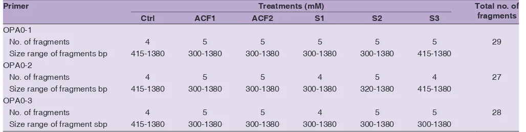
Randomly amplified polymorphic DNA (RAPD) was utilized to detect differences in DNA segments of the entire genomic DNA which were amplified by 3 different primers (10 single short oligonucleotide). After exposing date palm callus to biotic and abiotic stresses, a genomic DNA (gDNA) was extracted and amplified by OPA primers. The control of date palm callus (untreated callus) was included in all PCR reactions. A total of 84 amplicons were produced by 3 primers (Table 3; Fig. 3) within a size range of 300-1380 bp. Primer OPA0-1 revealed a total of 29 fragments, and within the control treatment, 4 fragments were amplified as 1380; 1100; 650 and 415 bp, whereas, at all other treatments (both ACF and NaCl treatments) 5 fragments were produced. Notably, 4 fragments were similar as seen in control products; new varied band (new appearance) at ACF treatment of 10 and 20% was observed with the size of 300 bp, additionally, at the NaCl of 205.34 mM treatment a new fragment of 490 bp was observed. Similar findings were analyzed with primer OPA0-2.
Regarding the DNA amplification by primer OPA0-3, appearance of a new fragment of the size 300 bp, and disappearance of the 415 bp band in ACF treatment (20%) occurred at a high concentration of ACF and NaCl. The entire amplified DNA fragments showed a polymorphism status in contrast with the control treatment, which indicated that at high concentration of ACF and NaCl, date palm callus, altered their genetic arrangement, which might be an explanation for the effects of biotic and abiotic stresses on callus morphology and biology.
Our results are in accordance with many other researchers, who revealed the suitability of RAPD technique to detect the genotoxicity of different biotic and abiotic effects, including salinity, heavy metals and fungal culture filtrate of F. oxysporum f.sp. albedinis (Esmaiel et al., 2012; Liu et al., 2012; Kurup et al., 2014). The appearance of new amplicons and the disappearance of existing fragments (compared to the control profile) could be attributed to point mutation associated with DNA damage (insertion or deletion mutations) (Atienzar and Jha, 2006; Liu et al., 2012). Not all the RAPD primers produced the same DNA
Fig 3. DNA fragments of exposed date palm Hilawii cv. callus to different
profile within same treatment; this could be explained by the primer sensitivity which depends on primer sequence (Liu et al., 2012; Kurup et al., 2014).
CONCLUSIONS
The results obtained herein indicate that date palm callus at the proliferation stage responds significantly to biotic and abiotic stresses; high concentration of ACF (20%) and NaCl (204.34 mM) led to a decrease of the fresh and dry weight growth, as well as increasing the browning intensity of Hillawi cv. callus. The decline in the growth parameters was accompanied with an increase of free proline and hydrogen peroxide productions, compared to a decrease in the activity of catalase enzyme which was observed at a high concentration of ACF and NaCl. The results of RAPD analysis revealed its feasibility as a method to evaluate the toxic impact of both ACF and NaCl on callus growth.
REFERENCES
Abass, M. H. 2011. Analysis of the role of the HvMPK4 gene in the barley biotic stress response, PhD Thesis, Heriot-Watt University, UK.
Abass, M. H. 2013a. Microbial contaminants of date palm (Phoenix dactylifera L.) in Iraqi tissue culture laboratories. Emirates. J. Food Agric. 25(11): 875-882.
Abass, M. H. 2013b. A PCR ITS-RFLP method for identifying fungal contamination of date palm (Phoenix dactylifera L.) tissue cultures. Afr. J. Biotechnol. 12(32): 5054-5059.
Abass, M. H., U. A. M. Al-Abadi and A. M. S. Al-Kaby. 2007. The efficiency of henna leaves extracts and some fungicides to reduce thefungal contamination of date palm (Phoenix dactylifera L.) tissue culture. Iraqi J. Biotech. 6(2): 1-40. Abass, M. H. and P. C. M. Morris. 2013. The Hordeum vulgare
signaling protein MAP Kinase 4 is a regulator of biotic and abiotic stress responses. J. Plant Physiol. 170: 1353-1359.
Abul-Soad, A. A., Z. Zaid, A. Slah and R. A. Sidky. 2002. Tissue culture of date palm (Phoenix dactylifera L.). “The 3rd. ed Scientific
Conference of Agricultural Sciences”, Assuit University, Egypt, Pp. 327-341.
Al-Bahrany, A. M. and J. M. Al-Khayri. 2012. In vitro responses of date palm cell suspensions under osmotic stress induced by sodium,
potassium and calcium salts at different exposure durations. Am. J. Plant Phys. 7(3): 120-134.
Al-Khayri, J. M. 2002. Growth, proline accumulation, and ion content in NaCl-stressed callus cultures of date palm (Phoenix dactylifera L.). In vitro cell. Dev. Biol. Plant 38: 79-82.
Al-Khayri, J. M. and Y. Ibraheem. 2014. In vitro selection of abiotic stress tolerant date palm (Phoenix dactylifera L.): A review. Emirates. J. Food Agric. 26(11): 921-933.
Al-Khayri, J. M., S. M. Jain and D. V. Johnson. (Eds.). 2015a. Date Palm Genetic Resources and Utilization, Africa and the Americas, Vol. 1. Springer, Dordrecht.
Al-Khayri, J. M., S. M. Jain and D. V. Johnson. (Eds.). 2015b. Date Palm Genetic Resources and Utilization, Asia and Europe, Vol. 2. Springer, Dordrecht.
Al-Mansoori, T. A. and M. N. A. Eldeen. 2007. Evaluation technique for salt tolerance in date palm. Acta Hortic. 736: 301-307. Al-Shahib, W. and R. J. Marshall. 2003. The fruit of the date palm:
it’s possible use as the best food for the future. Int. J. Food Sci. Nutr. 54: 247-259.
Al-Zubaydi, S., A. Jassim and H. Zair. 2013. Effect of sodium chloride and proline on embryo formation and germination through
in vitro micropropagation of date palm (Phoenix dactylifera L.) cv. Barhee. J. Agric. Sci. Technol. 3: 313-320.
Asada, K. 1999. The water-water cycle in chloroplast scavenging of active oxygen and dissipation of excess photons. Ann. Rev. Plant Physiol. Plant Mol. Biol. 50: 601-639.
Ashraf, M. and M. R. Foolad. 2005. Role of glycine betaine and proline in improving plant abiotic stress resistance. Environ. Exp. Bot. 59: 206-216.
Ashry, N. A. and H. I. Mohamed. 2011. Impact of secondary metabolites and related enzymes in flax resistance and or susceptibility to powdery mildew. World J. Agric. Sci. 7(1): 87-85.
Atienzar, F. A. and A. N. Jha.2006. The random amplified polymorphic DNA (RAPD) assay and related techniques applied to genotoxicity and carcinogenesis studies: A critical review. Mut. Res. 613: 76-102.
Bartlets, D. and R. Sunkar. 2005. Drought and salt tolerance in plants. Critic. Rev. Plant Sci. 24: 23-58.
Bassi, R. and S. S. Sharma. 1993. Changes in proline content accompanying the uptake of zinc and copper by Lema minor. Ann. Bot. 72: 151-154.
Bates, L. S., R. P. Waldernand and I. D. Teara. 1973. Rapid determination of free proline for water-stress studies. Plant Soil. 39: 205-207. Bor, M., F. Ozdemir and I. Turkan. 2003. The effect of salt stress on
lipid peroxidation and antioxidants in leaves of sugar beet Beta vulgaris L. and wild beet Beta maritime L. Plant Sci. 164: 77-84. Table 3: RAPD analysis of date palm callus exposed to ACF and NaCl treatment
Total no. of fragments Treatments (mM) Primer S3 S2 S1 ACF2 ACF1 Ctrl OPA0-1 29 5 5 5 5 5 4 No. of fragments
415-1380 300-1380 300-1380 300-1380 300-1380 415-1380
Size range of fragments bp OPA0-2 27 4 5 4 5 5 4 No. of fragments
415-1380 320-1380 300-1380 300-1380 300-1380 415-1380
Size range of fragments bp OPA0-3 28 5 5 4 5 5 4 No. of fragments
300-1380 300-1380 300-1380 300-1380 300-1380 415-1380
Size range of fragment sbp
Cvikrova, M., J. Mala, M. Hrubcova, J. Eder and S. Foretova. 2008. Induced changes in phenolic acid and stilbenes in embryogenic cell culture by culture filtrate of Ascocalyx abietina. J. Plant Dis. Prot. 115(2): 57-62.
Dahleen, L. S. and S. P. McCormic. 2001. Trichothecen toxins effect on barley callus and seedling growth. Cereal Res. Commun. 29: 115-120.
Dubey, R. S. and M. Rain. 1998. Influence of NaCl salinity on growth and metabolic status of protein and amino acids in rice seedlings. J. Agron. Crop. Sci. 97: 162-166.
El-Hadrami, A., F. Daayf and I. El-Hadrami. 2011. In vitro selection for abiotic stress in date palm. In: Jain, S. M., J. M. Al-Khayri and D. V. Johnson. (Eds.), Date Palm Biotechnology, Springer, Dordrecht, Pp. 237-252.
El-Sharabasy, S. F., W. H. Wanas and A. Y. Al-Kerdany. 2008. Effect of salinity stress on some date palm cultivars during proliferation stage in vitro. Arab J. Biotechnol. 11: 273-280.
Esmaiel, N. M., A. A. Al-Doss and M. N. Barakat. 2012. In vitro
selection for resistance to Fusarium oxysporum f.sp. dianthi
and detection of genetic polymorphism via RAPD analysis in carnation. J. Med. Plants Res. 6(23): 3997-4004.
FAO. 2012. FAOSTAT Agriculture. Available from: http://www.faostat. fao.org/site/567/default.aspx#ancor. [Last accesed on 2015 May 15]
Flowers, T. J. and A. Lauchli. 1983. Sodium versus potassium: Substitution and compartmentation. In: Pirson, A. and M. H. Zimmerman, (Eds.), Encyclopedia of Plant Physiology, New Series, Vol. 15. Springer-Verlag, Berlin, Pp. 651-681.
Gill, S. S. and N. Tuteja. 2010. Reactive oxygen species and antioxidant machinery in abiotic stress tolerance in crop plants. Plant Physiol. Biochem. 48: 909-930.
Girish, K., B. S. Shankaraand and K. A. Raveesha. 2009. Crude toxin extract from culture filtrate of Phomopsis azadirachtae infecting neem and its phytotoxicity. Int. J. Integr. Biol. 6(2): 79-84. Goicoechea, N., J. Aguirreolea, S. Cenoz and J. M. Garcia-Mina.
2000. Verticillium dahliae modifies the concentrations of proline, soluble sugars, starch, soluble protein and abscisic acid in pepper plants. Eur. J. Plant Pathol. 106: 19-25.
Grote, D., R. Schmitand and W. Claussen. 2006. Water uptake and proline index as indicators of predisposition in tomato plants to
Phytophthora nicotiana infection as influenced by abiotic stress. Physiol. Mol. Plant Pathol. 69: 121-130.
Hameed, M. A. and M. H. Abass. 2006. Study of cytological changes associated with contaminated date palm Phoenix dactylifera L. tissue cultures with fungi. Basra Res. J. 32: 1-27.
Hare, P., W. A. Cressand and V. J. Staden. 1998. Dissecting the roles of osmolyte accumulation during stress. Plant Cell Environ. 21: 535-553.
Htwe, N. N., M. Maziah, H. C. Ling, F. Q. Zaman and A. M. Zain. 2011. Responses of some selected Malaysian rice genotypes to callus induction under in vitro salt stress. Afr. J. Biotechnol. 10: 350-362. Ibraheem, Y. M., I. Pinker, M. Böhme and Z. Al-Hussin. 2012.
Screening of some date palm cultivars to salt stress in vitro. Acta Hortic. 961: 359-365.
Jasim, A. M., M. F. Abbas and B. H. Alzubaidy. 2010. Effect of salt stress and proline on chemical content of embryogenic callus and somatic embryos of date palm (Phoenix dactylifera L. ‘Ashkar’). Acta Hortic. 882: 219-224.
Jin, H., G. L. Hartman, C. D. Nickell and J. M. Widholm. 1996. Phytotoxicity of culture filtrates from Fusarium solani, the causal agent of sudden death syndrome of soybean. Plant Dis. 80(8): 922-927.
Kim, K., H. Kook, Y. Jang, W. Lee, S. Kamala-Kannan, J. Chae and K. Lee. 2013. The effect of blue-emitting-light diodes on antioxidant properties and resistance to Botrytis cinerea in tomato. Plant Pathol. Microbiol. 4(9): 203. doi:10.4172/2157.7471.1000203.
Koike, M., T. Murakami, Y. Katsumata, Y. Amemiya and T. Shimada. 1993. Use of culture filtrates of Verticillium dahlia as a bioassay for screening of disease tolerant eggplant. Plant Tiss. Cult. 10(1): 71-74.
Kosturkova, G., R. Rodeva, K. Tasheva, M. Dimitrova and D. Dimano. 2012. Effect of crude culture filtrates of the pathogenic fungus
Phoma medicaginis on in vitro cultures of pea. Agro Life Sci. J. 1: 126-131.
Kurup, S., M. A. M. Aly, G. Lekshmi and N. H. Tawfik. 2014. Rapid
in vitro regeneration of date palm (Phoenix dactylifera L.) cv. kheneizi using tender leaf explants. Emirates. J. Food Agric. 26(6): 539-544.
Liu, W., X. Hu, W. Zhang, W. J. Rogers and W. Cai. 2005. Hydrogen peroxide mediates defence responses induced by chitosans of different molecular weights in rice. J. Plant Physiol. 162: 937-944. Liu, W., L. Sun, M. Zhong, O. Zhou, Z. Gong, P. Li, P. Ti and X.
Li. 2012. Cadmium-induced DNA damage and mutations in Arabidopsis plantlet shoots identified by DNA fingerprinting. Chemo. 89: 1048-1055.
Mohanraj, D., P. Padmanaban and M. Karunakaran. 2003. Effect of phyototoxin of Colletotrichum falcatum Went. (Physalospora tucumanensis) on sugarcane in tissue culture. Acta Phytopathol. Entomol. Hung. 38: 21-28.
Munns, R. 2005. Genes and salt tolerance: bringing them together. New Phytol. 167: 645-663.
Munns, R. and M. Tester. 2008. Mechanism of salinity tolerance. Ann. Rev. Plant Biol. 59: 651-681.
Murashige, T. and F. Skoog. 1962. A revised medium for rapid growth and bioassays with tobacco tissue cultures. Physiol. Plant. 15: 473-497.
Naidu, B., L. Paleg, D. Aspinall, A. Jennings and G. Jones. 1991. Amino acid and glycine betaine accumulation in cold-stressed wheat seedlings. Phytochemistry. 30: 407-409.
Rao, S. and S. Ramgoapl. 2010. Effect of Alternaria helianthi culture filtrate on callus and regeneration of plantlets from tolerant callus in sunflower (Helianthus annuus L). Indian J. Biotechnol. 9: 187-191.
Rhodes, D., A. Nadolska-Orczyk and P.J. Rich. 2002. Salinity, osmolyte and compatible solutes. In: Lauchli, A. and U. Luttge, (Eds.), Salinity, Environment, Plant, Molecules, Kluwer Academic Publisher, Netherlands, Pp. 181-204.
Saxena, G., P. C. Verma, L. Rahman, S. Banerjee, R. S. Shukla and S. Kumar. 2008. Selection of leaf blight-resistant Pelargonium graveolens plants regenerated from callus resistant to a culture filtrate of Alternaria alternata. Crop Prot. 27: 558-565.
Starvareck, S. Y. and D. W Rains. 1984. The development of tolerance cell to mineral stress. Hortic. Sci. 19: 377-382.
Subhashini, K. and G. M. Reedy. 1991. Role of proline in callus growth and plant regeneration under salt stress in rice. Biol. Sci. 57: 81-83.
Taha, R. A. and M. M. Hassan. 2014. Using low levels of seawater to enhance growth and development of date palm embryogenic cultures. Asian J. Agric. Sci. 6(2): 69-74.
Virk, G. S. and A. Nagpal. 2011. In vitro selection of calli of Citrus jambhiri Lush. for tolerance to culture filtrate of Phytophthora parasitica and their regeneration. Physiol. Mol. Biol. Plants. 17(1): 41-47.
Wagh, P., S. Sinh, H. K. Singh and U. K. Khare. 2013. Pathogenic behaviour of Alternaria alternate and phytotoxicity of its culture filtrates on Lepidium sativum: a medicinal herb of immense pharmacological potential. Bioscan. 8(2): 643-647.
Yaish, M. W. 2015. Short communication: Proline accumulation is a general response to abiotic stress in the date palm tree (Phoenix dactylifera L.). Genet Mol. Res. 14(3): 9943-9950.
Yaish, M. W. and P. P. Kumar. 2015. Salt tolerance research in date palm tree (Phoenix dactylifera L.), past, present, and
future perspectives. Front Plant Sci. 6: 348. doi: 10.3389/ fpls.2015.00348.
Yaron, D. 1981. In: Yaron, D., (Ed.), Salinity in Irrigation and Water Resources, Marcel Dekker, Inc., New York, Pp. 1-20.
Yokoi, S., R. A. Bressan and P. M. Hasegawa. 2002. Salt Stress Tolerance in Plants, JIRCAS Working Reports, Pp. 25-33. Zolan, M. E. and P. J. Pukkila.1986. Inheritance of DNA methylation in
Coprinuscinceus. Mol. Cell Biol. 6: 195-200.
Zaid, A. (Ed.). 2002. Date Palm Cultivation, Rev. Ed. FAO, Rome. Zhou, B., J. Wang, Z. Guo, H. Tan and X. Zhu. 2006. A simple