ARTICLE
Volumetric MRI Study of Brain in Children With
Intrauterine Exposure to Cocaine, Alcohol, Tobacco,
and Marijuana
Michael J. Rivkin, MDa,b,c, Peter E. Davis, SBa, Jennifer L. Lemaster, BAa, Howard J. Cabral, PhDd, Simon K. Warfield, PhDb, Robert V. Mulkern, PhDb, Caroline D. Robson, MB, CHBb, Ruth Rose-Jacobs, ScDe, Deborah A. Frank, MDe
Departments ofaNeurology,bRadiology, andcPsychiatry, Children’s Hospital and Harvard Medical School, Boston, Massachusetts;dDepartment of Biostatistics, Boston University School of Public Health, Boston, Massachusetts;eDepartment of Pediatrics, Boston University School of Medicine, Boston, Massachusetts
The authors have indicated they have no financial relationships relevant to this article to disclose.
What’s Known on This Subject
MRI neuroimaging has been used to study the effect of prenatal exposure to alcohol on the human brain but comparatively little data have been gathered using these methods to study the effects of prenatal exposure to cigarettes, cocaine or marijuana.
What This Study Adds
This study adds MRI neuroimaging data on the lasting effect of prenatal exposures to alcohol, cigarettes, cocaine and marijuana on human brain structure in early adoles-cence. Moreover, it provides new information about the persisting potential effects of prenatal exposure to combinations of these substances.
ABSTRACT
OBJECTIVE.The objective of this study was to use volumetric MRI to study brain
volumes in 10- to 14-year-old children with and without intrauterine exposure to cocaine, alcohol, cigarettes, or marijuana.
METHODS.Volumetric MRI was performed on 35 children (mean age: 12.3 years; 14
with intrauterine exposure to cocaine, 21 with no intrauterine exposure to cocaine) to determine the effect of prenatal drug exposure on volumes of cortical gray matter; white matter; subcortical gray matter; cerebrospinal fluid; and total parenchymal volume. Head circumference was also obtained. Analyses of each individual sub-stance were adjusted for demographic characteristics and the remaining 3 prenatal substance exposures.
RESULTS.Regression analyses adjusted for demographic characteristics showed that
children with intrauterine exposure to cocaine had lower mean cortical gray matter and total parenchymal volumes and smaller mean head circumference than com-parison children. After adjustment for other prenatal exposures, these volumes remained smaller but lost statistical significance. Similar analyses conducted for prenatal ethanol exposure adjusted for demographics showed significant reduction in mean cortical gray matter; total parenchymal volumes; and head circumference, which remained smaller but lost statistical significance after adjustment for the remaining 3 exposures. Notably, prenatal cigarette exposure was associated with significant reductions in cortical gray matter and total parenchymal volumes and head circumference after adjustment for demographics that retained marginal sig-nificance after adjustment for the other 3 exposures. Finally, as the number of exposures to prenatal substances grew, cortical gray matter and total parenchymal volumes and head circumference declined significantly with smallest measures found among children exposed to all 4.
CONCLUSIONS.These data suggest that intrauterine exposures to cocaine, alcohol, and cigarettes are individually related to reduced head circumference; cortical gray matter; and total parenchymal volumes as measured by MRI at school age. Adjustment for other substance exposures precludes determination of statistically significant individual sub-stance effect on brain volume in this small sample; however, these subsub-stances may act cumulatively during gestation to exert lasting effects on brain size and volume.
S
CIENTIFIC UNCERTAINTY PERSISTSas to whether intrauterine cocaine exposure (IUCE) exerts a lasting effect on brain structure and development in humans. Early observation that head circumference (HC) at birth was reduced in children who were exposed to cocaine raised concern that structural development of brain in children wasadversely affected as a result of IUCE.1–3 Animal and nonhuman primate studies suggested that IUCE may exert
deleterious effects on the developing brain.4,5Postnatal reductions of cortical neuron number and lamination have
www.pediatrics.org/cgi/doi/10.1542/ peds.2007-1399
doi:10.1542/peds.2007-1399
Key Words
pregnancy, illicit substance, MRI, children, brain
Abbreviations
IUCE—intrauterine cocaine exposure HC— head circumference FAS—fetal alcohol syndrome DE— dual echo
CGM— cortical gray matter SCGM—subcortical gray matter WM—white matter CSF— cerebrospinal fluid TOT—total parenchymal volume TICV—total intracranial volume
been observed in rodent models of IUCE.5It is interesting that upregulation of proapoptotic proteins has been re-ported to occur in cerebral cortex of IUCE fetal mice
relative to controls.6–8 IUCE has been associated with
diminished neuronal density and altered cortical
organi-zation of postnatal brain in nonhuman primates9;
how-ever, the validity of extrapolating these results from mammalian models to humans remains unclear. Animal studies involve known dosages of single, pure drug prep-arations administered at precisely defined points in ges-tation. In contrast to animal studies, human studies of cocaine use by pregnant women present additional an-alytic challenges that consist not only of widely varying drug concentration, purity, and patterns of use during pregnancy but also of concurrent cigarette, alcohol, or
marijuana use during the course of the pregnancy10–12
The effect of variability in the pattern of human intra-uterine exposures is exacerbated by differences in social and clinical risk factors between cocaine-using and non-using pregnant women, which can be statistically but
not experimentally controlled.13
Concern about structural consequences of these in-trauterine exposures is heightened by an ample litera-ture that documents cognitive and behavioral conse-quences associated with prenatal exposures to cocaine, alcohol, cigarettes, or marijuana. Some, although not all, neurobehavioral studies of infants, toddlers, and children with IUCE reported deficits in language de-velopment, early executive function, learning, and
self-regulation.14–18 Prenatal exposure to alcohol has
been associated with cognitive and behavioral deficits in executive function, processing speed, and inatten-tiveness that persist into childhood even in children
without stigmata of fetal alcohol syndrome (FAS).19–22
Human newborns with intrauterine nicotine exposure demonstrated increased tone and excitability as
com-pared with control infants,23 delayed babbling at 8
months of age,24 increased impulsivity as young
chil-dren, and working memory and visuospatial skills
defi-cits throughout childhood.25–27 An inverse relationship
between prenatal cigarette exposure and general
intelli-gence persisted into adolescence.28Children with
intra-uterine marijuana exposure have demonstrated poorer
scores on intelligence tests,29,30increased impulsivity,
hy-peractivity, decreased attentiveness, increased rates of
delinquency, and externalizing problems26,31–35Thus,
in-vestigation of the effect of 1 prenatal exposure on sub-sequent brain structure and function must take into consideration the contributions of other prenatal expo-sures. In this study, we acquired volumetric MRI data and applied quantitative segmentation techniques to evaluate potential differences in brain tissue volumes of children with and without IUCE while adjusting for intrauterine exposures to alcohol, cigarettes, and mari-juana.
METHODS
Participants
The parent sample was recruited by trained interviewer/ recruiters who screened maternity and nursery charts 7
days a week on the postpartum floor of Boston City Hospital, from October 1990 to March 1993, using in-formed consent and protocols approved by the Boston Medical Center institutional review board. Potential par-ticipants for the IUCE group were initially identified by review of medical charts for mother’s report of cocaine use in pregnancy or positive prenatal or postnatal urine drug screen for cocaine metabolites from mother or in-fant. Unexposed dyads roughly comparable to cocaine-exposed mother–infant dyads in ethnicity (African American/African Caribbean versus other) were prefer-entially approached for recruitment soon after delivery. All mother–infant dyads met the following criteria: (1)
infant gestational ageⱖ36 weeks; (2) no NICU care; (3)
no major congenital malformation; (4) no diagnosis of FAS in the neonatal chart or physical examination; (5) no history of HIV seropositivity in mother’s or infant’s medical chart; (6) mother’s ability to communicate flu-ently in English; (7) no neonatal or maternal urine drug screen or medical chart evidence of mother’s use during pregnancy of illegal opiates, methadone, amphetamines, phencyclidine, barbiturates, or hallucinogens; and (8)
mother’s ageⱖ18 years. Additional details about sample
characteristics and recruitment are reported elsewhere.36
Mothers who participated in the study were identified as either lighter users, heavier users, or nonusers of cocaine by interview and biological markers obtained by clinicians and study personnel. Meconium specimens from enrolled infants were analyzed by radioimmuno-assay for the cocaine metabolite benzoylecgonine, opi-ates, amphetamines, benzodiazepines, and cannabinoids using a modified method of Ostrea, published in detail
elsewhere.36,37
During the period of study recruitment at Boston Medical Center, urine testing for metabolites of illicit drugs was performed for clinical indications at the dis-cretion of health care personnel but was not universal. Clinical indications at the time included no prenatal care, a jittery infant, unusual maternal behavior, or a known history of substance use. When available in the medical chart, we documented the results of urine drug enzyme-multiplied immunoassay technique assays ob-tained for clinical purposes during prenatal or perinatal care of mother or infant. After recruitment and written informed consent, we collected additional urine samples from study mothers to determine benzoylecgonine, opi-ates, amphetamines, benzodiazepines, and cannabinoids by radioimmunoassay using commercial kits (Abuscreen RIA; Roche Diagnostics Systems, Inc, Montclair, NJ). Participants were protected by a writ of confidentiality.
Exposure Classification
or 9 days of self-reported use during pregnancy. Before source study data were analyzed, a composite measure of heavier use was a priori defined as the top quartile of meconium concentration for cocaine metabolites
(ben-zoylecgonine⬎3314 ng/g meconium) and/or top
quar-tile days of self-reported use (⬎61 days) during the
entire pregnancy. All other use was classified as “lighter.” By this method, 45 users (33% of the users)
were classified as heavier users. All other users (n⫽93)
were classified as “lighter users.”
Pragmatic as well as scientific considerations influenced this either/or definition of exposure level. Women are more likely to underreport rather than overreport illicit
substance use during pregnancy38; therefore, women who
reported days of use in the top quartile were a priori considered heavier users, even if the meconium benzo-ylecgonine level was not in the top quartile because not all infants who are exposed to cocaine prenatally have
posi-tive meconium assays.39 Moreover, meconium samples
were not obtained from 14% of study infants; therefore, whichever indicator (self-report or meconium assay) dem-onstrated higher exposure was used to determine exposure category. For this study, we recruited participants from the parent sample of children had who IUCE and fit additional criteria listed next and whose intrauterine cocaine
expo-sure was⬎1100 ng/g meconium, the parent sample
me-dian.
A total of 123 infants were initially identified by perinatal clinicians as cocaine exposed on the basis of maternal antepartum or peripartum urine drug screen cocaine metabolite results. Five of 134 infants who were clinically considered to be cocaine unexposed were ex-cluded from the final sample because no confirmatory biological marker was available, and 15 others were reclassified because urine or meconium assays were pos-itive for cocaine metabolites, leaving 114 unexposed infants in the sample. In all, 138 infants were found to demonstrate evidence of IUCE. Together, the unexposed and exposed infants constituted a neonatal study sample of 252 initially consented. In all, 216 (86%) of the neo-natal sample had meconium assays performed. No infant demonstrated amphetamine, opiate, or phencyclidine exposure by urine or meconium assay or by maternal report.
The Addiction Severity Index, a validated tool for substance abuse report, as well as additional study-spe-cific interview questions was used to determine mater-nal alcohol, cigarette, and marijuana use during
preg-nancy.40 Alcohol, cigarette, and marijuana use was
reported as number of drinks per week; number of cig-arettes smoked per day; and number of days of use of alcohol, cigarettes, and marijuana, respectively, during the last 30 days of pregnancy. Data were acquired sim-ilarly for use of these substances during pregnancy be-fore the last 30 days. This permitted determination and use of average daily volume and average daily cigarettes for alcohol and cigarette use, respectively. Because mar-ijuana is stored for long periods in body fat, marmar-ijuana metabolite concentration in meconium constitutes a less valid indication of total dosage than that of cocaine
metabolites.37 In addition, one third of the marijuana
users identified on the basis of meconium/urine assay denied its use on interview. Consequently, construction of a reliable index of marijuana dosage proved difficult. In the following analyses, marijuana exposure was coded as exposed or unexposed on the basis of any positive self-report or any positive urine/meconium assay.
Cohort Selected for MRI
We recruited children for this study from the parent sample who were African American/African Caribbean until a sample size of 40 was attained, 16 with IUCE
⬎1100 ng/g meconium, the parent sample median, and
24 otherwise demographically comparable children who had no IUCE. All children were right-handed. In addi-tion to the initial exclusion criteria, the sample for this neuroimaging study excluded children with the follow-ing complications that might confound MRI interpreta-tion: (1) history of central nervous system radiation, (2) loss of consciousness after head trauma, (3) history of seizures, (4) history of lead poisoning requiring chela-tion therapy, (5) history of intracranial surgery, and (6) history of systemic illness or medical treatment that might adversely affect brain structure. Children were recruited after parent/guardian informed consent and child assent were obtained using protocols approved by the Boston Medical Center institutional review board and the Committee on Clinical Investigation of Chil-dren’s Hospital Boston.
MRI Scans
Each of the 40 children underwent imaging using a 1.5 T GE LX MR scanner (GE, Milwaukee, WI) operating at the GE 12.0 platform at Children’s Hospital Boston. No sedation was used for image acquisition, but patients were acclimatized to the scanner environment. After a T1-weighted localizing scan was obtained, a high-reso-lution 3-dimensional T1-weighted Fourier Transform Spoiled Gradient neuroanatomic whole-brain acquisi-tion was obtained (24 cm field of view, 1.5 mm contig-uous coronal slice thickness, 120 slices, repetition time/
echo time⫽40/4, matrix 256⫻192, flip angle ⫽20°,
scan time 11 minutes 20 seconds). Subsequently, whole-brain T2-weighted and proton density data were ac-quired using a dual echo (DE) fast spin echo pulse
se-quence (echo train length [ETL]⫽8, 3-mm skip, 3-mm
interleaved coronal slices, 2 acquisitions, repetition
time/echo time/echo time 2⫽4000/14/84, 192⫻256,
20 cm field of view, 1 number of excitations [NEX], scan time 10 minutes and 25 seconds).
MRI Quality
21 without IUCE; 88% of children scanned) produced data sets that were fully analyzed.
Postimaging Data Analysis
Postimaging data processing and quantitative volumetric analyses were conducted on computer workstations us-ing a LINUX platform. A sequence of established and validated image processing algorithms was used to segment each of the MRI slices into separate tissue
classes.41–46Briefly, each MRI slice in each data set was
segmented into cerebral cortical gray matter (CGM), subcortical gray matter (SCGM; to include caudate, pu-tamen, globus pallidus, and substantia nigra, bilaterally), white matter (WM), and cerebrospinal fluid (CSF; in-cluding ventricular and extra-axial CSF). These algo-rithms were designed to reduce imaging system noise, identify a linear transformation, and resample the DE spin-echo images according to the aforementioned lin-ear transform to align the coronal DE spin-echo images with the coronal spoiled gradient images to form a 3-channel data set to classify tissue types on the basis of the MR intensity in the 3 channels and identify tissue-class surfaces for 3-dimensional visualization. From tis-sue segmentation of each slice, whole-brain absolute tissue volumes were determined in milliliters for CGM, WM, SCGM, and CSF (Fig 1), permitting calculation of
total parenchymal volume (TOT; CGM⫹WM⫹SCGM)
and total intracranial volume (TICV; CGM ⫹ WM ⫹
SCGM ⫹ CSF). Data were examined both as volumes
referenced to TICV and as absolute volumes.
Segmentations and volumetric analyses were under-taken by 2 members of the research team (Mr Davis and Ms Lemaster) and reviewed by a third team member (Dr Rivkin), all of whom were masked to subject group membership. Reviewers’ consistency in tissue classifica-tion was assessed throughout the study with no more than 4% variance in tissue volumes found on quality certification data sets performed by both reviewers.
Data Analysis
All volumetric data and head circumference measure-ments were included in analysis. To ensure comparabil-ity with the cocaine variable, alcohol, tobacco, and mar-ijuana were also collapsed to exposed/unexposed. First, bivariate analyses were performed to examine the effect of each individual exposure on HC and brain tissue volumes without adjustment for demographic variables or other exposures. Second, an initial multiple linear regression analysis discerned the effect of each individ-ual exposure adjusted for child’s gender and age at scan
(demographic features). Such adjustment was per-formed because age and gender can affect brain size. Given that the children studied were both male and female and represented ages ranging from 10 to 13.8 years, adjustment for these features was essential to avoid misattribution of possible effects of age and gender to prenatal drug exposures. Because the impact of the birth mother’s level of highest educational attainment was tested and found not to correlate with any outcome, it was not retained in final models. Third, a second-level, multiple linear regression analysis for each exposure incorporated the first model and adjusted for the re-maining exposures. Thus, for comparison of children with IUCE and those with none, mean values were adjusted for gender and age at scan as well as for simul-taneous cigarette, alcohol, and marijuana exposure. Similarly, for comparison of children with intrauterine alcohol exposure and those with none, mean values were adjusted for demographic features as well as for simultaneous cocaine, cigarette, and marijuana expo-sure. Similar analyses were conducted for intrauterine cigarette exposure and for intrauterine marijuana
expo-sure. Associations with 2-tailedP⬍.05 were judged to
be statistically significant.
Small HC in bivariate analyses was associated with each of the 4 substance exposures studied (see “Results”). Be-cause HC has been demonstrated to correlate with brain volumes, analyses were performed using absolute
vol-umes.47–49Analyses were also performed using a ratio of
absolute tissue volumes divided by TICV, which yielded no statistically significant differences among groups for any of the cerebral tissue outcome variables; however, children who were exposed to cocaine, to alcohol, and to cigarettes each demonstrated reductions in HC at the time of mea-surement that were statistically significant as compared with control subjects. Furthermore, significant reduction of HC was found in children who were exposed prenatally to
ⱖ2 of these substances. Because the principal determinant
of head size is brain size and because reduction of HC accompanied prenatal drug exposure for the majority of the exposed children, we reasoned that adjustment for HC or its proxy, intracranial cavity volume, factored out the effect of the exposure as well. As a result, all analyses were performed using absolute tissue volumes.
RESULTS
Subjects
Thirty-five children were included for final analysis of MRI data. The group comprised 14 children with IUCE and 21
B
A C D
FIGURE 1
with no IUCE. Characteristics of children in the study sam-ple are provided in Table 1. Among the children with IUCE, prenatal exposures to maternally used alcohol, cigarettes, or marijuana were common; however, 8 (38%) children with no IUCE had prenatal exposures to alcohol, cigarettes, or marijuana as well. Multiple prenatal exposures occurred in 15 of the 35 children studied. Among children with
IUCE, 11 (78.5%) of the 14 had prenatal exposure toⱖ2 of
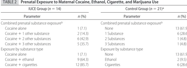
the other substances studied, whereas only 2 (9.5%) of the 21 children with no IUCE had such exposures. Finally, 43% of the children in the exposed group of this study had exposures to cocaine and 2 other substances, whereas 35% had exposures to cocaine and the 3 other substances (Ta-ble 2).
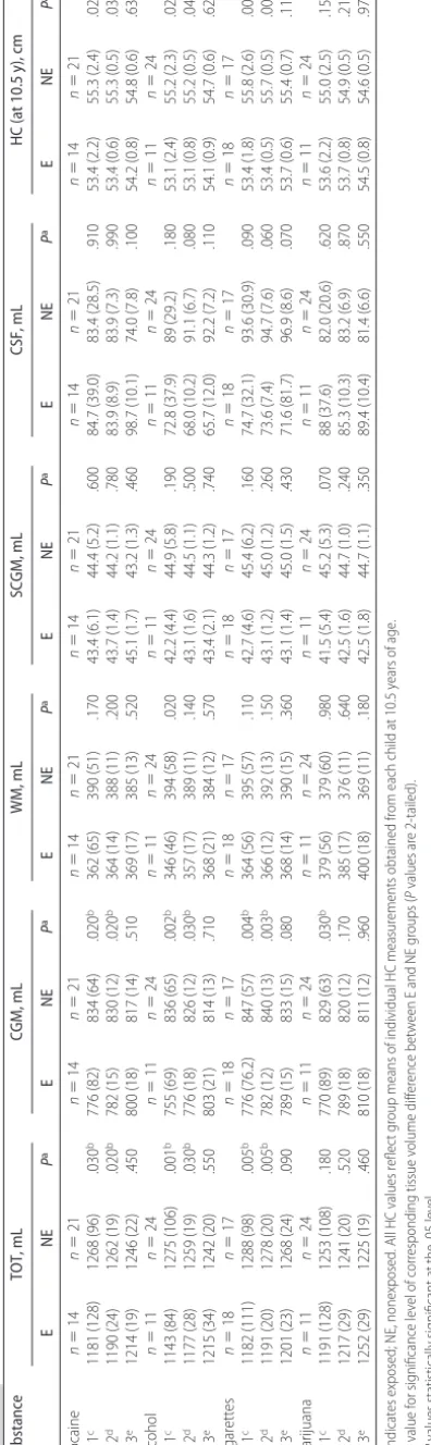
Children with IUCE demonstrated significantly
smaller HC than those with no IUCE in both the unad-justed analysis (53.4 cm [SE 2.2] vs 55.3 cm [SE: 2.4];
P ⫽.02) and the analysis that was adjusted for
demo-graphic features (53.4 cm [SE: 0.6] vs 55.3 cm [SE: 0.5];
P⫽.03). CGM and TOT were also significantly reduced
in both the unadjusted analysis (CGM: 776 mL [SE: 82]
vs 834 mL [SE: 64;P⫽.02]; TOT: 1181 mL [SE: 128] vs
1268 mL [SE: 96; P ⫽ .03]) and the analysis that was
adjusted for demographics (CGM: 782 mL [SE: 15] vs
830 mL [SE: 12;P⫽.02]; TOT: 1190 mL [SE: 24] vs 1262
mL [SE: 19;P ⫽.02]) for children with IUCE as
com-pared with children with no IUCE. No significant differ-ences in WM, SCGM, or CSF were found between chil-dren with and without IUCE in either of these analyses (Table 3).
Children with intrauterine alcohol exposure showed
significantly smaller HC compared with those with no prenatal alcohol exposure (53.1 cm [SE: 2.4] vs 55.2 cm
[SE: 2.3;P⫽.02]) for the unadjusted analysis and after
adjustment for demographic features (53.1 cm [SE: 0.8]
vs 55.2 cm [SE: 0.5; P ⫽ .04]). Volumetric analyses
revealed that CGM, WM, and TOT were significantly reduced in the unadjusted analysis (CGM: 755 mL [SE:
69] vs 836 [SE: 65;P⫽.002]; WM: 346 mL [SE: 46] vs
394 cm [SE: 58;P⫽.02]; TOT: 1143 mL [SE: 84] vs 1275
mL [SE: 106; P⫽.001]), whereas only CGM and TOT
were significantly reduced after adjustment for demo-graphic features (CGM: 776 mL [SE: 18] vs 826 mL [SE:
12;P⫽.03]; TOT: 1177 mL [SE: 28] vs 1259 mL [SE: 19;
P ⫽ .03]) among children with prenatal exposure to
alcohol as compared with those with no such exposure. No significant intergroup differences were observed be-tween groups in SCGM or CSF volumes for the unadjusted analysis or for SCGM, WM, or CSF volumes for the analysis adjusted for demographic features (Table 3).
Children with intrauterine cigarette exposure dem-onstrated significantly smaller HC (53.4 cm vs 55.8 cm) as compared with those with no prenatal cigarette ex-posure for both the unadjusted analysis (53.4 cm [SE:
1.8] vs 55.8 cm [SE: 2.6];P⫽.003) and analysis adjusted
for demographics (53.4 cm [SE 0.5] vs 55.7 cm [SE 0.5];
P⫽.005). Volumetric analyses revealed that CGM and
TOT were significantly reduced in both the unadjusted
analysis (CGM: 776 mL [SE: 76.2] vs 847 [SE 57; P⫽
.004]; TOT: 1182 mL [SE: 111] vs 1288 mL [SE: 98;P⫽
.005]) and the analysis adjusted for demographic
fea-tures (CGM: 782 mL [SE: 12] vs 840 [SE: 13;P⫽.003];
TOT: 1191 mL [SE: 20] vs 1278 mL [SE: 20;P⫽.005]).
No difference between groups was observed for WM, SCGM, or CSF volumes in both the unadjusted analysis and the analysis that was adjusted for demographics (Table 3).
Children with prenatal marijuana exposure demon-strated a trend toward smaller mean HC as compared with those with no prenatal marijuana exposure in both the unadjusted analysis and the analysis that was ad-justed for demographics, but the difference did not reach significance. Although CGM and TOT were reduced in the unadjusted analysis and the analysis that was ad-justed for demographic features, only in the unadad-justed TABLE 1 Study Sample Characteristics
Characteristic Children With
IUCE
Control Children
No. of children 14 21
Age, mean (range), y 12.3 (10.0–13.8) 12.4 (10.0–13.2)
Handedness All right-handed All right-handed
Gender (M/F) 8/6 12/9
Gestational age at birth, mean (SD), wk
39.7 (1.5) 40.3 (1.6)
IQ score at 10.5 y, mean (SD)a 90.3 (11.1) 85.9 (12.5)
aWechsler Intelligence Scale for Children full-scale IQ score.
TABLE 2 Prenatal Exposure to Maternal Cocaine, Ethanol, Cigarette, and Marijuana Use
IUCE Group (n⫽14) Control Group (n⫽21)a
Parameter n(%) Parameter n(%)
Combined prenatal substance exposureb Combined prenatal substance exposureb
Cocaine alone 1 (7.1) None 13 (61.9)
Cocaine⫹1 other substance 2 (14.3) 1 Substance 6 (28.6)
Cocaine⫹2 other substances 6 (42.9) 2 Substances 1 (4.8)
Cocaine⫹3 other substances 5 (35.7) 3 Substances 1 (4.8)
Exposure by substance type Exposure by substance type
Cocaine alone 1 (7.1) None 13 (61.9)
Cocaine⫹ethanol 9 (64.3) Ethanol 2 (9.5)
Cocaine⫹cigarettes 12 (85.7) Cigarettes 6 (28.6)
Cocaine⫹marijuana 8 (57.1) Marijuana 3 (14.3)
aControl group had no IUCE.
volumetric analysis did the CGM reduction reach
signif-icance (770 mL [SE: 89] vs 829 mL [SE 63];P⫽.03). No
significant difference in any other cerebral tissue volume was found in the analysis that was adjusted for demo-graphic features (Table 3).
After the foregoing unadjusted and first-level multi-ple regression analyses, a second multimulti-ple regression analysis for each exposure was performed and incorpo-rated the first model and added adjustments for the remaining exposures. It is interesting that although the trend for group differences in cerebral tissue volumes persisted, the individual effects of cocaine and alcohol, respectively, were no longer statistically significant after controlling for the other exposures. Notably, CGM and TOT remained reduced and retained marginal
signifi-cance (CGM:P⫽.08; TOT:P⫽.09) in the analysis of
intrauterine cigarette exposure after adjustment for si-multaneous exposures to cocaine, alcohol, and mari-juana (see Table 3 for summary of intergroup differences by substance for the unadjusted and both adjusted anal-yses).
Additional analysis was performed to compare
chil-dren who were exposed prenatally to ⱖ2 substances
with control subjects, adjusted for gender and age at examination. This analysis revealed significant
reduc-tions in CGM, TOT, and HC (776 mL vs 841 mL [P ⫽
.004]; 1175 mL vs 1269 mL [P⫽.007]; 53.4 cm vs 55.7
cm [P ⫽ .02], respectively; Table 4). Next, analysis of
variance was performed to examine the cumulative
ef-fect on CGM, TOT, and HC of prenatal exposure toⱖ1 of
these substances (Table 5) as compared with nonex-posed children with the independent variable the count of the number of substances to which the child was
exposed in utero. A global meanPvalue for the analysis
of variance models of .01 for both CGM and TOT indi-cated that reductions in these volumes relative to the unexposed were significantly associated with number of substances of exposure. A similar pattern was apparent
for HC, which demonstrated a global mean P ⫽ .07.
Table 5 demonstrates that CGM, TOT, and HC all de-clined as the number of substances to which study chil-dren were prenatally exposed exceeded 1. Indeed, this analysis of variance demonstrated that the smallest vol-umes of CGM, TOT, and HC were found in association TABLE 4 Adjusted Effect of Prenatal Exposure to>2 Substances on
MRI Volumes, HC
Parameter Adjusted Mean Adjusted for Gender and Age at Examination
P
Exposed toⱖ2 Substances (n⫽15)
Unexposed (n⫽13)
TOT, mL 1175 (22) 1269 (24) .007a
CGM, mL 776 (14) 841 (15) .004a
WM, mL 356 (13) 383 (14) .180
Basal ganglia, mL 43.1 (1.3) 45.4 (1.4) .260
CSF, mL 80.3 (8.8) 86.1 (9.5) .660
HC (at 10.5 y), cm 53.4 (0.6) 55.7 (0.7) .020a
AllPvalues are 2-tailed.
aPvalues statistically significant at the .05 level.
with prenatal exposures to all 4 substances: cocaine, cigarettes, alcohol, and marijuana (CGM: 731 mL
[ex-posed] vs 853 mL [unexposed;P⫽.002]; TOT: 1129 mL
[exposed] vs 1287 mL [unexposed;P⫽.006]; HC: 52.3
cm [exposed] vs 55.7 cm [unexposed;P⫽.008]).
DISCUSSION
In this study, multiple regression analyses of absolute brain tissue volumes and head size revealed that prenatal expo-sures to cocaine, alcohol, and cigarettes individually were associated with reductions in HC and CGM and TOT vol-umes in 10- to 14-year-old children relative to comparison subjects after controlling for age at scan and gender. Im-portantly, these reductions reached bivariate statistical sig-nificance for cocaine, alcohol, and cigarette exposures, in-dividually. Additional analyses of each exposure in which additional adjustment was made for the remaining 3 ex-posures revealed that the observed trend to reductions in CGM and TOT volumes associated with prenatal cocaine or alcohol exposure persisted but was no longer statistically significant. In distinction, the effects of prenatal cigarette exposure on CGM and TOT retained marginal significance
despite such adjustment (P ⫽.08 and .09, respectively).
Last, analysis of variance provided evidence of an inverse relationship between the number of substances to which these children were prenatally exposed and the reduction found in CGM, TOT, and HC.
To our knowledge, this is the first volumetric mag-netic resonance neuroimaging study to report whole-brain parenchymal volume and CGM volume reductions in older children that may be related to the individual effects of multiple intrauterine exposures, including co-caine. Our data indicate that, in addition to cocaine, prenatal exposures to tobacco and alcohol could play roles individually or in combination with IUCE in the observed brain tissue volume and HC reductions.
These findings are consistent with those of other in-vestigators. Diminished HC has been linked repeatedly to intrauterine exposure. Prenatal cocaine exposure has
been associated with reduced HC at birth14,50,51with both
an inverse dosage effect and trimester-specific effects of
prenatal cocaine exposure on HC reported.3,52,53
Simi-larly, maternal cigarette use has been associated with
significant reduction of newborn HC.54,55Finally,
prena-tal alcohol exposure during pregnancy has been associ-ated with reduced HC both at birth and as late in
child-hood as 14 years of age56,57 in children who do not
manifest physical features of FAS.
The reported reductions of both CGM and TOT
vol-umes are consistent with other observations about brain structure reported with respect to intrauterine exposures to cocaine, alcohol, cigarettes, or marijuana. Use of re-cently developed quantitative neuroimaging techniques to study the brain structure and function of children with IUCE has been limited. In the parent sample of this study, cranial ultrasonography revealed that neonates who were exposed to higher (top quartile) levels of prenatal cocaine demonstrated more frequent occur-rence of caudothalamic groove subependymal hemor-rhage than was found in lighter exposed or unexposed newborns after adjustment for a variety of confounding variables, including prenatal exposure to cigarettes,
al-cohol, and marijuana.36Other studies using similar
sam-ples have shown no such effects.58 Use of MRI
mor-phometry and magnetic resonance spectroscopy to examine the subsequent effect of IUCE on the brain of school-age children showed no morphometric difference between children with and without IUCE; however, frontal WM creatine was elevated by magnetic
reso-nance spectroscopy in children with IUCE.59 Although
concomitant exposures to cigarettes or alcohol were rec-ognized in both groups, statistical adjustment for these exposures was not attempted. Another study that sought correlation between cognitive performance and mean diffusivity measured with diffusion tensor imaging found higher mean diffusivity in anterior callosal and right frontal projection fibers and lower scores on exec-utive function measures among children who had IUCE
compared with those without such exposure.60Notably,
these investigators found that prenatal exposures to al-cohol and marijuana as well as an interaction between prenatal exposures to marijuana and cocaine affected prefrontal WM mean diffusivity. Although we found an association between alcohol exposure and WM volume in the bivariate analysis of this study, we did not find any group differences in WM volume associated with co-caine exposure; however, our data are volumetric and do not include diffusion tensor imaging and spectros-copy data.
Similarly, compelling evidence supports the deleteri-ous effect of prenatal alcohol exposure on brain devel-opment. Neuropathologic study of brain from children with FAS indicated alcohol-associated derailment of the migrational phase of normal brain development. These findings included microcephaly, widespread
leptomen-ingeal heterotopias, and schizencephaly.61,62In addition,
midline WM abnormalities such as agenesis and dysgen-esis of the corpus callosum as well as septo-optic dyspla-TABLE 5 One-Way ANOVA for Effect of Number of Substances in Prenatal Exposure on CGM, TOT,
and HC
Parameter No. of Substances in Prenatal Exposure Global
P
0 (n⫽13) 1 (n⫽7) 2 (n⫽3) 3 (n⫽7) 4 (n⫽5)
TOT, mL 1287 1292 (.91) 1131 (.02) 1194 (.06) 1129 (.006) .01
CGM volume, mL 853 827 (.42) 772 (.06) 788 (.04) 731 (.002) .01
HC, cm 55.7 54.9 (.46) 54.5 (.41) 53.6 (.05) 52.3 (.008) .07
sia have been found on autopsy and in vivo through
MRI study.63,64Volumetric MRI studies of children with
prenatal exposure to alcohol including children without stigmata of FAS revealed reduced intracranial vault size; volume reduction of corpus callosum, basal ganglia, and cerebellum; and abnormal ratios of gray matter to WM in temporoparietal regions of brain, persistent even in
adolescence.65–72
A growing body of evidence indicates a lasting adverse effect of prenatal exposure to maternal cigarette use. Mam-malian studies demonstrated that prenatal exposure to nic-otine can upregulate nicotinic cholinergic receptors in de-veloping brain, shortening the proliferative phase of brain development and thereby allowing earlier onset of neuro-nal differentiation as compared with comparison
sub-jects.73,74Volumetric MRI study of brain tissue volumes in
adult smokers as compared with nonsmokers revealed re-duced gray matter volume in prefrontal cortex in both
hemispheres and left anterior cingulate.75Although these
findings related to postnatal cigarette exposure could rep-resent effects of chronic smoking, predisposing traits that lead to smoking, or some combination of these factors, they raise the possibility that prenatal cigarette exposure may exert structural consequences on developing brain.
Data are less available regarding the structural conse-quences of prenatal marijuana exposure on brain
devel-opment. Wilson et al76 raised the question of
develop-mental sensitivity of brain to marijuana exposure in their volumetric MRI study that demonstrated propor-tionately less CGM and more WM in children who ini-tiated marijuana use before the age of 17 as compared with those who did not use marijuana or who initiated use after the 17 years of age.
The limitations of this study must be recognized. First, because not all cases had drug screens from both mem-bers of each dyad (urine from mothers and urine or meconium from control infants), we cannot exclude the possibility that in addition to those who were reclassified after recruitment because of positive screen results, there were other exposed children misclassified as “con-trols.” Second, our definition of heavy cocaine exposure
(top quartile for this sample,ⱖ61 days of reported use
during pregnancy) differed from the definition used by
others in other cohorts to define heavy cocaine use (ⱖ2
times per week throughout pregnancy), and it is, there-fore, possible that our inability to detect an effect of heavy exposure on HC or brain tissue volumes in the analysis adjusted for both demographic and other sub-stance exposures may have been attributable to a cohort that was overall less heavily exposed than some other
cohorts.77,78Comparisons across cohorts are difficult
be-cause the potency and contamination of illicit sub-stances, such as cocaine, vary across time and by geo-graphic location so that number of days of use can only approximate the actual “dosage” experienced by the fe-tus. Third, the total number of children studied was small. This small number prohibits determination of in-teractions among the prenatal exposures that may have affected the outcomes. Furthermore, stratification by gender was not possible. Finally, the sample studied was exclusively African American or African Caribbean and
poor; therefore, generalization of these findings to the general population of children in the United States is not possible at this time. It is possible that study of a larger sample would allow effects of individual and combined prenatal exposures on the MRI outcome measures to emerge that are not apparent now.
Our data indicate that prenatal exposures to cocaine, cigarettes, and alcohol may each individually exert an adverse effect on CGM and TOT volumes that can be detected subsequently in children who are 10 to 14 years of age using volumetric MRI. Furthermore, prenatal ex-posures to these substances in combination may exert deleterious and lasting consequences on brain structure. Prenatal exposure to increasing numbers of substances was associated with significant reduction in TOT, CGM, and HC. Although firm conclusions about the discrete individual effects of prenatal cocaine, alcohol, or ciga-rettes on brain volume in the children of our small sample cannot be made, these data are consistent with a possible, lasting effect of each and raise concern that exposure to combinations of these 4 substances during the prenatal period may have an enduring effect on brain structure in children.
The clinical implication of these results suggests that prenatal care and counsel of pregnant women not only should include emphasis on the potential lasting conse-quences on children of use of individual substances (whether legal or illegal) by their pregnant mothers but also should stress that substances such as cocaine, ciga-rettes, alcohol, and marijuana may produce cumulative effects on brain structure that are detectable at school age. Furthermore, assistance should be offered to reduce use of all of these substances. From a scientific perspective, addi-tional systematic neuroimaging studies of larger samples are needed to clarify the effect of dosage of each substance, threshold of effect, interaction of substances with each other, and potential moderating effects of demographic variables such as age and gender.
ACKNOWLEDGMENTS
This work was supported by National Institute on Drug Abuse grants R01 DA06532 (Dr Frank); R01 RR021885 (Dr Warfield), and R01 GM074068 (Dr Warfield), and Children’s Hospital MRDDRC HD018655 (Dr Rivkin).
REFERENCES
1. Behnke M, Eyler FD, Warner TD, Garvan CW, Hou W, Wobie K. Outcome from a prospective, longitudinal study of prenatal cocaine use: preschool development at 3 years of age.J Pediatr
Psychol.2006;31(1):41– 49
2. Singer LT, Salvator A, Arendt R, Minnes S, Farkas K, Kliegman R. Effects of cocaine/polydrug exposure and maternal psycho-logical distress on infant birth outcomes.Neurotoxicol Teratol. 2002;24(2):127–135
3. Richardson GA. Prenatal cocaine exposure. A longitudinal study of development.Ann N Y Acad Sci.1998;846:144 –152 4. Nassogne MC, Gressens P, Evrard P, Courtoy PJ. In contrast to
cocaine, prenatal exposure to methadone does not produce detectable alterations in the developing mouse brain.Brain Res
Dev Brain Res.1998;110(1):61– 67
distur-bances of corticogenesis in the developing murine brain.
Neu-rosci Lett.1992;140(1):113–116
6. Nassogne MC, Louahed J, Evrard P, Courtoy PJ. Cocaine in-duces apoptosis in cortical neurons of fetal mice.J Neurochem. 1997;68(6):2442–2450
7. Nassogne MC, Evrard P, Courtoy PJ. Selective neuronal toxic-ity of cocaine in embryonic mouse brain cocultures.Proc Natl
Acad Sci U S A.1995;92(24):11029 –11033
8. Novikova SI, He F, Bai J, Badan I, Lidow IA, Lidow MS. Cocaine-induced changes in the expression of apoptosis-related genes in the fetal mouse cerebral wall. Neurotoxicol
Teratol.2005;27(1):3–14
9. Lidow MS. Consequences of prenatal cocaine exposure in non-human primates.Brain Res Dev Brain Res.2003;147(1–2):23–36 10. Woods NS, Behnke M, Eyler FD. Cocaine use among pregnant women: socioeconomic, obstetrical, and psychological issues. In: Lewis M, Bendersky M, eds.Mothers, Babies, and Cocaine: The Role of Toxins in Development. Hillsdale, NJ: Erlbaum; 1995: 305–332
11. Bendersky M, Alessandri S, Gilbert P, Lewis M. Characteristics of pregnant substance abusers in two cities in the northeast.
Am J Drug Alcohol Abuse.1996;22(3):349 –362
12. NIDA. National Pregnancy and Health Survey: Drug Use Among Women Delivering Live Births, 1992. Rockville, MD: US Dept of Health and Human Services; 1993
13. Frank DA, Augustyn M, Knight WG, Pell T, Zuckerman B. Growth, development, and behavior in early childhood follow-ing prenatal cocaine exposure: a systematic review. JAMA. 2001;285(12):1613–1625
14. Bandstra ES, Morrow CE, Vogel AL, et al. Longitudinal influ-ence of prenatal cocaine exposure on child language
function-ing.Neurotoxicol Teratol.2002;24(3):297–308
15. Eyler FD, Behnke M, Conlon M, Woods NS, Wobie K. Birth outcome from a prospective, matched study of prenatal crack/ cocaine use: II—interactive and dose effects on neurobehav-ioral assessment.Pediatrics.1998;101(2):237–241
16. Potter SM, Zelazo PR, Stack DM, Papageorgiou AN. Adverse effects of fetal cocaine exposure on neonatal auditory informa-tion processing. Pediatrics. 2000;105(3). Available at: www. pediatrics.org/cgi/content/full/105/3/e40
17. Delaney-Black V, Covington C, Templin T, et al. Expressive language development of children exposed to cocaine prenatally: literature review and report of a prospective cohort study.J Commun Disord.2000;33(6):463– 480, quiz 480 – 481 18. Singer LT, Arendt R, Minnes S, et al. Cognitive and motor
outcomes of cocaine-exposed infants. JAMA. 2002;287(15): 1952–1960
19. Huizink AC, Mulder EJ. Maternal smoking, drinking or can-nabis use during pregnancy and neurobehavioral and cognitive functioning in human offspring.Neurosci Biobehav Rev. 2006; 30(1):24 – 41
20. Burden MJ, Jacobson SW, Jacobson JL. Relation of prenatal alcohol exposure to cognitive processing speed and efficiency in childhood.Alcohol Clin Exp Res.2005;29(8):1473–1483 21. Burden MJ, Jacobson SW, Sokol RJ, Jacobson JL. Effects of
prenatal alcohol exposure on attention and working memory at 7.5 years of age.Alcohol Clin Exp Res.2005;29(3):443– 452 22. Rasmussen C. Executive functioning and working memory in
fetal alcohol spectrum disorder. Alcohol Clin Exp Res. 2005; 29(8):1359 –1367
23. Law KL, Stroud LR, LaGasse LL, Niaura R, Liu J, Lester BM. Smoking during pregnancy and newborn neurobehavior.
Pe-diatrics.2003;111(6 pt 1):1318 –1323
24. Obel C, Henriksen TB, Hedegaard M, Secher NJ, Ostergaard J. Smoking during pregnancy and babbling abilities of the 8-month-old infant. Paediatr Perinat Epidemiol. 1998;12(1): 37– 48
25. Fried PA, Watkinson B. Differential effects on facets of atten-tion in adolescents prenatally exposed to cigarettes and mari-huana.Neurotoxicol Teratol.2001;23(5):421– 430
26. Fried PA, Watkinson B, Gray R. Differential effects on cognitive functioning in 9- to 12-year olds prenatally exposed to ciga-rettes and marihuana.Neurotoxicol Teratol.1998;20(3):293–306 27. Fried PA, Watkinson B. Visuoperceptual functioning differs in 9- to 12-year olds prenatally exposed to cigarettes and mari-huana.Neurotoxicol Teratol.2000;22(1):11–20
28. Fried PA, Watkinson B, Gray R. Differential effects on cognitive functioning in 13- to 16-year-olds prenatally exposed to ciga-rettes and marihuana.Neurotoxicol Teratol.2003;25(4):427– 436 29. Day NL, Richardson GA, Goldschmidt N, et al. Effect of prena-tal marijuana exposure on the cognitive development of off-spring at age three.Neurotoxicol Teratol.1994;16(2):169 –175 30. Fried PA. Behavioral outcomes in preschool and school-age
children exposed prenatally to marijuana: a review and spec-ulative interpretation.NIDA Res Monogr.1996;164:242–260 31. Fried PA, O’Connell CM, Watkinson B. 60- and 72-month
follow-up of children prenatally exposed to marijuana, ciga-rettes, and alcohol: cognitive and language assessment.J Dev
Behav Pediatr.1992;13(6):383–391
32. Gray KA, Day NL, Leech S, Richardson GA. Prenatal marijuana exposure: effect on child depressive symptoms at ten years of
age.Neurotoxicol Teratol.2005;27(3):439 – 448
33. Leech SL, Richardson GA, Goldschmidt L, Day NL. Prenatal substance exposure: effects on attention and impulsivity of 6-year-olds.Neurotoxicol Teratol.1999;21(2):109 –118 34. Richardson GA, Ryan C, Willford J, Day NL, Goldschmidt L.
Prenatal alcohol and marijuana exposure: effects on neuropsy-chological outcomes at 10 years. Neurotoxicol Teratol. 2002; 24(3):309 –320
35. Goldschmidt L, Day NL, Richardson GA. Effects of prenatal marijuana exposure on child behavior problems at age 10.
Neurotoxicol Teratol.2000;22(3):325–336
36. Frank DA, McCarten KM, Robson CD, et al. Level of in utero cocaine exposure and neonatal ultrasound findings.Pediatrics. 1999;104(5 pt 1):1101–1105
37. Ostrea EM, Knapp DK, Ostrea AR, Tannenbaum L, Saleri V. A prospective study comparing systematic interview and analysis of maternal hair and meconium to determine illicit drug use during pregnancy.Pediatr Res.1994;35:A245
38. Lester BM, ElSohly M, Wright LL, et al. The Maternal Lifestyle Study: drug use by meconium toxicology and maternal self-report.Pediatrics.2001;107(2):309 –317
39. Ostrea EM, Brady M, Gause S, Raymundo AL, Stevens M. Drug screening of newborns by meconium analysis: a large-scale, prospective, epidemiologic study. Pediatrics. 1992;89(1): 107–113
40. Kosten TR, Rounsaville BJ, Kleber HD. Concurrent validity of the addiction severity index.J Nerv Ment Dis. 1983;171(10): 606 – 610
41. Warfield SK, Kaus M, Jolesz FA, Kikinis R. Adaptive, template moderated, spatially varying statistical classification.Med Image
Anal.2000;4(1):43–55
42. Hu¨ppi PS, Maier SE, Peled S, et al. Microstructural develop-ment of human newborn cerebral white matter assessed in vivo by diffusion tensor magnetic resonance imaging.Pediatr Res.1998;44(4):584 –590
43. Inder TE, Warfield SK, Wang H, Huppi PS, Volpe JJ. Abnormal cerebral structure is present at term in premature infants.
Pediatrics.2005;115(2):286 –294
44. Shah DK, Guinane C, August P, et al. Reduced occipital re-gional volumes at term predict impaired visual function in early childhood in very low birth weight infants.Invest
Oph-thalmol Vis Sci.2006;47(8):3366 –3373
factors altering regional brain structure in the preterm infant.
Brain.2007;130(Pt 3):667– 677
46. Mewes AU, Huppi PS, Als H, et al. Regional brain development in serial magnetic resonance imaging of low-risk preterm in-fants.Pediatrics.2006;118(1):23–33
47. Cooke RW, Lucas A, Yudkin PL, Pryse-Davies J. Head circum-ference as an index of brain weight in the fetus and newborn.
Early Hum Dev.1977;1(2):145–149
48. Bray PF, Shields WD, Wolcott GJ, Madsen JA. Occipitofrontal head circumference: an accurate measure of intracranial
vol-ume.J Pediatr.1969;75(2):303–305
49. Wickett JC, Vernon PA, Lee DH. Relationships between factors of intelligence and brain volume. Person Individ Diff. 2000; 29(6):1095–1122
50. Sallee FR, Katikaneni LP, McArthur PD, Ibrahim HM, Nesbitt L, Sethuraman G. Head growth in cocaine-exposed infants: relationship to neonate hair level.J Dev Behav Pediatr.1995; 16(2):77– 81
51. Mirochnick M, Frank DA, Cabral H, Turner A, Zuckerman B. Relation between meconium concentration of the cocaine me-tabolite benzoylecgonine and fetal growth. J Pediatr. 1995; 126(4):636 – 638
52. Bateman DA, Chiriboga CA. Dose-response effect of cocaine on newborn head circumference.Pediatrics. 2000;106(3). Avail-able at: www.pediatrics.org/cgi/content/full/106/3/e33 53. Bada HS, Das A, Bauer CR, et al. Gestational cocaine exposure
and intrauterine growth: maternal lifestyle study. Obstet
Gynecol.2002;100(5 pt 1):916 –924
54. Ka¨lle´n K. Maternal smoking during pregnancy and infant head circumference at birth.Early Hum Dev.2000;58(3):197–204 55. Shankaran S, Das A, Bauer CR, et al. Association between
patterns of maternal substance use and infant birth weight, length, and head circumference.Pediatrics.2004;114(2). Avail-able at: www.pediatrics.org/cgi/content/full/114/2/e226 56. Day NL, Leech SL, Richardson GA, Cornelius MD, Robles N,
Larkby C. Prenatal alcohol exposure predicts continued deficits in offspring size at 14 years of age.Alcohol Clin Exp Res.2002; 26(10):1584 –1591
57. Smith IE, Coles CD, Lancaster J, Fernhoff PM, Falek A. The effect of volume and duration of prenatal ethanol exposure on neonatal physical and behavioral development. Neurobehav
Toxicol Teratol.1986;8(4):375–381
58. Behnke M, Davis Eyler F, Conlon M, Wobie K, Stewart Woods N, Cumming W. Incidence and description of structural brain abnormalities in newborns exposed to cocaine.J Pediatr.1998; 132(2):291–294
59. Smith LM, Chang L, Yonekura ML, et al. Brain proton mag-netic resonance spectroscopy and imaging in children exposed to cocaine in utero.Pediatrics.2001;107(2):227–231
60. Warner TD, Behnke M, Eyler FD, et al. Diffusion tensor imag-ing of frontal white matter and executive functionimag-ing in co-caine-exposed children.Pediatrics.2006;118(5):2014 –2024 61. Clarren SK, Alvord EC, Sumi SM, Streissguth AP, Smith DW.
Brain malformations related to prenatal exposure to ethanol.
J Pediatr.1978;92(1):64 – 67
62. Peiffer J, Majewski F, Fischbach H. Alcohol embryo and fe-topathy.J Neurol Sci.1979;41(2):125–137
63. Clarren SK. Central nervous system malformations in two offspring of alcoholic women.Birth Defects Orig Artic Ser.1977; 13(3D):151–153
64. Johnson VP, Swayze VW II, Sato Y, Andreasen NC. Fetal alcohol syndrome: craniofacial and central nervous system manifestations.Am J Med Genet.1996;61(4):329 –339 65. Autti-Ra¨mo¨ I, Autti T, Korkman M, Kettunen S, Salonen O,
Valanne L. MRI findings in children with school problems who had been exposed prenatally to alcohol.Dev Med Child Neurol. 2002;44(2):98 –106
66. Sowell ER, Thompson PM, Mattson SN, et al. Voxel-based morphometric analyses of the brain in children and adoles-cents prenatally exposed to alcohol.Neuroreport.2001;12(3): 515–523
67. Sowell ER, Mattson SN, Thompson PM, Jernigan TL, Riley EP, Toga AW. Mapping callosal morphology and cognitive correlates: effects of heavy prenatal alcohol exposure. Neurol-ogy.2001;57(2):235–244
68. Sowell ER, Jernigan TL, Mattson SN, Riley EP, Sobel DF, Jones KL. Abnormal development of the cerebellar vermis in chil-dren prenatally exposed to alcohol: size reduction in lobules
I-V.Alcohol Clin Exp Res.1996;20(1):31–34
69. Riley EP, McGee CL, Sowell ER. Teratogenic effects of alcohol: a decade of brain imaging.Am J Med Genet C Semin Med Genet. 2004;127(1):35– 41
70. Mattson SN, Riley EP, Sowell ER, Jernigan TL, Sobel DF, Jones KL. A decrease in the size of the basal ganglia in children with fetal alcohol syndrome. Alcohol Clin Exp Res. 1996;20(6): 1088 –1093
71. Sowell ER, Thompson PM, Peterson BS, et al. Mapping cortical gray matter asymmetry patterns in adolescents with heavy prenatal alcohol exposure.Neuroimage.2002;17(4):1807–1819 72. Sowell ER, Thompson PM, Mattson SN, et al. Regional brain shape abnormalities persist into adolescence after heavy pre-natal alcohol exposure.Cereb Cortex.2002;12(8):856 – 865 73. Levin ED, Wilkerson A, Jones JP, Christopher NC, Briggs SJ.
Prenatal nicotine effects on memory in rats: pharmacological and behavioral challenges.Brain Res Dev Brain Res.1996;97(2): 207–215
74. Slikker W Jr, Xu ZA, Slotkin TA. Mode of action: disruption of brain cell replication, second messenger, and neurotransmitter systems during development leading to cognitive dysfunction developmental neurotoxicity of nicotine.Crit Rev Toxicol.2005; 35(8 –9):703–711
75. Brody AL, Mandelkern MA, Jarvik ME, et al. Differences be-tween smokers and nonsmokers in regional gray matter vol-umes and densities.Biol Psychiatry.2004;55(1):77– 84 76. Wilson W, Mathew R, Turkington T, Hawk T, Coleman RE,
Provenzale J. Brain morphological changes and early mari-juana use: a magnetic resonance and positron emission tomog-raphy study.J Addict Dis.2000;19(1):1–22
77. Jacobson SW, Bihun JT, Chiodo LM. Effects of prenatal alcohol and cocaine exposure on infant cortisol levels.Dev Psychopathol. 1999;11(2):195–208
DOI: 10.1542/peds.2007-1399
2008;121;741
Pediatrics
Frank
Warfield, Robert V. Mulkern, Caroline D. Robson, Ruth Rose-Jacobs and Deborah A.
Michael J. Rivkin, Peter E. Davis, Jennifer L. Lemaster, Howard J. Cabral, Simon K.
Cocaine, Alcohol, Tobacco, and Marijuana
Volumetric MRI Study of Brain in Children With Intrauterine Exposure to
Services
Updated Information &
http://pediatrics.aappublications.org/content/121/4/741
including high resolution figures, can be found at:
References
http://pediatrics.aappublications.org/content/121/4/741#BIBL
This article cites 73 articles, 12 of which you can access for free at:
Subspecialty Collections
http://www.aappublications.org/cgi/collection/substance_abuse_sub Substance Use
sub
http://www.aappublications.org/cgi/collection/fetus:newborn_infant_ Fetus/Newborn Infant
following collection(s):
This article, along with others on similar topics, appears in the
Permissions & Licensing
http://www.aappublications.org/site/misc/Permissions.xhtml
in its entirety can be found online at:
Information about reproducing this article in parts (figures, tables) or
Reprints
http://www.aappublications.org/site/misc/reprints.xhtml
DOI: 10.1542/peds.2007-1399
2008;121;741
Pediatrics
Frank
Warfield, Robert V. Mulkern, Caroline D. Robson, Ruth Rose-Jacobs and Deborah A.
Michael J. Rivkin, Peter E. Davis, Jennifer L. Lemaster, Howard J. Cabral, Simon K.
Cocaine, Alcohol, Tobacco, and Marijuana
Volumetric MRI Study of Brain in Children With Intrauterine Exposure to
http://pediatrics.aappublications.org/content/121/4/741
located on the World Wide Web at:
The online version of this article, along with updated information and services, is
by the American Academy of Pediatrics. All rights reserved. Print ISSN: 1073-0397.