University of Pennsylvania
ScholarlyCommons
Publicly Accessible Penn Dissertations
Fall 11-22-2010
MicroBioRobots for Single Cell Manipulation
Mahmut Selman Sakar
University of Pennsylvania, [email protected]
Follow this and additional works at:http://repository.upenn.edu/edissertations
Part of theBiological Engineering Commons,Biomedical Commons,Controls and Control Theory Commons, and theNanoscience and Nanotechnology Commons
This paper is posted at ScholarlyCommons.http://repository.upenn.edu/edissertations/284
For more information, please [email protected].
Recommended Citation
Sakar, Mahmut Selman, "MicroBioRobots for Single Cell Manipulation" (2010).Publicly Accessible Penn Dissertations. 284.
MicroBioRobots for Single Cell Manipulation
Abstract
One of the great challenges in nano and micro scale science and engineering is the independent manipulation of biological cells and small man-made objects with active sensing. For such biomedical applications as single cell manipulation, telemetry, and localized targeted delivery of chemicals, it is important to fabricate
microstructures that can be powered and controlled without a tether in fluidic environments. These
microstructures can be used to develop microrobots that have the potential to make existing therapeutic and diagnostic procedures less invasive.
Actuation can be realized using various different organic and inorganic methods. Previous studies explored different forms of actuation and control with microorganisms. Bacteria, in particular, offer several advantages as controllable micro actuators: they draw chemical energy directly from their environment, they are genetically modifiable, and they are scalable and configurable in the sense that any number of bacteria can be selectively patterned. Additionally, the study of bacteria inspires inorganic schemes of actuation and control. For these reasons, we chose to employ bacteria while controlling their motility using optical and electrical stimuli.
In the first part of the thesis, we demonstrate a bio-integrated approach by introducing MicroBioRobots (MBRs). MBRs are negative photosensitive epoxy (SU8) microfabricated structures with typical feature sizes ranging from 1-100 μm coated with a monolayer of the swarming Serratia marcescens. The adherent bacterial cells naturally coordinate to propel the microstructures in fluidic environments, which we call Self-Actuation. First, we demonstrate the control of MBRs using self-actuation, DC electric fields and ultra-violet radiation and develop an experimentally-validated mathematical model for the MBRs. This model allows us to to steer the MBR to any position and orientation in a planar micro channel using visual feedback and an inverted microscope. Examples of sub-micron scale transport and assembly as well as computer-based closed-loop control of MBRs are presented. We demonstrate experimentally that vision-based feedback control allows a four-electrode experimental device to steer MBRs along arbitrary paths with micrometer precision. At each time instant, the system identifies the current location of the robot, a control algorithm determines the power supply voltages that will move the charged robot from its current location toward its next desired position, and the necessary electric field is then created. Second, we develop biosensors for the MBRs. Microscopic devices with sensing capabilities could significantly improve single cell analysis, especially in high-resolution detection of patterns of chemicals released from cells in vitro. Two different types of sensing mechanisms are employed. The first method is based on harnessing bacterial power, and in the second method we use genetically
engineered bacteria. The small size of the devices gives them access to individual cells, and their large numbers permit simultaneous monitoring of many cells.
In the second part, we describe the construction and operation of truly micron-sized, biocompatible ferromagnetic micro transporters driven by external magnetic fields capable of exerting forces at the pico Newton scale. We develop micro transporters using a simple, single step micro fabrication technique that allows us to produce large numbers in the same step. We also fabricate microgels to deliver drugs. We demonstrate that the micro transporters can be navigated to separate single cells with micron-size precision and localize microgels without disturbing the local environment.
Degree Type
Degree Name
Doctor of Philosophy (PhD)
Graduate Group
Electrical & Systems Engineering
First Advisor
George J Pappas
Second Advisor
Vijay Kumar
Keywords
single cell manipulation, microrobotics, biosensors, biointegrated microsystems
Subject Categories
MICROBIOROBOTS FOR SINGLE CELL MANIPULATION
Mahmut Selman Sakar
A DISSERTATION
in
Electrical and Systems Engineering
Presented to the Faculties of the University of Pennsylvania in Partial
Fulfillment of the Requirements for the Degree of Doctor of Philosophy
2010
George J. Pappas, Supervisor of Dissertation,
Professor of Electrical and Systems Engineering
Vijay Kumar, Co-Supervisor of Dissertation,
Professor of Mechanical Engineering and
Applied Mechanics
Roch Guerin, Graduate Group Chair,
Professor of Electrical and Systems Engineering
Dissertation Committee:
Alejandro Ribeiro, Asisstant Professor of Electrical and Systems Engineering
Min Jun Kim, Assisstant Professor of Mechanical Engineering and Mechanics
MICROBIOROBOTS FOR SINGLE CELL MANIPULATION
COPYRIGHT
2010
Acknowledgements
First, and foremost, I would like to express my gratitude to my advisor Prof George J
Pappas for his confidence in me and giving me the opportunity to conduct my own research
in microrobotics, and to my co-advisor Prof Vijay Kumar for his guidance and establishing
the collaborations that made the work presented here possible. I feel lucky for having the
privilege to observe these two great minds in action. I am also indebted my committee:
Professors Alejandro Ribeiro, Paulo Arratia, and Min Jun Kim for their suggestions and
encouragement. I specifically thank Professors Min Jun Kim and Paulo Arratia for treating
me as if I am one of their group members. In addition, I would like to thank the whole
GRASP Laboratory for making Penn such a fruitful experience to me.
During my time at Penn, I have collaborated with many researchers from various
dif-ferent departments, as well as faculty members working in difdif-ferent colleges. Among these
people, Dr Edward B Steager was more than a colleague to me. We worked on various
dif-ferent projects together and he always found a way out when we got stuck no matter how
hopeless the situation was. I thank him for all his help as well as for his friendship
through-out my years in graduate school. I also learned a lot from Prof Agung Julius during my first
years of my graduate work while we were working together. From our group members, I
would like to thank Prof. David Cappelleri, Dr Spring Berman, Quentin Lindsay, Prof Ani
Hsieh and Prof Adam Halasz. Additionally, I would like to thank our collaborators
Alberto Bemporad. I greatly benefited from the discussions I had with Professors Gary
Friedman, Christopher Chen, Casim Sarkar, Robert Kurzban, Mark Goulian, and Simon
Knight. Special thanks to Dr Sean Kim, Dr Shin Teh, Dr Anthony Cowley, Dr Sri Ram, Dr
Alireza Tahbaz Salehi, David Chow, Hoa Giang, Dalhyung Kim, UKei Chang, Xiaoning
Shen, and David Caselle for various contributions to my research that is presented in this
thesis. I also want to thank Peter Rockett from our machine shop for his help in the design
and fabrication of devices.
I am particularly grateful to my parents, my grandmother and my aunts for all the love
and support throughout my career. My brother Furkan has always been an inspiration to
me. Serdar Ozkan, my first roommate in US, has helped me a lot. For me, he will always
be my elder brother. I also shared a lot with my soulmate Selman Erol during our coffee
talks. The conversations I had with Fatih Karahan, Murat Ozturk, Omur Arslan, Fazil Pac,
Kamil Ciftci, Dr Mustafa Koksal, Dr Naci Yazicioglu, Umit Saglam, and Sami Akin greatly
shaped my thoughts and vision. They are exceptional friends with great personalities. My
younger brothers Sercan Onal, Soltan Cangoz and Hasim Gencer always pumped me with
energy and enthusiasm whenever I got bored or felt old. I also had great time with my
soccer buddies from Grasp Lab: Ben Sapp, Victor M. Preciado, Ceyhun Eksin, Alexander
Toshev, Chinwendu Enyioha, Praveen Srinivasan, Joo Graa, and Emmerich Davies. We
should have beaten medical school guys in the semi-final game tough. Additionally, I
would like to thank my roommates and all my Turkish friends from the Turkish Student
Association for their companionship during my stay at Penn that has made my studies
ABSTRACT
MICROBIOROBOTS FOR SINGLE CELL MANIPULATION
Mahmut Selman Sakar
Supervisor: George J. Pappas
One of the great challenges in nano and micro scale science and engineering is the
inde-pendent manipulation of biological cells and small man-made objects with active sensing.
For such biomedical applications as single cell manipulation, telemetry, and localized
tar-geted delivery of chemicals, it is important to fabricate microstructures that can be powered
and controlled without a tether in fluidic environments. These microstructures can be used
to develop microrobots that have the potential to make existing therapeutic and diagnostic
procedures less invasive.
Actuation can be realized using various different organic and inorganic methods.
Previ-ous studies explored different forms of actuation and control with microorganisms.
Bacte-ria, in particular, offer several advantages as controllable microactuators: they draw
chem-ical energy directly from their environment, they are genetchem-ically modifiable, and they are
scalable and configurable in the sense that any number of bacteria can be selectively
pat-terned. Additionally, the study of bacteria inspires inorganic schemes of actuation and
control. For these reasons, we chose to employ bacteria while controlling their motility
using optical and electrical stimuli.
In the first part of the thesis, we demonstrate a biointegrated approach by introducing
MicroBioRobots (MBRs). MBRs are negative photosensitive epoxy (SU8) microfabricated
structures with typical feature sizes ranging from 1-100µm coated with a monolayer of the
swarmingSerratia marcescens. The adherent bacterial cells naturally coordinate to propel
demon-strate the control of MBRs using self-actuation, DC electric fields and ultra-violet radiation
and develop an experimentally-validated mathematical model for the MBRs. This model
allows us to to steer the MBR to any position and orientation in a planar micro channel
using visual feedback and an inverted microscope. Examples of sub-micron scale transport
and assembly as well as computer-based closed-loop control of MBRs are presented. We
demonstrate experimentally that vision-based feedback control allows a four-electrode
ex-perimental device to steer MBRs along arbitrary paths with micrometer precision. At each
time instant, the system identifies the current location of the robot, a control algorithm
determines the power supply voltages that will move the charged robot from its current
location toward its next desired position, and the necessary electric field is then created.
Second, we develop biosensors for the MBRs. Microscopic devices with sensing
capabili-ties could significantly improve single cell analysis, especially in high-resolution detection
of patterns of chemicals released from cells in vitro. Two different types of sensing
mech-anisms are employed. The first method is based on harnessing bacterial power, and in the
second method we use genetically engineered bacteria. The small size of the devices gives
them access to individual cells, and their large numbers permit simultaneous monitoring of
many cells.
In the second part, we describe the construction and operation of truly micron-sized,
biocompatible ferromagnetic micro transporters driven by external magnetic fields capable
of exerting forces at the pico Newton scale. We develop micro transporters using a simple,
single step micro fabrication technique that allows us to produce large numbers in the same
step. We also fabricate microgels to deliver drugs. We demonstrate that the micro
trans-porters can be navigated to separate single cells with micron-size precision and localize
Contents
Acknowledgements iv
Abstract vi
Contents viii
List of Figures xii
1 Introduction 1
1.1 Problem Statement . . . 4
1.2 Approach . . . 6
1.3 Organization of this work . . . 8
2 Background 10 2.1 Life at Low Reynolds Number . . . 10
2.2 The hydrodynamics of swimming microorganisms . . . 11
2.2.1 Flagellar Dynamics in Viscous Fluids . . . 12
2.3 Controlling Biological Systems . . . 15
2.3.1 Stochasticity and Single Cell Studies . . . 15
3 Experimental Characterization and Stochastic Modeling of Bacterial
Actua-tion 20
3.1 Introduction . . . 20
3.2 Experimental Methods . . . 22
3.2.1 Cell Culturing . . . 22
3.2.2 Mask design . . . 23
3.2.3 Fabrication of Microstructures . . . 23
3.2.4 Microstructure tracking . . . 24
3.3 Experimental Characterization . . . 26
3.4 Mathematical modeling and analysis . . . 27
3.4.1 Stochastic kinematic model . . . 27
3.4.2 Quantitative analysis of the microbarge rotation . . . 30
3.5 Parameter estimation and model validation . . . 34
3.5.1 Parameter estimation . . . 34
3.5.2 Model validation . . . 35
3.5.3 The effect of orientation coherence on microbarge actuation . . . . 37
3.6 Discussion . . . 39
4 Electrokinetic and optical control of bacterial microrobots 40 4.1 Introduction . . . 40
4.2 Fabrication of experimental chamber . . . 41
4.3 Data acquisition and analysis . . . 42
4.4 Electrophysiology of bacteria . . . 43
4.5 Model for electrokinetic actuation . . . 44
4.6 System Characterization . . . 52
4.8 Discussion . . . 59
5 Microscale Manipulation, Transport and Biosensing using MBRs 61 5.1 Control of MBRs . . . 62
5.1.1 Control Law and Feedback . . . 62
5.1.2 Results . . . 63
5.2 Microassembly and Micromanipulation . . . 65
5.3 Biosensing . . . 72
5.3.1 Motility-based sensing . . . 72
5.3.2 Fluorescence-based sensing . . . 78
5.3.3 Stochastic Modeling of Lactose Sensing with Bacteria . . . 80
5.3.4 Discussion . . . 84
6 Single Cell Manipulation using Magnetic micro transporters and Microgels 86 6.1 Introduction . . . 86
6.2 Experimental Setup and Fabrication of Magnetic micro transporters . . . . 90
6.3 Motion Control and Visual Tracking . . . 92
6.3.1 Motion Control . . . 92
6.3.2 Visual Tracking . . . 93
6.4 Fabrication of Microgels . . . 96
6.5 Results . . . 98
6.5.1 Automated transport of Latex Microbeads . . . 98
6.5.2 Transport of Agarose Microbeads . . . 98
6.5.3 Manipulation ofTetrahymenacells . . . 101
6.5.4 Manipulation of Hippocampal Neurons . . . 103
7 Conclusions 110
7.1 Summary of Contributions . . . 110
7.2 Future Work . . . 111
List of Figures
2.1 (a) - (b) The gradual movement of the dye downwards while wrapping
around the filament and producing a fishscale-like pattern until it reaches
an unstable point at the tip of the helix. This instability forms due to the
helical shape of the flagellum and continues to be generated at the tip of
the helix as shown in (c). (d) Fully developed flow pattern. The flow in
the far field falls of inversely with distance. (e) A close-up of the tip of the
flagellum revealing complex flow patterns. . . 13
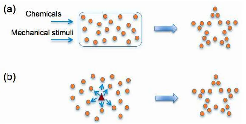
2.2 Controlling collective behavior of cells. (a) Using a global signal and/or
physical limitations. (b) Using an agent and local communication through
chemical and mechanical signals . . . 17
2.3 Control block diagram for the lactose regulation problem . . . 18
3.1 Experimental approach. We integrate motile microorganisms with
3.2 Microfabrication of biocompatible SU8 microstructures: (a) The glass slide
is coated with Dextran. (b) SU8 layer is spin coated onto the sacrificial
dextran layer. (c) UV light is transmitted through a photomask to create
an exposure pattern. (d) SU8 photoresist is developed. (e) Sections of the
glass slide each with many microstructures are inverted along the swarm
edge for bacterial attachment. (f) Individual microstructures are released
into motile buffer. . . 24
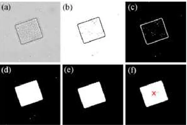
3.3 Image processing procedure: (a) Phase contrast capture. (b) Binary image
tuned threshold. (c) Inverted image. (d) Closed region filling. (e) Size
thresholding. (f) Centroid identification . . . 25
3.4 A rectangular MBR (50µmx 100µm) that is used in this paper. The
puter vision tracking system marks the trajectory of the MBR and its
com-puted interframe velocity with the arrows. . . 26
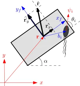
3.5 A schematic of a microbarge and a bacterium. The angle αis formed by
the main axis of the microbarge and thex axis. The vectorr denotes the
position of the microbarge’s center of mass. The vector bi denotes the
position of the i-th bacterium w.r.t the microbarge’s center of mass. The
vectorψiis a unit vector that denotes the orientation of thei-th bacterium.
The angleθiis formed by the microbarges main axis and the orientation of
thei-th bacterium. . . 28
3.6 A two-state continuous Markov chain model for the stochastic behavior of
the bacteria. The transition rates between the states are given as λ1 and
λ2. In chemical attractant free environment, measurements in biological
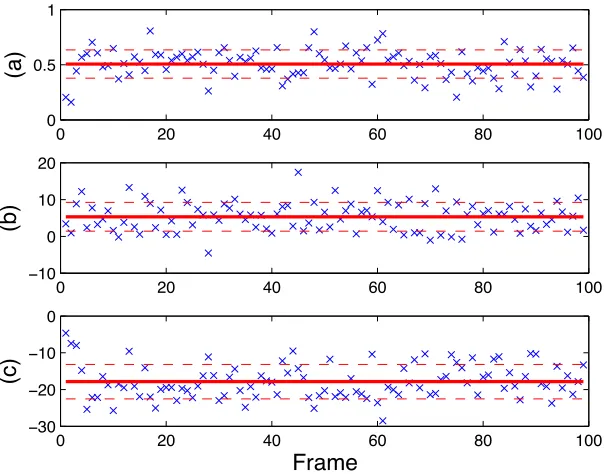
3.7 The computed data for a rectangular microbarge (50 µm×100 µm). (a)
{ω¯i}in rad/s,(b){v¯x,i} inµm/s, (c){¯vy,i} inµm/s. The solid lines show
the averages of the data, while the gaps between the solid lines and the
dashed lines represent the standard deviations. . . 33
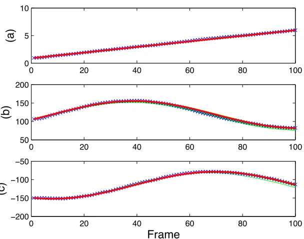
3.8 The comparison between the experimental data (x), the deterministic model
prediction (thick line), and stochastic simulations (solid lines) for a
rectan-gular microbarge (50µm×100µm). (a) αin rad/s,(b)xinµm, (c)yin
µm. . . 36
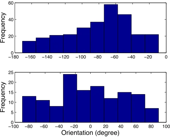
3.9 Histograms of the orientation of the bacteria on Microbarge A (top) and
Microbarge B(bottom). . . 38
4.1 Photograph of the PDMS galvanotaxis chamber. All experimental
obser-vations were performed in the central portion of the control chamber. . . 41
4.2 Histograms of the cell body orientation ofS. marcescensat electrics fields
of 4.3 and 8.9 V/cm. Electric fields were coincident with zero degrees.
The individual cells do not exhibit galvanotaxis as might be expected, and
distributions are relatively uniform across the range of angles. . . 44
4.3 MBR speed is directly proportional to applied electric field which shows
electrophoresis is the dominant electrokinetic phenomenon. The
compo-nent of speed due to self actuation appears as an offset along the vertical
4.4 A schematic of an MBR. The angle α is formed by the main axis of the
MBR and the x axis. The vector r denotes the position of the MBR’s
center of mass. The vector bi denotes the position of the i-th bacterium
w.r.t the MBR’s center of mass. The vectorni is a unit vector that denotes
the orientation of thei-th bacterium. The angleθi is formed by the MBR’s
main axis and the orientation of the i-th bacterium. The angle Ψ is the
angle between the direction of electrophoretic forceuand thexaxis. . . 47
4.5 The comparison between the experimental data (blue line) and the model
prediction (red line) for a rectangular MBR (40µm×45µm) showing self
actuation. (a)αin rad, (b)xinµm, (c)yinµm . . . 49
4.6 The comparison between the experimental data (blue line) and the model
prediction (red line) for a rectangular MBR (40 µm×45µm). 10V /cm
was applied to the MBR in+ydirection. (a)αin rad, (b)xinµm, (c)yin
µm . . . 50
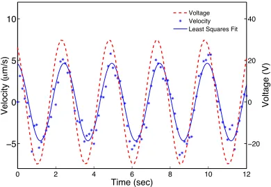
4.7 The velocity of 20×22µm2rectangular MBR as a response to a sinusoidal
input voltage (dashed line) with an amplitude of 30 V and a frequency of
0.4 Hz. A least squares minimization was used to fit a sine curve (solid
line) representing the velocity data (*) . . . 52
4.8 Bode plot of the system. The MBR follows the input signal quite well up
to a cutoff frequency near 3 Hz, where the magnitude drops off considerably 54
4.9 Sequence of cell patterning using a PDMS sieve. (a) PDMS sieve is
at-tached to the SU8 microstructures (b) Cell suspension is poured and the
solution is degassed in a vacuum chamber. (c)10X phase contrast image
of PDMS sieve in contact with microstructures (d) 63X phase contrast
im-age of microchannels filled with bacteria suspension (e)-(g) Bacteria were
4.10 Photoexposure characterization. (a) Shown is a representative selection of
results from several trials of exposure of MBRs to UV light, as well as a
trend lines before and after UV exposure. White regions represent UV
ex-posure. The results of the angular orientation (position) were normalized
and averaged to reveal characteristic trends. The final, motionless
orien-tation of the MBRs were normalized as zero radians. Angular roorien-tation is
constant before exposure, varies near zero during the few seconds
imme-diately after exposure, and decreases exponentially afterward. (b) When
exposing repeatedly, angular speed may be adjusted lower, as reflected by
slope of the curve. Angular velocity remains constant when there is no UV
exposure, even after several repetitions. . . 58
5.1 Block diagram for vision-based computer control of MBRs. The vision
system informs the control algorithm of the current position of the robot.
The control algorithm calculates the distance between the current and the
desired position and finds the power supply voltages that will create the
electric field required to steer the robot towards its next destination. . . 62
5.2 Steering of a 20×22 µm rectangular MBR along a star-shaped path. The
MBR passes through destinations 1-4 before stopping at its initial
posi-tion.The scale bar represents50µm . . . 64
5.3 The voltage applied to the system and the corresponding velocity of the
MBR inx(top) andy(bottom) direction during the experiment. The robot
responds to the changes in voltage immediately as expected. . . 66
5.4 The comparison between the experimental data (blue line) and the model
5.5 Steering of a 20×22µm rectangular MBR along a circular and a diamond
shaped path. The MBR passes through destinations and returns to its
orig-inal position. The scale bar represents50µm. . . 68
5.6 An MBR is directed through the entrance of a C-shaped microfabricated
goal using tele-operation. Scale bar represents time (2 min) as well as
length (100µm). . . 68
5.7 Micromanipulation experiment (a) Initial position of U-shaped MBR
trans-porter and target. (b) Transtrans-porter is moved to the right and down while
rota-tion continues. (c) Rotarota-tion is stopped in proper orientarota-tion upon exposure
to UV light. (d) Transporter engages the target object. . . 70
5.8 At left is shown a summary of the complete path of an MBR transporter
moving a target load described in detail in parts A-D. Total time is 2 min,
and scale bar is 25µm. (A) The transporter initially rotates clockwise due
to the self-coordination of the bacterial carpet. Electric fields are applied
to move the transporter to the left, then up. (B) With the application of
UV light, the transporter stops rotating in 6 s. As rotation is stopped,
elec-tric fields are applied to position the transporter close to the target. (C)
The target is engaged and transported to the right. (D) The target is
disen-gaged/reengaged by switching field polarity. . . 71
5.9 Fabrication process of the microdevices for motility-based and
5.10 Motility-based sensing. (a) Schematic of the setup used for the copper
sensing experiment. (b) Sensing of copper ions is observed as a loss of
rotation. The translational movement due to applied electric field persists.
(c) Angular position and velocity vs time. Fluctuations in angular velocity
are caused by the torque applied by the electric field. Scale bar represents
time (100 sec) as well as length (50µm). . . 77
5.11 Fluorescence-based sensing. (a) Schematic description of the setup.
Mi-crostructures were patterned in the center of the glass slide so that they
could be trapped inside the PDMS microchannel. (b) Fluorescence image
visualizing GFP proteins produced by induced E.coli cells. The scale bar
is 100µm. . . 80
5.12 Diagram of the lactose utilization network. The fluorescent reporter GFP
integrated in the genome is expressed in parallel with LacY under control
of the lac promoter and reports the induction level of the cell [1]. . . 81
5.13 Overlayed green fluorescence and inverted phase-contrast images of cells
that are initially uninduced for lac expression, then grown for 20 h in a
solution with (a) no TMG (b) 10 µM TMG (c) 100 µM TMG (d)
Steady-state solutions of the system. The induced Steady-state is shown as the upper
dark line whereas the uninduced state is shown as the lower dark line. The
intermediate unstable steady state is shown as a dashed line. . . 82
5.14 (a) Phase contrast image of the microstructure showing the attachedE.coli
cells (b) Phase contrast image of a monolayer of the mixed population. S.
Marcescens cells fill all the gaps on the microstructure. (c) Fluorescence
image visualized onlyE.colicells as they express GFP whileSerratiacells
6.1 Electromagnetic coils mounted on an optical microscope to actuate the
mi-cro transporters. . . 89
6.2 Single step microfabrication of large numbers of biocompatible
micro-robots. (a) The glass slide is fcoated with Dextran. (b) First pure SU8
layer and then ferromagnetic composite SU8 layer are spin coated onto the
sacrificial dextran layer. Microrotransporters are magnetized with a
per-manent magnet. (c) Photoresist is developed and micro transporters are
released into experimental chamber. (d) Phase-contrast image of 30×30
×10µm3U-shaped micro transporters. Scale bar is 30µm . . . 91
6.3 Micro transporter velocity as a function of pulsing frequency. Each data
point represents five measurements and error bars indicate one standard error. 92
6.4 Micro transporter velocity as a function of magnetic field strength. Error
bars indicate one standard error. . . 94
6.5 Microtransporter visual tracking output. The tracker estimates the
posi-tion and orientaposi-tion of the manipulator in 2D, as well as the posiposi-tions of
polystyrene beads. Several stages of processing are used to refine the
es-timate, resulting in a tracker capable of providing stable pose estimates at
30Hz. Figure by Anthony Cowley. . . . 94
6.6 (a)-(c) Microfabricated hydrogels in different shape and sizes. (d)
Phase-contrast and (e) fluorescent images of a fluorescein doped microgel. (f)
6.7 Automated transport of a 10µm latex microbead. (a) The position of
tar-get bead and the micro transporter is detected and used to plan a two-step
trajectory. (b) The transporter successfully follows the pre-planned path
and engages the target. (c) When the transporter approaches the target,
non-contact manipulation is observed. The target bead moves slower than
the transport until the transporters comes into contact. (d) The bead is
released by moving the transporter back in the same orientation. Again,
target moves with the transporter for a while due to fluid coupling. Scale
bar is 20µm. . . 99
6.8 The velocity of the transporter plus microbead as a function of the size of
the microbead. Agorose microbeads in different size and the microrobot
are shown in the inset figure. Scale bar is 30µm . . . 100
6.9 Transport of target cells. (a) The orientation of the transporter is adjusted
according to the position of the target cell. (b) With the application of an
out-of-plane time-varying magnetic field, the transporter starts translating
towards the target. The pulsing frequency is 100 Hz. (c) The target is
engaged and transported out of the field of view. The average velocity of
the transporter is 350µm/s. The scale bar is 100µm. . . 102
6.10 Phase-contrast images of rat hippocampal neurons. (a) After 10 days in
culture, an extensive, intertwined network of neurons develops on glass
slides. (b) Trypsinized neurons. Scale bars, 25µm . . . 104
6.11 Transport of trypsinized neurons. (a) A cell is detected and targeted for
manipulation. (b) The target is engaged and transported. (c) Transported
cell is released by moving the micro transporter to the left with the same
6.12 Delivery of microgels to the hippocampal cultures. (a)-(c) A microgel is
transported from its initial position to its target location. micro transporters
can be teleoperated on the neuron-coated surface without causing structural
damage to cells. (d) After releasing the microgel, another target is detected
Chapter 1
Introduction
The field of microrobotics can be defined as the design and fabrication of robotic agents
in the micrometer range and the robotic manipulation of objects with characteristic
dimen-sions in a similar size range [2]. Applications of micromanipulation include manipulation
of biological cells and assembly of micro-sized parts. The dominant forces at these length
scales are considerably different than those in typical macroscale systems. As length scale
(L) decreases, surface forces (L2) begin to dominate body forces (L3). Gravitational and
inertial forces become less influential, and adhesive interactions as well as viscous fluid
forces become more significant [3]. In addition, traditional fabrication methods become
unfeasible and novel technologies must be considered in the design of microrobots.
The fundamental challenge with decreasing robot size is providing wireless actuation
and power to the robot. Microrobots can receive both power and instructions through a
patterned surface with a scratch drive actuation technique [4]. However, microrobots that
do not rely on specialized surfaces for power delivery and control are needed to perform
complicated tasks like single cell manipulation. It is well understood that non-reciprocal
movements are necessary for propulsion in low Reynolds number fluidic environments [3],
shape-varying stroke of the cilium have garnered considerable attention [5–9]. Such biomimetic
microactuators can be manufactured using inorganic materials. Inspired by the flagella of
bacteria, helical microrobots have been demonstrated to swim in low Reynolds number
regimes by using rotating magnetic fields. In one line of work, artificial bacteria with a
helical semiconductor tail and soft magnetic head are controlled using magnetic fields [6].
In a related work, artificial bacteria are fabricated from silicon dioxide using a glancing
angle deposition technique [7]. In this technique, silicon dioxide is vapor deposited on an
array of beads using a shadow growth method. However, the ability to fabricate the required
geometries is practically limited by the planar nature of microfabrication processes. Hybrid
organic/inorganic schemes can also be realized. Magnetic particles linked by DNA are
attached to a red blood cell, and created a propulsive, beating motion [5].
The use of magnetic fields provides an attractive source of energy for untethered
wire-lessly controlled microrobotic agents. Systems have been proposed that rely on field
gradi-ents to propel the robots [10]. Gradient propulsion requires relatively large magnetic fields
as magnetic force scales with both distance and agent volume. These restrictions place
strong limits on the minimum size of the robots [11]. Torques induced on ferromagnetic
materials in a magnetic field scale more favorably than the gradient force, which has led to
a number of different approaches for microrobotic locomotion. Miniature robotic systems
with wireless magnetic end effectors have been proposed for biomanipulation [12]. In a
similar approach, hard magnetic materials such as NdFeB have been employed to induce a
stick slip motion on different substrates [13]. Another class of actuators utilizes magnetic
energy from the environment and transforms it to impact-driven mechanical force. This
concept is called wireless resonant magnetic micro-actuation (WRMMa) for untethered
mobile microrobots and it has been introduced in [14] while their application and driving
performance were demonstrated by [15]. In another line of work, untethered
release clusters of fibroblast cells [36].
Using biomolecular motors is another option [16, 17]. The integration of parallel motor
assemblies, such as muscle bundles, with MEMS technologies is very attractive because the
developed system can be highly scalable. Besides, these devices have onboard actuators
that can be powered by glucose present in physiological liquids. Neonatal rat ventricular
cardiomyocytes [18, 19] and insect dorsal vessel tissue [20] are assembled on
microfabri-cated structures to develop muscle-powered controllable autonomous microdevices.
As an alternative solution, previous studies explored different forms of actuation and
control with microorganisms. Several groups reported attempts to integrate living
or-ganisms into micromechanical devices and presented methods for harnessing the power
produced by biological motors in efforts to realize microrobots or autonomous
microde-vices [21–24]. Such cyborg microrobots could use chemical gradients, light or other stimuli
to passively or actively control the motion of the cell component. The earliest
demonstra-tion of control of microorganisms for microrobotics involved the galvanotactic
(electrode-seeking) control of the protozoan Paramecium [25]. Similar work has recently
demon-strated steeringTetrahymena pyriformiscells using galvanotaxis while using phototaxis for
temporary cell trapping [26].
Bacteria, in particular, offer several advantages as controllable microactuators: they
draw chemical energy directly from their environment, they are genetically modifiable, and
they are scalable and configurable in the sense that the cells can be selectively patterned.
Bacteria are a key component of several biointegrated hybrid organic/inorganic MEMS
devices, including a host of actuators and sensors. In one of the first instances of
bioin-tegrated, mechanical actuators, microscale rotors were driven using the gliding bacteria
Mycoplasma mobile, which were directionally attached to the rotor teeth [21].
Lab-on-a-chip fluid pumping and mixing was also demonstrated with Serratia marcescens and E.
gears have also been shown to produce useful work given the proper choice of device
ge-ometry [28,29]. Other work that shows considerable promise has focused on using bacteria
for optical or chemical read-out techniques to develop hybrid sensors.
Much of the current work on harnessing the mechanical energy of bacteria has been
directed by researchers with interest in robotics. As such, there has been a focus on the
ability to control groups of cells with the goal of directing and harnessing their energy to
accomplish tasks such as manipulation and assembly. Magnetotactic bacteria may be
con-trolled to swim en massein the direction of magnetic field lines, and have been used as
collectives to move microscale structures [30]. On/off microbead propulsion in response
to chemical stimulus has been demonstrated using the bacteriumSerratia marcescens[31],
but controlled actuation in response to chemical gradients is inherently limited by the
de-velopment of the chemical concentrations, as governed by the diffusion equation. On/off
control of microstructure movement powered by swarming S. marcescens has also been
investigated using short-term exposure to ultraviolet light [32].
1.1 Problem Statement
Autonomous smart microdevices with sensing and information processing capabilities have
great potential use in drug delivery and single cell analysis [33]. For the specific application
of automatic, remotely controlled manipulation of cells or microobjects, control of both
rotation and translation is desired. Rotational control is of particular interest where the
device has a nonsymmetrical geometry designed for engagement and trapping. Since many
potential applications for micromanipulation are performed on a glass slide in a single focal
plane using standard light microscopy, the manipulation techniques presented in this work
are restricted to two-dimensional planar motion. There is still no microrobot that has a
of sensing biologically relevant chemicals. The ability to monitor the behavior of these
microrobots in response to biologically relevant chemicals is an important requirement for
further development.
As the length scales of robotic systems continue to decrease, one of the clear emerging
applications is the manipulation of single biological cells in fluid environments. Single-cell
manipulation has traditionally been achieved with pipettes, optical tweezers, or specialized
microfluidic channel designs [34]. Magnetic control of microrobots and microgrippers
has also been established as an effective means of microobject manipulation [15, 35–37].
However, significant challenges remain for applications relating to single cell manipulation
mainly due to appropriate scaling of robot size and geometry of existing designs.
To define the appropriate design constraints for robotic single cell manipulation, it is
assumed that the most appropriate workspace is on the stage of existing inverted or
up-right light microscopes. Such microscopes are ubiquitous in life science research
laborato-ries, and include essential capabilities such as phase contrast and fluorescence microscopy.
Therefore, the integration of the full design necessarily includes not only an appropriate
robot design, but also a compact controller that is compatible with the stage of existing
microscopes. By integrating the design into existing microscopes, imaging capture
capa-bilities of the microscopes may also be harnessed.
One of the most important length scales to consider for the system is the workspace for
the robot. When working with single cells, fine details of individual cells must be resolved.
It is essential to have microstructures with sizes in the same order of target cells in order
to transport and position them with some precision. The mammalian cell is an entity with
typical dimensions of tens of microns. This requires a magnification of at least 40X. The
workspace is then 150µm×150µm. Based on this, it becomes clear that the robot must
not only be small relative to the workspace, but also that precise control of movement is
unwanted disturbances to the microenvironment.
Biocompatibility is another essential consideration for the design of a microrobotic cell
manipulator. For experiments with living cells, the idea of biocompatibility must be
ex-tended from the basic concept of not causing injury to cells to not influencing the behavior
of cells due to the chemical composition of the robot. Furthermore, the biocompatibility of
the overall design should include any chemicals released in the process of introducing the
robot to the cellular microenvironment.
Robotic manipulators on the scale of cells themselves offers significant potential
ben-efits beyond simply moving cells. Wirelessly controlled (i.e. untethered) cell-sized robots
are highly noninvasive. At this length scale, where viscous fluid forces dominate inertial
forces, motile microrobots cause very little mixing or agitation of the surrounding
environ-ment. This is a significant advantage over suction pipetting for life scientists, since pipettes
cause relatively large fluid disturbances. Traditionally, the focus of robotic manipulators
has been centered on applying mechanical forces. However, on the scale of individual cells,
the understanding of the word manipulation itself must be expanded to include chemical
manipulation of local microenvironments. To a great extent, research in single cell life
sci-ences is concerned with biochemistry. Due to this, a system for the delivery of chemicals in
the microenvironment would also greatly enhance the potential of a microrobotic system.
1.2 Approach
In the first part of this thesis (Chapters 3,4 and 5), we investigate a hybrid solution for the
controlled manipulation of microscale components with a biointegrated approach. Bacteria
attached to the surface of microfabricated parts, referred to as microbiorobots (MBRs),
nat-urally impart a predominantly rotational motion, largely due to the distribution of bacterial
rota-tion can be adjusted using optical stimuli. By harnessing both the collective work and the
electrostatic potential of bacteria, MBRs are actuated and their motion is controlled using
a combination of external stimuli. These robots are steered in a fully-automated fashion
using computer control, used to transport and manipulate micron-size objects. To take
ad-vantage of integrated live cells, they can be genetically modified and employed as sensing
elements. We describe the development of self-sustained mobile biohybrid microdevices
that harness bacterial cells for biosensors. We demonstrate two different approaches for
biosensing: motility-based sensing and fluorescent-based sensing.
In the second part of the thesis (Chapter 6), we develop a microrobotic manipulation
system using electromagnetic actuation supported with visual feedback to meet these
chal-lenges. The robot, which is only slightly larger than the rat hippocampal neurons which
we are interested in manipulating, has been designed to work on a scale appropriate for
the working space of a standard optical microscope. It is aligned by magnetic fields and
pulled by field gradients. An oscillating out-of-plane magnetic field induces a stick/slip
mechanism that enhances control of the robot [38]. This is useful not only for adjusting the
velocity of the robot [35], but also for traversing irregular microscale topographies such as
surfaces densely patterned with adherent cells. Composed of iron oxide nanoparticles
em-bedded in a polymer, the robot is fully biocompatible and is patterned using a single-mask
photolithographic process. The robot is similar in density to the working fluid. Thus, very
small magnetic forces are required for movement. Furthermore, due to the sub-micron
res-olution of the photolithographic micromachining process, the robot’s shape can be tailored
to and scaled appropriately for geometric compatibility with different cell types. Release
in the microenvironment is enabled by a biocompatible, water-soluble etch process.
A five-coiled magnetic controller was designed for rapid integration with existing
mi-croscopes. This is essential due to the fact that many features of single cells are nearly
ser-voing was incorporated for either teleoperation or fully automated manipulation, and was
demonstrated using latex microbeads,Tetrahymenacells and rat hippocampal neurons.
Finally, we present results on the integration of microscale hydrogels designed for the
localized delivery of chemicals using the microrobot. Hydrogels have been established as
an effective means of encapsulating and delivering drugs, and their design may be
specifi-cally tailored for customized time-based release [39] or even release in response to
environ-mental triggers such as pH and temperature [40]. The gels are capable of creating localized
complex gradients and transporting drugs or chemicals to specific positions of target cells.
1.3 Organization of this work
To understand microrobotics, we must begin with a discussion of how physical effects
man-ifest at the microscale. In the first part of the thesis, fabrication, mathematical modeling,
control and experimental characterization of MBRs is described. In Chapter 3, a
stochas-tic mathemastochas-tical model for the system is constructed, based on the assumption that the
behavior of each bacterium is random and independent of that of its neighbors. In
addi-tion to developing the stochastic model, parameters of the model are identified, based on
experimental data. Then it is shown that that the model with the estimated parameters is
able to predict the behavior of the system very well. In Chapter 4, two-dimensional
con-trol of MBRs is demonstrated using DC electric fields. A novel electrotaxis chamber is
designed and fabricated to harness the electrical potential of the cells. Further, the system
is rigorously analyzed and a comprehensive understanding of the fundamental physics and
a complete model are developed. In Chapter 5, the control techniques are applied to orient
and steer bacterial microbiorobots as well as to transport target loads. We also integrate
genetically engineered bacterial cells with our robots and show the feasibility of
and operation of truly micron-sized, biocompatible ferromagnetic microtransporters driven
by external magnetic fields is described. We also describe the fabrication of microgels
to deliver drugs. We demonstrate that the microtransporters can be navigated to separate
single cells with micron-size precision and localize microgels without disturbing the local
Chapter 2
Background
2.1 Life at Low Reynolds Number
The physics governing swimming at the micron scale is fundamentally different from the
physics of swimming at the macroscopic scale. The world of microrobots is the world of
low Reynolds number, a world where inertial forces are small compared to viscous drag
forces. The response of the fluid to the motion of boundaries is instantaneous and the rate
at which the momentum of a low Reynolds number swimmer changes is negligible when
compared with the typical magnitude of the forces from the surrounding viscous fluid. As a
result, Newtons law becomes a simple statement of instantaneous balance between external
and fluid forces and moments.
The Reynolds number Re is defined as Re = ρUL/η, where ρ is the fluid density,
η is the viscosity and U and L are characteristic velocity and length scales of the flow,
respectively. In water (ρ≈103kg m−3,η≈10−3Pa s), a swimming bacterium such asE. coliwithU ≈10µm s−1 andL≈1−10µm has a Reynolds numberRe≈10−5−10−4. At these low Reynolds numbers, it is appropriate to study the limitRe = 0, for which
−∇p+η∇2u= 0, ∇·u= 0. (2.1)
where uis the velocity of the swimmer,pis the pressure andη is the viscosity of the
fluid.
The Stokes equation (2.1) is linear and independent of time. When applied to low
Reynolds number locomotion, the linearity and time-independence of Stokes equation of
motion lead to two important properties. The first is rate independence: if a body undergoes
surface deformation, the distance travelled by the swimmer between two different surface
configurations does not depend on the rate at which the surface deformation occurs but
only on its geometry. The second important property of swimming without inertia is the
so-called scallop theorem: if the sequence of shapes displayed by a swimmer deforming in
a time periodic fashion is identical when viewed after a time-reversal transformation, then
the swimmer cannot move on average. As a result, locomotion results from non-reciprocal
deformations in order to break time-reversal symmetry [3].
2.2 The hydrodynamics of swimming microorganisms
Swimming strategies employed by larger organisms that operate at high Reynolds number,
such as fish, birds or insects, are ineffective at the small scale. For example, any attempt
to move by imparting momentum to the fluid, as is done in paddling, will be foiled by the
large viscous damping. Therefore microorganisms have evolved propulsion strategies that
successfully overcome and exploit drag [41].
Examples of lowReswimmers include bacteria, sperm cells, and various kinds of
proto-zoa. Flagellated bacteria, such asEscherichia coli, swim by rotating thin helical filaments,
each driven at its base by a rotary motor. The filament has a diameter of≈20nm and traces
the helix is left-handed with a pitch≈2.5µm and a helical diameter≈0.5µm [42]. There
are usually several flagella per cell. When the motor turns counter-clockwise (when viewed
from outside the cell body), the filaments wrap into a bundle that pushes the cell along at
speeds of 25-35µm s−1 [43]. When one or more of the motors reverse, the corresponding filaments leave the bundle and undergo polymorphic transformations in which the
hand-edness of the helix changes; these polymorphic transformations can change the swimming
direction of the cell.
2.2.1 Flagellar Dynamics in Viscous Fluids
Direct visualization of the flow patterns around individual flagellar filaments is quite
chal-lenging due to the filament small length scale (≈ 20 nm) and its high rotation rates (≈
100Hz).
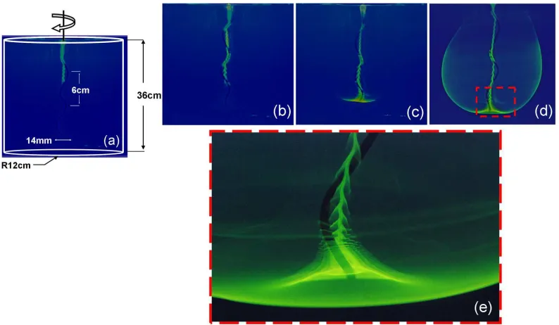
In our previous work, we investigated the flow behavior of a helical impeller rotating in
a viscous fluid at low Reynolds number, defined as Re = ρΩλ2/µ, using a
macroscopic-scale model system [44, 45]. Here, Ωis the angular velocity,λ is the helical pitch, andρ
and µare the fluid density and viscosity, respectively. Experiments were performed in a
transparent flat-bottom cylindrical vessel placed in a large cubic chamber made of acrylic
to correct for optical distortion. Both chamber and tank were filled with the same working
fluid in order to match the refraction index. The working fluid was pure glycerol (ρ= 1.2
g/cm3,µ= 800cP). The tank height and diameter were 36 cm and 24 cm, respectively. A
rigid helical filament, which was attached to an electric motor, was immersed in the fluid.
The motor typically rotated at 1.2 Hz and the helical pitch is 6 cm. Under such conditions,
Re≈0.8. The flow was visualized using ultra-violet(UV) fluorescence. The flow behavior
was assessed by the location of a neutrally buoyant dye as a function of time using a digital
camera (see Figure 2.1). The geometry of the cylindrical tank seems to be the main factor
Figure 2.1: (a) - (b) The gradual movement of the dye downwards while wrapping around the filament and producing a fishscale-like pattern until it reaches an unstable point at the tip of the helix. This instability forms due to the helical shape of the flagellum and continues to be generated at the tip of the helix as shown in (c). (d) Fully developed flow pattern. The flow in the far field falls of inversely with distance. (e) A close-up of the tip of the flagellum revealing complex flow patterns.
cyclic.
Eukaryotic flagella and cilia are much larger than bacterial flagella, with a typical
di-ameter of≈200 nm. There is a vast diversity in the beat pattern and length of eukaryotic
flagella. The cilia and flagella of sperm cells and protozoa are autonomously active
struc-tures that propagate bending waves from their base to the tip. The sperm of many organisms
consists of a head containing the genetic material together with enzymes that enable
fer-tilization to occur propelled by a filament with a planar sinusoidal or helical beat pattern,
depending on the species. The length of the flagellum is≈ 40µm for humans. In ciliate
protozoa, hundreds of cilia can coordinate and self-organize to produce a collective thrust.
Parameciumis one classic example of a ciliated microorganism. Its surface is covered by
≈500µm s−1. Tetrahymena pyriformis (T. pyriformis) also swim using cilia and there are approximately 600 cilia, both oral and locomotive, covering the cell body. Cilia closely
resemble the eukaryotic flagella in internal structure and mode of action. They are also
active organelles but their waveform is more complex consisting of a power stroke and a
recovery stroke. The cell rotates as swims due to a slight time lag between the beating of
successive rows of cilia and moves through water with a net rate of up to several
millime-ters per second. Chlamydomonas reinhardtii is an alga with two flagella that can exhibit
both ciliary and flagellar beat patterns. A cell swimming in the dark stochastically switches
between synchronous and asynchronous flagellar beating. These regimes lead to nearly
straight swimming and to abrupt large reorientations, which yield a eukaryotic version of
the ”run-and-tumble” motion of peritrichously flagellated bacteria [47].
Many cell types travel from one location to another throughout their life. The direction
of this movement is intricately linked to their local environment. This directed movement,
or taxis, may be an active signaling response such as chemotaxis, whereby the cells sense
and process the composition of their chemical environment. When a cell moves toward
higher concentrations of attractant or lower concentrations of repellent, clockwise flagellar
rotation and, hence, tumbling are suppressed. The random walk is thereby biased so that the
cell migrates up an attractant gradient or down a repellent gradient. Gradients are sensed as
temporal changes in concentration by comparing the instantaneous concentration with the
concentration the cell experienced a few seconds earlier [48].
The taxis may also be passive, where the stimuli impose a change on the motion. As an
example of such passive movement, the motion of magnetotactic bacteria such as MC-1 is
heavily influenced by weak magnetic fields. The cell bodies align with external magnetic
fields as they carry chains of iron-rich magnetosomes [49]. This kind of directed movement
is a form of adaptation enabling these cells to move to oxygen-rich environments. There
of bacteria have been shown to have several mechanisms to move toward or away from
light. Multiple mechanisms for this movement have been discovered, and sensitivities to
various wavelengths have been reported. The unicellular algaeChlamydomonas reinhardtii
is phototactic and can be guided using visible light. Cells have been controlled by switching
light emitting diodes on or off at either end of a microfluidic channel. This mechanism has
been exploited to direct and transport microscale loads [23].
2.3 Controlling Biological Systems
Biological systems exhibit many features of complex engineering systems. The origin
of system complexity is generally the presence of multiple regulatory mechanisms such
as feedback. Among several possible control strategies, feedback seems to be favored in
biological systems [50]. Hierarchies of feedback loops result in system robustness,
per-formance, and noise rejection, which are the properties of almost every biological system.
Another key feature of biological systems is emergence, aggregate behaviors that may not
be predicted by only investigating the individual components or subsystems. The existence
of emergent properties can also be explained by the presence of control mechanisms [51].
2.3.1 Stochasticity and Single Cell Studies
Cellular function often involves small numbers of molecules like DNA. These molecules
give organisms their unique genetic identity. Even genetically identical organisms grown
in homogenous environments can be very different. One of the main sources of this
vari-ability is noise in gene expression - the random fluctuations in the expression of individual
genes. This is because the expression of a gene involves discrete and random biochemical
reactions involved in the production of mRNAs and proteins. The lox copy numbers further
stochastic phenomena may have a crucial role in the fate of individual cells [52]. Stochastic
gene expression has important consequences for cellular function, being beneficial in some
contexts by producing adaptive phenotypes and harmful in others. As an example,
stud-ies have found that the decisions by E. colito enter a quiescent state to survive antibiotic
exposure (persistence) are stochastically controlled [53]. The most prominent adaptive
ex-planation for this strategy is bet-hedging. In microorganisms, a population might enhance
its fitness in fluctuating environments by allowing individual cells to stochastically
transi-tion among multiple phenotypes, thus ensuring that some cells are always prepared [54].
Without the need to sense the environment, cells could blindly anticipate and survive
en-vironmental changes with this strategy assuming that each phenotype fit to a particular
environment. Persistence has a direct benefit to the population as it allows survival during
catastrophes. Furthermore, persistent cells can provide an indirect benefit to other
individ-uals, as the reduced growth rate can reduce competition for limiting resources.
Cells in the population can exist in a continuum of phetoypes too, as occurs in
swim-ming in E. coli. In complex habitats with a sparse distribution of nutrients, variability
among individual cells in the time periods between motility switches can be expected to
form the basis of an ideal search strategy. In addition, variable responsiveness to
chemoat-tractants could help individual bacterial cells avoid predators, where attractant release is a
mechanism for luring bacterial prey [55].
2.3.2 Performing Collective Tasks with Cells and Microorganisms
There is a recent interest in the scientific community to integrate microorganisms and
liv-ing cells with engineered systems to develop microrobots, novel biosensors and intelligent
microdevices. The overall system can be compact, fast and inexpensive. Furthermore, the
cellular propulsion or actuation component could be multifunctional, perhaps serving as a
Figure 2.2: Controlling collective behavior of cells. (a) Using a global signal and/or phys-ical limitations. (b) Using an agent and local communication through chemphys-ical and me-chanical signals
often stochastic nature of cellular motion as described in the previous section. As a result,
novel strategies have to be employed to harness and control ensembles of cells. There are
two different methods control methodologies as shown in Figure 2.2. In the first approach,
the collective behavior of cells are controlled by applying a global signal: a mechanical
and/or chemical signal that affects all the cells in the environment.
In our previous work, we explored such a scenario which involves pure chemical
con-trol [57]. The architecture of the concon-trol system is illustrated in Figure 2.3. The plant to
be controlled is a large colony of E. coli bacteria. We constructed an abstraction of the
stochastic model that is simple enough to allow for fast computation. This is particularly
desirable, for example, when we want to simulate the behavior of a colony of bacteria.
Without the abstraction, we would have to run multiple copies of the stochastic simulation,
which can be computationally prohibitive.
The controller affects the plant by adjusting the external concentration of thio-methyl
Figure 2.3: Control block diagram for the lactose regulation problem
the form of a global quantity, which we consider as the output of the control system. By
this, we mean the controller does not have any information about the individual cells in the
colony. Rather, the controller relies on sensing a global quantity, for example, the fraction
of induced cells in the population. The control goal is to make the output track a given
reference trajectory or attain a desired level. Similar control architecture, where feedback
control of a group of Markov chains by adjusting the transition rates has been studied,
for example in [58]. There, the plant is a group of artificial muscle cells that can switch
between contracting and non-contracting states.
In the second method, a wirelessly-controlled synthetic agent is introduced into a colony
of cells. The agent uses the same mechanical and chemical signals that the cells use and
communicate with the members of the community in its vicinity to initiate a series of
inter-connected interactions among other members. As a result, a collective task is performed
by all the members even tough the agent has limited manipulation capability. In one
exper-imental study, authors showed collective decision-making by mixed groups of cockroaches
Chapter 3
Experimental Characterization and
Stochastic Modeling of Bacterial
Actuation
The work in this chapter was first presented in [60] and was done in collaboration with
Edward Steager, Agung Julius, UKei Cheang and Min Jun Kim
3.1 Introduction
The main challenges that need to be addressed in realizing the idea of using bacterial power
to actuate microstructures are
1) how to fabricate the structures and integrate the bacteria to them, and
2) what is the behavior of the swarm of bacteria under certain environmental conditions
and how to regulate it.
We focus our attention to the chemotactic behavior of flagellated bacteria, such as
Figure 3.1: Experimental approach. We integrate motile microorganisms with microfabri-cated structures using a chemical treatment if needed to develop MBRs
extensively studied since the 1970’s (c.f. the seminal paper by Howard Berg [61], and a
more recent book [62]). It has been established that these bacteria use their flagella to
gen-erate propulsion by rotating them [63] and that the motile behavior of the bacteria is similar
to a biased random walk toward higher concentration of chemotactic attractant.
We build buoyancy-neutral plate-like microstructures, which we callmicrobarges. We
then blot flagellated bacteria on the surface of the microbarge, which is then released to the
medium (see Figure 3.1). The motion of the microbarge is carefully tracked and compared
with model prediction.
We construct a stochastic mathematical model for the system, based on the assumption
that the behavior of each bacterium is random and independent of that of its neighbors.
In a recent paper [64], the authors proposed an approximate stochastic model to study
the diffusion or random walk properties of microbeads with bacterial propulsion. In this
paper, we study smooth and regular propulsion that is potentially more beneficial than
random walk. The study of actuation by using a large number of random actuators has
also been reported elsewhere, e.g. [58]. In addition to developing the stochastic model, we
also perform parameter identification for the model, based on experimental data. We then
the system very well. One of the key findings in this paper is that although the system is
inherently distributed, in the sense that there are a large number of independent actuators,
we can construct an accurate model with only a few parameters representing the distribution
of the bacteria.
3.2 Experimental Methods
To accomplish effective actuation of custom designed microstructures several processes are
necessary. These processes include culturing bacteriaS. marcescensusing the swarm plate
technique, fabricating microstructures, blotting and manipulating microstructures with
bac-teria into the working fluid, and finally tracking the microstructures using an algorithm to
quantify the magnitude and direction of motion.
3.2.1 Cell Culturing
SwarmingS. marcescens were cultured on a 0.6% agar plate. To prepare agar plates for
swarming, 5 g Difco Bacto tryptone, 2.5 g yeast extract, 2.5 g NaCl and 3 g Difco Bacto
agar are dissolved into 500 ml of deionized water. After autoclaving the solution was
poured into smaller bottles for later redistribution to Petri dishes. Before pouring individual
agar plates, the agar solution was mixed with 25 % glucose solution by adding 1 ml glucose
solution for 100 ml of prepared agar solution. Then, 50 ml of this new agar solution was
pipetted into large 14 cm Petri dishes. The swarm plate was inoculated on one edge with
2 µl of S. marcescens saturated culture. Agar plates were incubated at 30−34◦C, and
3.2.2 Mask design
Masks are an integral component in the photolithographic process of microstructure
fabri-cation. Using AutoCAD, the designed two-dimensional micro-geometry was drawn with
precision, and printed onto a transparency film (CAD/Art Service, Inc, Bandon, OR) with
high resolution (18,000 dpi). A dark field mask design for microstructures was generated
with 50× 100µm2 rectangles placed in an array. The distance between each individual
pattern was approximately 40µm to allow working space for extraction of individual
mi-crostructures.
3.2.3 Fabrication of Microstructures
The fabricated structures should be biocompatible, i.e. the structure material should
pre-serve and promote bacterial motility and provide a surface to which bacteria can attach
readily. Additionally, the composite specific gravity of the structure should be similar to
the working fluid and provide both chemical and thermal stability. It is additionally
help-ful if the fabricated structures are transparent and have a high refractive index to provide
clearly defined boundaries which can be readily discerned by a tracking algorithm.
SU8 Series 2 (MicroChem, Newton, MA) negative photoresist forms strong cross links
on exposure to ultraviolet (UV) light, and the unexposed regions are easily removed using
a developer solution. The SU8 microfabrication and development procedure is compatible
with a technique of release using a water-soluble sacrificial dextran layer (27). Traditional
techniques for release of structures using a sacrificial layer have required toxic chemicals.
Using dextran for the release layer, the motility medium in which the studies are performed
acted as an agent of release.
The chosen substrate for the patterning of SU8 microstructures is glass. The fabrication
Figure 3.2: Microfabrication of biocompatible SU8 microstructures: (a) The glass slide is coated with Dextran. (b) SU8 layer is spin coated onto the sacrificial dextran layer. (c) UV light is transmitted through a photomask to create an exposure pattern. (d) SU8 photoresist is developed. (e) Sections of the glass slide each with many microstructures are inverted along the swarm edge for bacterial attachment. (f) Individual microstructures are released into motile buffer.
the water-soluble sacrificial dextran layer. An aqueous solution of 5% (w/v) dextran 50-70
kDa was prepared. The solution was dispensed onto the glass slide, spin-coated, and baked.
Next, a 5µm layer of SU8-2 was spin-coated. The exposed substrate was post-baked and
developed in Propylene Glycol Monomethyl Ether Acetate (PGMEA). The wafer was then
blow dried with a jet of Nitrogen gas, and the SU8 pattern was ready for blotting and then
extraction.
3.2.4 Microstructure tracking
A tracking algorithm was designed to analyze the motion of the MBRs in motility buffer.
The current study analyzed two distinct motions of rigid bodies, translation and rotation.
To characterize the motion of the bacteria-driven microstructures, the geometric centroid
and orientation angle was traced. The algorithm was validated by testing the motion and
velocity of a theoretical test structure with predetermined shape and velocity.