Liver
Vı´ctor M. Pin˜eiro-Carrero, MD*, and Eric O. Pin˜eiro, MS‡
ABSTRACT. The liver’s unique metabolism and rela-tionship to the gastrointestinal tract make it an important target of the toxicity of drugs and xenobiotics. The de-velopmental changes that occur in the liver’s metabolic activity from birth to adolescence contribute to the varied sensitivity to toxins seen in the pediatric population. Hepatic drug metabolism, often with an imbalance be-tween the generation of toxic metabolites and detoxifica-tion processes, can influence the degree of hepatotoxic-ity. The decreased capacity of the neonatal liver to metabolize, detoxify, and excrete xenobiotics explains the prolonged action of drugs such as phenobarbital, theophyline, and phenytoin. The reduced capacity of glucuronide conjugation in the neonate not only predis-poses them to physiologic jaundice but also is probably responsible for the chloramphenicol-induced gray infant syndrome. Age-related sensitivity to drugs is attributable in part to differences in metabolic activity. For example, young children are more resistant to acetaminophen hep-atotoxicity when compared with adults, whereas children are more susceptible to valproic acid–induced toxicity. The resistance to acetaminophen toxicity is attributable to biochemical differences in young children. In chil-dren, sulfation predominates over glucuronidation, lead-ing to decreased formation of toxic intermediates. In addition, infants have a greater capacity to synthesize glutathione, thereby inactivating toxic metabolites of acetaminophen more effectively. Hepatic toxicity as a result of drugs and environmental toxins presents a wide spectrum of clinical disease. Hepatitis is the most com-mon presentation, but every major type of liver pathol-ogy can occur. Most drug reactions are attributable to idiosyncratic hepatotoxins; therefore, liver injury occurs rarely. The diagnosis of toxin-induced liver disease re-quires a high index of suspicion and often entails the exclusion of other causes of liver disease in children. Drug or environmental xenobiotic-induced hepatotoxic-ity should be considered in the setting of identified exposure or when other causes of childhood liver disease are excluded. Children who take medications that are known to be hepatotoxic, such as anticonvulsants and antineoplastic drugs, need frequent monitoring for evi-dence of hepatic toxicity. The treatment is often nonspe-cific; the most important intervention is the prompt dis-continuation of the drug or removal of the environmental toxin. A specific antidote is available only for acetamin-ophen intoxication. In cases of severe toxicity, the patient may develop liver failure. Liver transplantation may be necessary for patients whose liver failure does not
resolve.Pediatrics2004;113:1097–1106;hepatotoxicity, xe-nobiotic, drug metabolism.
ABBREVIATIONS. CYP, cytochrome P450; ALT, alanine amino-transferase; AFB, aflatoxin B; ALP, alkaline phosphatase; VOD, veno-occlusive disease; PCB, polychlorinated biphenyl; PCP, pen-tachlorophenol; TCHQ, tetrachlorohydroquinone.
T
he liver’s main function is to synthesize anarray of body proteins and to act as the detox-ifying center for the multiple toxic metabolic byproducts endogenous to the body and the toxins ingested daily by the organism. The liver undergoes dramatic changes in structure and function during development. The developmental changes that occur in the liver determine the rate and metabolic path-ways used in the disposition of drugs and other xenobiotics. The resultant metabolic intermediates may in themselves be toxic to the liver but may also cause detrimental effects to other organs of the body. This article discusses some of those xenobiotics that are hepatotoxic, with particular emphasis on sub-stances found to be toxic in the pediatric age group. For understanding the variable effects of environ-mental xenobiotic exposures in children, a basic re-view of liver anatomy, physiology, and development is necessary.
MORPHOLOGY AND FUNCTION OF THE LIVER Microscopic Anatomy and Liver Physiology
The liver performs many essential functions, in-cluding the production of bile, regulation of plasma proteins and glucose, and biotransformation of drugs and toxins. The liver is the first organ that comes into contact with enterally absorbed nutrients and xenobiotics via the portal vein. Other products of metabolism—substances that enter the body through other pathways and substances that are not extracted from the portal blood during the first pass—reach the liver by the hepatic artery.1 The
newborn liver manifests many unique physiologic traits that are likely part of the normal developmen-tal process and may predispose the liver in infants and children to the toxic effect of xenobiotics at levels that may be safe for the adult.2 The neonate has
⬍20% of the hepatocytes that are present in the adult liver, and liver growth continues after birth until it reaches its mature size.1,2The liver consists of 4 main
types of cells. The hepatocytes are the biosynthetic engines of the liver. Their prominent Golgi system and rough endoplasmic reticulum enable them to synthesize and secrete a variety of proteins. The
en-From the *Alfred I. duPont Hospital for Children, Wilmington, Delaware; and ‡Ohio State Environmental Protection Agency, Columbus, Ohio. Received for publication Oct 7, 2003; accepted Oct 20, 2003.
Reprints requests to (V.M.P.-C.) Alfred I. duPont Hospital for Children, 1600RocklandRd,Box269,Wilmington,DE19899.E-mail:vpineiro@nemours. org
dothelial cells line the sinusoids and serve as a bar-rier (interface) between the blood and hepatocytes. Two other cell types line the sinusoids: the Kupffer cells, which function as macrophages, and the stel-late cells, which store fat and vitamin A.1,3
From a functional standpoint, the liver has been described as a collection of acini that are present by the third month of gestation. Each acinus is defined as the tissue supplied by the terminal branches of the portal vein and hepatic artery and drained by the terminal branches of the hepatic vein. The paren-chyma is divided into 3 zones according to proximity to the portal triads. The hepatocytes closest to the portal areas (zone 1) receive the richest oxygen and nutrient supply and have a high concentration of enzymes involved in cell respiration; they mostly synthesize glycogen and other proteins. The hepato-cytes in zone 3 are closest to the central veins (ter-minal branches of the hepatic veins). In zone 3, little oxygen is available and the hepatocytes are involved in glycolytic energy production and contain cyto-chromes P450 (CYP), a class of enzymes responsible for metabolizing many xenobiotics. Therefore, the hepatocytes in zone 3 are more specialized in bio-transformation reactions.4,5Zone 2 is the
intermedi-ate area of hepatocytes between zones 1 and 3. Cells more distant from the portal supply (acinar zones 2 and 3) have a different enzymatic phenotype and respond differently to hypoxia and toxin exposure.
The liver performs multiple functions: bile forma-tion and excreforma-tion, synthesis of liver proteins, detox-ification of xenobiotic and endogenous compounds, and regulation of blood glucose. Toxicity caused by xenobiotics therefore can cause derangement in any of these functions and can be detected by laboratory tests used to measure these functions. Bilirubin and bile acids are the 2 primary components of bile and the best-known products of liver metabolism. Bile formation is essential for the excretion of endoge-nous waste products and the glucuronide and gluta-thione conjugates of many xenobiotics.6The capacity
to synthesize and excrete bile is immature in the neonate, making the neonate susceptible to signifi-cant cholestasis from toxic injury (Table 1).2
Functional Development of the Liver: Differential Vulnerabilities of the Liver at Different Stages of Development
The functional development of the liver has been studied extensively in the rat, less so in humans.7It
involves complex changes in liver function in the embryo and the fetus. Some enzyme activity is high in the fetus and falls during postnatal development (thymidine kinase and ornithine decarboxylase), whereas other enzymes are expressed in the fetus and increase postnatally (fructose-1,6-diphosphatase and aspartate aminotransferase). Another group of enzymes is expressed perinatally and continues to increase postnatally (uridine 5⬘-diphosphate glucu-ronyl transferase). Finally, some enzymes are ex-pressed at birth and peak at the time of weaning in the rat (alanine aminotransferase [ALT] and alcohol dehydrogenase).2 The development of physiologic
jaundice in the newborn may be caused in part by low glucuronidation activity in the liver (Table 1). These developmental changes most likely place the developing fetus and infant at differential risk from environmental toxins. For example, the reduced ca-pacity of glucuronide conjugation in the neonate is probably responsible for the gray infant syndrome from chloramphenicol.8 Unfortunately, few studies
are available in the literature exploring the effects of environmental toxins on the liver at various stages of development. This is further complicated by the lack of appropriate experimental models available to ex-amine the effect of xenobiotics at different develop-mental stages.
The structural and functional development of the liver can influence the absorption, excretion, and metabolism of drugs and other xenobiotics. Most of the knowledge regarding the differential hepatic me-tabolism is based on studies of drugs. Some of these observed differences in drug metabolism highlight potential susceptibilities of the developing human. Hepatic biotransformation is divided into 2 broad categories: phase I, or activation reactions (oxida-tions-reductions and hydrolysis), and phase II, or detoxification reactions (synthetic conjugations with sulfate, glucuronic acid, glutathione, acetate, and glycine). Many phase I and phase II enzymes that are
TABLE 1. Developmental Changes in Hepatic Metabolism, Biotransformation, and Enterohepatic Circulation
Low rates of gluconeogenesis and glucose use by the fetal liver
Amino acids are an important energy source for fetal liver (transamination and oxidative degradation)
Decreased capacity of the neonatal liver to metabolize, detoxify, and excrete xenobiotics Decreased levels of many enzymes involved in oxidation, reduction, and conjugation reactions Early expression of many CYP enzymes in the embryo and fetus (eg, CYP3A7, involved in steroid
metabolism)
Delayed expression of other CYP enzymes important for xenobiotic metabolism (eg, CYP1A2, important in drug metabolism)
Reduced activity of phase II enzymes (eg, UGT, NAT2) in the fetus and neonate Decreased hepatocyte bile acid uptake, binding, and transport in the fetus and newborn Altered conjugation and sulfation of bile acids
Decreased bile acid pool size (mostly in the premature infant) Decreased bile flow rates and intraluminal bile acid concentration
important for drug metabolism are polymorphically expressed and subject to developmental regulation. The balance between activation and detoxification reactions is critical in determining the hepatotoxic risk of drugs and toxins. For example, toxicity of benzene most likely results from oxidative metabo-lism of benzene to reactive products. A recent study showed that both phase I and phase II pathways influence the relative risk from exposure to benzene, underscoring the importance of considering the bal-ance between activation and detoxification reactions in the elimination of toxicants.9This balance is
influ-enced by stage of development, state of nutrition, exposure to multiple drugs or chemicals, and immu-nomodulators resulting from viral infections. Some enzymatic inducers may affect phase I and phase II reactions disproportionately. In addition, polymor-phisms of CYP (the major phase I enzymes) also influence this balance.10Finally, drugs and
xenobiot-ics utilize energy-dependent pathways for the excre-tion of the drug metabolites and their conjugates. These pathways, recently referred to as phase III of hepatic drug metabolism, include the multidrug
re-sistance protein and the multidrug rere-sistance-related proteins that transport drugs and chemicals into bile or into the sinusoidal circulation.11Depending on the
dose and on the metabolic and excretory pathway of xenobiotics, metabolic intermediates that can lead to varied manifestations of hepatic toxicity are formed (Fig 1). Thus, it is clear that multiple and complex interactions can alter the hepatic susceptibility of infants and children to environmental toxins.
Developmental Changes in Bioactivation and Detoxification
The ontogenic (developmental) changes in metab-olism interact with the genetic determinants of drug metabolism (pharmacogenetics) to regulate the bio-transformation of xenobiotics. The development of phase I enzymes, specifically the expression of CYP in the fetus, infant, and child, has received consider-able attention.12–14Fourteen CYP families have been
described in humans, although mostly members in CYP families 1 to 3 are important with respect to drug and xenobiotic metabolism and toxicity in hu-mans.10Several examples of developmental changes
in the functional capacity of the liver will be men-tioned. Although the fetal liver can metabolize many xenobiotics, the neonate has a prolonged half-life for most drugs. Significant and rapid maturation occurs in the first year of life; the most rapid elimination of drugs is found in school-age children and adoles-cents, and thereafter plasma clearance slows with age.14 The total hepatic microsomal CYP content
ranges from 0.3 nmol/mg microsomal protein in the fetus to 0.5 in the adult. There is a tendency for CYP content to increase with age, but the specific transi-tion age is unknown. Numerous xenobiotics are transformed to toxic intermediates in the liver. The presence of CYP in the liver and in the placenta may contribute to the toxic effects of known teratogens such as thalidomide, phenytoin, and ethanol. Of par-ticular interest is CYP3A7, the major CYP constituent in fetal liver that is not present in adult liver. CYP3A7 metabolizes many foreign compounds and plays a major role in the fetal metabolism of xenobi-otics, including potential hepatotoxins such as afla-toxin, aniline, and diethylnitrosamine.15
Microsomal epoxide hydrolase is a critical bio-transformation enzyme that catalyzes the hydrolysis of a wide variety of xenobiotic epoxides, including hydrocarbons, aromatic amines, benzene, and afla-toxin B (AFB). Studies of transplacental transfer of AFB suggest that the developing human fetus may be a sensitive target for AFB injury. Currently, there are no data on the function of this enzyme with increasing age, making it impossible to determine at which age adult levels are reached or whether the microsomal epoxide hydrolase activity in infants and children exceeds that of adults.13 An important
phase II enzyme that undergoes dramatic ontogenic and polymorphic change is N-acetyltransferase 2. This enzyme mediates the biotransformation of a large number of drugs and chemicals, including many carcinogenic arylamides. Before 15 months of age, approximately 50% of infants are slow acetyla-tors. By the age of 3 years, N-acetyltransferase 2 activity is fully expressed, although possible compe-tence (compared with adult values) is reached by 12 months of age.13 Additional research into the
onto-genic development of metabolizing enzymes is needed, in particular the changes that occur in in-fants and children.
ENVIRONMENTAL CHEMICALS, DRUGS, AND PHYSICAL AGENTS THAT ARE TOXIC
TO THE LIVER Classification of Toxic Liver Injury Intrinsic Versus Idiosyncratic
Hepatotoxicity is defined as liver injury caused by drugs or chemicals. Several classifications of hepato-toxic agents are used in the medical and hepato-toxicologic literature. Classification may focus on the source and the chemical class of the toxicant, on the circum-stances of exposure, on the type of hepatic lesion produced, on the cell structure damaged, or on the molecular or cellular mechanisms involved. Liver toxins are often classified as intrinsic (obligatory) or facultative (idiosyncratic) toxins. Intrinsic liver
tox-icity is dose dependent, is reproducible in animal models, and occurs in every person who is exposed to a sufficient dose. This type of hepatotoxicity is found in occupational, environmental, or household chemicals. Only a few drugs in clinical use are in-trinsically toxic (eg, chemotherapeutic agents, acet-aminophen); the toxicity often is seen above thera-peutic dose levels. Idiosyncratic reactions are unpredictable and are caused by the inability of sin-gle individuals to tolerate the compound. They can be hypersensitivity reactions accompanied by clini-cal symptoms as eosinophilia, fever, and rash. An-other type of idiosyncratic reaction is attributable to pharmacogenetic differences between individuals (genetic polymorphism in the metabolism of com-pounds). These individuals are not able to detoxify the parent compound, or there is an accumulation of toxic metabolites. As these pharmacogenetic mecha-nisms are elucidated, animal models can be designed in which the metabolic alterations are mimicked, therefore allowing prediction of hepatotoxicity for these susceptible individuals.16
Acute Versus Chronic Hepatic Injury
Another classification of hepatic injury is based on mode of presentation (acute vs chronic) and on the type of injury caused (Table 2). Acute hepatic injury may be cytotoxic or cholestatic. Cytotoxic injury re-sembles acute hepatitis and is characterized by dam-age to the hepatocytes with prominent elevation of aminotransferases. Severe cases may result in fulmi-nant liver failure. Depending on the agent, cell death of hepatocytes may occur by either necrosis or by apoptosis (programmed cell death). Apoptosis is a controlled form of cell death, whereby mitochondrial function is maintained and it does not induce an immune response. This lack of inflammatory re-sponse in apoptosis is advantageous because it al-lows the tissue to regenerate. Oxidative stress is one of the important mechanisms that mediate xenobi-otic-induced cell death. Many chemicals lead to the production of free radicals that can cause oxidative stress, leading to apoptosis of hepatocytes.17 In
ad-dition, free radicals can lead to lipid peroxidation of cellular membranes and cause cell death. Carbon tetrachloride, a widely known hepatotoxin, causes lipid peroxidation.18 Inhibition of protein synthesis
can result in hepatocellular necrosis. Mushroom in-toxication as a result of ingestion ofAmanitaspecies causes severe liver necrosis and is the prototype for this mechanism of action. Amatoxin in the mush-room inhibits RNA polymerase and therefore mRNA synthesis, leading to inhibition of protein synthesis.5
Cholestatic injury resembles obstructive jaundice. Aminotransferase levels are modestly elevated, whereas the alkaline phosphatase (ALP),␥-glutamyl transferase, and bilirubin elevations are more prom-inent. Cholestatic injury has a better prognosis over-all than cytotoxic injury.
Chronic hepatic injury may also be cytotoxic and cholestatic; in addition, it may cause vascular lesions such as hepatic vein thrombosis and veno-occlusive disease (VOD).19 Another important mechanism of
alterations. Immunosuppression is seen with aflatox-ins, leading to an increase risk of hepatocellular car-cinoma in areas of the world endemic to hepatitis B. Immunoallergic response has been reported with an-tibiotics such as sulfonamides, amoxicillin/clavu-lanic acid, and halothane or with chlorofluorocar-bons, still widely available as refrigerants.20
Carcinogens
A variety of xenobiotics can increase the incidence, multiplicity, or type of onset of hepatic cancer. These compounds can either damage the DNA (genotoxic) or produce cancer through nongenotoxic mecha-nisms. A single exposure to a genotoxic hepatocar-cinogen can be sufficient to produce neoplasia. In contrast, a number of drugs and chemicals may in-duce cancer in laboratory animals when adminis-tered at high doses for prolonged periods through nongenotoxic mechanisms.21 In addition,
peroxi-some proliferation has been implicated to play a role in the induction of liver cancer in rodents. Com-pounds that have been shown to cause peroxisome proliferation include hypolipidemic drugs (fibrates), phthalate ester plasticizers, and several herbicides (phenoxy acid herbicides, tridiphane, and fome-san).22 Many herbicides and pesticides are found
at low levels in the water supply. Despite the con-taminant exposure levels used (drinking water stan-dards established by the Environmental Protection Agency), these exposure levels are often not tested for their long-term effect in infants and children. The recent report23 of mutagenic potential in frogs of
another commonly used herbicide, atrazine, and its synergistic interaction with a trematode infection raises the possibility that yet-to-be-determined inter-actions could predispose susceptible individuals to genotoxic or mutagenic effects even at levels that are now deemed to be safe. Indeed, in vitro coexposure
to atrazine potentiates arsenic trioxide–induced cy-totoxicity and transcriptional activation of stress genes in transformed human hepatocytes.24 The
en-hanced toxicity of arsenic when coexposed to widely used herbicides is especially concerning, given the wide distribution of arsenic in treated wood.
Most available studies report the carcinogenic po-tential of chemicals in laboratory animals. The risk for humans, specifically for children, is not well known. Most information on cancer risk is based on epidemiologic studies. Several epidemiologic studies of cancer in young children have implicated a num-ber of environmental factors, but most studies have shown negative findings, thereby excluding poten-tial risk factors in a healthy population. The inability to identify environmental causes could mean either that the environment does not substantially affect cancer incidence in young children or that other risk factors, such as chronic hepatitis and genetic liver diseases, are not being considered.25The synergistic
effect of coexposures to AFB1and hepatitis B
infec-tion have recently been documented. Individuals who have chronic hepatitis B infection and are ex-posed to AFB1have a 3-fold increased risk for
hep-atocellular carcinoma.26 In addition, the role of
ge-netic polymorphism of detoxifying enzymes in liver cancer has only recently been documented. A recent study showed a significant association of hepatocel-lular carcinoma with the uridine 5⬘ -diphosphate-glu-curonosyltransferase UGT1A7*3 allele encoding a low detoxification activity protein.27 The increased
risk of malignancy after chronic xenobiotic exposure may not be apparent for decades. For example, the carcinogenic potential of vinyl chloride was noted after 30 years of extensive polyvinyl chloride produc-tion. Careful epidemiologic studies in addition to appropriate laboratory data are needed to determine TABLE 2. Pathologic and Biochemical Changes in Environmental Toxin–Induced Injury
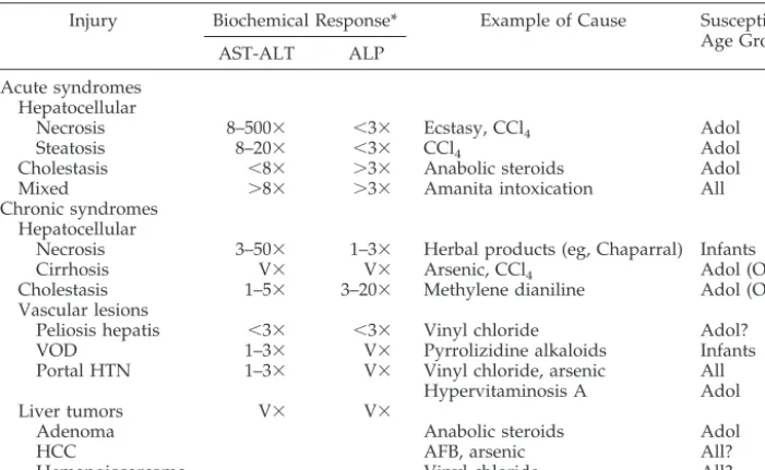
Injury Biochemical Response* Example of Cause Susceptible Age Group
AST-ALT ALP
Acute syndromes Hepatocellular
Necrosis 8–500⫻ ⬍3⫻ Ecstasy, CCl4 Adol
Steatosis 8–20⫻ ⬍3⫻ CCl4 Adol
Cholestasis ⬍8⫻ ⬎3⫻ Anabolic steroids Adol
Mixed ⬎8⫻ ⬎3⫻ Amanita intoxication All
Chronic syndromes Hepatocellular
Necrosis 3–50⫻ 1–3⫻ Herbal products (eg, Chaparral) Infants
Cirrhosis V⫻ V⫻ Arsenic, CCl4 Adol (OE?)
Cholestasis 1–5⫻ 3–20⫻ Methylene dianiline Adol (OE?)
Vascular lesions
Peliosis hepatis ⬍3⫻ ⬍3⫻ Vinyl chloride Adol?
VOD 1–3⫻ V⫻ Pyrrolizidine alkaloids Infants
Portal HTN 1–3⫻ V⫻ Vinyl chloride, arsenic All
Hypervitaminosis A Adol
Liver tumors V⫻ V⫻
Adenoma Anabolic steroids Adol
HCC AFB, arsenic All?
Hemangiosarcoma Vinyl chloride All?
Modified from Zimmerman.19
HTN indicates hypertension; HCC, hepatocellular carcinoma; AST, aspartate aminotransferase; V⫻, variable; CCl4, carbon tetrachloride; Adol, adolescents; OE, occupational exposure.
accurately the long-term effects of environmental toxins.
Environmental Toxins
There are very few reports of hepatic injury in children caused by environmental toxins. Because most of these hepatotoxins are industrial or agricul-tural products, adolescent and adults are at higher risk. Few pesticides are reported to cause hepatotox-icity. Among them, chlordecone can cause mild hep-atocellular injury. Arsenic, used as a pesticide and wood preservative, can cause hepatitis, cirrhosis, and angiosarcoma (see above and Table 2). Carbon tetra-chloride, found in many industrial applications, is a cause of hepatocellular necrosis and steatosis (Table 2). Industrial agents that are reported to cause hep-atitis include dioxane, picric acid, tetrachloroethane, and tetrachloroethylene. Polychlorinated biphenyls (PCBs), used in electrical equipment and other indus-trial applications, can cause hepatitis and may cause cirrhosis. Trinitrotoluene and phosphorus, used in explosives, can also cause hepatitis. Vinyl chloride, used in solvents and in the production of polyvinyl chloride, is a potent hepatotoxin that can cause fibro-sis, portal hypertension, and hemangiosarcoma (Ta-ble 2). Other environmental toxins that are not asso-ciated with hepatotoxicity include lead, mercury, and tobacco smoke.11
Several environmental hepatotoxins are ubiqui-tous in nature and more likely to affect children. Among them, the pyrrolizidine alkaloids found in herbal and bush teas are a recognized cause of VOD in children. This toxin causes sudden onset of portal hypertension, with very prominent hepatomegaly and ascites in a previously healthy infant or child. A recent report documented VOD in a preterm neonate whose mother had been exposed to pyrrolizidine alkaloids. Post mortem examination confirmed the presence of pyrrolizidine alkaloids in the liver.28
Vi-tamin A is a dose-dependent hepatotoxin. Hypervi-taminosis A can cause hepatic fibrosis and portal hypertension. Accidental ingestion of Amanita phal-loides and other toxic mushrooms can cause fulmi-nant liver failure. Finally, aflatoxin found in contam-inated crops is a widely recognized cause of hepatocellular carcinoma.11
Drugs
Many drugs are known to be hepatotoxic, ranging from mild, asymptomatic elevation of aminotrans-ferases to fulminant liver failure and death. Most drugs are more commonly toxic to adults, as a result of either a lower risk of toxicity in the younger pa-tient or the increased exposure to drugs in the adult and the elderly population. Most drugs that are known to cause hepatotoxicity in children fall into several categories: analgesics, antibiotics, anticonvul-sants, and antineoplastic drugs. These and several other miscellaneous drugs that are known to cause hepatotoxicity in children are listed in Table 3. Tox-icity by antineoplastic drugs deserves special consid-eration. The diagnosis of hepatotoxicity induced by antineoplastic drugs can be complicated by the fact that these patients often are treated with multiple
drugs and may also receive irradiation that can en-hance the toxicity of the drugs. Nitrosoureas, 6-mer-captopurine, cytosine arabinoside, cis-platinum, cy-clophosphamide, and dacarbazine (DTIC) may cause mild hepatitis with asymptomatic elevation of serum aminotransferases. Adriamycin, dactinomycin, and vinca alkaloids are infrequently associated with hep-atotoxicity.l-Asparaginase has been associated with more severe damage characterized by severe steato-sis, hepatocellular necrosteato-sis, and fibrosis. VOD can be seen in patients who receive thioguanine, cytosine arabinoside, DTIC, busulfan, and carmustine. Most often, VOD presents acutely with a tender, enlarged liver; ascites; and unexplained weight gain. Most cases of VOD are seen in patients after bone marrow transplantation, often in patients who also receive irradiation.
Acetaminophen is 1 of the most common causes of drug-induced hepatic toxicity in children. It is a dose-dependent toxin involving the formation of a toxic metabolite. There are 2 main clinical syn-dromes: acute overdose, either accidental in a tod-dler or intentional in adolescents, and a subacute form seen in a child who receives moderately large doses administered at regular intervals. Young chil-dren are more resistant to acetaminophen hepatotox-icity. The incidence of hepatotoxicity was 5.5% in a study of 417 children younger than 5 years, com-pared with 29% in adolescents and adults at compa-rable toxic levels.29 Several studies have
demon-strated that these age differences are attributable to biochemical differences in young children. The met-abolic profile differs greatly in early childhood. For the phase II detoxification reactions, sulfation pre-dominates over glucuronidation, probably contribut-ing to less formation of toxic intermediates. The switch to the adult pattern occurs at approximately 12 years of age. In addition, infants have a greater capacity to synthesize glutathione, thereby inactivat-ing toxic metabolites of acetaminophen more effec-tively.10 Conversely, fasting, which enhances
hepa-totoxicity to many chemicals, is known to deplete glutathione stores.17Other hepatotoxins, such as
bro-mobenzene, can also lead to glutathione depletion.18
These developmental differences lead to a decreased susceptibility to acetaminophen toxicity in young children. Other drugs that frequently are reported to cause hepatotoxicity in children are listed in Table 3. For a detailed discussion of drug-induced liver dis-ease, the reader is referred to recent reviews.10,11
VULNERABILITY OF CHILDREN TO HEPATOTOXICANTS
who receive aspirin for symptomatic treatment of a viral infection (mostly influenza and varicella; Table 3). Both valproic acid and salicylates may cause mi-tochondrial toxicity. The specific reasons for this lower risk to drug hepatotoxicity in children is prob-ably multifactorial and depends on the specific mechanisms of drug toxicity. The overall increased frequency of adverse drug reactions in adults is probably the result of increased exposure, drug in-teractions, and altered drug disposition. The lower incidence of documented hepatic toxicity from xeno-biotics in children is attributable not only to less exposure to environmental toxicants but also to their relative resistance to hepatic toxicity.
The syndrome known as Yusho disease exempli-fies the risk to the fetus. Infants who are born to mothers who were poisoned with PCB developed a congenital syndrome that included dysmorphism, skin changes, and hepatic dysfunction.30
Hepatotox-icity from low-level fetal exposure to PCBs has not been demonstrated. The risk for liver injury as a result of placental transfer of xenobiotics is also exemplified by a report of neonatal hepatitis in a newborn whose mother was taking propylthiouracil during pregnancy.31 The risk of toxicity from
con-taminated breast milk has received considerable at-tention. Specific guidelines are available regarding use of medications by lactating mothers. There are few cases of hepatic toxicity to breastfed infants caused by xenobiotics. The most important charac-teristics that determine the rate of transfer of chem-icals to breast milk are lipid solubility, ionization, and molecular weight. Chemicals that are most likely to be present in breast milk are neutral, are lipophilic, and have low molecular weight. Breastfed infants from mothers who were exposed to organic solvents are at potential risk. There is 1 report of obstructive jaundice and hepatomegaly in a 6-week-old infant who was exposed to breast milk that was contami-nated with tetrachloroethylene, a dry-cleaning sol-vent. Rapid clinical and biochemical improvement followed breastfeeding discontinuation.32 Breast
milk contains other environmental pollutants, such as PCBs, dioxin, and lead. Although a Canadian study found that only PCBs and dioxins are present at higher-than-acceptable levels in breast milk, low-level exposure and the risk for cancer are ill de-fined.33There are several reported epidemics of
per-cutaneous absorption of xenobiotics, including cases of neonatal jaundice as a result of the use of a phe-nolic disinfectant detergent.34
The preschool- and school-aged child begins to explore the neighborhood beyond the immediate confines of the home. Exposures in the school setting and play areas are the most likely sources of toxi-cants. Significant exposure to hepatotoxicants may occur in the playground areas, including exposure to organic pesticides and playground equipment treated with preservatives, such as arsenic, penta-chlorophenol, or chromium that may be toxic if in-gested. Pentachlorophenol (PCP) is a pesticide used worldwide in industrial and domestic applications as a wood preservative. Recent metabolic studies conducted in rodents and human liver homogenates
have indicated that PCP undergoes oxidative dechlo-rination to form tetrachlorohydroquinone (TCHQ). The results indicated that more toxic effects could be observed in both rats and human hepatoma cell line treated with TCHQ than its parent compound, PCP. Reactive oxygen species may be involved in the mechanism of TCHQ intoxication, suggesting that the risk of intoxication will depend on the metabolic rate of the exposed individual and on their endoge-nous antioxidant protective capacity.35
Adolescents often engage in risky behaviors such as solvent sniffing or the use of illicit drugs that can be hepatotoxic, such as ecstasy.36 In addition,
ado-lescents may have jobs that may expose them to pesticides (farm workers and lawn care) or to organic solvents (most commonly in food service and auto-motive services). They are often not properly trained or may not receive adequate protective clothing or gear, which increases their risk. Changes in CYP expression, which may occur in response to growth hormone, may lead to decreased metabolic capacity for some xenobiotics.37,38
DIAGNOSIS AND TREATMENT Detection of Liver Injury
Because there are no specific diagnostic tests or pathologic findings, the diagnosis requires a high index of suspicion and a careful drug and environ-mental exposure history, including over-the-counter and herbal preparations. Always consider the possi-bility of a child’s taking the parent’s or grandparent’s medication. The most important clue is the temporal pattern of disease evolution in relation to exposure to toxins or drugs. A brief environmental history taken at every patient encounter should document the oc-cupations of the patient and the parents and some information about the community where they live.38
Often the patient has nonspecific symptoms of gen-eral malaise, anorexia, nausea, and vomiting. The patient may have systemic features of drug hyper-sensitivity, such as fever, rash, lymphadenopathy, or mucositis. The patient with VOD may present with features of paotal hypertension in the absence of signs of chronic liver disease. Tender hepatomegaly, ascites, jaundice, and mild elevation of aminotrans-ferases is characteristic. Occasionally, the only evi-dence of liver disease is a finding of elevated amin-otransferases, ALP, or bilirubin in an asymptomatic patient. The detection of liver injury in the clinical setting is often accomplished by the use of a battery of tests for liver function. Although most of these are not specific to the liver, if several of these are abnor-mal, then a hepatic cause is likely. These tests include serum aspartate aminotransferase and ALT, which measure the integrity of the hepatocyte and the si-nusoidal plasma membrane; serum albumin and he-patic clotting factors measure the biosynthetic
capac-ity; and serum bilirubin, ALP, and ␥-glutamyl
cytomegalovirus, and Epstein-Barr virus) should be done and as well as serologic testing for autoimmune hepatitis (antinuclear antibody and anti–smooth muscle antibody). Metabolic diseases to be consid-ered include Wilson’s disease and␣1-antitrypsin de-ficiency. If a dose-dependent hepatotoxin is sus-pected (aspirin and acetaminophen), then blood levels should be obtained. Additional evaluation should include a liver ultrasound to evaluate for cholelithiasis, cholecystitis, and evidence of cirrhosis or a liver mass. In cases of poorly explained liver disease, possible drug or xenobiotic toxicity should be considered. Most often, an environmental toxin will be difficult to identify. Referral to a pediatric gastroenterologist may be necessary if no cause for the liver disease is identified. In some cases, a liver biopsy may be indicated to exclude other diseases and to help make a specific diagnosis.10,11
Treatment
With the exception of acetaminophen hepatotoxic-ity, there is little effective treatment for most cases of toxin- or drug-induced liver disease. Most often, the liver disease resolves once the offending agent is stopped. Early detection is important to ensure prompt withdrawal of the offending agent. A spe-cific antidote is available only for acetaminophen. N-acetylcysteine is most effective when given within 10 hours of acetaminophen ingestion. The decision to use it is based on plotting the blood level on a widely available toxicity nomogram. The risk of hepatotox-icity correlates with the plasma acetaminophen level and the time after ingestion. In cases of a recognized acute overdose, a poison center should be contacted for other specific guidelines (eg, gastric lavage, char-coal use). The use of corticosteroids in drug-induced liver disease is controversial. They are often used when severe acute hepatitis is part of a multisystem hypersensitivity reaction, as with phenytoin, pheno-barbital, carbamazepine, or sulfa. The treatment of fulminant liver failure as a result of drug hepatotox-icity is similar to failure caused by viral hepatitis. Deterioration of mental status and sustained impair-ment of clotting studies in conjunction with a falling ALT indicate poor outcome and require prompt re-ferral to a liver transplant center. Liver transplanta-tion may be necessary and has been reported for acetaminophen and mushroom intoxication, among others.
CONCLUSION
Hepatic toxicity as a result of drugs and environ-mental toxins presents a wide spectrum of clinical disease. Hepatitis is the most common presentation, but every major type of liver pathology can occur. Developmental changes in xenobiotic metabolism add to the complexity of hepatotoxicity as a result of drugs and environmental toxins in children. Hepatic drug metabolism, often with an imbalance between the generation of toxic metabolites and detoxification processes, can influence the degree of hepatotoxicity. Most drug reactions are attributable to idiosyncratic hepatotoxins; therefore, liver injury occurs rarely. Making the diagnosis of xenobiotic-induced
hepato-toxicity in children requires a high index of suspi-cion. Drug or environmental xenobiotic-induced hepatotoxicity should be considered in the setting of identified exposure or when other causes of child-hood liver disease are excluded. Children who take medications that are known to be hepatotoxic, such as anticonvulsants and antineoplastic drugs, need frequent monitoring for evidence of hepatic toxicity.
REFERENCES
1. Wanless IR. Anatomy, histology, embryology, and developmental anomalies of the liver. In: Feldman M, Friedman LS, Sleisenger MH, eds.Sleisenger & Fordtran’s Gastrointestinal and Liver Disease. 7th ed. Philadelphia, PA: WB Saunders; 2002:1195–1201
2. Karpen SJ, Suchy FJ. Structural and functional development of the liver. In: Suchy FJ, Sokol RJ, Balistreri WF, eds.Liver Disease in Children.2nd ed. Philadelphia, PA: Lippincot Williams & Wilkins; 2001:3–21 3. Barriault C, Desmoulire A, Costa AMA. Evaluation of chemical-induced
bile duct proliferation. In: Plaa GL, Hewitt, WR, eds.Toxicology of the Liver.2nd ed. Washington, DC: Taylor and Francis; 1998:401– 416 4. Stolz A. Liver physiology and metabolic function. In: Feldman M,
Friedman LS, Sleisenger MH, eds.Sleisenger & Fordtran’s Gastrointestinal and Liver Disease. 7th ed. Philadelphia, PA: WB Saunders; 2002: 1202–1226
5. Kahl R. The liver. In: Marquart H, Scha¨fer S, McClellan RO, Welsch F, eds.Toxicology.San Francisco, CA: Academic Press; 1999:273–296 6. Nathanson MH, Boyer JL. Mechanisms and regulation of bile formation.
Hepatology.1991;14:551–566
7. Greengard O. Enzymatic differentiation of human liver: comparison with the rat model.Pediatr Res. 1977;11:669 – 676
8. de Wildt SN, Kearns GL, Leeder JS, van den Anker JN. Glucuronidation in humans. Pharmacogenetic and developmental aspects.Clin Phama-cokinet. 1999;36:439 – 452
9. Seaton MJ, Schlosser P, Medinsky MA. In vitro conjugation of benzene metabolites by human liver: potential influence of interindividual vari-ability on benzene toxicity.Carcinogenesis. 1995;16:1519 –1527 10. Roberts EA. Drug-induced liver disease. In: Suchy FJ, Sokol RJ,
Balis-treri WF, eds.Liver Disease in Children.2nd ed. Philadelphia, PA: Lip-pincot Williams & Wilkins; 2001:463– 491
11. Farrell GC. Liver disease caused by drugs, anesthetics and toxins. In: Feldman M, Friedman LS, Sleisenger MH, eds.Sleisenger & Fordtran’s Gastrointestinal and Liver Disease.7th ed. Philadelphia, PA: WB Saunders; 2002:1403–1447
12. Hakkola J, Tanaka E, Pelkonen O. Developmental expression of cyto-chrome P450 enzymes in human liver. Pharmacol Toxicol. 1998;82: 209 –217
13. Kearns GL. Pharmacogenetics and development: are infants and chil-dren at increased risk for adverse outcomes?Curr Opin Pediatr. 1995;7: 220 –233
14. Klinger W. Biotransformation of drugs and other xenobiotics during postnatal development.Exp Toxicol Pathol. 1996;48(suppl 1):1– 88 15. Hakkola J, Pelkonen O, Pasanen M, Raunio H. Xenobiotic-metabolizing
cytochrome P450 enzymes in the human feto-placental unit: role in intrauterine toxicity.Crit Rev Toxicol. 1998;28:35–72
16. Kahl R. Toxic liver injury. In: Bircher J, Benhamou J-P, McIntyre N, Rizzetto M, Rodes J, eds.Oxford Textbook of Clinical Hepatology.2nd ed. Oxford, England: Oxford University Press; 1999:1319 –1334
17. Reed DJ. Evaluation of chemical-induced oxidative stress as a mecha-nism of hepatocyte death. In: Plaa GL, Hewitt WR, eds.Toxicology of the Liver.2nd ed. Washington, DC: Taylor and Francis; 1998:187–220 18. Comporti M. Lipid peroxidation as a mediator of chemical-induced
hepatocyte death. In: Plaa GL, Hewitt, WR, eds.Toxicology of the Liver.
2nd ed. Washington, DC: Taylor and Francis; 1998:221–257
19. Zimmerman HJ. Drug-induced hepatic disease. In: Plaa GL, Hewitt WR, eds.Toxicology of the Liver.2nd ed. Washington, DC: Taylor and Francis; 1998:3– 60
20. Furst SM, Gandolfi AJ. Immunologic mediation of chemical-induced hepatotoxicity. In: Plaa GL, Hewitt WR, eds.Toxicology of the Liver.2nd ed. Washington, DC: Taylor and Francis; 1998:259 –296
21. Klaunig JE, Kolaja KL. Chemical-induced hepatocarcinogenesis. In: Plaa GL, Hewitt WR, eds.Toxicology of the Liver.2nd ed. Washington, DC: Taylor and Francis; 1998:93–121
23. Kiesecker JM. Synergism between trematode infection and pesticide exposure: a link to amphibian limb deformities in nature?Proc Natl Acad Sci U S A. 2002;99:9900 –9904
24. Tchounwou PB, Wilson BA, Ishaque AB, Scheneider J. Atrazine poten-tiation of arsenic trioxide-induced cytotoxicity and gene expression in human liver carcinoma cells (HepG2).Mol Cell Biochem.2001;222:49 –59 25. Buckley JD. The aetiology of cancer in the very young.Br J Cancer.
1992;18:S8 –S12
26. Sun Z, Lu P, Gail MH, et al. Increased risk of hepatocellular carcinoma in male hepatitis B surface antigen carriers with chronic hepatitis who have detectable urinary aflatoxin metabolite M1.Hepatology.1999;30: 379 –383
27. Vogel A, Kneip S, Barut A, et al. Genetic link of hepatocellular carci-noma with polymorphisms of the UDP-glucuronosyltransferase UGT1A7 gene.Gastroenterology. 2001;121:1136 –1144
28. Rasenack R, Muller C, Kleinschmidt M, Rasenack J, Wiedenfeld H. Veno-occlusive disease in a fetus caused by pyrrolizidine alkaloids of food origin.Fetal Diagn Ther. 2003;18:223–225
29. Rumack BH. Acetaminophen overdose in young children. Treatment and effect of alcohol and other additional ingestants in 417 cases.Am J Dis Child. 1984;138:428 – 433
30. Masudo Y. Health status of Japanese and Taiwanese after exposure to
contaminated rice oil.Environ Health Perspect. 1985;60:321–325 31. Hayashida CY, Duarte AJS, Sato AE, Yamashiro-Kanashiro WH.
Neo-natal hepatitis and lymphocyte sensitization by placental transfer of propylthiouracil.J Endocrinol Invest. 1990;13:937–941
32. Bagnell PC, Ellenberg HA. Obstructive jaundice due to a chlorinated hydrocarbon in breast milk.Can Med Assoc J. 1977;117:1047–1048 33. Schreiber JS. Parents worried about breast milk contamination.Pediatr
Clin North Am. 2001;48:1113–1127
34. Wysowski DK, Flynt JW, Goldfield M, Altman R, Davis AT. Epidemic neonatal hyperbilirubinemia and use of a phenolic disinfectant deter-gent.Pediatrics. 1978;61:165–170
35. Wang YJ, Lee CC, Chang WC, Liou HB, Ho YS. Oxidative stress and liver toxicity in rats and human hepatoma cell line induced by penta-chlorophenol and its major metabolite tetrachlorohydroquinone.Toxicol Lett. 2001;122:157–169
36. Andreu V, Mas A, Bruguera M, et al. Ecstasy: a common cause of severe acute hepatotoxicity.J Hepatol.1998;29:394 –397
37. Pollack SA. Adolescent occupational exposures and pediatric-adolescent take-home exposures. Pediatr Clin North Am. 2001;48: 1267–1289
2004;113;1097
Pediatrics
Víctor M. Piñeiro-Carrero and Eric O. Piñeiro
Liver
Services
Updated Information &
http://pediatrics.aappublications.org/content/113/Supplement_3/1097 including high resolution figures, can be found at:
References
#BIBL
http://pediatrics.aappublications.org/content/113/Supplement_3/1097 This article cites 24 articles, 3 of which you can access for free at:
Subspecialty Collections
http://www.aappublications.org/cgi/collection/gastroenterology_sub Gastroenterology
sub
http://www.aappublications.org/cgi/collection/environmental_health_ Environmental Health
following collection(s):
This article, along with others on similar topics, appears in the
Permissions & Licensing
http://www.aappublications.org/site/misc/Permissions.xhtml in its entirety can be found online at:
Information about reproducing this article in parts (figures, tables) or
Reprints
2004;113;1097
Pediatrics
Víctor M. Piñeiro-Carrero and Eric O. Piñeiro
Liver
http://pediatrics.aappublications.org/content/113/Supplement_3/1097
located on the World Wide Web at:
The online version of this article, along with updated information and services, is
by the American Academy of Pediatrics. All rights reserved. Print ISSN: 1073-0397.