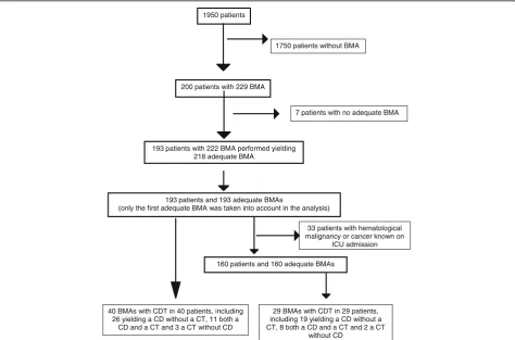
Contribution to diagnosis and treatment of bone marrow aspirate results in critically ill patients undergoing bone marrow aspiration: a retrospective study of 193 consecutive patients
9
0
0
Full text
Figure

![Fig. 2 Indications for bone marrow aspiration and pathological: Megakaryocyte depletion, one or fewer megakaryocytes per 5 to 10 low-powerfields on bone marrow examination [15] Other, suspicion of tuberculosis (N=5), undetermined (N=5); Reactive bone marrow changes, non-specificbone marrow modifications associated with acute inflammation including hypercellularity, an increased myeloid-to-erythroid ratio with a largenumber of myeloid precursors and mature segmented neutrophils, with or without megakaryocyte hyperplasia, monocytosis and a slight increasein normal plasmocytes [14]; Vitamin B12/folate deficiency -like features, cytological signs classically associated with vitamin B12/folate deficiencyincluding hypersegmented neutrophil granulocytes, giant metamyelocytes, erythroid asynchronism maturation](https://thumb-us.123doks.com/thumbv2/123dok_us/721096.1568559/5.595.56.541.87.256/megakaryocyte-megakaryocytes-modifications-hypercellularity-megakaryocyte-deficiencyincluding-hypersegmented-metamyelocytes.webp)

+2

Related documents