R E S E A R C H A R T I C L E
Open Access
Distal tibial distraction osteogenesis
—
an
alternative approach to addressing limb
length discrepancy with concurrent
hindfoot and ankle reconstruction
Todd M. Chappell
1*, Casey C. Ebert
2, Kevin M. McCann
3, Byron L. Hutchinson
4and Edgardo Rodriguez-Collazo
5Abstract
Background:Limb length discrepancy (LLD) in the setting of concurrent hindfoot and ankle deformity poses an added level of complexity to the reconstructive surgeon. Regardless of etiology, a clinically significant LLD poses additional challenges without a forthright and validated solution. The purpose of the current study is to determine whether reconstructive hindfoot and ankle surgery with concurrent lengthening through a distal tibial corticotomy is comparable to other treatment alternatives in the literature.
Patients and methods:A retrospective review of hindfoot and ankle deformity correction utilizing Ilizarov circular external fixation with concurrent distal tibial distraction osteogenesis from July 2009 to September 2014 was conducted.
Results: This study included 19 patients with a mean age of 47.47 ± 13.36 years with a mean follow up of 576.13 ± 341.89 days. The mean preoperative LLD was 2.70 ± 1.22 cm and the mean operatively induced LLD was 2.53 ± 0.59 cm. The mean latency period was 9.33 ± 3.47 days and distraction rate was 0.55 ± 0.16 mm/day. The mean distraction length was 2.14 ± 0.83 cm and mean duration of external fixation was 146.42 ± 58.69 days. The time to union of all hindfoot and ankle fusions was 121.00 ± 25.66 days with an overall fusion rate of 85.71%.
Conclusions: The successful treatment of hindfoot and ankle deformity correction in the setting of LLD using the technique of a distal tibial corticotomy and distraction osteogenesis is reported and illustrates an additional treatment technique with comparable measured outcomes to those previously described. We urge that each patient presentation be evaluated with consideration of all described approaches and associated literature to determine the current best reconstructive approach as future studies may validate or replace the accepted options at present.
Keywords:Distraction osteogenesis, Ankle replacement, Avascular necrosis, Talus, Limb length discrepancy, Distal tibial corticotomy
© The Author(s). 2019Open AccessThis article is distributed under the terms of the Creative Commons Attribution 4.0 International License (http://creativecommons.org/licenses/by/4.0/), which permits unrestricted use, distribution, and reproduction in any medium, provided you give appropriate credit to the original author(s) and the source, provide a link to the Creative Commons license, and indicate if changes were made. The Creative Commons Public Domain Dedication waiver
(http://creativecommons.org/publicdomain/zero/1.0/) applies to the data made available in this article, unless otherwise stated.
* Correspondence:[email protected]
1Franciscan Foot & Ankle Associates, 1608 S J St., 4th Floor, Tacoma, WA
98405, USA
Background
Orthopedic pathology affecting the hindfoot and ankle can be debilitating to the ambulatory patient, especially when accompanied by limb length discrepancy (LLD). Patients with a broad array of diagnoses may be catego-rized as having hindfoot and ankle deformity with ac-companying LLD, these include talar avascular necrosis (AVN), failed total ankle replacement (TAR), congenital or post-traumatic LLD, and those with unintended se-quela of surgical interventions or failed hindfoot and ankle reconstructive attempts, e.g., nonunion and/or malunion. Regardless of the etiology, these pathologies involving LLD can lead to functional and anatomical changes that may increase patient morbidity [1–4]. While it has been suggested that a LLD greater than 2– 2.5 cm is poorly tolerated [5], a LLD of 0.5–1 cm is per-haps desirable in situations of preexisting or concurrent hindfoot fusions [6]. It has, however, also been reported that as little as a 3-mm LLD can cause postural changes, which over time, may lead to degenerative changes and adaptations of the kinetic chain [7–10]. Deficient limb lengths not amenable to conservative management may require surgical intervention.
A resultant bone deficit and/or LLD has been ad-dressed in multiple ways including fusion of the hindfoot and ankle with accepted loss of length [1, 4, 11], bone grafting procedures of many varieties [12–16], im-plantable trabecular metal grafts [17–21], distraction osteogenesis [22–25], the use of internal and/or ex-ternal hardware, and any combination of the above [3, 6, 13, 26–28]. Despite the growing number of treat-ment alternatives and the various suggested treattreat-ment al-gorithms that are present in the literature, [29] there is no proven gold standard to date.
This study proposes an application of a sparsely published approach to addressing LLD in the distal tibia and ankle. Distal tibial corticotomy (DTC) followed by distraction osteogenesis has been employed to address LLD when faced with complicated ankle fusion. [24] The approach described in this study is similar to the previously reported technique and in-cludes the concomitant arthrodesis of the hindfoot followed by lengthening through a DTC utilizing Ili-zarov circular external fixation (ICEF). There is a paucity of reports on distal tibial distraction osteogen-esis (DTDO), and as far as the authors are aware, this approach to hindfoot deformity and LLD has not been described in the literature. These surgical tech-niques have been employed for the past several years to address lower limb deformity correction in the set-ting of LLD post-reconstruction with the desired out-come of limb salvage and restoration of limb length to prevent the comorbidities associated with limb length inequality.
This approach to treating bone defects of the hindfoot is equivalent to, and in some cases, superior to other treatment alternatives in the literature as it pertains to limb salvage, osseous union, and restoration of limb length. The purpose is to review the outcomes of such reconstructions in terms of radiographic deformity cor-rection and limb length restoration to determine whether or not this technique is comparable to other treatment alternatives in the literature.
Patients and methods
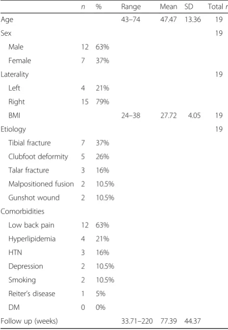
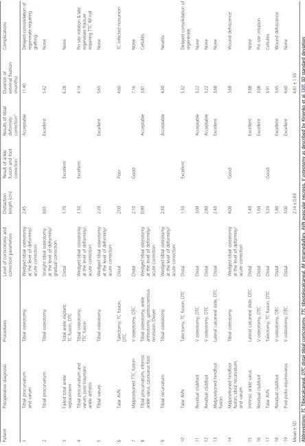
Following Institutional Review Board (IRB) approval, a retrospective chart review of hindfoot, ankle, and lower limb deformity correction utilizing ICEF with concurrent DTDO from July 2009 to September 2014 was con-ducted. Current Procedural Terminology (CPT) codes (apply bone fixations device, 20692; osteotomy, tibia, 27705; osteotomy of tibia fibula, 27709; lengthening tibia/fibula, 27715) were searched for patients undergo-ing reconstructive surgery at our facilities. This search revealed 172 results. Operative reports and clinical and radiographic charts were then reviewed for all cases to included patients who underwent surgical reconstruc-tion of hindfoot and ankle deformity with simultaneous DTDO utilizing ICEF to address LLD as a component of their overall deformity. Patients were excluded if they were less than 18 years of age, had inadequate radio-graphic imaging, or less than 6 months of follow-up. This review resulted in a total of 19 patients. A clinical and radiographic review of patient records was then conducted to determine demographic data, (Table 1) preoperative diagnosis, concurrent procedures, distrac-tion length, duradistrac-tion of external fixadistrac-tion, minor compli-cations, major complicompli-cations, union rates, and objective deformity correction outcomes (Table2).
Preoperative LLD was evaluated using 51-in. bipedal radiographs and/or scanograms for those patients with-out suspected intraoperatively induced LLD. For those patients with an operatively induced LLD, defined as LLD due to intraoperative extraction of failed TAR hard-ware or bone extraction due to AVN, this was calculated based off of intraoperative measurements and postopera-tive assessment. All cases experienced less than 3 cm of shortening and segmental bone loss due to extraction of pathologic elements and therefore underwent acute shortening intraoperatively. Of note, none of these pa-tients experienced neurovascular compromise.
Orthobiologics
the prepared distal fibular resection combined with a preparation of allogeneic bone matrix containing vi-able osteogenic cells (Trinity Elite® Orthofix, Inc., Verona, Italy).
External fixation construct
Following reconstructive procedures of the hindfoot and ankle, application of an ICEF stabilized the correction and was the final step in preparation for the DTC. The construct for the ICEF varied depending on patient dif-ferences and pathology addressed; however, it most com-monly consisted of a three-ring tibial block spanning the DTC site fixed to the extremity with a combination of 4-mm half pins and tensioned wires in the two proximal tibial rings for biplane stabilization. The distal tibial ring was fixed with two simultaneously tensioned opposing olive wires and this tibial block was connected to a foot-plate secured to the tibial block with compression rods and fixed to the foot with two simultaneously tensioned opposing olive wires in the calcaneus and two additional wires in the mid to forefoot. The hindfoot fusion sites were compressed as appropriate depending on concur-rent procedures.
Surgical technique
The DTC was completed through an anterior approach at the level of the deformity apex in cases of distal tibial angular deformity. Acute correction at the site of angu-lar tibial deformity was conducted in 6/7 (85.71%) pa-tients via a wedge osteotomy due to its relatively small angular deviation. The remaining case of tibial deformity was corrected gradually due to its high angle of deform-ity and potential of neurovascular injury with acute cor-rection. In cases with an absence of distal tibial angular deformity, the corticotomy was conducted in similar fashion as close to the distal tibial meta-diaphyseal junc-tion as possible without interfering with fixajunc-tion ele-ments for stability. Due to initial success lengthening distally at the site of tibial angular deformity, this method was applied to patients who required < 4 cm of lengthening concurrent with their hindfoot and ankle deformity correction. Fixation elements prohibiting DTC as close as possible to the distal tibial meta-diaphyseal junction, and patients requiring > 4 cm of length were not considered candidates for this technique and may be better served by a proximal tibial osteotomy.
The DTC was initiated through the anterior cortex using a drill bit with the use of cold saline irrigation. The DTC was then completed using a series of drill holes connected by sharp osteotomes taking care to not disturb the medullary canal. Once the corticotomy was completed, the ICEF was used to compress the site and intraoperative fluoroscopy was used to verify maintained alignment of the distal extremity. Patients were instructed to remain non-weight bearing and follow up within 5–10 days at which time alignment and pin sites were examined and documentation of the acquired LLD was performed as appropriate.
Clinical monitoring
Depending upon the concurrent procedures for hindfoot and ankle reconstruction, the LLD, and patient comor-bidities, various methods were used to determine the timing of weight bearing and dynamization. Limb length was monitored during treatment by clinical evaluation and radiographic monitoring during distraction until LLD was less than 1 cm. Observation of regenerate and serial radiographs to determine the time to fusion were also collected. Regardless of the variability in each case, the time for consolidation was estimated to be twice the duration of distraction and dynamization was initiated based off of clinical judgment and the appearance of radiographic signs of fusion and consolidation of the regenerate.
Objective radiographic parameters
Objective measures of osseous correction were evaluated using previously published parameters for distal tibial
Table 1Patient demographics
n % Range Mean SD Totaln
Age 43–74 47.47 13.36 19
Sex 19
Male 12 63%
Female 7 37%
Laterality 19
Left 4 21%
Right 15 79%
BMI 24–38 27.72 4.05 19
Etiology 19
Tibial fracture 7 37%
Clubfoot deformity 5 26%
Talar fracture 3 16%
Malpositioned fusion 2 10.5%
Gunshot wound 2 10.5%
Comorbidities
Low back pain 12 63%
Hyperlipidemia 4 21%
HTN 3 16%
Depression 2 10.5%
Smoking 2 10.5%
Reiter’s disease 1 5%
DM 0 0%
Follow up (weeks) 33.71–220 77.39 44.37
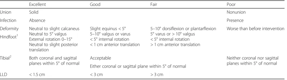
[2], hindfoot and ankle [9], deformity correction includ-ing union, presence or absence of infection, position of fusion, and residual LLD post lengthening with less than 1.5 cm being excellent. The goals for position of fusion of the hindfoot and ankle were deemed excellent when the hindfoot was in neutral to slight calcaneus, neutral to 5° of valgus, neutral to slight posterior translation, and 0–15° of external rotation [9]. In situations of tibial deformity alone, the same parameters were evaluated in terms of union, absence or presence of infection, and re-sidual LLD post lengthening according to Schoenleber et al. [2]. When both coronal and sagittal planes were less than 5° from the normal anatomic axis of the tibia, the correction was deemed excellent; however, the position of the hindfoot was not assessed in his cri-teria [2] which is the rationale for using the com-bined criteria for objective radiographic evaluation of osseous outcomes (Table 3).
Means and standard deviations were performed for all parameters using Microsoft Excel (Palo Alto, CA, USA). Outcome comparison statistics were not evaluated due to the small sample size of the hindfoot and tibial de-formity groups as well as the variability of the concur-rent hindfoot and ankle procedures making comparison between groups and cases challenging.
Results
This study included 12 males and 7 females with a mean age of 47.47 ± 13.36 years (range 22 to 74 years) with a mean body mass index (BMI) of 27.72 ± 4.05. The mean follow-up was 576.13 ± 341.89 days (range 236–1667 days). The most common cause of deformity was tibial fracture 7/19 (36.84%) with 2/7 (28.57%) being open at the time of initial injury. The second most common cause of deformity was history of clubfoot deformity with a remote history of correction 5/19 (26.32%) (Fig.1) followed by talar fracture 3/19 (15.79%) (Fig.2) malposi-tioned hindfoot fusions, and gunshot wounds with 2/19
(10.53%) each. Although the abovementioned diagnoses were the initial cause of deformity, 16/19 (84.21%) had undergone at least one surgery prior to their presentation. The most surgical procedures undergone by a single pa-tient was five.
The mean preoperative LLD was 2.70 ± 1.22 cm and the mean operatively induced LLD was 2.53 ± 0.59 cm. The most common procedure to correct deformity of the hindfoot and ankle was a V osteotomy as described by Kirienko et al. [30] 5/9 (55.56%) followed by tibiocal-caneal fusion 4/9 (44.44%) following explant of TAR or talectomy for AVN. The mean latency period was 9.33 ± 3.47 days. The mean distraction length was 2.14 ± 0.83 cm and mean duration of external fixation was 146.42 ± 58.69 days.
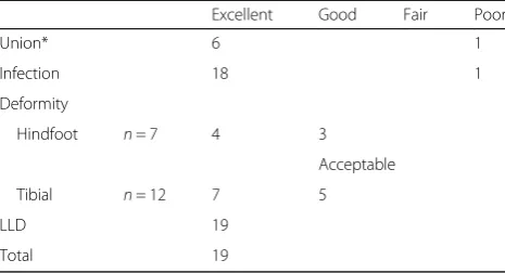
The time to union of hindfoot/ankle fusions was 121.00 ± 25.66 days as determined radiographically by routine postoperative radiographs demonstrating bridg-ing trabeculation of the fusion site and also clinically by the sustained absence of pain and swelling at the surgical site. The overall fusion rate was 6/7 (85.71%). Following the parameters outlined by Schoenleber et al. for tibial deformity correction and Katsenis et al. for hindfoot and ankle deformity correction, excellent union results in 6/ 7 (85.71%) patients, excellent infection results in 18/19 (94.74%) patients, excellent postoperative LLD results in 19/19 (100%) patients, excellent hindfoot and ankle de-formity correction results in 4/7 (57.14%) patients, and excellent tibial alignment results in 7/12 (58.33%) pa-tients were achieved (Table4).
Complications were evaluated and categorized as major and minor. Major complications required surgical intervention for resolution whereas minor complications were addressed non-surgically. There were four major complications and seven minor complications. Major complications included delayed consolidation of regener-ate in 2/19 (10.53%), lregener-ate fracture of regenerregener-ate in 1/19 (5.26%), and infected nonunion of the tibiocalcaneal
Table 3Objective measures of osseous correction
Excellent Good Fair Poor
Union Solid Nonunion
Infection Absence Presence
Deformity Neutral to slight calcaneus Neutral to 5° valgus External rotation 0–15° Neutral to slight posterior translation
Slight equinus < 5° 5–10° valgus or varus < 5° internal rotation < 1 cm anterior translation
5–10° dorsiflexion or plantarflexion 5° varus or > 10° valgus
< 5° internal rotation > 1 cm anterior translation
Worse than before intervention
Hindfoot1
Tibial2 Both coronal and sagittal
planes within 5° of normal
Acceptable Neither coronal nor sagittal
planes within 5° of normal Either coronal or sagittal plane within 5° of normal
LLD < 1.5 cm < 3 cm > 3 cm
Abbreviations:LLDlimb length discrepancy 1
Post surgical hindfoot and ankle alignment grading as proposed by Katsenis et al. [9] 2
fusion site in 1/19 (5.26%). The delayed consolidation was treated with intramedullary (IM) nailing and bone marrow stimulation consisting of autologous BMAC in 1/2 (50%) and the other with bone grafting and internal plate fixation. Both procedures resulted in successful healing. The late fracture of regenerate was treated with tibiotalocalcaneal (TTC) fusion using IM nailing and re-sulted in a successful outcome. The infected nonunion was treated by debridement, an extended course of intravenous (IV) antibiotics, and definitive placement of an antibiotic cement spacer. This patient is currently ambulating with the assistance of a leg brace (Exosym™ Hanger, Inc., TX, USA) which offloads the extremity and there are currently no plans of revision surgery. Of the
seven minor complications, cellulitis, pin site infection, and wound dehiscence were the most common occur-ring in 2/19 (10.53%) patients each. Neuritis was the sec-ond most common minor complication affecting 1/19 (5.26%) patients. Minor complications of an infectious nature were all treated with oral antibiotics to full resolution.
Discussion
Hindfoot and ankle deformity is a complex pathology and a challenge for the reconstructive surgeon; this complexity is increased in the setting of LLD. The etiology of LLD is varied and often is a result of trauma or prior surgical interventions and/or failures
Fig. 1Case #7. Fifty-eight-year-old F with history of clubfoot correction as a teenager.aPreoperative appearance of foot.b,cPreoperative
radiographs of cavoadductovarus foot with evidence of prior surgery.dIntraoperative fluoroscopic image of V osteotomy.e–gClinical
and radiographic appearance of frame construct for gradual foot correction and DTC demonstrating regenerate formation.h–jPostoperative
anteroposterior and lateral radiographs demonstrating foot correction and consolidated distal tibial corticotomy site.k,lPostoperative appearance of
the foot and corrected LLD
Fig. 2Case #10. Seventy-four-year-old F 9 months s/p motor vehicle accident with closed talus fracture.a,bPreoperative radiographs
demonstrating talar collapse and AVN.c Intraoperative image post talectomy.d,eIntraoperative fluoroscopy of tibiocalcaneal fusion site
alignment.f,gAP and lateral radiographs demonstrating frame construct for tibiocalcaneal fusion and DTC.h–jClinical and radiographic
appearance of frame construct demonstrating regenerate formation.k,lFinal postoperative AP and lateral radiographs following distal
of such interventions. Regardless of the cause of the LLD, there is no consensus as to the most successful method of treatment and several approaches have demonstrated success.
Based off of our experience with distal tibial lengthen-ing, we propose an additional alternative to the previ-ously described methods. The time to union in this study was 121.00 ± 25.66 days with an overall fusion rate of 85.71%. The mean distraction length was 2.14 ± 0.83 cm and mean duration of external fixation was 146.42 ± 58.69 days resulting in an external fixation index of 68.42 days/cm. These results are comparable to other described methods in the literature making this tech-nique a successful alternative when used by surgeons with appropriate experience and technical expertise.
The consistent search for alternate treatment options for this challenging pathology has resulted in multiple studies reporting on various methods to achieve success-ful hindfoot and ankle fusions in the setting of LLD. In a review of published studies, it was suggested that the most straightforward method of treatment is direct fu-sion with loss of length when the resultant LLD is < 2 cm and acceptable to the patient [11]. A similar conclu-sion was made in a study reviewing cases of talar AVN which demonstrated that successful tibiocalcaneal fusion can be achieved without the use of structural bone graft [4]. Fragomen et al. reported on complex ankle fusion in 91 patients utilizing the Ilizarov method and noted greater LLD was associated with a higher risk of non-union although the majority of their patients did not undergo lengthening to normalize limb lengths. They recommended that patients with a LLD > 2.5 cm and < 70 years old be considered candidates for lengthening. Their results indicate that the 24/91 (26.37%) patients who underwent lengthening resulted with an 83% rate of union [1].
Although it has been shown that correction of LLD is not necessary to obtain successful fusion or positive pa-tient outcomes, the majority of the current literature aims for correction of LLD. Some of the more recent
literature has been directed toward implantation of metal spacers in an attempt to limit the post treatment LLD. There has been a myriad of reportedly successful implantations; however, they are commonly single case reports or small case series. In 2008, the use of Harms cages was reported in three patients after failed TAR. All required revision surgery with IM retrograde nail fix-ation after removal of the Harms cages, and 2/3 (66.66%) successfully fused the second attempt [17]. The use of a titanium cage with morselized cancellous graft was also reported; however, retrograde IM nail fixation was added for stability and successful results were re-ported in two patients [19]. Sagherian et al. has reported on the successful use of porous tantalum metal spacers in ankle and hindfoot surgery as well as various other foot pathologies with good results reporting 100% fusion rate of the implant-bone interface. Fusion was defined as an absence of lucency at the bone-tantalum interface on plain radiography and maintenance of correction with the clinical absence of pain, tenderness or swelling [18, 21]. Although there may be some potential for successful implementation of these devices in the fu-ture, it would require increased patient numbers and sustained evidence of successful incorporation into bone for widespread adoption.
One of the most widely reported methods for address-ing hindfoot and ankle deformity in the settaddress-ing of LLD has been the use of structural bone grafting techniques. These have included tricortical iliac crest autograft, fresh-frozen femoral head allograft (FFFHA), free vascu-larized bone autografts, and fibular strut autografts to name a few. In 2009, the use of an anterior double plat-ing system for tibiotalar arthrodesis was reported on 29 patients. Amongst this group, 9 patients had failed TAR for which they utilized FFFHA. Although the mean fol-low up for this study was 43.9 months, it appears that they elected not to comment on whether or not these grafts underwent late collapse, it was reported that they had a 100% union rate with no evidence of collapse dur-ing the first 12 months. They did comment however that they could not conclude that such grafts will not col-lapse in the future [15].
Others have reported on their experience with FFFHA in patients treated for failed TAR. In a review of 9 pa-tients treated with FFFHA and internal fixation, it was reported as a 55.6% union rate with 4/9 patients failing at the graft subtalar joint (STJ) interface. They believed that it was their selected difficult approach to prepar-ation of the STJ through the anterior incision to blame for their poor result and therefore their revision cases were approached through a sinus tarsi incision and eventually healed the remainder of the patients. They also reported a late fracture of the FFFHA [12]. In 2013, Jeng et al. reported on the use of FFFHA for TTC fusion
Table 4Osseous correction outcomes
Excellent Good Fair Poor
Union* 6 1
Infection 18 1
Deformity
Hindfoot n= 7 4 3
Acceptable
Tibial n= 12 7 5
LLD 19
Total 19
in 32 patients. The average size of the FFFHA implanted was 38 mm, and IM rod fixation was the primary method of choice although there were 6 patients that re-ceived internal plate fixation. Only 50% of these grafts went on to heal and the average collapse of the FFFHA at final follow up was measured at 3.6 mm [14]. A simi-lar study in 2014 utilized FFFHA and a combination of internal plate and rod fixation in 17 patients for failed TAR. Their results are superior to Jeng et al. with a 76.5% union rate initially at 3.7 months and 3/4 healed after a second attempt at fusion [13].
In a deviation from the use of the traditional FFFHA in the treatment of failed TAR and talar AVN with bone void, another group utilized autogenous fibula and IM nailing with an anterior plate in 6 patients. Their tech-nique involved resection of the distal fibula and subse-quent splitting into medial, lateral, anterior, and posterior quarters of sufficient length to fill the bony void. These segments were placed circumferentially around the intramedullary (IM) nail and although pa-tient numbers are low, they achieved a 100% union rate at final follow up (26 months) [16]. It is obvious that there remains room for improvement in these difficult cases. The above reports are examples of the expecta-tions with regard to the use of structural bone grafting techniques and internal fixation to preserve limb length.
The technique of external fixation provides a myriad of options in the treatment of skeletal defects and these appli-cations have been employed in the treatment of foot and ankle deformity correction and segmental defects. In a dir-ect comparison of bone grafting versus distraction osteo-genesis, Green et al. reported similar outcomes between the two groups. The average defect treated by Papineau grafting was 4 cm as compared to the average defect treated by bone transport of 5 cm. They compared the duration of ex-ternal fixation and this resulted in equivalency at 1.9 months/cm. The outcomes revealed that 2/15 (13.33%) in the Papineau grafting required additional procedures for nonunion as compared to 7/17 (41.18%) in the distraction osteogenesis group with all of these affecting the docking site [22]. More recently, 11 talectomy and TTC fusion pro-cedures were reviewed with concurrent proximal tibial lengthening in 8/11 patients. An 81.82% union of the fusion site was achieved and a mean length of 4 cm over 7 months in external fixation [23]. McCoy et al. reviewed 7 patients with failed TAR with an average follow up of 58 months. Due to the loss of length after implant removal and residual talus debridement, four patients elected to undergo prox-imal tibial lengthening. These patients were lengthened an average of 4.6 cm and resulted with an external fixation index of 42.6 days/cm with 100% union of the distal fusion site [6]. These studies all point to the success of proximal tibial lengthening to address LLD when faced with distal tibial and hindfoot pathology.
Similar to the approach reported in this current study, Sakurakichi et al. performed ankle fusion and tibial lengthening through a distal corticotomy. Their review consisted of six patients separated into two distinct treatment groups based on defect size. Three patients underwent distal corticotomy for defects < 3 cm and the remaining three underwent bone trans-port for defects > 5 cm. The average length obtained in the compression distraction group was 1.93 cm with an external fixation index of 144 days/cm while the bone transport group was 4.1 cm and an external fixation index of 90 days/cm [24]. Similar to this study, we do not recommend this approach for de-fects > 3–4 cm, but it does demonstrate the ability to lengthen through a DTC to obtain successful fusion and deformity correction of complicated hindfoot and ankle deformity with LLD.
As evidenced in the literature, there have been many approaches to the treatment of hindfoot and ankle path-ology in the setting of LLD, whether that be operatively induced or present preoperatively. The literature also demonstrates that there is no gold standard approach to treatment and further confirms the limb-threatening challenge that this group of pathologies presents to the reconstructive surgeon. The consecutive case selection of the current study and evaluation of deformity correc-tion outcomes based off of previously reported methods of osseous correction is a strength of the study.
Although the authors of the current study have ported a successful method for treatment, there re-main limitations to this study. Due to the infrequency of this presenting pathology, the authors reviewed pa-tients treated at two different sites by two different surgeons, and although the treatment algorithm is uniform, there are minor differences in technique that cannot be ignored. However, the success of treatment evidenced by these results indicates the reproducibil-ity of this technique. It would have been ideal to in-crease the minimum follow up time of the patients; however, doing so likely would not have altered the radiographic measurements reported. Finally, there was a lack of patient function and satisfaction scoring due to the retrospective nature of this study design. A proposal for further works describing and compar-ing reconstructive techniques with both subjective and objective measures for these unique and challen-ging presentations is made in an effort to allow for better understanding and reproducibility of the most appropriate surgical approaches and their outcomes.
Conclusions
and distraction osteogenesis. Although latency period and distraction rate are variable and unique to each patient, the authors experience indicates that a la-tency period of 7–10 days and a distraction rate of < 1 mm/day improves regenerate formation in the dis-tal tibia. While the strength of the current study is not sufficient to recommend this technique for all patients presenting with the indications for this pro-cedure, it does provide an additional alternative for these challenging deformities.
Abbreviations
AVN:Avascular necrosis; BMAC: Bone marrow aspirate concentrate; BMI: Body
mass index; CPT: Current procedural terminology; DTC: Distal tibial corticotomy; DTDO: Distal tibial distraction osteogenesis; FFFHA: Fresh-frozen femoral head allograft; ICEF: Ilizarov circular external fixation; IM: Intramedullary; LLD: Limb length discrepancy; STJ: Subtalar joint; TAR: Total ankle replacement
Acknowledgments
Not Applicable
Authors’contributions
TC, CE, KM, BH, and ERC made substantial contributions to the conception and design, acquisition of data, or analysis and interpretation of data. TC, CE, KM, BH, and ERC had been involved in drafting the manuscript or revising it critically for important intellectual content. TC, CE, KM, BH, and ERC had given final approval of the version to be published. Each author participated sufficiently in the work to take public responsibility for appropriate portions of the content, and TC, CE, KM, BH, and ERC agreed to be accountable for all aspects of the work in ensuring that questions related to the accuracy or integrity of any part of the work are appropriately investigated and resolved.
Funding
There were no sources of funding for any portion of this study.
Availability of data and materials
The datasets generated and/or analyzed during the current study are not publicly available due to sensitivity of patient information contained in the data sets but portions are available from the corresponding author on reasonable request.
Ethics approval and consent to participate
CHI Franciscan Health Medical Research Evaluation Committee Institutional Review Board approval was obtained February 27, 2018. No requirement for participant consent was needed.
Consent for publication
Not Applicable
Competing interests
The authors declare that they have no competing interests.
Author details
1Franciscan Foot & Ankle Associates, 1608 S J St., 4th Floor, Tacoma, WA
98405, USA.2Department of Veterans Affairs, 2360 E Pershing Blvd,
Cheyenne, WY 82001, USA.3St. Cloud Orthopedics, 1901 Connecticut Ave
South, Sartell, MN 56377, USA.4Franciscan Foot & Ankle Institute, 34509 9th Ave S, Ste 306, Federal Way, WA 98003, USA.5Department of Surgery, AMITA
Health St. Joseph Hospital, Chicago Foot & Ankle Deformity Correction Center, 875 N. Dearborn St. Ste 400, Chicago, IL 60610, USA.
Received: 24 November 2018 Accepted: 8 July 2019
References
1. Fragomen AT, Borst E, Schachter L, Lyman S, Rozbruch SR. Complex ankle
arthrodesis using the Ilizarov method yields high rate of fusion. Clin Orthop
Relat Res. 2012;470(10):2864–73.
2. Schoenleber SJ, Hutson JJ. Treatment of hypertrophic distal tibial nonunion
and early malunion with callus distraction. Foot Ankle Int. 2015;36(4):400–7.
3. Makwana NK, Morrison P, Jones CB, Kirkup J. Salvage operations after failed
total ankle replacement. Foot. 1995;5:180–4.
4. Tenenbaum S, Stockton KG, Bariteau JT, Brodsky JW. Salvage of avascular
necrosis of the talus by combined ankle and hindfoot arthrodesis without
structural bone graft. Foot Ankle Int. 2015;36(3):282–7.
5. Papakostidis C, Bhandari M, Giannoudis PV. Distraction osteogenesis in the
treatment of long bone defects of the lower limbs. Bone Joint J.
2013;95-B(12):1673–80.
6. McCoy TH, Goldman V, Fragomen AT, Rozbruch SR. Circular external
fixator-assisted ankle arthrodesis following failed total ankle arthroplasty. Foot
Ankle Int. 2008;33(11):947–55.
7. Tellisi N, Fragomen AT, Ilizarov S, Rozbruch SR. Limb salvage reconstruction
of the ankle with fusion and simultaneous tibial lengthening using the
Ilizarov/Taylor spatial frame. HSS J. 2008;4:32–42.
8. Defrin R, Benjamin SB, Aldubi RD, Pick CG. Conservative correction of
leg-length discrepancies of 10mm or less for the relief of chronic low Back pain.
Arch Phys Med Rehabil. 2005;86:2075–80.
9. Katsenis D, Bhave A, Paley D, Herzenberg JE. Treatment of malunion and
nonunion at the site of an ankle fusion with the Ilizarov apparatus. J Bone
Joint Surg. 2005;87-A(2):302–9.
10. Sabharwal S, Kumar A. Methods for assessing leg length discrepancy. Clin
Orthop Relat Res. 2008;466:2910–22.
11. Berkowitz MJ, Sanders RW, Walling AK. Salvage arthrodesis after failed ankle
replacement: surgical decision making. Foot Ankle Clin N Am. 2012;17:725–40.
12. Berkowitz MJ, Clare MP, Walling AK, Sanders RW. Salvage of failed total
ankle arthroplasty with fusion using structural allograft and internal fixation.
Foot Ankle Int. 2011;32(5):493–502.
13. Deleu PA, Bevernage BD, Maldague P, Gombault V, Leemrijse T. Arthrodesis
after failed total ankle replacement. Foot Ankle Int. 2014;35(6):549–57.
14. Jeng CL, Campbell JT, Tang EY, Cerrato RA, Myerson MS. Tibiotalocalcaneal
arthrodesis with bulk femoral head allograft for salvage of large defects in
the ankle. Foot Ankle Int. 2013;34(9):1256–66.
15. Plaass C, Knupp M, Barg A, Hintermann B. Anterior double plating for rigid
fixation of isolated tibiotalar arthrodesis. Foot Ankle Int. 2009;30:631–9.
16. Paul J, Barg A, Horisberger M, Herrera M, Henninger H, Valderrabano V.
Ankle salvage surgery with autologous circular pillar fibula augmentation
and intramedullary hindfoot nail. J Foot Ankle Surg. 2014;53:601–5.
17. Carlsson A. Unsuccessful use of a titanium mesh cage in ankle arthrodesis: a
report on three cases operated due to a failed ankle replacement. J Foot
Ankle Surg. 2008;47(4):337–42.
18. Sagherian BH, Claridge RJ. Salvage of failed total ankle replacement using
tantalum trabecular metal: case series. Foot Ankle Int. 2015;36(3):318–24.
19. Bullens P, de Waal MM, Louwerens JW. Conversion of failed ankle arthroplasty
to an arthrodesis technique using an arthrodesis nail and a cage filled with
morselized bone graft. Foot Ankle Surg. 2010;16:101–4.
20. Cohen MM, Kazak M. Tibiocalcaneal arthrodesis with a porous tantalum
spacer and locked intramedullary nail for post-traumatic global avascular
necrosis of the talus. J Foot Ankle Surg. 2015;54:1172–7.
21. Sagherian BH, Claridge RJ. Porous tantalum as a structural graft in foot and
ankle surgery. Foot Ankle Int. 2012;33(3):179–89.
22. Green SA. Skeletal defects: a comparison of bone grafting and bone
transport for segmental skeletal defects. Clin Orthop Relat Res. 1994;301:
111–7.
23. Rochman R, Hutson JJ, Alade O. Tibiocalcaneal arthrodesis using the Ilizarov
technique in the presence of bone loss and infection of the talus. Foot
Ankle Int. 2008;29(10):1001–8.
24. Sakurakichi K, Tsuchiya H, Uehara K, Kabata T, Yamashiro T. Ankle arthrodesis
combined with tibial lengthening using the Ilizarov apparatus. J Orthop Sci.
2003;8:20–5.
25. Schottel PC, Muthusamy S, Rozbruch R. Distal tibial periarticular
nonunions: ankle salvage with bone transport. J Orthop Trauma. 2014;
28(6):e146–52.
26. Rahm S, Klammer G, Benninger E, Gerber F, Farshad M, Espinosa M. Inferior
results of salvage arthrodesis after failed ankle replacement compared to
primary arthrodesis. Foot Ankle Int. 2015;36(4):349–59.
27. Bruggeman NB, Kitaoka HB. Arthrodesis after failed total ankle arthroplasty.
Tech Foot Ankle Surg. 2002;1(1):60–8.
28. Myerson MS, Won HY. Primary and revision total ankle replacement using
29. DeCoster TA, Gehlert RJ, Mikola EA, Pirela-Cruz MA. Management of posttraumatic segmental bone defects. J Am Acad Orthop Surg. 2004;
12:28–38.
30. Kirienko A, Villa A, Calhoun JH. The Equinus Foot. In: Overgaard K, editor.
Ilizarov technique for complex foot and ankle deformities, vol. 46. New York:
Marcel Dekker; 2004. p. 13–4.
Publisher’s Note