Volume 19 Number 3 pp. 197–206 C The Author(s) 2016 doi:10.1017/thg.2016.28
Laser Treatment of Twin–to–Twin Transfusion
Syndrome
Rub ´en A. Quintero,1,2Eftichia Kontopoulos,1,2and Ramen H. Chmait3
1Elizabeth J. Ferrell Fetal Health Center, Children’s Mercy Hospital, Kansas City, Missouri, USA
2Division of Maternal-Fetal Medicine, Department of Obstetrics and Gynecology, Truman Medical Center, University of
Missouri Kansas City, Kansas City, Missouri, USA
3Division of Maternal-Fetal Medicine, Department of Obstetrics and Gynecology, Keck School of Medicine, University of
Southern California, Los Angeles, California, USA
Objective: Laser ablation of all placental vascular anastomoses is the optimal treatment for twin–twin transfusion syndrome (TTTS). However, two important controversies are apparent in the literature: (a) a gap between concept and performance, and (b) controversy regarding whether all the anastomoses can be identified endoscopically and whether blind lasering of healthy placenta is justified. The purpose of this article is: (a) to address the potential source of the gap between concept and performance by analyzing the fundamental steps needed to successfully accomplish the surgery, and (b) to discuss the resulting competency benchmarks reported with the different surgical techniques. Materials and Methods: Laser surgery for TTTS can be broken down into two fundamental steps: (1) endoscopic identification of the placental vascular anastomoses, (2) laser ablation of the anastomoses. The two steps are not synonymous: (a) regarding the endoscopic identification of the anastomoses, the non-selective technique is based upon lasering all vessels crossing the dividing membrane, whether anastomotic or not. The selective technique identifies and lasers only placental vascular anastomoses. The Solomon technique is based on the theory that not all anastomoses are endoscopically visible and thus involves lasering healthy areas of the placenta between lasered anastomoses, (b) regarding the actual laser ablation of the anastomoses, successful completion of the surgery (i.e., lasering all the anastomoses) can be measured by the rate of persistent or reverse TTTS (PRTTTS) and how often a selective technique can be achieved. Articles representing the different techniques are discussed.Results: The non-selective technique is associated with the lowest double survival rate (35%), compared with 60–75% of the Solomon or the Quintero selective techniques. The Solomon technique is associated with a 20% rate of residual patent placental vascular anastomoses, compared to 3.5–5% for the selective technique (p < .05). Both the Solomon and the selective technique are associated with a 1% risk of PRTTTS. Adequate placental assessment is highest with the selective technique (99%) compared with the Solomon (80%) or the ‘standard’ (60%) techniques (p<.05). A surgical performance index is proposed. Conclusion: The Quintero selective technique was associated with the highest rate of successful ablation and lowest rate of PRTTTS. The Solomon technique represents a historical backward movement in the identification of placental vascular anastomoses and is associated with higher rate of residual patent vascular communications. The reported outcomes of the Quintero selective technique do not lend support to the existence of invisible anastomoses or justify lasering healthy placental tissue.
Keywords:twin–twin transfusion syndrome, TTTS, laser therapy, fetal therapy
The treatment of TTTS has evolved through the years and has included expectant-medical management (Jones et. al., 1993),sectio parva(Urig et al.,1988), serial amniocentesis (Dennis & Winkler,1997; Saunders et al.,1992; Wax et al., 1991), and the current treatment using laser photocoagu-lation of the placental vascular anastomoses (De Lia et al., 1990; De Lia et al.,1995; Hecher et al.,2000; Quintero et al., 1998; Ville et al.,1992; Ville et al.,1995). The rationale for the use of laser photocoagulation in TTTS stems from the
RECEIVED8 March 2016;ACCEPTED10 March 2016.
fact that: (1) TTTS occurs via placental vascular anasto-moses, which are responsible for the net imbalance sharing of blood volume between two (or more) fetuses; (b) as a corollary, TTTS does not occur in the absence of placental vascular anastomoses (e.g., dichorionic twins); (c) TTTS should disappear if the anastomoses are ablated.
Therefore, the goal of the laser surgery is to correctly identify and ablate all of the vascular anastomoses. This raises two issues: (1) can all of the placental vascular anas-tomoses be identified? (2) can all of the placental vascular anastomoses be ablated?
Step 1: Identification of the Placental
Vascular Anastomoses
Classic placental pathology studies have shown the pres-ence of three different types of placental vascular anasto-moses: arterio-venous (A-V; so-called ‘deep anastomoses’) and arterio-arterial (A-A) or veno-venous (V-V; so-called ‘superficial’) anastomoses (Benirschke & Kaufmann,1995). All of these anastomoses can be seen on the surface of the placenta, even if the actual anastomotic exchange occurs deep within the substance of the placenta. While some au-thors have suggested that in fact there are anastomoses deep within the placental parenchyma that are not visible from the surface (Lewi et al.,2006), these surgical pathology argu-ments have been raised more to explain the failure of laser surgery from certain centers, rather than plausible surgi-cal pathology explanations. In actuality, all of the different types of vascular anastomoses should be visible endoscopi-cally from the fetal surface, with exceedingly few exceptions, provided obviously that they are within the reach of the en-doscope.
How can the anastomoses be identified? In the early days of laser surgery, this was an issue of contention. The original reports suggested that the anastomoses could be identified based on their appearance, that is, based on specific pat-terns, angles, with drawings intended to aid in this process (identification based on pattern recognition; De Lia et al., 1993). Unfortunately, this was not conducive, given the myriad of patterns that the anastomoses can actually have. Furthermore, a pair of vessels could look exactly like an anastomosis, yet they could both trace back to the same fe-tus and thus not be an anastomosis. The lack of a practical way of identifying placental vascular anastomoses was one of the most important hindrances in the beginning of the era of laser surgery for TTTS. This led to the development of the so-called ’non-selective technique’.
The Non-Selective Technique
The non-selective technique was based on lasering all of the vessels that would cross the dividing membrane. By definition, this technique did not attempt to differentiate anastomotic from non-anastomotic vessels, but rather to catch as many anastomoses as possible, based on three
as-sumptions: (1) the dividing membrane lies parallel to the vascular equator; (2) all of the vessels crossing the divid-ing membrane are vascular anastomoses; (3) the vascular anastomoses (vascular equator) are all within the sac of the recipient twin (where the endoscope is inserted).
In actuality, although many cases could fall within these three assumptions, the assumptions do not guarantee that only anastomotic vessels will be targeted. First, the location of the dividing membrane may or may not be parallel to the so-called vascular equator, the area of the placenta where the anastomoses are located. Indeed, the location of the dividing membrane relative to the fetal placental surface may be: (a) at an angle to the vascular equator (such that some of the anastomoses may be in the sac of the donor twin and some in the sac of the recipient twin); (b) completely within the sac of the donor twin, such that all of the anastomoses are inside the sac of the donor twin.
Second, and as a corollary, not all of the vessels crossing the dividing membrane are anastomotic vessels. Therefore, by lasering all of the vessels that would cross the dividing membrane, the risk of ablating normal vessels (i.e., non-anastomotic) could be high. Third, in the rare cases where all of the anastomoses are within the sac of the donor twin, lasering of all the vessels crossing the dividing membrane from within the sac of the recipient twin would aim only at recipient vessels (with or without anastomoses), with certain demise of this fetus. Clinical proof of the poten-tial for harm to either twin from the use of a non-selective technique was shown in the report by Ville et al. (1995), where the use of the non-selective technique was associ-ated with a dual fetal survival of only 35%, and a rate of single intrauterine fetal demise of 35% as well (Ville et al., 1995). Subsequent clinical studies showed that the use of a non-selective technique, which unnecessarily targeted ves-sels that were not involved in blood exchange between the fetuses, was associated with an increased risk for demise of one or both twins (Ville et al.,1995). Clearly, using the di-viding membrane as a surrogate for the identification of the placental vascular anastomoses was suboptimal, albeit an improvement over the previously non-descriptive reports. Thus, a better way of identifying the actual anastomoses was needed.
The Selective Technique
to the same fetus, this was labeled as a non-anastomotic pair. On the other hand, if the terminal end of an artery was accompanied by a returning vein to the other twin, this was labeled as an A-V anastomosis. A-A anastomoses were apparent, since the artery of one twin would continue as an artery to the other twin. Similarly, V-V anastomoses could be identified by following a vein from one twin to the other. The identification of these anastomoses did not rely on the location of the dividing membrane on the surface of the placenta. This avoided missing anastomoses located in the sac of the donor, whether this meant a few or even all of the anastomoses. Visualization of the anastomoses through the dividing membrane within the sac of the donor was also shown to be possible. This was in contrast to previous reports, where presumably the anastomoses within the sac of the donor were not visible due to the presence of two layers of amnion (De Lia et al.,1993). In fact, the two layers of amnion do not preclude visualization of the anastomoses within the sac of the donor twin, particularly when there is severe oligohydramnios or anhydramnios in the sac of that fetus. The selective technique provided a reproducible way of identifying all of the vascular anastomoses, independent of their location, a first step in the proper performance of the laser surgical technique. R. Quintero, L. Quintero L., (2000) also reported how to document the vascular anastomoses. This included mentioning the size (hair, small, medium, large, extra-large) of the anastomoses, as well as their direc-tion (from donor to recipient or from recipient to donor). The direction of flow in A-A or V-V anastomoses cannot be determined in most cases, unless significant discoloration is present in these vessels from differences in fetal SpO2 (Quintero et al., 2008). In cases with significant discol-oration differences, the collision front between the two fetal circulations within an A-A or a V-V anastomosis led to the concept of the ‘hemodynamic equator’ (HE; Chang et al., 2006). The HE allowed, for the first time, a better under-standing of the actual behavior of A-A or V-V anastomoses, which until then were thought to be bidirectional in all cases. Indeed, if the HE moves back and forth between draining vessels of either twin, the behavior of the A-A or V-V anas-tomosis is truly bidirectional. On the other hand, if the HE only reaches a draining vessel of one twin, the function of the superficial vessel is essentially no different than an AV anastomosis (and, as such, labeled as fAVDR if from donor to recipient, or fAVRD if from recipient to donor). Lastly, if the HE does not reach any draining vessel, the net exchange of blood between the fetuses through that vessel is zero.
The selective technique also assumed that once the vas-cular equator was entirely mapped, the anastomoses could all be photocoagulated (a two-step process).
The acronym used for the selective technique, that is, SLPCV, defined the systematic approach that needs to take place to identify and photocoagulate all of the vascular anastomoses. Other acronyms used to describe the per-formance of the laser surgery may or may not be similar
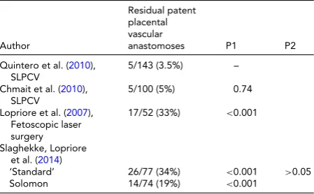
TABLE 1
Reported Incidence of Residual Patent Placental Vascular Anastomoses After Laser Surgery on Surgical Pathology Analysis of the Placentas. Comparisons Made Relative to the Lowest Reported Rate (P1) or Between the ‘Standard’ and the Solomon Techniques (P2)
Author
Residual patent placental vascular
anastomoses P1 P2
Quintero et al. (2010),
SLPCV
5/143 (3.5%) –
Chmait et al. (2010),
SLPCV
5/100 (5%) 0.74
Lopriore et al. (2007),
Fetoscopic laser surgery
17/52 (33%) <0.001
Slaghekke, Lopriore et al. (2014)
‘Standard’ 26/77 (34%) <0.001 >0.05
Solomon 14/74 (19%) <0.001
to the Quintero SLPCV technique. For example, while per-forming SLPCV, the dividing membrane is always respected. Purposeful injury to the dividing membrane, or so-called ‘septostomy’, is not part of the SLPCV technique (Harkness & Crombleholme,2005). Though not implicit, the perfor-mance of a laparotomy to access the amniotic cavity is also not part of the SLPCV technique. While general anesthesia was originally used in our cases (Rossi et al.,2008), surgery can be best performed under local anesthesia (Ville et al., 1998). Therefore, the acronym SLPCV should apply only to those surgeries in which access to the amniotic cavity is performed under local anesthesia, percutaneously, and fol-lowing a systematic and thorough identification and obliter-ation only of placental vascular anastomoses (Chmait et al., 2011; Quintero,2002; Quintero et al.,1998).
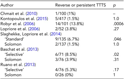
TABLE 2
Reported Incidence of Clinical Outcomes After Laser Therapy Reflecting Residual Patent Placental Vascular Anastomoses. Comparisons Made Relative to the Lowest Reported Rate
Author Reverse or persistent TTTS p
Chmait et al. (2010) 1/100 (1%) –
Kontopoulos et al. (2015) 5/417 (1.5%) 1.0
Robyr et al. (2006) 14/101 (13.8%) .0006
Lopriore et al. (2006) 2/52 (3.8%) .27
Slaghekke, Lopriore et al. (2014)
‘Standard’ 9/135 (6.7%) .046
Solomon 2/137 (1.5%) 1.0
Baschat et al. (2013)
‘Selective’ 6/71 (8.5%) .02
Solomon 3/76 (3.9%) .31
Ruano et al. (2013)
‘Selective’ 4/76 (5.3%) .17
Solomon 0/26 (0%) 1
consistently been less than 5%, with no anemia after demise of the co-twin, and an incidence of reverse or persistent TTTS of only 1–1.5% (USFetus Consortium; Quintero, 2007).
In view of the relatively high incidence of residual patent placental vascular anastomoses seen by some groups, some authors proposed ‘connecting the dots’ between photoco-agulated areas on the surface of the placenta (Chalouhi et al.,2011). The premise behind this idea was that by laser-ing areas between laser-ablated placental vascular anasto-moses, such ‘blind lasering’ would capture ‘anastomoses’ that would otherwise be missed (i.e., ‘not visible’; Lewi et al.,2006). Such authors believed that, in fact, not all of the placental vascular anastomoses can be identified endo-scopically (Lewi et al.,2006), and that therefore, ablating only the visible ones using the selective technique could miss vascular anastomoses and explain their high rate of residual patent placental vascular anastomoses. The result-ing surgical technique of ‘connectresult-ing the dots’ was dubbed ‘the Solomon technique’ (Chalouhi et al.,2011), in reference to the biblical passage where, in order to resolve a dispute between two alleging mothers of a child, King Solomon pro-posed to cut the baby in half (1 Kings 3:16–28, NIV). The analogy, therefore, is that by lasering the areas of the pla-centa between endoscopically identified and laser-ablated vascular anastomoses, the placenta would be ‘cut in half’. Recent studies suggest that indeed, relative to the surgical groups’ prior experience with the selective technique, the use of the Solomon technique was associated with improved perinatal outcomes (Baschat et al.,2013; Ruano et al.,2013). To test whether the Solomon technique could in-deed reduce the rate of residual patent placental vascu-lar anastomoses, an open-label randomized clinical trial was conducted in Europe (the Solomon trial) compar-ing the Solomon technique with the ‘standard’ technique (Slaghekke, Lopriore et al.,2014). The latter, although not directly mentioned, was intended to refer to the selective
technique (SLPCV). Interestingly, the actual rate of persis-tent residual papersis-tent placental vascular anastomoses was no different between the two techniques (14/74, 19% vs. 23/77, 29.8%, Solomon vs. ‘standard’, respectively,p=.12). How-ever, the study did show a decreased rate of PRTTTS (2/137, 1% vs. 9/135, 7%, Solomon vs. ‘standard’, respectively,p=
.03) and of twin-anemia-polycythemia syndrome (TAPS; 4/137, 2.9% vs. 21/135, 15.5%, Solomon vs. ‘standard’, re-spectively,p=<.001). Although the rationale and primary outcome of the study (to reduce the rate of residual patent placental vascular anastomoses) was no different between the groups, the authors concluded that the Solomon tech-nique was superior to the ‘standard’ techtech-nique. Two addi-tional observaaddi-tional studies comparing the Solomon tech-nique with the selective techtech-nique also appeared to show favorable results with the former technique (Baschat et al., 2013; Ruano et al.,2013).
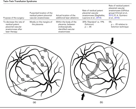
TABLE 3
Principles and Results of the Use of the Solomon Technique to Identify and Ablate All Placental Vascular Anastomoses in Twin–Twin Transfusion Syndrome
Purpose of the surgery:
Purported location of the residual patent placental vascular anastomoses:
Actual location of the additional laser ablations:
Rate of residual patent placental vascular anastomoses (Slaghekke,
Lopriore et al.,2014):
Rate of residual patent placental vascular anastomoses (USFetus group) (Chmait et al.,
2010; R. A. Quintero
et al.,2010):
To decrease the rate of residual patent placental vascular anastomoses after laser therapy
Mostly on the margins of the placenta
Within the body of the placenta, between endoscopically identified vascular anastomoses
30% (‘Standard’ vs. 19% (Solomon)
p>.05
5%
p<.05 relative to
Solomon technique
FIGURE 1
(a). Selective photocoagulation of communicating vessels (SLPCV). All of the anastomoses are photocoagulated, regardless of their loca-tion relative to the dividing membrane, while sparing non-anastomotic vessels. Rate of residual patent placental vascular anastomoses: 3.5–5%. (Chmait et al.,2010; Kontopoulos et al.,2015). (b) Solomon modification of the SLPCV technique. The fetal surface of the placenta between endoscopically identified anastomoses is also lasered, to occlude ‘anastomoses’ not visible by the endoscope. Note that marginal anastomoses may be missed, as they are not between laser shots. Rate of residual patent vascular anastomoses: 20% (Slaghekke, Lopriore et al., 2014).
Step Two: Ablation of the Placental
Vascular Anastomoses
Assuming that there is a conceptual agreement on how to identify the anastomoses, the next step consists of being able to ablate them. Successful ablation of the placental vascu-lar anastomoses assumes that the surgeon can adapt to the different clinical scenarios and overcome the various chal-lenges that may be present in each case. Particular known surgical challenges include:
1. the location of the placenta (anterior, posterior); 2. interference with the visualization of the anastomoses
by the donor twin that is ‘stuck’ along the vascular equa-tor from severe oligohydramnios (unmovable donor);
3. the presence of discolored fluid within the sac of the recipient twin from prior procedures (amniocenteses) or from prior intra-amniotic bleeding;
4. triplet (or higher order multiple) gestations, whether monochorionic or not;
5. close proximity of the umbilical cord placental inser-tions;
6. large anastomotic vessels;
7. tangential access to the placenta (whether the placenta is anterior or posterior);
8. anastomoses located behind the entry of the trocar; 9. high maternal BMI.
fluid management systems, blunt probes, trocar assistance (Quintero et al.,2010), and special laser photocoagulation techniques, which are beyond the scope of this article (Quin-tero,2007). A recent Delphi study outlined the multitude of steps needed to perform the surgery. Most authors agreed on the basic surgical goal (the purpose of the Delphi sur-vey), including the ablation of all of the placental vascular anastomoses along the vascular equator, without injuring healthy placenta or non-anastomotic vessels (Peeters et al., 2015). The question is: How often can these goals be accom-plished while overcoming the various challenges mentioned above? One way to address this question is by noting both how often the placenta can be adequately assessed, as well as how often the vessels can be selectively ablated.
Adequate Placental Assessment
Adequate placental assessment refers to the ability of the surgeon to survey the entire vascular equator. For example, in a subanalysis of the Solomon trial, the authors reported that they were able to adequately assess the placenta in only 65 out of 74 patients in the Solomon group (87%) com-pared to also only 69 out of 77 (89%) in the ‘standard’ group (Slaghekke, Lewi et al.,2014). The reasons behind the inability of the surgeons to adequately assess the pla-centa in more than 10% of the cases in each group were not stated. Obviously, if the placenta cannot be adequately as-sessed, this may result in missing anastomoses and thus an increased likelihood for adverse clinical outcomes, includ-ing PRTTTS. In contrast, our group has shown consistently the ability to assess the placenta adequately in over 99% of the patients (Crisan et al.,2010).
Selective Ablation of the Anastomoses
Another surgical competency benchmark refers to the abil-ity of the surgeon to selectively ablate the vascular anasto-moses without including non-anastomotic vessels. A ‘selec-tivity index’ (SI) was proposed by Stirnemann et al. (2008), as SI=log(SC+1)/(NSC+1), where SC are the selec-tively coagulated vessels and NSC are the non-anastomotic vessels. NSC were also vessels that were considered pre-sumed anastomoses, but that could not be followed to their terminal end and were nonetheless lasered. In their expe-rience, most surgeries involved lasering both anastomotic and non-anastomotic vessels (Chalouhi et al.,2011; Stirne-mann et al.,2008). A ‘high’ SI, defined as -0.25, was re-ported by the authors to correlate with improved postnatal survival at 28 days of at least one twin and both twins. Crisan et al. (2010) have shown that such an index is math-ematically inaccurate and should not be used to assess the adequacy of the laser surgery. Instead, a simpler index con-sisting of a ratio between how often the surgery can be done using the Quintero selective technique versus non-selectively, is more representative and easier to understand: (QSI=100∗SLPCV−NSLPCV
SLPCV ), where QSI is the Quintero SI, SLPCV is a surgery performed selectively, NSLPCV is a
surgery where at least one vessel was not clearly identified as an anastomosis but was lasered. For example, in that same article, the authors showed that they were able to perform a selective surgery in only 34% of cases (Stirnemann et al., 2008). In another report of 123 patients, surgery could not be completed in five cases for a stuck twin obscuring the equator (2), poor visualization (2) and a large anastomotic vessel (1) (Lopriore et al.,2006). Obviously, the goal is to try to perform the surgery selectively as close to 100% of the time as possible (Chmait et al.,2011; Crisan et al.,2010; Kontopoulos et al.,2015).
Accuracy of Laser Therapy
Theoretically, one could combine the rate of adequate pla-cental assessment and of selective laser surgery with the rate of either residual patent placental vascular anastomoses (when available) and the rate of PRTTTS to determine how accurate the laser surgery is being performed at a given cen-ter or by a given surgeon. Accuracy of SLPCV could thus be defined as:
AccSLPCV=QSI∗ (1−RPPVA)∗ (1−PRTTTS),
where AccSLPCV is the accuracy in performing SLPCV, QSI is the rate of Quintero selectively performed surgeries, RP-PVA is the rate of residual patent placental vascular anasto-moses (when available) and PRTTTS is the rate of PRTTTS. Table 4shows such a theoretical calculation and its use to compare different reports.
Should the Rate of TAPS be Included as a Benchmark for the Performance of Laser Therapy in TTTS? The rate of TAPS has been included as a measure of failed laser therapy for TTTS, in addition to the rate of residual patent placental vascular anastomoses and persis-tent/reverse TTTS (Baschat et al.,2013; Chmait et al.,2010; Ruano et al.,2013; Slaghekke, Lewi, et al.,2014; Slaghekke, Lopriore, et al.,2014). The decision stems from the pur-ported etiology of TAPS, which presumably results from the transfer of blood between two monochorionic twins through small placental vascular anastomoses in such a way that one fetus develops anemia and the other twin develops polycythemia, but without the net blood volume inequali-ties typical of TTTS (Lopriore et al.,2007). Presumably, the syndrome occurs through small caliber AV anastomoses, in contrast to larger size vessels typically seen in TTTS. In actu-ality, such a theory is even less plausible than the contested theory of TTTS (in which all indirect and circumstantial evidence does point to placental vascular anastomoses as being the etiological factor for TTTS, and indirectly con-firmed by resolution of the syndrome with occlusion of all placental vascular anastomoses). The proposed theory of TAPS is thus suspect for many reasons:
TABLE 4
Accuracy of Laser Surgery for Twin–Twin Transfusion Syndrome
Author
Rate of selectively performed surgeries
1−rate of residual
patent placental vascular anastomoses
1−rate of
persistent/reverse TTTS
Accuracy of SLPCV
USFetus group (Crisan et al.,2010; Kontopoulos et al.,2015) 98.7% 0.95 0.99 92%
Solomon (Slaghekke, Lopriore et al.,2014) 87% 0.81 0.99 69.7%
Solomon (Slaghekke, Lopriore et al.,2014) ‘standard’ 89% 0.66 0.93 54%
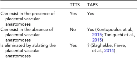
TABLE 5
Role of Placental Vascular Anastomoses in the Etiology of Twin–Twin Transfusion Syndrome and
Twin-Anemia-Polycythemia Syndrome
TTTS TAPS
Can exist in the presence of placental vascular anastomoses
Yes Yes
Can exist in the absence of placental vascular anastomoses
No Yes (Kontopoulos et al.,
2015; Taniguchi et al.,
2015) Is eliminated by ablating the
placental vascular anastomoses
Yes ? (Slaghekke, Favre,
et al.,2014)
Note: TTTS =twin–twin transfusion syndrome; TAPS =
twin-anemia-polycythemia syndrome.
incidence of residual patent placental vascular anasto-moses reported from such groups (20–33%), it is not possible to discern what role, if any, these anastomoses play in the etiology of TAPS.
2. while TAPS has been described in the presence of resid-ual patent placental vascular anastomoses, they are not indispensable. We and others have recently reported on the presence of TAPS without apparent placental vas-cular anastomoses (Kontopoulos et al.,2015; Taniguchi et al.,2015).
3. contrary to TTTS, ablation of residual patent placental anastomoses in cases of TAPS does not necessarily elim-inate the condition.Table 5compares TTTS with TAPS relative to the role of placental vascular anastomoses.
Therefore, at this point, the assumption that TAPS is the result of unsuccessful laser therapy constitutes a leap of faith in the understanding of both the etiology of TAPS and the role that laser therapy may play in the occurrence of this condition. In our opinion, the inclusion of TAPS as a benchmark of failed laser therapy should be reconsidered.
Functional Ablation of the Placental
Vascular Anastomoses: The Sequential
Technique
The development of the selective technique represents an important historical step in the surgical treatment of TTTS.
SLPCV is indeed an anatomical surgical technique, which involves the identification and selective laser obliteration of the placental vascular anastomoses. However, intrauterine fetal demise of one of the two fetuses after SLPCV would still occur in approximately 9–29% of cases with this technique (Quintero et al.,1998; R. Quintero, C. Comas,2000). Be-cause TTTS presumably occurs from an excessive transfer of blood from the donor twin to the recipient twin, the se-quence with which the anastomoses are obliterated during surgery could have prognostic implications. Indeed, if the anastomoses from the donor to the recipient are obliterated first, that would immediately stop the transfer of blood from this twin to the recipient. Furthermore, during this interval, however brief, the recipient twin would be transfusing the donor twin back. As a result, the donor twin, which is pre-sumably hypotensive, would stop losing blood as soon as the laser process starts, while at the same time would start re-ceiving additional blood back from the recipient twin. The photocoagulation of the vascular anastomoses from donor-to-recipient first, followed by from recipient-to-donor sec-ond was called the ‘sequential technique’ or SQLPCV. Using a sequential technique, our group showed a reduction in the rate of intrauterine fetal demise of the donor twin from 21% to 7%, and an increase in the double survival rate from 56% to 75% (Quintero et al.,2007). Our group is currently assess-ing the merits of performassess-ing a sequential technique through a randomized clinical trial of the USFetus group (Chmait et al.,2014). While a sequential technique may not necessar-ily be required in all cases, it could have an indication in pa-tients where the condition of the donor twin would be most compromised.
Is There a Role for Umbilical Cord
Occlusion in TTTS?
et al.,2011). Umbilical cord occlusion should not be of-fered as an alternative to laser because of limitations of the surgeon or the center, unless the patient cannot be re-ferred to another center capable of offering laser. Umbilical cord occlusion should be offered to patients with TTTS in which additional complicating circumstances may exist. This may be the case of patients with a severe congenital anomaly of one of the twins, or a moribund hydropic fe-tus. Since such cases are rare, the performance of selective feticide via umbilical cord occlusion in TTTS should be an exception, rather than the rule. Indeed, the counseling of our patients involves mentioning a survival rate of ap-proximately 90% with a 5% risk of neurological damage if laser therapy is chosen, compared to 90% survival and a 5% risk of neurological damage to the surviving co-twin if umbilical cord occlusion is chosen. Therefore, since both survival and morbidity statistics are similar between the two procedures, but with umbilical cord occlusion one of the fe-tuses is denied the chance to survive, the justification is not there to offer feticide to an otherwise anatomically normal fetus.
Conclusion
It has been more than 20 years since laser therapy was first proposed for the treatment of TTTS. Significant strides have been made both in establishing the scientific merit of using laser to ablate the placental vascular anastomoses to treat the condition (Senat et al.,2004) as well as in the vari-ous steps, techniques, and other technical aspects that allow such therapy. Selectively interrupting the placental vascular anastomoses without injuring healthy portions of the pla-centa using the Quintero selective technique, while obvious as a concept and as a surgical goal, has been associated with markedly different outcome results between centers. The Solomon technique has been proposed as a way to miti-gate such differences, but has not shown to lower the high rate of residual patent placental vascular anastomoses that prompted its development. A properly performed Quin-tero SLPCV technique is associated with the highest rate of clinical success and with the lowest rate of failed ther-apy either by surgical pathology or clinical criteria. Further improvements in clinical outcomes with the use of the se-quential technique, particularly for specific situations in which the donor may be at a unique disadvantage, could be expected and is being addressed in the ongoing randomized clinical trial conducted by our groups comparing SLPCV with SQLPCV. Selective feticide via umbilical cord occlu-sion should be the exception rather than the rule for severe cases of TTTS, and should not be performed to compensate for physician or surgical center limitations. Improvements in the surgical equipment and other ancillary technology, while difficult to pursue, should continue to remain among the objectives of caregivers in this field. The education of the next generation of surgeons using the wealth of information
thus far gathered by the different centers should also be a main focus of all programs.
References
Baschat, A. A., Barber, J., Pedersen, N., Turan, O. M., & Harman, C. R. (2013). Outcome after fetoscopic selective laser ablation of placental anastomoses versus equatorial laser dichorionization for the treatment of twin-to-twin transfusion syndrome.American Journal of Obstetrics and Gynecology,209, 234.e1–e8.
Benirschke, K., & Kaufmann, P. (1995).Pathology of human placenta. New York: Springer-Verlag.
Chalouhi, G. E., Essaoui, M., Stirnemann, J., Quibel, T., Deloison, B., Salomon, L., & Ville, Y. (2011). Laser therapy for twin-to-twin transfusion syndrome (TTTS).Prenatal Diagnosis, 31, 637–646.
Chalouhi, G. E., Stirnemann, J. J., Salomon, L. J., Essaoui, M., Quibel, T., & Ville, Y. (2010). Specific complications of monochorionic twin pregnancies: Twin-twin transfu-sion syndrome and twin reversed arterial perfutransfu-sion se-quence.Seminars in Fetal and Neonatal Medicine, 15, 349– 356.
Chang, Y. L., Chmait, R. H., Bornick, P. W., Allen, M. H., & Quintero, R. A. (2006). The role of laser surgery in dissect-ing the etiology of absent or reverse end-diastolic veloc-ity in the umbilical artery of the donor twin in twin-twin transfusion syndrome.American Journal of Obstetrics and Gynecology, 195, 478–483.
Chmait, R. H., Assaf, S. A., & Benirschke, K. (2010). Resid-ual vascular communications in twin-twin transfusion syn-drome treated with sequential laser surgery: Frequency and clinical implications.Placenta, 31, 611–614.
Chmait, R. H., Kontopoulos, E. V., Korst, L. M., Llanes, A., Petisco, I., & Quintero, R. A. (2011). Stage-based outcomes of 682 consecutive cases of twin-twin transfusion syndrome treated with laser surgery: The USFetus experience. Ameri-can Journal of Obstetrics and Gynecology, 204, e391–e396. Chmait, R. H., Kontopoulos, E. V., & Quintero, R. A. (2014).
Sequential laser surgery for twin-twin transfusion syn-drome.American Journal of Perinatology, 31(Suppl. 1), S13– S18.
Crisan, L. S., Kontopoulos, E. V., & Quintero, R. A. (2010). Appraisal of the selectivity index in a cohort of patients treated with laser surgery for twin-twin transfusion syn-drome.American Journal of Obstetrics and Gynecology, 202, 157 e151–e155.
De Lia, J. E., Kuhlmann, R. S., Cruikshank, D. P., & O’Bee, L. R. (1993). Current topic: Placental surgery: A new frontier. Placenta, 14, 477–485.
De Lia, J. E., Cruikshank, D. P., & Keye, W. R. Jr. (1990). Feto-scopic neodymium: YAG laser occlusion of placental vessels in severe twin-twin transfusion syndrome.Obstetrics & Gy-necology, 75, 1046–1053.
Journal of Obstetrics and Gynecology, 172(4 Pt 1), 1202– 1208, discussion 1208–1211.
Dennis, L. G., & Winkler, C. L. (1997). Twin-to-twin trans-fusion syndrome: Aggressive therapeutic amniocentesis. American Journal of Obstetrics and Gynecology, 177, 342– 347, discussion 347–349.
Harkness, U. F., & Crombleholme, T. M. (2005). Twin-twin transfusion syndrome: Where do we go from here?Seminars in Perinatology, 29, 296–304.
Hecher, K., Diehl, W., Zikulnig, L., Vetter, M., & Hackeloer, B. J. (2000). Endoscopic laser coagulation of placental anasto-moses in 200 pregnancies with severe mid-trimester twin-to-twin transfusion syndrome.European Journal of Obstet-rics & Gynecology and Reproductive Biology, 92, 135–139. Jones, J., Sbarra, A., Dilillo, L., Cetrulo, C. L., & D’Alton, M. E.
(1993). Indomethacin in severe twin-to-twin transfusion syndrome.American Journal of Perinatology 10, 24–26. Kontopoulos, E., Chmait, R., Baker, B., Llanes, A., & Quintero,
R. (2015). USFetus surgical benchmarks of selective laser therapy for twin-twin transfusion syndrome. American Journal of Obstetrics and Gynecology, 212, S91–S92. Lewi, L., Jani, J., Cannie, M., Robyr, R., Ville, Y., Hecher, K., . . .
Deprest, J. (2006). Intertwin anastomoses in monochori-onic placentas after fetoscopic laser coagulation for twin-to-twin transfusion syndrome: Is there more than meets the eye?American Journal of Obstetrics and Gynecology, 194, 790–795.
Lopriore, E., Middeldorp, J. M., Oepkes, D., Kanhai, H. H., Walther, F. J., & Vandenbussche, F. P. (2007). Twin anemia-polycythemia sequence in two monochorionic twin pairs without oligo-polyhydramnios sequence.Placenta, 28, 47– 51.
Lopriore, E., Middeldorp, J. M., Oepkes, D., Klumper, F. J., Walther, F. J., & Vandenbussche, F. P. (2006). Residual anas-tomoses after fetoscopic laser surgery in twin-to-twin trans-fusion syndrome: Frequency, associated risks and outcome. Placenta, 28, 204–208.
Peeters, S. H., Akkermans, J., Westra, M., Lopriore, E., Middeldorp, J. M., Klumper, F. J., . . . Oepkes, D. (2015). Identification of essential steps in laser procedure for twin-twin transfusion syndrome using the Delphi methodology: SILICONE study.Ultrasound in Obstetrics and Gynecology, 45, 439–446.
Quintero, R. (2002). Selective laser photocoagulation of com-municating vessels in twin-twin transfusion syndrome. In R. Quintero (Ed.),Diagnostic and operative fetoscopy. (pp. 43–54). New York: The Parthenon Publishing Group. Quintero, R. (2007). Laser treatment for twin-twin transfusion
syndrome. In R. Quintero (Ed.),Twin-twin transfusion syn-drome. (pp. 99–120). London: Informahealth.
Quintero, R., Morales, W., Mendoza, G., Allen, M., Kalter, C., Giannina, G., & Angel, J. (1998). Selective photocoagula-tion of placental vessels in twin-twin transfusion syndrome: Evolution of a surgical technique.Obstetrical and Gyneco-logical Survey, 53, s97–s103.
Quintero, R., Quintero, L., Pivatelli, A., Bornick, P., Allen, M., & Johnson, P. (2000). The donor-recipient (D-R) score: In vivo endoscopic evidence to support the hypothesis of a
net transfer of blood from donor to recipient in twin-twin transfusion syndrome.Prenatal and Neonatal Medicine, 5, 84–91.
Quintero, R. A., Chmait, R. H., Bornick, P. W., & Kontopoulos, E. V. (2010). Trocar-assisted selective laser photocoagula-tion of communicating vessels: A technique for the laser treatment of patients with twin-twin transfusion syndrome with inaccessible anterior placentas.Journal of Maternal-Fetal and Neonatal Medicine, 23, 330–334.
Quintero, R. A., Chmait, R. H., Carver, J., Bornick, P. W., Allen, M. H., & Kontopoulos, E. V. (2008). In utero fetal oximetry via visible light spectroscopy in twin-twin transfusion syn-drome.American Journal of Obstetrics and Gynecology,199, 639.e1–e4.
Quintero, R. A., Comas, C., Bornick, P. W., Allen, M. H., & Kruger, M. (2000). Selective versus non-selective laser pho-tocoagulation of placental vessels in twin-twin transfusion syndrome. Ultrasound in Obstetrics and Gynecology, 16, 230–236.
Quintero, R. A., Ishii, K., Chmait, R. H., Bornick, P. W., Allen, M. H., & Kontopoulos, E. V. (2007). Sequential selective laser photocoagulation of communicating vessels in twin-twin transfusion syndrome.Journal of Maternal-Fetal and Neonatal Medicine, 20, 763–768.
Robyr, R., Lewi, L., Salomon, L. J., Yamamoto, M., Bernard, J. P., Deprest, J., & Ville, Y. (2006). Prevalence and man-agement of late fetal complications following successful se-lective laser coagulation of chorionic plate anastomoses in twin-to-twin transfusion syndrome.American Journal of Obstetrics and Gynecology, 194, 796–803.
Rossi, A. C., Kaufman, M. A., Bornick, P. W., & Quintero, R. A. (2008). General versus local anesthesia for the percutaneous laser treatment of twin-twin transfusion syndrome. Amer-ican Journal of Obstetrics and Gynecology,199, 137.e1–e7. Ruano, R., Rodo, C., Peiro, J. L., Shamshirsaz, A., Haeri, S.,
Nomura, M. L., . . . Belfort, M. A. (2013). Fetoscopic laser ablation of the placental anastomoses in twin-twin transfu-sion syndrome using the ‘Solomon technique’.Ultrasound in Obstetrics and Gynecology, 42, 434–439.
Saunders, N. J., Snijders, R. J., & Nicolaides, K. H. (1992). Therapeutic amniocentesis in twin-twin transfusion syn-drome appearing in the second trimester of pregnancy. American Journal of Obstetrics and Gynecology, 166, 820– 824.
Senat, M. V., Deprest, J., Boulvain, M., Paupe, A., Winer, N., & Ville, Y. (2004). Endoscopic laser surgery versus serial amnioreduction for severe twin-to-twin transfusion syn-drome.New England Journal of Medicine, 351, 136–144. Slaghekke, F., Favre, R., Peeters, S. H., Middeldorp, J. M.,
Weingertner, A. S., van Zwet, E. W., . . . Lopriore, E. (2014). Laser surgery as a management option for twin anemia-polycythemia sequence.Ultrasound in Obstetrics and Gyne-cology, 44, 304–310.
Slaghekke, F., Lopriore, E., Lewi, L., Middeldorp, J. M., van Zwet, E. W., Weingertner, A. S., . . . Oepkes, D. (2014). Fetoscopic laser coagulation of the vascular equator ver-sus selective coagulation for twin-to-twin transfusion syn-drome: An open-label randomised controlled trial.Lancet, 383, 2144–2151.
Stirnemann, J. J., Nasr, B., Quarello, E., Ortqvist, L., Nassar, M., Bernard, J. P., & Ville, Y. (2008). A definition of selectivity in laser coagulation of chorionic plate anastomoses in twin-to-twin transfusion syndrome and its relationship to perinatal outcome.American Journal of Obstetrics and Gynecology, 198, 62e61–e66.
Taniguchi, K., Sumie, M., Sugibayashi, R., Wada, S., Matsuoka, K., & Sago, H. (2015). Twin anemia-polycythemia sequence after laser surgery for twin-twin transfusion syndrome and maternal morbidity.Fetal Diagnosis and Therapy, 37, 148– 153.
Urig, M. A., Simpson, G. F., Elliott, J. P., & Clewell, W. H. (1988). Twin-twin transfusion syndrome: The surgical
removal of one twin as a treatment option.Fetal Diagnosis and Therapy, 3, 185–188.
Ville, Y., Hecher, K., Gagnon, A., Sebire, N., Hyett, J., & Nicolaides, K. (1998). Endoscopic laser coagulation in the management of severe twin-to-twin transfusion syn-drome.British Journal of Obstetrics and Gynaecology, 105, 446–453.
Ville, Y., Hecher, K., Ogg, D., Warren, R., & Nicolaides, K. (1992). Successful outcome after Nd : YAG laser separation of chorioangiopagus-twins under sonoendoscopic control. Ultrasound in Obstetrics and Gynecology, 2, 429–431. Ville, Y., Hyett, J., Hecher, K., & Nicolaides, K. (1995).
Prelim-inary experience with endoscopic laser surgery for severe twin-twin transfusion syndrome.New England Journal of Medicine, 332, 224–227.