INTRODUCTION
It is well established fact that microbial natural products remain one of the most important sources of lead compounds for the pharmaceutical industry. Among microorganisms, the actinomycetes are a fascinating group that they are sources of most of the antibiotics used in medicine today. They also produce metabolites that are used as anticancer drugs, antihelminthics and drugs that suppress the immune system in patients who have received organ transplants. This becomes apparent in 1940, following Selman Waksman’s discovery of actinomycin from S. gresous1 and was fully realized by the 1980s, when actinomycetes accounted for almost 70% of the world’s naturally occurring antibiotics2.
Screening and Isolation of antibiotic producing
Actinomycetes from marine samples using rifampicin
FELEKE MOGES
1,2*, T. PRABHAKAR
1, T. RAMANA
2and
G. SANKAR
11Pharmaceutical Biotechnology Division,
A.U. College of Pharmaceutical Sciences, Andhra University (India). 2Department of Biotechnology, College of Science and Technology,
Andhra University, Visakhapatnam - 530 003 (India).
(Received: July 24, 2009; Accepted: September 29, 2009)
ABSTRACT
The total number of actinomycetes recovered from different samples varied between a maximum of 3.5 X 10⁵ CFU/gm of samples of sea weeds and algae to a minimum of 6.2×10² CFU/ml of sea water. A total of 132 strains of actinomycetes were isolated, of these 76 (57.6%) exhibit antimicrobial activity to one and/ or more of bacterial, filamentous fungi or yeast strains. Most of the inhibitory activity was directed against bacteria than filamentous fungi and yeast. Among bacterial group Bacillus subtilis (39.4%) was the most susceptible while Proteus vulgaris (11.4%) was the least. Among fungal strains, Penicillium chryseogenum (34.1%) was the most susceptible while Aspergillus oryzae (10.6%) was the least. Thirty five (26.5%) of the total isolates exhibited broad spectrum activity. The result showed that, better recovery of actinomycetes was observed using rifampicin than using naldixic acid. Therefore, in the search for screening and isolation of bioactive producing actinomycetes, rifampicin might be used as a better drug of choice than naldixic acid for suppressing and inhibiting growth of non-filamentous bacterial contaminants in marine samples.
Key words: Actinomycetes, marine samples and Rifampicin.
The goal of this study were to screen and isolate antibiotic producing actinomycetes that are found in different samples of marine environment in the East Coast of Bay of Bengal using refampicin or naldxic acid as a selective inhibitor of bacterial contaminant.
MATERIAL AND METHODS Sample collection and processing
A total of 37 different samples were taken from marine environment (10 sea water, 20 sediments, 1sample each from sponges, hard corals and snails, and 4 algae and sea weeds) at a various depth using sterile screw capped bottle and sterile plastic bags.
Samples were processed as soon as collected and some of the samples collected from distant site were processed in the following days of collection by preserving the samples with a sterile plastic bag in an ice box. Algae and sea weeds samples were dried overnight in a laminar flow hood and crushed with an alcohol sterilized mortar and pestle. Other samples like sponges, corals and shell of snail samples were processed as described by Jensen et. al. 8. These samples were subjected overnight in a laminar flow hood and the samples were scrapped with a sterile spatula generating a powder.
About 1gm of the above samples (sediments, dried powdered samples) was suspended aseptically and were homogenized with sterile sea water and kept in 250 ml flask having 50 ml of sterile sea water and incubated in an orbital shaker at 26 0C with shaking at 140 rpm for 30 minutes. Mixtures were allowed to settle then ten fold serial dilutions were prepared. Depending on the condition water samples were either directly or serially diluted.
One millilitre of each of these dilutions was added to 50ml of starch casein agar (g/l: starch 10, casein 0.3, KNO3 2, NaCl 2, K2HPO4 2, MgSO4.7H2O 0.05, CaCO3 0.02, FeSO4.7H2O 0.01, agar 20) and oat meal agar (g/l: oat meal 20, agar 20, trace salt solution 1ml) when the temperature is about 40-45 0C. Samples were thoroughly mixed and poured in to a sterile Petri-plate (6 diam). One
copy of the samples was plated following spread plating technique using sterile L-shaped glass rod. Inoculated samples were incubated at 28°C for 21 days. To inhibit growth of non-filamentous bacteria and fungi medium was supplemented with 5µg/ml rifampicin8 or 20µg/ml naldixic acid and 50µg/ml cyclohexamide 9 respectively.
Identification of actinomycetes
Actinomycetes colonies were recognized by their characteristics tough leathery colonies that adhered to the agar surface, branched vegetative mycelia, and when present, aerial mycelia and spore formation 8,10. Actinomycetes colonies were counted on different plates after 10-21 days incubation at 28 0C. Data are expressed as colony forming unit (CFU/ml) for water or CFU/gm for dry weight samples. Actinomycetes colonies were characterized morphologically and physiologically following the methods given in the International Streptomyces Project (ISP) 11. Species were identified by the morphological characteristics of colonies, substrate and aerial mycelium, structure of spore chains and pigment production on different ISP media. Detailed physiological and biochemical character ization of promising isolates were performed following the standard procedure12. Determination of antimicrobial activity
Pr imary screening of antimicrobial activities of actinomycetes isolated from different samples were screened following the streak plating technique. The test organisms employed in this study were supplied by the National Chemical Laboratory (NCIM), Pune and Microbial Type Culture Collection and Gene bank (MTCC), Chandigarh. The following test organisms were used for antimicrobial activities. Bacillus pimilus (NCIM-2327), Bacillus subtilis (NCIM-2063), Staphylococcus aureus (NCIM-2079), Escherichia coli (NCIM-2065), Shigella flexinari (MTCC-1457), Salmonella typhimerium (MTCC-98), Aspergillus niger (NCIM-548), Aspergillus oryzae (NCIM-643), Penicillium cryseogenum (NCIM-738), Candidia albicans, and Saccharomyces cervisae.
over 1.5ml chloroform for 40 minutes. The dead colonies were overlaid with 5ml of nutrient agar (bacteria) and 5 ml of potato dextrose agar (yeast and fungi) that has been inoculated with the test organism. Zone of inhibition around the colonies was recorded after 24 hours at 37 °C for bacteria and 48 hours at 28°C for yeast and filamentous fungi.
RESULT AND DISCUSSION
The total number of actinomycetes recovered from different samples showed that the maximum population density was recorded in samples of sea weeds and algae, 3.5×10⁵ CFU/gm followed by samples of sponges, corals and snails, 4.7 ×10⁴ CFU/ml; sediments 2.6×10³ CFU/ml and
water with least population counts, 6.2×10²CFU/ml. (Table 1). Sahu et.al14 observed similar patterns of population density of marine actinomycetes from sediment and water samples collected in the East coast of India. The maximum count of actinomycetes in algae and sea weeds samples may be because of nutrient availability. That is, nutrient availability in plant and animal residues is higher and this may create a fertile ground for spores of actinomycetes to germinate. However, the population of actinomycetes decrease in water samples than other samples, this is also because dormancy of spore imposed by lack of nutrients in water and a spore population will decline15 and results low number of actinomycetes in water sample than other samples.
Table 1: Total microbial count obtained in different samples using Rifampcin Type of sample Total Average total Medium used Antifungal and
number of count of antibacterial agents
samples actinomycetes
Water 10 6.2×10² CFU/ml Starch casein agar Cyclohexamide 50µg/ml
and rifampicin5µg/ml
Sediment 20 2.6×10³ CFU/gm Starch casein agar Cyclohexamide 50µg/ml
and rifampicin5µg/ml
Sponges, corals 3 4.7×10⁴CFU/gm Starch casein agar Cyclohexamide 50µg/ml
and snails and rifampicin5µg/ml
Algae and sea 4 3.5×10⁵ CFU/gm Starch casein agar Cyclohexamide 50µg/ml
weeds and rifampicin5µg/ml
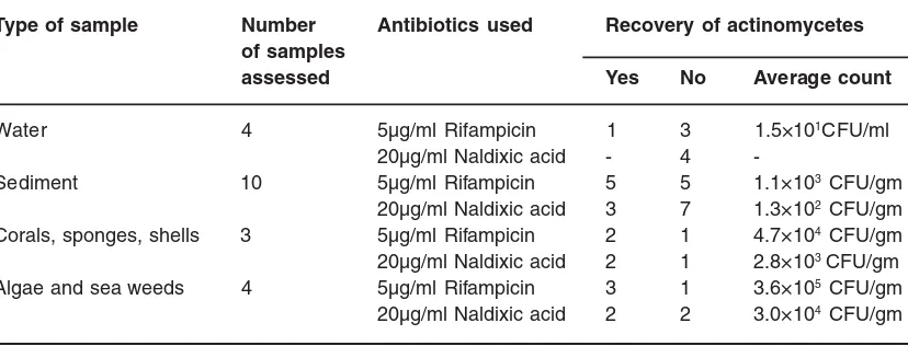
Table 2: Comparison of actinomycetes recovery from same samples using rifampicin and naldixic acid as inhibitors of bacterial contaminants. (Only 21 samples were compared) Type of sample Number Antibiotics used Recovery of actinomycetes
of samples
assessed Yes No Average count
Water 4 5µg/ml Rifampicin 1 3 1.5×101CFU/ml
20µg/ml Naldixic acid - 4
-Sediment 10 5µg/ml Rifampicin 5 5 1.1×10³ CFU/gm
20µg/ml Naldixic acid 3 7 1.3×10² CFU/gm
Corals, sponges, shells 3 5µg/ml Rifampicin 2 1 4.7×10⁴ CFU/gm
20µg/ml Naldixic acid 2 1 2.8×10³ CFU/gm
Algae and sea weeds 4 5µg/ml Rifampicin 3 1 3.6×10⁵ CFU/gm
To avoid bacter ial contaminant from different samples, 5µg/ml rifampicin or 20µg/ml naldixic acid have been used for comparison. Comparing to naldixic acid, the rate of recovery of antibiotic producing actinoomycetes using rifampicin
is relatively high (Table 2). Similar report by Pisano et al.16 showed that, in pre-treated samples 16 % of the actinomycetes produced bioactivity while in r ifampicin treated mar ine samples 46% of actinomycetes showed bioactivity17.
Table 3: Distribution of actinomycets exhibiting antimicrobial activities
Type of test organisms Number of actinomycetes %
Gram positive 27 20.5
Bacteria Gram negative 5 3.8
Gram positive and negative 32 24.2
Total number of isolates 64 48.9
Filamentous fungi 19 14.4
Fungi Yeast 2 1.5
Filamentous fungi & yeast 29 22.0
Total number of isolates 50 37.9
Number of isolates with broad spectrum activity (bacteria,
filamentous fungi and yeast) 35 26.5
Total actinomycetes showing activities 76 57.6
Total actinomycetes with weak and/or no activities 56 42.4
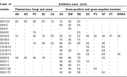
Table 4: Activity of promising isolates on different test organisms using an agar overlay method
Code of Inhibition zone (mm)
isolate Filamentous fungi and yeast Gram-positive and gram-negative bacteria
AN AO PC SC CA SA BM BS EC PV SF ST MRSA
WS1/31 25 20 28 25 15 15 25 20 - - - -
-SS4/2 - - - 50 20 55 - - - -
-SS4/4 - _ _ _ _ - 20 - - -
-SS4/61 _ _ 15 _ _ - - 20 - - - -
-SS16/2 15 - 35 12 23 32 15 57 45 25 45 47 32
SS18/2 - - - 10 25 15 - - - -
-SS18/3 - - 10 30 23 60 25 65 25 - 30 -
-CS19/10 - - - 55 - - 23 - 55 -
-CS19/11 - - - 40 50 - 30 - 28 -
-CS19/13 - - - 55 - 45 23 - 28 - 30
SS23/2 18 20 40 18 - 60 60 70 - - 23 -
-SS31/1 - - - 30 45 25 10 - - -
-SS31/2 - - - 48 50 41 - - - -
-SS31/32 - - - 47 45 42 - 40 - -
-SS31/T2 - - - 42 45 38 - - 55 -
As indicated in table-2, four water samples were taken for comparison of rifampicin and naldixic acid. It was found that actinomycetes colonies were recovered only in one sample using rifampicin (1.5×101CFU/ml). Out of 10 sediment samples taken for comparison, actinomycetes were recovered in 5 samples for rifampicin (1.1×10³ CFU/gm) and 3 samples for naldixic acid (1.3×10² CFU/gm). One sample each from corals, sponges and shells were also compared, both rifampicin (4.7 ×10⁴ CFU/gm) and nalixic acid (2.8×10³ CFU/gm) were showing similar recovery with high population density in rifampicin. Four samples from algae and sea weeds were also compared, 3 of the samples were showing high density count using rifampicin (3.6×10⁵ CFU/ gm) and 2 of the samples showing also good count using naldixic acid (3.0×10⁴ CFU/gm). As naldixic acid, rifampicin is also effective against some gram-positive and gram-negative bacteria. However, rifampicin is mostly effective when there is combination of other drugs (Examples, treatment of tuberculosis, leprosy); this might be the reason that using rifampicin alone may favour recovery of more actinomycetes populations than using the broad spectrum antibiotic, naldixic acid.
During screening of 37 different marine samples, 420 suspected actinomycetes colonies were recovered. Among these, on the basis of their colony morphology, colour of their aerial mycelium 150 colonies suspected to be different were selected
and looping out. Although rifampicin can suppress the growth of some of the strains of actinomycetes, 5µg/ml concentration added to the sample during primary screening found to be effective to protect rapidly growing members of non-filamentous bacteria.
After detail study of their morphology and characteristics of aerial and substrate mycelium, a total of 132 isolates were confir med to be actinomycetes (Table 3). Among these, 76 (57.6%) exhibit antimicrobial activity to one and/ or more test organisms of bacterial, and filamentous fungi or yeast strains while the other 56 (42.4%) isolates don’t show and/or poor activities. This result was lower than reported by Pisano et.al.18, 73% of chitinolytic actinomyctes were showing bioactivity. The reason may be due to correlation between chitinolysis and bioactivity of actinomycetes18.
Most of the inhibitory activity was directed against bacteria than filamentous fungi and yeast. As shown in table-3, 64 (48.9%) of the total number isolated, proved to be active to one or more bacteria, while 50 (37.9%) were active to filamentous fungi and yeast.
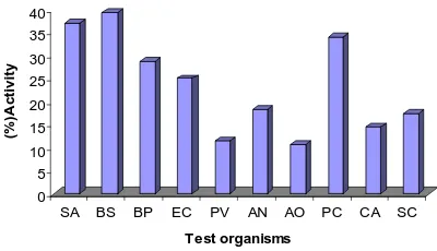
Among bacterial group B. subtilis (39.4%) was the most susceptible organism followed by S. aureus (37.1%), B. pimilus (28.8%), E. coli (25%); P. vulgaris (11.4%) was the least susceptible. Among fungal group Penicillium cryseogenum (34.1%) was the most susceptible followed by Aspergillus niger (18.2%), Saccharomyces cer visae (17.4%), Candidia albicans (14.4%); Aspergillus oryzae (10.6%) was the least susceptible (Figure-1). Total number of actinomycetes exhibiting broad spectrum activity (bacteria and fungi) were 35(26.5%).
Fifteen promising isolate were selected and their detailed biochemical and physiological study showed that 10 of them were under the Sterptomyces species and 5 of them were under the group Micromonospora species. This result was in agreement with reports of Das et.al., 19 and Peela et.al., 20 as the major genera isolated in the bay of
Bengal were the Streptomyces and
Micromonospora species. However, this result is different from repor ts by Bredholt et.al.21 that Micromonospora species are dominant. The reason 0
5 10 15 20 25 30 35 40
(
%
)A
c
tiv
it
y
SA BS BP EC PV AN AO PC CA SC
Test organisms
Fig. 1: Actinomycetes isolated from different marine samples showing bioactivity against bacteria and fungi
may be because of difference in depth of the sample collected. The deep water sediments contained a higher relative amount of Micromonospora compared to the shallow water sediments22. Although, the other isolate showed strong antagonistic activities against different test organisms two of the Streptomyces species were also strongly active against methicillin resistant S. aureus (MRSA (Table 4). The antimicrobial spectrums of these species were also evaluated by submerged fermentation and their activities are still very promising against the test organism of bacterial and fungal strains. Thediversity of these promising isolates and their content of biological activities are understudy.
CONCLUSION
As it is well documented drug resistance pathogens such as methicillin resistant S. aureus become the most problematic gram positive
bacterium in public health not only because it is highly prevalent but also it has reported that, the organism shows reduced susceptibility to the last resort antibiotics of vancomycin and teicoplanin. Therefore, despite the challenges of identifying new natural product antibiotics; searching and screening of antibiotic producing actinomycetes from new unexplored area using different pre-treatment of marine samples such as rifampicin as selective inhibitors for non filamentous bacterial contaminants during primary screening may help to identify new novel bioactive molecules.
ACKNOWLEDGMENTS
The authors are thankful to DBT, New Delhi for the financial support to carry out this work. We are also grateful to Dr. B. Narasinga Rao (King George Hospital) for providing methicillin-resistant S. aureus.
1. Waksman, S.A. and Woodr uff, H.B.
Bacteriostatic and bactericidal substances produced by soil actinomycetes. Proc. Soc. Exp. Biol.Med. 45:609-614 (1940).
2. Okami,Y. and Hotta K. Search and discovery of new antibiotics, P.33-67. In:M. Goodfellow, S.T.Williams and M. Mordarski (ed), Actinomycetes in Biotechnology, Academic press. Inc., New York (1988).
3. Baltz, R.H. Antimicrobials from
actinomycetes: Back from the future. Microbe. 2:125-130 (2007).
4. Bull A.T., Ward, A.C. and Goodfellow M. Search discover y strategies for biotechnology: The paradigm shift. Micobiol. Mol. Biol. Rev. 64:573-606 (2000).
5. Proksch,P., Edrada, R.A., Ebel, R. Drug from the seas-current status and micobiological
implications. Applied Micobiol
Biotechnol.59:125-134. (2002).
6. Newman, D.J. and Cragg, G.M. Marine natural products and related compounds in clinical and advanced preclinical trials. J.
Natural products. 67: 1216-1238 (2004). 7. Lam, K.S. Discovery of novel metabolites
from marine actinomycetes. Curr. Opin. Microbiol. 9:245-251(2006).
8. Jensen, P.R, Gontang, E., Mafnas, C., Mincer, T.J., Fenical, W. Culturable marine actinomycets diversity from tropical Pacific Ocean sediments. Environ Microbiol. 7: 1039-1048 (2005).
9. Imada C., Noseki N., Kamata M., Kobayashi T. and Hamada-Sato N. Isolation and characterization of antibacterial substances produced by marine actinomycetes in the presence of sea water. Actinomycetologica. 21: 27-31. (2007)
10. Ghanem, N.M.,Sabry, S.A., El-Sherif, Z.M. and Abu El-Ela, G.A. Isolation and Enumeration of marine actinomycetes from sea water and sediments in Alexanderia. J. Gen. Appl. Micobiol. 46: 105-111 (2000). 11. Shirling , E.B., Gottlieb, D. Methods for
12. Salle, A.J. “ Laborator y Manual on Fundamental Principles of Bacteriology” 3rd ed. Mc Graw-Hill Book Co. (1948).
13. Williams, S.T., Goodfellow, m., Alderson, G.,Wellington, E.M.H., Sneath, P.H.A. and Sackin, M.J. Numerical classification of streptomyces and related genera. J. Gen. Microbiol. 129: 1743-1843 (1983a). 14. Sahu MK., Kumar KS and Kannan L. Isolation
of actinomycetes from various samples of the Vellar Estuary, Southeast Coast of India. Poll Res. 24: 45-48 (2005).
15. Cross, T. Acquatic Actinomycetes: A critical survey of the occurrence, growth and role of actinomycetes in aquatic habitats. J. Appl. Bacteriol. 50: 397-423 (1981).
16. Pisano MA, Sommer MJ, Lopez MM.
Application of pre-treatments for the isolation of bioactive actinomycetes from marine sediments. Appl Microbiol. Biotechnol. 25: 285-288 (1986).
17. Pisano MA,Sommer MJ, Brancaccio L.
Isolation of bioactive actinomycetes from marine sediments using rifampicin. Appl Microbiol. Biotechnol. 31: 609-612 (1989). 18. Pisano MA, Sommer MJ. and Taras, L.
Bioactivity of chitinolytic actinomycetes of marine origin. Appl Microbiol Biotechnol. 36: 553-555 (1992).
19. Das S., Lyla P.S. and Ajmalkhan S. Distribution and generic composition of culturable marine actinomycetes from the sediments of Indian continental slope of Bay of Bengal. Chinese J. Oceanology and Limnology. 26: 166-177 (2008).
20. Peela S. Kurada V. and T. Ramana. Studies on antagonstic marine actinomycetes from the Bay of Bengal. World J. Microbiol. Biotechnol. 21: 583-585 (2005).