OncoTargets and Therapy 2018:11 1285–1292
OncoTargets and Therapy
Dove
press
submit your manuscript | www.dovepress.com 1285
O r i g i n a l r e s e a r c h
open access to scientific and medical research
Open access Full Text article
Overexpression of c-kit(cD117), relevant with
microvessel density, is an independent survival
prognostic factor for patients with hBV-related
hepatocellular carcinoma
Weiwei Yan1,*
Zhenyu Zhu2,*
Fei Pan3
ang huang4
guang-hai Dai1
1Medical Oncology Department, chinese Pla general hospital, Beijing, china; 2Department of liver Metastasis, hepatobiliary surgery center, Beijing 302 hospital, Beijing, china; 3Department of gastroenterology and hepatology, chinese Pla general hospital, Beijing, china; 4Department of alcoholic liver Disease and autoimmune liver Disease, center of non-infectious liver Diseases, Beijing 302 hospital, Beijing, china
*These authors contributed equally to this work
Background: To explore new biomarkers for indicating the recurrence and prognosis in hepatitis B virus (HBV)-related hepatocellular carcinoma (HCC) patients after tumor resection, we investigated the expression and prognostic value of c-kit(CD117) in HBV-related HCC.
Materials and methods: Immunohistochemistry was used to estimate the expression of c-kit(CD117) and CD34 in the liver cancer tissues. The correlations between the expression of these biomarkers and the clinicopathologic characteristics were analyzed.
Results: The positive rate of c-kit(CD117) expression in 206 HCC cases was 48.1%, and c-kit expression was significantly related with CD34-positive microvessel density. CD34-microvessel density numbers were much higher in c-kit(+) HCC tissues than in c-kit(-) HCC tissues (44.13±17.01 vs 26.87±13.16, P=0.003). The expression of c-kit was significantly higher in patients with Edmondson grade III–IV (P,0.001) and TNM stage III (P,0.001). Moreover, Kaplan–Meier survival analysis showed that c-kit (P,0.001) expression was correlated with reduced disease-free survival (DFS). Multivariate analysis identified c-kit as an independent poor prognostic factor of DFS in HCC patients (P,0.001).
Conclusion: Increased c-kit expression could be considered as an independent unfavorable prog-nostic factor for predicting DFS in HBV-related HCC patients after surgery. These results could be used to identify patients at a higher risk of early tumor recurrence and poor prognosis.
Keywords: c-kit, CD34, hepatocellular carcinoma, prognosis, immunohistochemistry
Introduction
Hepatocellular carcinoma (HCC) is the fifth most common malignant tumor in the world and the second leading cause of cancer-related death in men. HCC occurs all over the world, but is mainly prevalent in East Asia, Central Africa, and parts of West Africa, with China alone accounting for about 50% of the total number of cases and deaths.1 The most effective treatment for HCC is early curative resection or liver
trans-plantation. However, the rate of tumor recurrence and metastasis after liver resection remains high, mainly due to the persistence of chronic hepatitis and cirrhosis. For those who undergo hepatic resection, the recurrence rate is about 50% after 2 years.2
There-fore, risk factors and prognostic biomarkers for prognosis must be clarified to manage patients after curative hepatectomy. There have been some investigations of cancerous tissues of HCC to predict intrahepatic recurrence, especially intrahepatic metastasis.3–5
In these investigations, the most frequent risk factors related with HCC were alcohol intake and hepatitis C virus (HCV). However, up to 80% of HCC cases in China are correspondence: guang-hai Dai
Medical Oncology Department, chinese Pla general hospital, Beijing 100853, china Tel +86 106 693 7291 email daigh301@vip.sina.com
ang huang
center of non-infectious liver Diseases, Beijing 302 hospital, Beijing 100039, china Tel +86 106 693 3129 email huangangwin@aliyun.com
Journal name: OncoTargets and Therapy Article Designation: Original Research Year: 2018
Volume: 11
Running head verso: Yan et al
Running head recto: c-kit in hepatocellular carcinoma DOI: 157545
OncoTargets and Therapy downloaded from https://www.dovepress.com/ by 118.70.13.36 on 25-Aug-2020
For personal use only.
Dovepress
Yan et al
attributable to hepatitis B virus (HBV).6 Therefore, the field
of prognostic biomarkers for HBV-related HCC remains open and requires further extensive investigation.
Angiogenesis is the basic and important stage in the process of tumorigenesis, invasion, and metastasis. Tumor angiogenesis also contributes to the aggressiveness and poor prognosis of HCC.7 Neovascularization not only supplies
oxygen and nutrients to the proliferative tumor cells, but also serves as the conduit for migration.8 CD34 is an antigen
associated with neovascularization and can be detected in some kinds of tumors that originate from blood vessels or mesenchymal tissue.9 Thus, at present, CD34 is used to label
microvessel density (MVD).10 MVD is the most recognized
indicator to evaluate angiogenesis of solid tumors.11
The proto-oncogene c-kit is a member of the platelet-derived growth factor receptor family and encodes a receptor tyrosine kinase. It has been reported that c-kit is expressed in many tumors and is well known to be highly expressed in gastrointestinal stromal tumors.12 Imatinib, targeting c-kit,
a small compound that selectively inhibits the activity of a limited number of receptor tyrosine kinases, has been shown to exert marked clinical activity in malignant gastrointestinal stromal tumors (GISTs).13 But only little data exist regarding
the overexpression of c-kit in HCC. The positive expres-sion of c-kit was observed in the mitotic, proliferating, and also dysplastic hepatic cells.14 Others have reported that the
c-kit(+) tumor cells might be present in human HBV-associ-ated HCC.15 Furthermore, previous studies have revealed that
CD34 and c-kit may be used as indicators closely related to the invasion and metastasis of malignant tumors.11,14
How-ever, paradoxical results have also been noted.3,16,17 Becker
et al3 found that the overall percentage of c-kit-positive
immunohistochemical (IHC) staining of HCC (related with alcohol in about 50% of patients) was 2.3% (6/258) and there seems to be no role for the use of imatinib. CD34 expres-sion also cannot be used singly as a prognostic indicator for pancreatic cancer and HCC patients.16,18
In this study, IHC was used to evaluate the expression of CD34 and c-kit in patients with HCC undergoing radical hepatectomy. The expression of c-kit in HCC and its rela-tionship with clinicopathologic features and prognosis were calculated and analyzed.
Materials and methods
subjects and samples
We identified 206 cases of HCC and 23 cases of liver cirrhosis without HCC who had undergone segmentectomy or lobec-tomy of the liver at the hepatobiliary surgery center of Beijing 302 Hospital (Beijing, China). All HCC patients were staged
at the time of surgery according to the guidelines of the American Association for the Study of Liver Diseases.19 All
HCC patients underwent initial curative hepatectomy between May 2010 and October 2013, with a total follow-up time of 48 months. Curative hepatectomy was defined as a resection in which all of the tumors were macroscopically resected at the time of surgery. All diagnoses of HCC were confirmed pathologically. All HCC patients had a history of .10 years of chronic HBV infection. All patients were hospitalized or followed up in Beijing 302 Hospital. All the patients included in the study had received antiviral therapy (nucleoside/ nucleotide analogs) .1 year. All 206 cases of HCC patients were hepatitis B virus surface antigen positive. Seventy-nine patients were serum hepatitis B e-antigen (HBeAg) positive. All the patients were serum HBV-DNA negative. Exclusion criteria included: 1) patients with intrahepatic cholangio-carcioma or other concomitant tumors; 2) patients with past history of alcohol addiction; 3) patients who received pre-operative interventional treatment, local ablation treatment, radiotherapy, or chemotherapy; 4) patients with coinfections of HCV, hepatitis D virus, and human immunodeficiency virus infections; and 5) patients who had concurrent autoim-mune liver disease or alcoholic liver disease. The clinico-pathologic characters of patients, such as gender, age, tumor diameter, serum alpha-fetoprotein (AFP) level, HBeAg status, liver cirrhosis, vascular invasion, Edmondson grade, and TNM stage, were summarized. The patients enrolled in this retrospective study consisted of 136 males and 70 females with a mean age of 56.3 years (range 32–65 years). The preoperative liver function of all patients was classified as Child–Pugh class A. Tumor differentiation grades were defined according to the Edmondson–Steiner classification system. Tumor clinical staging followed the sixth edition TNM classification standard revised by the International Union against Cancer. All 206 patients were followed up and all relevant data were collected. Disease-free survival (DFS) was analyzed as the length of time from the date of surgery to the date of recurrence. Diagnosis of HCC recurrence is based on noninvasive criteria or biopsy.19,20 Noninvasive criteria can
only be applied to cirrhotic patients and are based on imaging techniques obtained by four-phase multidetector computed tomography scan or dynamic contrast-enhanced magnetic resonance imaging.20 Healthy liver tissues were collected
from fifteen donation after cardiac death (DCD) donors whose livers were subsequently used for liver transplantation. The study protocol was in accordance with the ethical guidelines of the 1964 Declaration of Helsinki and was approved by the Ethics Committee of Beijing 302 Hospital. Written informed consent was obtained from all participants.
OncoTargets and Therapy downloaded from https://www.dovepress.com/ by 118.70.13.36 on 25-Aug-2020
Dovepress c-kit in hepatocellular carcinoma
ihc staining
IHC staining adopted a two-step method according to PV-9000 instructions, and the main protocols are as follows. Surgically resected tissue samples were soaked in formalin, embedded in paraffin, and cut into 4 µm slices. Slices were baked at 65°C for 30 min and then soaked in xylene (10 min×2) and decreasing concentrations of ethanol (100% 5 min×2, 95% 3 min×2, 90% 2 min×1, 80% 2 min×1). After that, the slices were washed with tap water (2 min×2) and distilled water (2 min×2). The antigen was retrieved in the heated citrate buffer (92°C–98°C, pH 6.0) for 15 min and then cooled to room temperature for 2 hours. After performing distilled washing for two times, 3% H2O2 was used to elimi-nate endogenous peroxidase at room temperature for 30 min. Next, the slices were washed with tap water (3 min×3), dis-tilled water (3 min×2), and PBS (2 min×2). After blocking with 5% concentration of sheep serum for 30 min at room temperature, the slices were incubated with primary anti-bodies CD34 (dilution 1:100, MA1-10202; Thermo Fisher Scientific, Rockford, IL, USA) and CD117 (dilution 1:100, MA5-12944; Thermo Fisher Scientific) at 4°C overnight. The next day, the slices were incubated at room temperature for 5–10 min and then washed with PBS (3 min×3). They were incubated with Second Antibody Enhancer at 37°C for 20 min and washed with PBS (3 min×3). They were then incubated with secondary antibody at 37°C for 20 min and washed with PBS (3 min×3). The coloration was observed by apply-ing DAB for 5 min. Slices were dipped in tap water to stop coloration and washed for three times. Slices were soaked in hematoxylin solution for 1 min and then washed with tap water for three times. Next, the slices were dehydrated in an increasing concentration of ethanol (80% 2 min×1, 90% 2 min×1, 95% 2 min×2, 100% 2 min×2) and soaked in xylene
(2 min×2). After mounting the slices, they were scanned by the slice scanning instrument (Nano Zoomer) and scored by two independent pathologists. The positive controls came from the pathology, and the negative controls were treated with PBS instead of primary antibody.
ihc scoring method
Quantitative IHC scores of c-kit(CD117) were composed of the proportion of positively stained cells (0–4) and the staining intensity (0–3). The proportion score was calculated as 0 (,5%), 1 (6%–25%), 2 (26%–50%), 3 (51%–75%), and 4 (.76%). As show in Figure 1, the intensity score was calculated as 0 (-), 1 (+), 2 (++), and 3 (+++). The final IHC score was the product of proportion score and intensity score.21 It is positive when the final score is $1.
The location of CD34 staining was vascular endothelial cells. Thus, the IHC result of CD34 staining is represented by the MVD. First, the regions with the highest positive rate (hot spots) were selected at low magnification (100×). Then, three regions in the hot spots were selected randomly at high magnification (200×) and the average of the number of positive vascular endothelial cells was calculated as MVD, expressed by the mean+SD.17,18 The median MVD (30.14)
was used as the cutoff for CD34 high or low expression.
statistical analysis
Quantitative data were expressed as mean±SD. The Mann– Whitney U test was used for quantitative analysis of data between two groups. Categorical data were expressed as frequency with percentage. Categorical variables were statistically analyzed using the Pearson’s χ2 test or Fisher’s
exact test. DFS was calculated using the Kaplan–Meier method, and comparisons between the groups were made
î î
FNLW±
$
%
&
FNLW&'
&'
'
î î
(
)
FNLW
*
+
î î
,
-FNLW
.
/
î î
0
1
FNLW
2
3
Figure 1 representative ihc images of c-kit and cD34 staining according to the intensities of c-kit and cD34 staining in the consecutive clinical specimens of hcc. Notes: (A, B) c-kit was scored as 0, with negative expression (100×, 200×). (C, D) cD34 staining in the continuous slices corresponding to A, B (100×, 200×). (E, F) c-kit
was scored as 1, defined as (+) expression (100×, 200×). (G, H) cD34 staining in the continuous slices corresponding to E, F (100×, 200×). (I, J) c-kit was scored as 2, defined
as (++) expression (100×, 200×). (K, L) cD34 staining in the continuous slices corresponding to I, J (100×, 200×). (M, N) c-kit was scored as 3, defined as (+++) expression (100×, 200×). (O, P) cD34 staining in the continuous slices corresponding to I, J (100×, 200×).
Abbreviations: hcc, hepatocellular carcinoma; ihc, immunohistochemistry.
OncoTargets and Therapy downloaded from https://www.dovepress.com/ by 118.70.13.36 on 25-Aug-2020
Dovepress
Yan et al
using log-rank tests. Multivariate survival analysis was performed using Cox proportional hazards regression model. All data were analyzed using SPSS version 16.0 for Windows (SPSS, Chicago, IL, USA). Tests were two-sided and a probability (P) value of ,0.05 was considered statistically significant.
Results
c-kit(cD117) and cD34 expression
by ihc
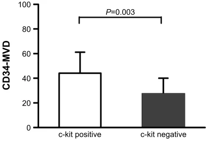
In healthy liver tissues, none or indefinite c-kit positivity was seen (0/15) (Figure 2A–B). In healthy liver tissues, 2 cases were positive for CD34 expression (2/15) (Figure 2C–D). Only 2 of 23 cirrhosis cases showed c-kit-positive staining (Figure 2E–F). Only 5 of 23 cirrhotic cases showed CD34-positive staining (Figure 2G–H). The greatest number of the c-kit positive cells was found in the liver cancer tissues of patients with HCC. The positive rate of c-kit expression in 206 HCC cases was 48.1% (99/206), with positive expression observed in the cellular membrane or endochylema (Figure 2I and 2J). We observed that the positive rate of CD34 expres-sion in HCC was 100%(206/206), with positive expresexpres-sion observed in the peribiliary capillaries and periportal and perilobular sinusoids (Figure 2K–L). Both c-kit- and CD34-positive expression were significantly higher in HCC tissues than in other groups (Table 1). The MVD of CD34-positive cells in HCC tissues ranged from 11.24 to 108.53. More-over, CD34-MVD numbers were much higher in c-kit(+) HCC tissues than in c-kit(-) HCC tissues (44.13±17.01 vs 26.87±13.16, P=0.003; Figure 3).
correlation between c-kit(cD117) or
cD34 and clinicopathologic features
The correlations of c-kit(CD117) expression with clinico-pathologic parameters are listed in Table 2. The expression of c-kit(CD117) was significantly related to Edmondson grade (P,0.001) and TNM stage (P,0.001), but was not significantly associated with age, gender, tumor diameter, vascular invasion, liver cirrhosis, HBeAg status, or AFP level (P.0.05; Table 2). When the median value (30.14) of MVD was used as the cutoff point for CD34 expression, all the 206 HCC patients were divided into two subgroups: high CD34 group (MVD.30.14) and low CD34 group (MVD30.14). Increased expression of CD34 was correlated with vascular invasion (P=0.042) and Edmondson grade (P,0.001). How-ever, CD34 expression was not significantly related with age, gender, tumor diameter, cirrhosis, HBeAg status, AFP level, or TNM stage (P.0.05; Table 2).
relationship between cD34 or c-kit
expression and prognosis
The median DFS was 37 months in the entire cohort of 206 patients. It was found that the expression of c-kit in HCC patients was negatively correlated with DFS. HCC patients with positive expression of c-kit had significantly shorter DFS than those with negative expression of c-kit (26 vs 40 months, P,0.001; Figure 4A). However, Kaplan–Meier analysis revealed no significant difference between patients with CD34-high expression, that is, MVD.30.14, and those with CD34-low expression, that is, MVD30.14 (38 vs 35 months, P=0.670; Figure 4B).
î
&'
FNLW&'
î î î î î
$
%
(
)
,
-&
'
*
+
.
/
+& /& +&&
Figure 2 representative ihc images of c-kit and cD34 staining in the consecutive clinical samples from one hc, one patient with lc, and one patient with hcc,
respectively.
Notes: (A, B) c-kit staining in the hc (100×, 200×). (C, D) cD34 staining in the hc with continuous slices according to A, B (100×, 200×). (E, F) c-kit staining in the lc
(100×, 200×). (G, H) cD34 staining in the lc with continuous slices according to E, F (100×, 200×). (I, J) c-kit staining in the hcc (100×, 200×). (K, L) cD34 staining in
the hcc with continuous slices according to I, J (100×, 200×).
Abbreviations: hcc, hepatocellular carcinoma; ihc, immunohistochemistry; lc, liver cirrhosis; hc, healthy control.
OncoTargets and Therapy downloaded from https://www.dovepress.com/ by 118.70.13.36 on 25-Aug-2020
Dovepress c-kit in hepatocellular carcinoma
Univariate and multivariate survival
analyses of prognostic variables in
hcc patients
Table 3 shows the results of the univariate and multivariate survival analyses for variables measured at baseline. Param-eters associated with DFS were c-kit expression, age, tumor diameter, vascular invasion, Edmondson grade, AFP level, and TNM stage. Subsequent multivariate survival analysis was conducted by incorporating all the above factors as covariates. In multivariate analysis, c-kit expression was an independent prognostic factor for DFS (hazard ratio 3.583, 95% CI 2.170–5.917; P,0.001). Additionally, Edmondson grade, AFP level, and TNM stage were also of significant value in predicting DFS. Detailed results are shown in Table 3. To further elucidate the validity of c-kit expression level in predicting the postoperative recurrence, Kaplan– Meier survival analysis was used to compare DFS of patients with weak and strong positive c-kit. The median value (2) of IHC score of c-kit-positive cells was used as the cutoff. According to the median value, these patients were divided into weak positive c-kit group (2) and strong positive c-kit group (.2). Both strong positive c-kit (16 vs 40 months,
P,0.001; Figure 5) and weak positive c-kit (34 vs 40 months,
P,0.001) groups had a shorter DFS time than those with negative expression of c-kit. Notably, the strong positive c-kit group also had a shorter DFS time compared to the weak positive c-kit group (16 vs 34 months, P,0.001; Figure 5).
Discussion
The recurrence rate of HCC after curative surgical resec-tion remained very high. In China, up to 80% of HCC cases are attributable to HBV and ~20% of HCC patients test positive for HCV-RNA.22 Long-term follow-up data after
surgery showed that the recurrence rate of HBV-positive HCC patients was 59%.23 Therefore, high-quality prognostic
markers are very important in clinical use.
The positive expression rate of CD34 in 206 HCC cases was 100%, which is consistent with the multivascular supply of HCC and the ability related to tumoral vascular formation.24
Wang et al25 found CD34 was an independent prognostic
factor for overall survival and recurrence-free survival in HCC. However, in our study, CD34 expression was negatively correlated with Edmondson grade. In addition, univariate and multivariate analyses showed that CD34 expression had no independent impact on DFS in HCC patients. Sup-porting our data, Wu et al17 have shown that positive CD34
expression has no significant effect on the prognosis of HCC patients. Therefore, it remains elusive whether CD34 can be used as a prognostic indicator for HCC patients.
c-kit(CD117) is a transmembrane tyrosine kinase that acts as a receptor for mast cell growth factor.26 It belongs
to the receptor kinases type III family and can be detected in some normal cells, including hematopoietic stem cells.27
Different studies assessing c-kit expression in HCC have shown contradictory results, with some showing increased expression and others reporting no or weak expression.3,14,15
Our study of 206 HBV-related HCC tissue sections showed that c-kit receptors were expressed in 48.1%. This result confirms the results of previous studies. Kara et al14 have
shown the overall percentage of c-kit-positive IHC staining of HBV-related HCC was 82.6%. In addition, Lee et al15 found
40 of 50 HBV-associated HCC tissue sections (80%) positive for c-kit by IHC staining. In contrast to these results, Becker et al3 found that the overall percentage of c-kit-positive
IHC staining of HCC (related with alcohol in about 50% of patients) was 2.3% (6/258). In their study, only 1/90 patient with hepatitis was positive for c-kit. We also found that the overall percentage of c-kit-positive IHC staining of HCV-related HCC samples was ,15% (Figure 6). We have no valid explanation for the contradiction between the results of
Table 1 Differential expression of c-kit and cD34 in healthy liver
tissues, cirrhosis tissues, and hcc tissues
Tissues Healthy
liver tissues
Cirrhosis tissues
HCC tissues
case number 15 23 206
c-kit-positive expression 0 (0%)a 2 (8.7%)a 99 (48.1%)b,c cD34-positive expression 2 (13.3%)a 5 (21.7%)a 206 (100%)b,c Notes: aP,0.05 compared to hcc tissues group; bP,0.05 compared to cirrhosis
tissues group; cP,0.05 compared to healthy liver tissues group. Data are expressed
as number with percentage.
Abbreviation: hcc, hepatocellular carcinoma.
3
FNLWSRVLWLYH FNLWQHJDWLYH
&'09'
Figure 3 immunohistochemical staining of cD34, labeled by MVD, in hcc tissues. Note: MVD numbers were higher in hcc with positive c-kit expression compared
to that with negative c-kit expression (44.13±17.01 vs 26.87±13.16, P=0.003).
Abbreviations: hcc, hepatocellular carcinoma; MVD, microvessel density.
OncoTargets and Therapy downloaded from https://www.dovepress.com/ by 118.70.13.36 on 25-Aug-2020
Dovepress
Yan et al
Table 2 relationship between c-kit(cD117), cD34 expression, and clinicopathologic parameters in 206 patients with hcc
Parameters c-kit(+) c-kit(-) χ2 P-value
CD34-MVD high
CD34-MVD low
χ2 P-value
n (%) 99 (48.1%) 107 (51.9%) 102 (49.5%) 104 (50.5%)
age (years)
#60 67 (45.3%) 81 (54.7%) 1.264 0.261 74 (50%) 74 (50%) 0.050 0.824
.60 32 (55.2%) 26 (44.8%) 28 (48.3%) 30 (51.7%)
gender
Male 59 (43.4%) 77 (56.6%) 2.976 0.085 71 (52.2%) 65 (47.8%) 0.864 0.352
Female 40 (57.1%) 30 (42.9%) 31 (44.3%) 39 (55.7%)
Tumor diameter (cm)
#5 57 (44.2%) 72 (55.8%) 1.679 0.195 66 (51.2%) 63 (48.8%) 0.219 0.639
.5 42 (54.5%) 35 (45.5%) 36 (46.8%) 41 (53.2%)
Vascular invasion
Present 19 (57.6%) 14 (42.4%) 1.008 0.315 11 (33.3%) 22 (66.7%) 4.116 0.042
absent 80 (46.2%) 93 (53.8%) 91 (52.6%) 82 (47.4%)
edmondson grade
i–ii 20 (27.8%) 52 (72.2%) 17.010 ,0.001 54 (75.0%) 18 (25.0%) 27.213 ,0.001
iii–iV 79 (59.0%) 55 (41.0%) 48 (35.8%) 86 (64.2%)
liver cirrhosis
Present 86 (47.3%) 96 (52.7%) 0.176 0.675 93 (51.1%) 89 (48.9%) 1.072 0.301
absent 13 (54.2%) 11 (45.8%) 9 (37.5%) 15 (62.5%)
hBeag
Positive 40 (50.6%) 39 (49.4%) 0.194 0.660 35 (44.3%) 44 (55.7%) 1.074 0.300
negative 59 (46.5%) 68 (53.5%) 67 (52.8%) 60 (47.2%)
aFP level (µg/l)
#400 23 (45.1%) 28 (54.9%) 0.106 0.744 27 (52.9%) 24 (47.1%) 0.162 0.687
.400 76 (49.0%) 79 (51.0%) 75 (48.4%) 80 (51.6%)
TnM stage
i–ii 35 (27.3%) 93 (72.7%) 55.939 ,0.001 62 (48.4%) 66 (51.6%) 0.064 0.801
iii 64 (82.1%) 14 (17.9%) 40 (51.3%) 38 (48.7%)
Note: Data are expressed as number with percentage.
Abbreviations: aFP, alpha-fetoprotein; hBeag, hepatitis B e-antigen; hcc, hepatocellular carcinoma; MVD, microvessel density.
0RQWKVDIWHURSHUDWLRQ
')6UDWH
Q Q
FNLWH[SUHVVLRQ
1HJDWLYH 3RVLWLYH
3RVLWLYHFHQVRUHG 1HJDWLYHFHQVRUHG
0RQWKVDIWHURSHUDWLRQ
')6UDWH
Q Q
&'H[SUHVVLRQ
09'ORZ 09'KLJK
09'KLJKFHQVRUHG 09'ORZFHQVRUHG
$
%
Figure 4 Kaplan–Meier curves for patients regarding DFs are plotted. c-kit(cD117) expression was divided into a positive group (n=99) and a negative group (n=107), and cD34 expression was divided into an MVD-high group (n=102) and an MVD-low group (n=104). A log-rank test was used to calculate significance.
Notes: (A) hcc patients with c-kit positivity showed notably worse DFs rates than those with c-kit-negativity (26 vs 40 months, P,0.001). (B) There was no significant
difference between hcc patients with high cD34-MVD and those with low cD34-MVD (38 vs 35 months, P=0.670).
Abbreviations: DFs, disease-free survival; hcc, hepatocellular carcinoma; MVD, microvessel density. c-kit staining of HCC in different studies. Our results further
demonstrated that c-kit overexpression was significantly associated with poor Edmondson grade and advanced TNM stage. These findings suggest an important role for c-kit in tumor development and progression.
In our study, all HCC patients had a history of .10 years of chronic HBV infection and had received antiviral therapy (nucleoside/nucleotide analogs) .1 year. All patients were hepatitis B virus surface antigen positive and serum HBV-DNA negative. Seventy-nine patients were serum
OncoTargets and Therapy downloaded from https://www.dovepress.com/ by 118.70.13.36 on 25-Aug-2020
Dovepress c-kit in hepatocellular carcinoma
1.0
0.8
0.6
0.4
0.2
0.0
0.00 10.00 20.00 30.00
Months after operation
DFS rate (%
)
40.00 50.00
n=48 n=107
n=51
c-kit expression
Weak positive-censored Negative-censored
Strong positive-censored
Negative Weak positive Strong positive
Figure 5 Kaplan–Meier analysis of DFs curves of patients with hcc based on c-kit
expression as strong positive (n=51), weak positive (n=48), or negative (n=107).
Notes: The hcc patients with c-kit positivity showed much worse DFs rates
than those with c-kit-negativity. The survival of patients with strong positive c-kit expression was poorest.
Abbreviations: DFs, disease-free survival; hcc, hepatocellular carcinoma.
î î
FNLWQHJDWLYH
$
FNLW&'
&'
î
FNLWSRVLWLYH
î
( )
%
& ' * +
Figure 6 representative ihc images of c-kit and cD34 staining in the continuous
slices of clinical hcV-related hcc.
Notes: (A, B) c-kit staining was marked as negative (100×, 200×). (C, D) cD34
staining in the continuous slices corresponding to A, B (100×, 200×). (E, F) c-kit
staining was marked as weak positive (100×, 200×). (G, H) cD34 staining in the
continuous slices corresponding to E, F (100×, 200×).
Abbreviations: hcc, hepatocellular carcinoma; hcV, hepatitis c virus; ihc,
immunohistochemistry.
HBeAg positive. However, c-kit expression was not sig-nificantly correlated with HBeAg status (P=0.660). Further researches are needed to investigate the relationship between HBV and c-kit in hepatic cells.
In our study, it was found that DFS was significantly different between patient groups with positive and nega-tive expression of c-kit. We further observed that c-kit was an independent poor predictor of DFS in the multivari-able analysis. Therefore, we believe that c-kit(CD117) can be used as an indicator of prognosis for resectable HBV-related HCC patients.
Angiogenesis is critical for the growth and progression of HCC. It was found that the CD34 antigen is related to vascular formation.25 Interestingly, we found that there
was a significantly positive correlation between c-kit and CD34 expression. This raises a possibility that c-kit might be involved in angiogenesis in HCC. However, additional researches are required to explore the “true” roles of c-kit in tumor angiogenesis in HCC.
A few limitations deserve further discussion. First of all, because data were retrospectively recorded from patients, this study may be associated with a potential selection bias. Second, in our study, there is no direct evidence that c-kit plays a key role in the development and progression of HCC. Despite these shortcomings, the advantage of our study is that we examined the expression of c-kit in HCC tissue developed in HBV infection progress.
Table 3 Univariate and multivariate analysis of factors associated with DFs
Parameters Univariate
analysis
95% CI P-value Multivariate
analysis
95% CI P-value
Hazard ratio Lower–upper Hazard ratio Lower–upper
c-kit(positive/negative) 2.710 1.845–3.980 ,0.001 3.583 2.170–5.917 ,0.001
cD34-MVD(high/low) 0.925 0.638–1.340 0.679
age (.60/#60 years) 1.712 1.114–2.633 0.014
gender (male/female) 0.923 0.631–1.788 0.474
Tumor diameter (.5/#5 cm) 1.995 1.362–2.922 0.001
Vascular invasion (present/absent) 4.091 2.546–6.575 ,0.001
edmondson grade (iii–V/i–ii) 2.531 1.679–3.815 ,0.001 1.912 1.144–3.196 0.013
liver cirrhosis (present/absent) 0.973 0.490–1.934 0.938
hBeag (positive/negative) 0.812 0.546–1.208 0.304
aFP level (.400/#400 µg/l) 2.769 1.751–4.379 ,0.001 2.705 1.550–4.719 ,0.001
TnM stage (iii/i–ii) 7.081 4.668–10.742 ,0.001 2.978 1.707–5.195 ,0.001
Abbreviations: aFP, alpha-fetoprotein; DFs, disease-free survival; hBeag, hepatitis B e-antigen; MVD, microvessel density.
OncoTargets and Therapy downloaded from https://www.dovepress.com/ by 118.70.13.36 on 25-Aug-2020
OncoTargets and Therapy
Publish your work in this journal
Submit your manuscript here: http://www.dovepress.com/oncotargets-and-therapy-journal
OncoTargets and Therapy is an international, peer-reviewed, open access journal focusing on the pathological basis of all cancers, potential targets for therapy and treatment protocols employed to improve the management of cancer patients. The journal also focuses on the impact of management programs and new therapeutic agents and protocols on
patient perspectives such as quality of life, adherence and satisfaction. The manuscript management system is completely online and includes a very quick and fair peer-review system, which is all easy to use. Visit http://www.dovepress.com/testimonials.php to read real quotes from published authors.
Dovepress
Dove
press
Yan et al
Taken together, our data demonstrated, for the first time, that upregulation of c-kit is an independent prognostic factor for predicting early tumor recurrence and poor prognosis in HBV-related HCC. Therefore, c-kit might be used to identify patients at a higher risk of early tumor recurrence and repre-sents a potential therapeutic target for this disease.28
Acknowledgments
This study was supported by grants from the National Natural Science Foundation of China (Nos. 81372286 and 31671298), Beijing Nova Program (No. Z171100001117114), the Military Medical Science and Technology Project of Youth Develop-ment (Nos. 14QNP112 and 15QNP084), the Doctorial Innova-tion Fund of Chinese PLA Medical School (No. B14015), and the 302 Hospital Foundation (No. QNPY2015007).
Disclosure
The authors report no conflicts of interest in this work.
References
1. Torre LA, Bray F, Siegel RL, Ferlay J, Lortet-Tieulent J, Jemal A. Global cancer statistics, 2012. CA Cancer J Clin. 2015;65(2):87–108. 2. Tung-Ping Poon R, Fan ST, Wong J. Risk factors, prevention, and
management of postoperative recurrence after resection of hepatocel-lular carcinoma. Ann Surg. 2000;232(1):10–24.
3. Becker G, Schmitt-Graeff A, Ertelt V, Blum HE, Allgaier HP. CD117 (c-kit) expression in human hepatocellular carcinoma. Clin Oncol (R Coll Radiol). 2007;19(3):204–208.
4. Kim JH, Sohn BH, Lee HS, et al. Genomic predictors for recurrence patterns of hepatocellular carcinoma: model derivation and validation. PLoS medicine. 2014;11(12):e1001770.
5. Yamamoto T, Uenishi T, Ogawa M, et al. Immunohistologic attempt to find carcinogenesis from hepatic progenitor cell in hepatocellular carcinoma. Dig Surg. 2005;22(5):364–370.
6. Wang FS, Fan JG, Zhang Z, Gao B, Wang HY. The global burden of liver disease: the major impact of China. Hepatology. 2014;60(6): 2099–2108.
7. Muto J, Shirabe K, Sugimachi K, Maehara Y. Review of angiogenesis in hepatocellular carcinoma. Hepatol Res. 2015;45(1):1–9.
8. Carmeliet P, Jain RK. Molecular mechanisms and clinical applications of angiogenesis. Nature. 2011;473(7347):298–307.
9. Di Carlo I, Fraggetta F, Lombardo R, Azzarello G, Vasquez E, Puleo S. CD 34 expression in chronic and neoplastic liver diseases. Panminerva Med. 2002;44(4):365–367.
10. Poon RT, Ng IO, Lau C, et al. Tumor microvessel density as a predictor of recurrence after resection of hepatocellular carcinoma: a prospective study. J Clin Oncol. 2002;20(7):1775–1785.
11. Uzzan B, Nicolas P, Cucherat M, Perret GY. Microvessel density as a prognostic factor in women with breast cancer: a systematic review of the literature and meta-analysis. Cancer Res. 2004;64(9):2941–2955.
12. Wiedmann MW, Caca K. Molecularly targeted therapy for gastroin-testinal cancer. Curr Cancer Drug Targets. 2005;5(3):171–193. 13. Cohen MH, Johnson JR, Justice R, Pazdur R. Approval summary:
imatinib mesylate for one or three years in the adjuvant treatment of gastrointestinal stromal tumors. The oncologist. 2012;17(7):992–997. 14. Kara B, Doran F, Kara IO, Akkiz H, Sandikci M. Expression of c-kit
protooncogen in hepatitis B virus-induced chronic hepatitis, cirrhosis and hepatocellular carcinoma: has it a diagnostic role? Int J Clin Pract. 2008;62(8):1206–1211.
15. Lee ES, Han EM, Kim YS, et al. Occurrence of c-kit+ tumor cells in hepatitis B virus-associated hepatocellular carcinoma. Am J Clin Pathol. 2005;124(1):31–36.
16. van der Zee JA, van Eijck CH, Hop WC, et al. Angiogenesis: a prog-nostic determinant in pancreatic cancer? Eur J Cancer. 2011;47(17): 2576–2584.
17. Wu LQ, Lu Y, Lu HJ, Lv ZH. Can E-cadherin and CD34 be used as indicators of prognosis for hepatocellular carcinoma patients? Clin Chem Lab Med. 2008;46(8):1122–1126.
18. Murakami K, Kasajima A, Kawagishi N, Ohuchi N, Sasano H. Microvessel density in hepatocellular carcinoma: prognostic significance and review of the previous published work. Hepatol Res. 2015;45(12):1185–1194. 19. Bruix J, Sherman M. American Association for the Study of Liver D.
Management of hepatocellular carcinoma: an update. Hepatology. 2011;53(3):1020–1022.
20. European Association for the Study of the Liver, European Organisation for Research and Treatment of Cancer. EASL-EORTC clinical prac-tice guidelines: management of hepatocellular carcinoma. J Hepatol. 2012;56(4):908–943.
21. Chen L, Shi Y, Jiang CY, et al. Coexpression of alpha, PDGFR-beta and VEGF as a prognostic factor in patients with hepatocellular carcinoma. Int J Biol Markers. 2011;26(2):108–116.
22. Nguyen VT, Law MG, Dore GJ. Hepatitis B-related hepatocellular carcinoma: epidemiological characteristics and disease burden. J Viral Hepat. 2009;16(7):453–463.
23. Sasaki Y, Yamada T, Tanaka H, et al. Risk of recurrence in a long-term follow-up after surgery in 417 patients with hepatitis B- or hepatitis C-related hepatocellular carcinoma. Ann Surg. 2006;244(5): 771–780.
24. Salizzoni M, Romagnoli R, Lupo F, et al. Microscopic vascular inva-sion detected by anti-CD34 immunohistochemistry as a predictor of recurrence of hepatocellular carcinoma after liver transplantation. Transplantation. 2003;76(5):844–848.
25. Wang W-Q, Liu L, Xu H-X, et al. Intratumoral alpha-SMA enhances the prognostic potency of CD34 associated with maintenance of microvessel integrity in hepatocellular carcinoma and pancreatic cancer. PLoS One. 2013;8(8):e71189.
26. Went PT, Dirnhofer S, Bundi M, et al. Prevalence of KIT expression in human tumors. J Clin Oncol. 2004;22(22):4514–4522.
27. Morini M, Bettini G, Preziosi R, Mandrioli L. C-kit gene product (CD117) immunoreactivity in canine and feline paraffin sections. J Histochem Cytochem. 2004;52(5):705–708.
28. Knight B, Tirnitz-Parker JE, Olynyk JK. C-kit inhibition by imatinib mesylate attenuates progenitor cell expansion and inhibits liver tumor formation in mice. Gastroenterology. 2008;135(3):969–979, 979 e961.
OncoTargets and Therapy downloaded from https://www.dovepress.com/ by 118.70.13.36 on 25-Aug-2020