*Corresponding author: H. S. Mohamed, Email: h_gendy_2010@yahoo.com page 183 of 232
Reviewing of Synthesis and Computational Studies of Pyrazolo
Pyrimidine Derivatives
Receive Date: 11 Jun 2019, Revise Date: 24 June 2019, Accept Date: 24 June 2019
Abstract: In this review we synthesized and conducted a computational studies on Pyrazolo pyrimidine’s derivatives that were carried out through density functional theory level utilizing HF/6−311+G**and B3LYP/6−311+G**. Charge transfer occured through molecule was shown by the calculation of HOMO and LUMO energies. The electric dipole moment values (l) of the molecule were counted calculations of DFT. Some geometrical and structural parameters such as total energies (E), relative energies (DE), (bond length in Å, angles in degree), energy gap, relative Gibbs free energy, dipole moment, and molecular electrostatic potentials (MEP) were studied.
DOI: 10.33945/SAMI/JCR.2019.3.3
Keywords: GAUSSIAN09, DFT, HOMO-LUMO, pyrazolo pyrimidine derivatives, quantum chemical calculation.
Graphical Abstract:
Hussein Shaban Hussein Mohamed a,
*
, Sayed Abdelkader Ahmed
ba Research Institute of Medicinal and Aromatic Plants (RIMAP), Beni-Suef University, Beni-Suef, Egypt b Chemistry Department, Faculty of Science, Beni-Suef University, Beni-Suef, Egypt
Biography:
Hussein S. H. Mohamed Was born Beni-Suef (Egypt) in 1980, obtained his Ph.D. degree in Organic chemistry in 2015 under the supervisor of Prof. Dr. Ahmed Elghandour and Prof. Dr. Sayed A. Ahmed. Later on, he is currently working as Senior Researcher in Natural Products of Research Institute of Medicinal and Aromatic Plants (RIMAP), Beni-Suef University. He has been actively involved Synthesis of Heterocyclic compounds and its applications in biological activities, recently he interested in Removal of dyes and heavy metals from wastewater by different synthesized compounds.
Sayed A. Ahmed Has completed his Ph.D under the supervision of Professor Galal Elgemie and Prof. Dr. Ahmed Elghandour in the Department of Chemistry at Beni-Suef University, Egypt. He currently works as a full professor in Organic Chemistry at Beni-Suef University Egypt. His studies focused on organic synthesis of Organic compounds and he has published more than 100 papers. He is a Dean of Faculty of Earth Sciences, Beni-Suef University Egypt. He has received several national and international awards.
1. Introduction
Heterocyclic pyrimidine nucleus includes four carbon and two nitrogen atoms and is pharmacologically in active; however, its synthetic derivatives have important roles in pharmaceutical applications [1-3]. The properties are demonstrated by their hydrogen and bonding systems. They belong to the family of nucleic acids. Nucleic acids are of great interest, since, they have role in the manufacture of proteins and the functions of the cells in living organisms [4-6]. For example Pyrazolo[3,4-d]pyrimidine derivatives are shown to exhibit anti-amoebic activity [7-13] and are potential anti-inflammatory agents [14, 15], anti-coagulation inhibitors [16], xanthine oxidase inhibitors [17], antiproliferative and proapoptotic agents in various tumor types [18,19]. They are called SRC kinase inhibitors [20] and are beneficial in human cancers treating sustaining oncogenic activation of RET [21]. After the knowing of scaled quantum mechanical (SQM) calculations, the philosophy of computational methods of vibrational spectroscopy [22-25] were changed significantly. The most applied spectroscopic methods by organic chemists for the characterization of compounds are ultra-violet, IR, NMR, and mass spectroscopy. IR measurement through liquid mixtures supplies an excellent tool to investigate inter and intramolecular interactions between like and unlike molecules. The density functional theory has become a great method for the description of vibrational spectra and molecular structure. Completed by a visualization program, the assignments can accurately be made [26-28]. The advance in the graphic and computational devices has increased the significance of MEP
surfaces, highest occupied molecular orbital (HOMO) and lowest unoccupied molecular orbital (LUMO) as tools for studying molecular reactivities, interactions, charge transfers and other molecular properties [29-34]. These characteristics are the highest occupied molecular orbital energy EHOMO, the lowest
unoccupied molecular orbital energy Elumo, energy gap
ΔE, dipole moment μ, total energy ET, activation
energy Ea, absorption maximum λmax and factor of
oscillation f(SO). The geometry of the systems has been improved at the B3LYP /6-31G (d) [35-38] computational level. As per the computations, the molecules concerned to energy minima on the Potential Energy Surface (PES). The Gaussian-09 program was used for all this calculations [39-42].The density functional used this form:
Exc= (1-α0 )EXLSDA +α0EXHF +αX ∆EXB88+αCEcLYP +
(1-αC)ECVWN
2.
Synthesis and Computational Studies of Pyrazolo[3,4-d]Pyrimidine2.1. 4- Aminopyrazolo[3,4-d]pyrimidine
The theoretical UV–Vis spectrum is used to detect this compound using CIS method and the electronic properties, such as HOMO and LUMO energies, were perfected by time-dependent DFT (TD-DFT) approach. Charge transfer occurs within the molecule was shown by HOMO and LUMO energies calculations. The first order hyperpolarizability (β0) of
this molecular system and related properties (β, α0 and
charges, the values of electric dipole moment (µ) of the molecule were calculated utilizing DFT calculations. They used natural bond (NBO) analysis to compute The diversity in electron density (ED) in the antibonding orbitals and stabilization energies giving clear evidence of stabilization originating in the hyper conjugation of hydrogen-bonded interactions [43].
2.1.1. Structure of 4-aminopyrazolo[3,4-d]pyrimidine
Scheme 1. Structure of 4-aminopyrazolo[3,4-d]pyrimidine
2.1.2. Computational
Gaussian 09W software package used to calculate the energy and dipole moment of tautomer [44], using the
B3LYP functional [45,46] combined with standard 6-31G* basis set.
In the Table 1 Prabavathi, et al. reported that the total energies, dipole moment and their relative energies of various tautomer of 4-APP were computed at B3LYP/6-31G* level.
We can realize from Table 1 that for 4-APP the energies of the tautomers are in the following order
(1a) < (1b) < (1c) < (1d) < (1e) < (1f) < (1g) < (1h) < (1i) < (1j) < (1k) < (1L) < (1m) < (1n) < (1o) < (1p).
It was found that the amino-N(1)H tautomer has the lowest energy and is the most stable form.
Xue et al. utilizing the computed geometrical parameters for 4-APP (as it contains both a pyrimidine ring and an imidazole ring) which are parallel with some of the purine derivatives [47,48] (6-aminopurine and adenine) for the possible bond lengths and bond angles. So it was predicted that the bond length and bond angle agree with adenine parameters obtained in Table 3.
fgjhhhhhhhhhhhhhhhhhhhhhhhhhhhhhhhhhhhhhhhhhhhhhhhhhhhhhkkkkkkkkkkkkkkk
Through Table 4 the values of the polarizabilities (α) and hyperpolarizability (β) of the Gaussian 09 output, Data et al. found that the extent of ᴨ electron delocalization control the ground-state dipole moment and the hyperpolarizability, which relies on the structural details of the molecules [49].
The six peaks of 4-APP appeared at 1587, 1580, 1478, 725 cm-1 in both IR and Raman spectra and the
IR bands at 1606, and 1512 cm-1 are attributed to ring
C–N stretching vibrations. Analogical bands are theoretically calculated at 1633, 1612, 1563, 1512, 1466, 705 cm-1 with a PED contribution in the range
58–35% at 6-31G** level.
Table 1. Energy and dipole moment of 4-aminoPyrazolo[3,4-d]pyrimidine.
Tautomera Total energy
(Hartree) B3LYP/
631_G*
Dipole moment
(Debye) B3LYP/
631_G*
Relative
energies
(kJ mol-1)
4-Aminopyrazolo [3 4-d]pyrimidine
1a -467.238374 1.7170 0.0
1b -467.218458 5.5250 52.3
1c -467.215798 6.2607 59.3
1d -467.198521 11.2556 104.6
1e -467.197957 4.8652 106.1
1f -467.185872 1.9407 137.8
1g -467.185169 4.5492 139.7
1h -467.184202 7.9895 142.2
1i -467.180848 3.8361 151.0
1j -467.166858 1.1979 187.8
1k -467.157364 8.5522 212.4
1l -467.154937 10.8967 219.1
1m -467.154623 8.6543 219.9
1n -467.154076 6.1545 221.3
1o -467.138857 9.2026 261.3
1p -467.126886 10.6410 292.7
a For the tautomeric figures.
Table 2. Theoretical computed energies (a.u.), energies (Kcal Mol-1), Rotational constant (GHZ), entropy (Cal mol-1 k-1)
and Dipole moment (Debye).
Structural parameter B3LYP/6-31G* 4-APP
B3LYP/6-31G** 4-APP
Total energy(thermal) Etotal 75.22 75.17
Heat capacity at const. Volume ,Cv 27.84 27.86
Entropy ,S 83.15 83.11
Vibrational energy Evib 73.45 73.40
Zero-point vibrational Energy , E0 70.82 70.78
Rotational constant
A
B
C
2.403 1.493 0.922
2.404 1.493 0.922
Dipole moment
µx
µy
µz
µtotal
-0.4060 -1.4263 1.3911 2.0332
Table 3. Optimized geometrical parameters of 4-aminoPyrazolo[3,4-d]pyrimidine obtained by B3LYP/6-31G* and B3LYP/6-31G** density functional calculations.
Bond length Value (Å) Bond angle Value (o)
6-31G* 6-31G** 6-31G* 6-31G**
N1–N2 1.36 1.36 N1–N2–C3 105.81 105.85
C3–N2 1.32 1.32 N2–C3–C4 111.19 111.12
C4–C3 1.42 1.42 C3–C4–C5 140.36 140.33
C5–C4 1.41 1.41 C4–C5–N6 119.81 119.78
N6–C5 1.33 1.33 C5–N6–C7 118.25 118.29
C7–N6 1.34 1.34 N6–C7–N8 128.61 128.57
N8–C7 1.32 1.32 C7–N8–C9 111.80 111.82
C9–N8 1.34 1.34 C9–N1–H10 127.62 127.49
H10–N1 1.00 1.00 N2–C3–H11 119.38 119.46
H11–C3 1.08 1.08 C4–C5–N12 122.43 122.44
N12–C5 1.38 1.38 C5–N12–H13 111.79 111.76
H13–N12 1.01 1.01 C5–N12–H14 108.53 108.48
H14–N12 1.01 1.01 N6–C7–H15 115.20 115.22
H15–C7 1.08 1.08 C4–C3–N2–N1 0.0 0.0
C5–C4–C3–N2 180.0 180.0
N6–C5–C4–C3 -180.0 -180.0
C7–N6–C5–C4 0.0 0.0
N8–C7–N6–C5 0.0 0.0
C9–N8–C7–N6 0.0 0.0
H10–N1–C9–N8 0.0 0.0
H11–C3–N2–N1 180.0 180.0
N12–C5–C4–C3 6.0 6.0
H13–N12–C5–C4 -46.0 -46.0
H14–N12–C5–C4 -161.0 -161.0
H15–C7–N6–C5 180.0 180.0
a For the tautomeric figures.
Table 4. Vibrational spectral data (cm-1)
No Observed Calculated frequency (cm-1) with B3LYP/6-31G** force field TED (%) among
type of internal coordinatesc Infrared Raman Unscaled Scaled IRa(Ai) Ramanb(Ii)
1 3318 _ 3644 3318 14.89 1.07 NــH Stretching
2 3135 _ 3553 3135 94.00 3.83 NH2ass(95)
3 _ 3102 3504 3102 22.60 72.71 NH2ss(95)
4 3060 _ 3337 3079 4.02 10.57 CــH(99)
5 3030 _ 3261 3010 29.03 1.14 CــH(99)
6 1677 1675 1670 1668 220.69 17.93 NH2sci(54)
7 1606 _ 1630 1633 190.18 29.66 CــN(51), NH2Sci(26)
9 1580 1580 1511 1563 4.36 25.17 CــN(54), bCCH(29)
10 1512 _ 1488 1512 165.42 19.41 CــN(58), CــC(16)
11 1478 1478 1450 1466 84.69 26.12 CــN(49), bCCH(22)
12 1398 _ 1389 1407 4.18 4.10 bCCH(52), CــN(16)
13 _ 1394 1368 1387 9.63 23.17 CــN(57), CــC(19)
14 1340 1337 1336 1335 8.02 10.30 CــC(42), CــN(35)
15 1339 _ 1332 1325 180.20 3.14 bNNH(47), CــN(16)
16 1265 _ 1283 1265 45.52 3.62 bCCH (59), bNNH
(15)
17 1199 _ 1211 1200 40.66 9.27 CــC(41), CــN(22)
18 _ 1154 1160 1147 11.30 12.00 bring2(35), CــN(27)
19 1067 1066 1075 1069 34.15 8.14 NــN(78), bNNH(9)
20 1029 1031 1053 1035 22.96 8.31 NH2roc(54), CــN(37)
21 962 939 981 972 150.30 1.43 CــC(53), bring2(18)
22 903 898 947 914 100.47 2.83 bring1(54), CــC(17)
23 900 900 906 895 11.79 2.12 gCH(59), bring2 (17)
24 870 _ 871 856 70.71 1.02 gHN(77), tring2(15)
25 796 796 853 805 15.64 1.94 gCH(31), tring1(27)
26 785 _ 800 775 115.94 1.51 gCH(30), tring1(29)
27 725 725 713 705 2.87 18.67 CــN(30), CــC(24)
28 693 _ 681 701 17.42 0.08 tring2(82), tButt(7)
29 636 _ 659 633 30.89 1.29 gCN(39), tring1(30)
30 609 615 608 594 0.23 8.34 bring2(31), CــC (29)
31 549 _ 573 554 57.46 2.13 tring1(46), gNH(23)
32 527 527 525 526 0.56 4.51 bring1(74), CــN(12)
33 _ _ 511 508 3.66 2.77 bring1(32), bCN(31)
34 _ _ 499 488 34.03 2.12 gNH(58), tring2(30)
35 _ _ 353 337 69.94 1.98 tNH2(68), gHN(19)
36 _ _ 299 292 3.05 0.06 tring1(35), tring2(25)
37 _ _ 285 280 2.64 0.58 bCN(45), bring1(15)
38 _ _ 209 209 2.64 0.43 tbutt(48), tring1(31)
39 _ _ 164 155 1.81 0.37 tring1(72), tring2(12)
Abbreviations used: b, bending; g, deformation; ass, asymmetric; ss, symmetric; t, torsion; sci, scissoring; gHN, twisting.
a Relative absorption intensities normalized with highest peak absorption. b Relative Raman intensities calculated and normalized to 100.
c For the notations used.
Chemical shifts of the carbon atoms in 4-APP theoretically are calculated, lies between 105 and 165
the electron distribution in its bond to close carbon atom and lowering the electron density of the molecule [50].
Table 5. Theoretical isotropic chemical shifts calculated using HF/6-31G* (with respect to TMS, all values in ppm) for 4-aminopyrazolo[3,4-d]pyrimidine [43].
Calculated chemical shift (ppm)
Atom HF/6-31G*
N1 180.7
N2 308.5
C3 134.8
C4 109.2
C5 162.1
N6 248.3
C7 160.2
N8 235.3
C9 157.4
H10 9.1
H11 7.9
N12 78.3
H13 3.6
H14 3.9
H15 8.9
All the exocyclic atoms have atomic charges corresponds to their electro-negativity while the charges at all the C atoms of the ring are corresponds to the net flow of π electrons (delocalization of electron density) [51]. The Mulliken atomic charges of every atomic site of 4-APP are gathered in Table 6. The visible absorption maximum was found to be a function of electron availability, oscillator strength, and theoretical electronic excitation energies (as shown in Table 7).
Depending on the calculated potential energy distribution results, assignments of the fundamental vibrational frequencies have been made
unambiguously. The lesser value of HOMO–LUMO energy gap has essential effect on the intramolecular bioactivity of the molecule and charge transfer. The energies of essential MOs and the λmax of the
compounds were also evaluated from TD-DFT strategy [43].
2.2. 4-Aminopyrazolo[3,4-d]Pyrimidine &
Computational details
One electron excitation from the highest occupied molecular orbital (HOMO) to the lowest unoccupied molecular orbital (LUMO) describe the electronic absorption related to the transition from the ground to the first excited state.
Table 6. Atomic charges for optimized geometry of 4-APP using DFT B3LYP/6-31G(d,p).
Atomic number 4-APP
Mulliken charges Natural atomic charges
N1 -0.522 -0.37630
N2 -0.242 -0.25642
C3 -0.002 -0.02723
C4 0.044 -0.24706
C5 0.369 0.45339
N6 -0.480 -0.55445
C7 0.169 0.26115
N8 -0.478 -0.51883
C9 0.554 0.38261
H10 0.350 0.44117
H11 0.153 0.23834
N12 -0.723 -0.82628
H13 0.322 0.40198
H14 0.331 0.40839
H15 0.153 0.21953
Table 7. Theoretical electronic absorption spectra values of 4-APP using TD-DFT/B3LYP/6-31G.
Excited state Wavelength λ (nm) Excitation energies (eV) Oscillator strengths (f)
4-APP 4-APP 4-APP
S1 206.37 6.0079 0.1907
S2 204.00 6.0777 0.1014
S3 187.27 6.6204 0.0082
S4 173.42 7.1494 0.0095
Table 8. The HOMO–LUMO energy gap of 4-aminopyrazolo(3,4-d)pyrimidine[4AP(3,4-D)P] was calculated at ab initio HF/6−31+G** (d, p) and B3LYP/6−31+G** (d, p)".
Parameters B3LYP/6−311+
G** (d,p)
HF/6−311+ G**(d,p)
HOMO energy (a.u.) −0.285697 −0.31699
LUMO energy (a.u.) −0.162230 −0.19304 HOMO–LUMO
energy gap (a.u.)
−0.123467 −0.12395
ab initio HF and density functional methods utilizing 6−31+G (d,p) demonstrate various thermodynamic properties as heat capacity, zero point energy, entropy along with the global minimum energy of [4AP(3,4-D) P]. As shown in Table 9, we present the total energy and the change in the total entropy of [4AP(3,4-D)P] at room temperature.
The C–H out-of-plane bending modes were given within the characteristic region and are shown in Table 10.
The vibrational properties of 4AP(3,4-d)P have been investigated by FTIR and Laser Raman spectroscopy and were performed accordingly to the SQM force field method based on the ab initio HF/6−311+G**(d,p) and B3LYP/6−311+G**(d,p) levels. It was discussed the importance of amino and other groups in the vibrational frequencies of 4AP(3,4-d)P. The various techniques of vibrations
were unambiguously assigned depend on the results of the TED output [26].
Figure 2. Atomic orbital (a) HOMO and (b) LUMO compositions of the frontier. Molecular Orbital for pyrazolo(3,4-d)pyrimidine.
Table 9. Thermodynamic parameters of 4-aminoPyrazolo (3,4-d)pyrimidine calculated at HF/6−311+G** (d, p) and
B3LYP/6−31+G**(d,p) methods.
Structural parameter Method/basis set
HF/6−311+G**(d,p) B3LYP/6−311+G**(d,p)
Optimized global minimum Energy (Hartrees)
−467.6102. −467.4267
Total energy (thermal), Etotal (kcal mol−1 ) 79.822 74.560
Heat capacity, Cv (kcal mol−1 k−1 ) 0.0369 0.0399
Entropy, S (cal mol−1 k−1 ) 26.800 29.023
Total 82.824 84.609
Translational 40.614 40.614
Rotational 28.893 28.951
Vibrational 13.318 15.044
Vibrational energy, Evib (mol−1 k−1 ) 91.859 85.698
Zero point vibrational energy, (Kcal mol−1 ) 75.492 69.927
Rotational constants (GHZ)
A 2.4673 2.4186
B 1.5277 1.4989
C 0.9436 0.9255
Table 10. Vibrational assignments of fundamental frequencies in cm−1 and wave numbers (characterized by TED) obtained for 4-aminoPyrazolo(3,4-d)pyrimidine at HF/6−311+G**(d,p) and B3LYP/6 311+G**(d,p) level: [wave numbers (cm-1);
IR intensities (km mol-1); reduced mass (amu), FORCE constant (m dyn A-1); Raman activity (A4/amu)] [26].
S.
no. Symm
species
Cs
Observed
frequencies
(cm−1 )
B3LYP/6−311+G**(d,p) HF/6−311+G**(d,p) Assignment
(% TED)
FTIR Laser
Raman
Calculated
frequencies
(cm−1 ) IR
Intensity Raman
activity Force
constant
Reduced
mass
Calculated
frequencies
(cm−1 ) IR
intensity Raman
Activity Force
constant
Reduced
Mass
1 A' 3472(w) – 3730 3.03 46.06 9.051 5.791 3961 5.96 37.36 6.861 7.095 NH2 ass(98)
2 A' – 3406(w) 3661 18.23 130.86 8.539 7.271 3913 25.71 99.45 4.042 9.917 NH2 ss(96)
3 A' 3306 (w) – 3601 509.74 180.33 7.990 8.101 3829 539.33 123.80 9.728 7.454 Ѵ NH(94)
4 A' 3125 (w) – 3229 46.73 34.29 10.868 2.002 3385 60.79 19.96 10.216 4.324 Ѵ CH(92)
5 A' – 3110(w) 3167 113.29 44.74 4.356 2.001 3343 146.89 4.55 9.771 1.432 Ѵ CH(90)
6 A' – 1680(vw) 1651 86.48 32.80 4.724 5.619 1800 162.43 59.30 9.039 6.589 Ѵ NN(89)
7 A' 1674(ms) – 1630 19.09 2.86 1.945 1.377 1794 23.33 84.38 3.133 2.789 NH2
sciss(86)
8 A' – 1610(vw) 1611 0.55 31.11 1.739 4.830 1776 1.45 116.01 2.509 2.553 Ѵ CN(85)
9 A' 1593 (s) – 1508 49.67 14.89 4.499 3.961 1699 58.06 26.41 5.571 6.009 Ѵ CN(84)
10 A' – 1580(w) 1496 33.68 24.80 5.631 1.312 1635 1.62 24.31 7.418 3.617 Ѵ CN(82)
11 A' 1493(ms) 1491(w) 1449 34.37 3.82 2.771 6.446 1585 1.41 25.20 7.201 2.174 Ѵ CN(81)
12 A' – 1478(w) 1399 35.99 14.02 3.255 3.858 1532 53.97 21.48 5.520 5.717 Ѵ CN(82)
13 A' – 1462(w) 1373 1.20 98.01 4.6153 6.946 1481 221.07 0.28 6.144 1.266 Ѵ CN(84)
14 A' – 1394(ms) 1347 12.61 138.17 3.747 2.851 1452 22.44 0.37 3.856 5.619 Ѵ CN(82)
15 A' – 1351(w) 1311 32.98 9.87 3.072 3.527 1398 37.97 16.69 3.975 3.777 Ѵ CC(83)
16 A' 1333(ms) – 1284 9.91 4.56 6.734 5.068 1381 67.5 5.89 2.684 1.365 Ѵ CC(80)
17 A' – 1298(w) 1214 7.48 6.64 6.445 2.591 1291 10.04 6.74 0.819 5.721 Ѵ CC(79)
18 A' 1248(w) – 1166 20.91 9.66 2.908 3.210 1254 34.37 13.09 3.756 3.532 b NH(78)
19 A' 1220(vw) – 1092 113.02 0.47 2.198 3.502 1184 556.26 44.93 5.391 1.099 b CH(76)
20 A' – 1202(vw) 1016 1.54 0.75 2.219 3.027 1129 57.89 3.84 2.202 1.093 b CN(75)
21 A' 1184(vw) – 985 17.56 1.91 2.299 2.664 1107 23.21 8.96 2.628 2.891 b CH(72)
22 A' 1151(vw) 1154(w) 952 195.81 0.13 2.666 3.562 1026 86.95 7.94 3.355 3.901 R symd(70)
23 A' 1075(w) – 905 101.76 8.04 1.147 6.072 986 37.19 0.25 3.545 2.603 Rt rigd(69)
24 A' – 1067(w) 855 16.47 1.06 0.828 2.395 984 21.78 4.74 2.596 2.875 R bend 1(71)
25 A' 1049(w) – 803 55.03 4.53 2.284 8.335 896 260.39 5.55 2.977 3.575 R bend 2(70)
26 A' – 1043(w) 720 26.81 2.16 3.938 1.957 779 5.43 0.94 1.106 6.028 R asymd(78)
27 A' – 1030(w) 694 15.18 0.75 0.605 1.859 769 9.75 1.38 1.385 2.356 NH2
rocking(65)
28 A" 873(w) – 668 222.51 5.59 0.937 1.096 722 28.33 0.28 1.097 4.172 tR symd(63)
29 A" 760(w) – 613 0.26 1.95 1.342 1.091 658 0.93 0.19 1.539 1.773 tR
asymd(64)
30 A" – 724(w) 566 5.89 0.36 0.451 1.811 603 6.17 1.25 0.506 2.283 tR trigd(62)
31 A" – 651(w) 531 25.37 0.11 1.382 3.793 570 12.95 0.32 1.596 3.605 tR bend
1(63)
2(64)
33 A" 600(w) – 505 5.12 0.33 0.279 1.449 537 3.62 0.75 0.234 1.917 ω CH(61)
34 A" 561(w) – 494 8.16 2.38 0.197 4.278 525 47.76 2.95 0.206 8.327 ω CH(60)
35 A" 544(w) – 293 8.13 0.13 0.244 8.153 315 4.82 0.32 0.329 3.331 ω NH(62)
36 A" – 538(w) 291 5.08 0.17 0.198 1.404 314 3.37 0.36 0.219 1.375 NH2
wagging(59)
37 A" 432(w) – 235 13.63 9.41 0.043 1.104 246 34.82 2.37 0.049 1.105 ω CN(60)
38 A" 354(w) – 202 5.47 5.25 0.155 1.082 217 3.71 2.57 0.159 1.083 NH2
twisting(59)
39 A" 129(w) – 154 3.51 0.11 0.054 1.046 174 3.07 0.99 0.063 1.047 Butterfly(58)
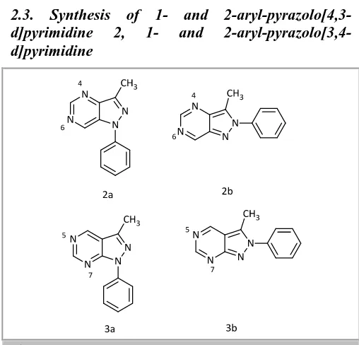
2.3. Synthesis of 1- and
2-aryl-pyrazolo[4,3-d]pyrimidine 2, 1- and
2-aryl-pyrazolo[3,4-d]pyrimidine
Scheme 2. pyrazolo[3,4-d]pyrimidine derivatives
2.3.1. Computational details
The system uses the B3LYP /6-31G (d) [35] computational level. Relatively the calculations, the molecules with minimum energy where λ is the number of negative eigenvalues of the Hessian matrix for a given stationary point) on the Potential Energy Surface.
It was found, that the Relative energies (kJ/mol) of isomers, classified by families (without aza, monoaza, diaza and triaza). Relative energy minima in bold (the compounds of only an isomer calculated have no reltive energy value). The negative values of the last column mean that a are more stable than b.
Table 11. Relative energy of isomers (Kj/mol)
Compound Isomer a Isomer b a vs. B
2 (diaza-4,6) 21.5 1.9 -22.3
3 (diaza-5,7) 0.0 0.0 -41.8
The geometries of compounds in the solid state it is useful to use theoretical calculations in the gas phase.
This allows to have a complete set of related structures and to differentiate the intrinsic effects from crystal field ones [52-55].
None the less , from the present survey, several conclusions can be drawn: i) N internal angles are more reliable than NC distances for structural characterization; ii) 1- and 2-aryl series form two separated sets; iii) a rationale has been found for the N-aryl torsion angles [56].
2.4. Synthesis of
4-Alkylidenehydrazino-1H-pyrazolo[3,4-d]pyrimidines and arylmethylidene
hydrazino-1H-pyrazolo[3,4-d]pyrimidines
4-Alkylidenehydrazino-1H-pyrazolo[3,4-d ]pyrimidin-es, 4-arylmethylidenehydrazino-1H-pyrazolo[3,4-d ]-pyrimidines, and 2-substituted 7H-pyrazolo[4,3-e]-1,2,4-triazolo-[1,5-c]-pyrimidines as potential xanthine oxidase inhibitors were docked into the active site of the bovine milk xanthine dehydrogenase using two scoring functions involved in the CAChe 6.1.10 and Auto Dock 3.05.
Through the docking investigation into the active site of XDH (PDB codes it was obtained Structures of different bicyclic pyrazolo pyrimidines 4–6 and tricyclic Pyrazolo triazolo pyrimidines 7a–s.
Figure 3. b)
and log IC50 of allopurinol, oxipurinol, 6b, e–k, m–r, and 7a–s docked against
Scheme 3. Structure of pyrazolo[3,4-d]pyrimidine derivatives
Table 12. The best docking results based on the binding free energies (ΔGb) of the compounds docked against XDH (PDB
code: 1n5x), the distance and angle of hydrogen bonds between compounds and amino acid residues involved in XDH, and RMSD from the crystallized TEI- 6720 ligand [57].
Compound IC50 (µM)a b (kcal/mol)b Hydrogen bonds between atoms of compounds and amino acids RMSD (A°)d
Atom of compound Amino acid Distance (A° ) Angle ( )
Allopurinol 24.3 -7.39 N8–N HN of Thr 1010 2.05 167.0 5.55
C6–OH OE2 of Glu 802 2.17 155.5
Oxipurinol 22.1 -7.54 C2–Oxo HO of Ser 876 2.31 146.7 3.93
C6–Oxo HN of Thr 1010 2.06 135.4
C6–Oxo Hn(HH22) of Arg 880 1.76 162.8
6a –d -7.72 C
C6–Oxo HN(HH22) of Arg 880 1.87 151.3
6b 4.670 -8.07 N7–H OH of Thr 1010 2.19 138.9 3.90
C6–Oxo HN(HH22) of Arg 880 1.71 146.1
6c – -8.53 C6–Oxo HN(HH22) of Arg 880 1.72 141.5 4.38
6d – -9.23 N2–N HE of Arg 880 1.99 138.0 4.86
N2–N HH22 of Arg 880 1.72 145.8
6f 7.894 -9.34 C6–Oxo HN of Ala 1079 2.21 164.6 2.14
6g 0.305 -9.88 N2–N HN(HE) of Arg 880 2.27 135.9 4.83
N2–N HN(HH22) of Arg 880 1.67 159.1
N1–H OH of Thr 1010 1.84 147.2
6h 0.373 -9.35 N2–N HN Ala 1079 2.07 145.9 2.86
C4–NH OE1 of Glu 802 1.86 136.1
C6–Oxo HN(HH22) of Arg 880 2.03 157.7
C6–Oxo HN of Thr 1010 1.90 141.1
6i 0.077 -9.79 C6–Oxo HN of Thr 1010 2.05 137.3 3.08
C6–Oxo HN(HH22) of Arg 880 1.87 153.8
N2–N HN of Ala 1079 2.19 148.8
6j 0.223 -9.35 N2–N HN Ala 1079 2.27 147.5 3.10
C6–Oxo HN(HH22) of Arg 880 1.83 150.8
6k 0.224 -9.55 N2–N HN Ala 1079 2.22 143.4 2.93
C6–Oxo HN(HH22) of Arg 880 2.06 155.3
C6–Oxo HN of Thr 1010 1.92 141.5
6le >10 -10.39 C
6–Oxo HN of Thr 1010 1.94 138.4 3.09
C6–Oxo HN(HH22) of Arg 880 1.91 162.1
C1–N HN of Ala 1079 2.26 147.0
6m 0.247 -10.34 C6–Oxo HN of Thr 1010 1.96 140.0 2.95
C6–Oxo HN(HH22) of Arg 880 1.96 158.9
C4–NH OE1 of Glu 802 1.74 137.7
N2–N HN of Ala 1079 2.24 146.4
C6–Oxo HN(HH22) of Arg 880 2.05 154.7
6o 0.385 -10.96 C6–Oxo HN of Thr 1010 2.08 143.0 3.04
C6–Oxo HN(HH22) of Arg 880 2.07 152.8
N2–N HN of Ala 1079 1.97 144.6
6p 0.399 -10.55 p–COO HN(HZ1) of Lys 771 1.99 145.2 2.14
C6–Oxo HN(HH22) of Arg 880 1.92 171.0
N2–N HN of Ala 1079 2.23 152.6
6q 0.359 -9.81 C6–Oxo HN of Thr 1010 2.07 143.4 2.82
C6–Oxo HN(HH22) of Arg 880 2.13 150.8
6r 1.925 -10.23 p–N=O HN(HE) of Arg 880 2.14 137.5 1.21
p–N=O HN(HH22) of Arg 880 2.49 139.1
p–N=O HN of Thr 1010 1.71 155.1
p_N+_O- HN(HH22) of Arg 880
6s – -11.85 C6–Oxo HN of Thr 1010 1.88 143.2 2.16
C6–Oxo HN(HH22) of Arg 880 2.12 156.3
N2–N HN of Ala 1079 2.07 143.1
7a 0.184 -8.86 C5–Oxo HH22 of Arg 880 1.85 158.2 5.59
7b 0.250 -9.40 C5–Oxo C5–Oxo HN of Thr 1010 1.96 138.2 5.35
HN(HH22) of Arg 880 1.90 158.6
7c 0.782 -9.33 C5–Oxo HN(HH22) of Arg 880 1.77 153.5 4.82
N8–N HN of Ala 1079 2.24 147.0
7d 0.529 -10.15 C5–Oxo HN(HH22) of Arg 1.83 138.2 4.32
N6–H OH of Thr 1010 1.85 149.9
7e 0.069 -10.84 C5–Oxo HN(HH22) of Arg 880 1.67 152.3 3.49
N8–N HN of Ala 1079 2.33 146.7
7f 0.117 -11.11 C5–Oxo HN(HH22) of Arg 880 1.87 136.2 2.27
N8–N HN of Ala 1079 1.93 145.8
7g 0.103 -10.53 C5–Oxo HN of Thr 1010 1.95 139.8 3.92
C5–Oxo HN(HH22) of Arg 880 1.95 159.9
7h 0.062 -10.68 C5–Oxo HN of Thr 1010 1.96 142.9 3.58
C5–Oxo HN(HH22) of Arg 880 2.06 158.4
7i 0.070 -10.90 C5–Oxo HN of Thr 1010 1.95 140.7 3.75
C5–Oxo HN(HH22) of Arg 880 1.98 160.8
7j 0.038 -11.21 C5–Oxo HN of Thr 1010 1.94 142.5 3.62
C5–Oxo HN(HH22) of Arg 880 2.05 158.6
7k 0.032 -10.97 C5–Oxo HN of Thr 1010 1.99 142.1 3.54
C5–Oxo HN(HH22) of Arg 880 2.04 155.7
7l 0.034 -11.08 C5–Oxo HN of Thr 1010 1.99 140.8 3.55
C5–Oxo HN(HH22) of Arg 880 1.98 156.7
N8–N HN of Ala 1079 2.28 146.5
7m 0.041 -10.98 C5–Oxo HN of Thr 1010 2.00 141.6 3.55
C5–Oxo HN(HH22) of Arg 880 2.02 155.7
N8–N HN of Ala 1079 2.23 146.3
7n 0.053 -10.60 C5–Oxo HN of Thr 1010 1.98 141.9 3.23
C5–Oxo HN(HH22) of Arg 880 2.03 156.4
N8–N HN of Ala 1079 2.27 146.0
7o 0.041 -10.89 C5–Oxo HN of Arg Thr 1010 1.97 144.1 2.88
C5–Oxo HN(HH22) of Arg 2.03 157.3
7q 0.055 -10.49 C5–Oxo HN of Arg Thr 1010 1.98 141.4 3.26
C5–Oxo HN(HH22) of Arg 880 2.01 156.5
N8–N HN of Ala 1079 2.29 146.1
7r 0.060 -11.19 C5–Oxo HN of Thr 1010 1.89 142.8 2.52
C5–Oxo HN(HH22) of Arg 880 2.06 161.4
N8–N HN of Ala 1079 2.34 145.4
7s 0.037 -11.78 N5–H OH of Thr 1010 1.75 140.7 1.30
a Through reference . b Binding free energy. c Root mean square deviation. d Not determined.
e This value is inaccurate because of the insolubility in DMSO.
2.5. Pyrazolo[3,4-d]pyrimidines
Pyrazolo[3,4-d]pyrimidines have a class of naturally occurring fused uracils that appear diverse biological
pyrazolo[3,4-d]pyrimidine molecules considered for docking studies are 1,3-bis(4,6-diethylthio-1H
-pyrazolo[3,4-d]pyrimidin-1-yl)propane [58] 8; 1,3-bis(4-ethoxy-6-methyl-sulfanyl-1H-pyrazolo[3,4-d] pyrimidin-1-yl)propane 9 [59]; 1,10-(1,3-propanediyl)bis(5-methyl-6-methylthio-4,5-Pyrazolo[3,4-d]pyrimidines as inhib-itor of viper Phospholipase A2 421
dihydro-1H-pyrazolo[3,4-d]pyrimidin-4-one) 10 [60]; 1,10-
(1,3-propanediyl)bis(5-ethyl-6-methylthio-4,5-dihydro-1H-pyrazolo[3,4-d]pyrimidin-4-one) 11 [61]; 1,10-(1,3- propanediyl)bis(5-benzyl-6-methylsulfanyl-4,5-dihy-dro-1H-pyrazolo[3,4-d]pyrimidin-4-one) 12
[62]; 1-(4,6-dimethylsulfanyl-1H
-pyrazolo[3,4-d ]pyrimidin-1-yl)-3-(5-methyl-6-methylsulfanyl-4-oxo-1,5-dihydropyra-zolo[3,4-d ]pyrimidin-1-yl)prop-ane 13 [63]; 4,6-bis(methylsulfanyl)-1-phthalimidopropyl-1H-pyrazo-lo[3,4-d]-pyrimidine 14
[64]; 6-methylsulfanyl-1-phthalimidopropyl-4(pyrrolidin-1-yl)-1H-pyrazolo-[3,4-d]pyrimidine 15
[64]; and 6-methylsulfanyl-1-(3-phenylpropyl)-4,5-dihydro-1H-pyrazolo[3,4-d]pyrimidine-4-one 16 [65].
2.5.1. Computational details
The Conformational search and docking studies using AUTODOCK4.2 show that electronic interaction has an important role in ligand–channel interaction. Table 13 show the result. For the best degree of the docked poses, with comparable to the indomethacin binding to PLA2, molecules 8, 9, 11 and 13 have poor binding
energies., molecule 15 has the comparable energy while molecules 10, 12, 14 and 16 have better bound energy values.
Table 13. Inhibition coefficient (KI) and different energy values of the ligands as obtained through the docking with 3H1X using AUTODOCK4.2.
Molecules Rank Kl (μM) Intramolecular
energy
(kcal/mol)
Internal
energy
(kcal/mol)
Torsional
energy
(kcal/mol)
Binding
Energy
(kcal/mol)
8 1-1 74.69 −9.21 0.19 3.58 −5.63
2-1 131.66 −8.87 −0.83 3.58 −5.29
3-1 160.42 −8.76 −0.55 3.58 −5.18
3-2 388.60 −8.23 −0.60 3.58 −4.65
4-1 249.63 −8.49 −0.80 3.58 −4.91
9 1-1 290.50 −7.80 −0.59 2.98 −4.83
2-1 295.25 −7.80 −0.23 2.98 −4.82
3-1 368.95 −7.67 −0.35 2.98 −4.68
3-2 375.14 −7.66 −0.66 2.98 −4.67
4-1 504.85 −7.48 −0.70 2.98 −4.50
10 1-1 9.21 −8.66 −0.12 1.79 −6.87
1-2 10.95 −8.56 −0.14 1.79 −6.77
1-3 20.45 −8.19 −0.20 1.79 −6.40
2-1 10.76 −8.57 0.30 1.79 −6.78
2-2 32.88 −7.91 −0.25 1.79 −6.12
11 1-1 91.64 −7.90 −0.50 2.39 −5.51
2-1 115.41 −7.76 −0.54 2.39 −5.37
2-2 178.43 −7.50 −0.25 2.39 −5.11
3-1 233.02 −7.34 −0.49 2.39 −4.96
4-1 282.48 −7.23 −0.73 2.39 −4.84
12 1-1 18.62 −9.44 −0.74 2.98 −6.45
2-1 34.87 −9.06 −1.44 2.98 −6.08
3-1 79.36 −8.58 −1.11 2.98 −5.59
4-1 84.09 −8.54 −1.29 2.98 −5.56
5-1 142.00 −8.23 −1.11 2.98 −5.25
13 1-1 81.41 −7.67 −0.18 2.09 −5.58
2-1 333.24 −6.83 −0.88 2.09 −4.74
3-1 349.47 −6.80 −0.61 2.09 −4.72
4-1 456.35 −6.65 −0.16 2.09 −4.56
5-1 480.47 −6.62 −0.60 2.09 −4.53
14 1-1 8.56 −8.70 −0.29 1.79 −6.91
1-2 11.47 −8.53 −0.31 1.79 −6.74
1-3 13.87 −8.42 −0.15 1.79 −6.63
2-1 69.00 −7.47 −0.38 1.79 −5.68
3-1 74.44 −7.42 −1.23 1.79 −5.63
15 1-1 27.26 −8.02 −0.63 1.79 −6.23
1-2 38.61 −7.81 −0.82 1.79 −6.02
2-1 118.32 −7.15 −1.50 1.79 −5.36
3-1 128.14 −7.10 −1.36 1.79 −5.31
16 1-1 13.88 −8.12 −0.6 1.49 −6.63
2-1 45.30 −7.42 −0.32 1.49 −5.93
2-2 58.17 −7.27 −0.32 1.49 −5.78
3-1 51.53 −7.34 −0.51 1.49 −5.85
3-2 58.36 −7.27 −0.48 1.49 −5.78
IMN 1-1 22.90 −7.52 −0.66 1.19 −6.33
2-1 24.65 −7.48 −0.70 1.19 −6.29
2-2 27.00 −7.43 −0.68 1.19 −6.23
3-1 27.26 −7.42 −0.66 1.19 −6.23
3-2 32.31 −7.32 −0.69 1.19 −6.13
Table 14. Glide scores and average electrostatic (coul), van der Waals (vdw), site energy (site) and Glide energy obtained through Glide SP docking.
Molecules
(SP)
Entry
ID
Glide
score
Ecoul
(kcal/mol)
Evdw
(kcal/mol) Esite
(kcal/mol)
Glide
energy
(kcal/mol)
8 32 −5.565 −3.270 −48.226 −0.157 −51.496
40 −5.367 −0.968 −49.591 −0.167 −50.559
44 −5.307 −2.830 −48.543 −0.185 −51.372
47 −5.247 −2.134 −49.201 −0.180 −51.335
56 −5.187 −1.967 −48.731 −0.183 −50.698
9 69 −4.856 −2.183 −43.014 −0.163 −45.197
76 −4.791 −3.310 −42.334 −0.193 −45.644
86 −4.694 −2.842 −42.245 −0.204 −45.087
93 −4.565 −3.323 −44.350 −0.142 −47.673
110 −4.073 −4.202 −43.388 −0.170 −47.589
10 8 −6.417 −4.752 −48.490 −0.017 −53.243
13 −5.887 −5.426 −47.934 −0.111 −53.360
14 −5.884 −5.442 −47.828 −0.017 −53.270
24 −5.787 −5.894 −43.697 −0.091 −49.592
25 −5.757 −5.637 −44.583 −0.090 −50.221
11 2 −6.705 −4.248 −50.535 −0.010 −54.783
3 −6.688 −4.189 −50.629 −0.010 −54.819
4 −6.668 −4.200 −50.783 −0.012 −54.983
5 −6.493 −4.304 −50.249 −0.010 −54.554
6 −6.435 −0.814 −52.373 0.0000 −53.187
12 89 −4.636 −2.338 −46.642 0.000 −48.980
100 −4.318 −7.057 −42.092 −0.042 −49.149
102 −4.283 −2.417 −43.022 −0.046 −45.439
103 −4.273 −3.476 −42.849 −0.002 −46.325
105 −4.216 −6.989 −39.835 −0.045 −46.823
13 7 −6.433 −1.882 −52.926 0.0000 −54.809
36 −5.429 −3.008 −43.402 −0.162 −46.410
37 −5.420 −2.630 −43.489 −0.173 −46.119
42 −5.380 −3.307 −41.347 −0.168 −44.655
14 17 −5.822 −3.086 −44.766 −0.018 −47.852
19 −5.818 −2.983 −45.060 −0.018 −48.043
20 −5.817 −2.871 −45.000 −0.017 −47.872
21 −5.815 −2.970 −45.107 −0.018 −48.077
22 −5.812 −2.854 −45.048 −0.017 −47.902
15 15 −5.866 −2.921 −46.758 −0.017 −49.679
34 −5.489 −1.316 −44.899 −0.043 −46.215
92 −4.591 −0.780 −41.055 −0.029 −41.836
94 −4.543 −1.039 −41.186 −0.045 −42.225
97 −4.396 −0.641 −40.911 −0.032 −41.553
16 71 −4.828 −3.552 −31.002 0.000 −34.554
74 −4.819 −3.611 −30.910 0.000 −34.521
75 −4.809 −3.538 −30.922 0.000 −34.460
77 −4.786 −3.340 −31.093 0.000 −34.432
78 −4.776 −3.606 −30.638 0.000 −34.244
IMN 84 −4.709 −8.409 −24.405 −0.087 −32.814
87 −4.672 −8.357 −24.488 −0.088 −32.846
111 −4.064 −6.586 −21.505 −0.100 −28.091
121 −3.243 −5.559 −23.459 −0.100 −29.018
In Table 14, the results of Glide docking in Standard Precision mode are summarized. Docking of Pyrazolo[3, 4-d]pyrimidine ligands and IMN appeared a great variation in their binding energy.
From Autodock calculations, it was noted that molecules 10, 12, 14 and 16 have better binding capabilities and minimum inhibitory concentrations than indomethacin. almost all the
Pyrazolo[3,4-d]pyrimidine molecules have better binding energies than indomethacin by obtaining Docking simulations through Standard Precision and Extra Precision protocols of Glide [16].
2.6. Synthesis of pyrazolo[3,4-d]pyrimidine
derivatives
By using the density functional B3LYP/6-31G** and dispersion corrected density functional DFT-D/B97D methods, the conformational stabilities and computations of optimized geometrical parameters of 1,3-bis(4,6-diethylthio-1H-pyrazolo[3,4-d ]pyrimidin-1-yl)propane 18; 1,3-bis(4-ethoxy-6-methylsulfanyl-1H-pyrazolo[3,4-d]pyrimidin-1-yl)propane 19; 4,6-bis(methylsulfanyl)-1-phthalimidopropyl-1H
-pyrazolo[3,4-d]pyrimidine 20 and 6-methylsulfanyl-1-phthalimidopropyl-4(pyrollidin-1-yl)-1H
-pyrazolo[3,4-d]pyrimidine 21 molecules have been detected.
Geometries at the minimum energies were fully optimized without any constraints. Since B3LYP
method does not include the dispersion term, M06-2X/6-311++G** method is used to define the energies of the B3LYP optimized geometries, which is known to better convenient for non-covalent interactions.
Figure 4. Chemical Structures with atomic numbering scheme of molecules 18, 19, 20 and 21. (Hydrogen atoms have been omitted for clarity. Torsion angles chosen for conformational variations are N1-C8-C9-C10 (τ1) and C8-C9-C10-N11
(τ2).
2.6.1. Computational details
Table 15a. Total energies (E), relative energies (ΔE) and torsion angles (τ1, τ2) corresponding to different conformations of
molecule 18. The crystallographic value of torsion angles are (-52.26, -52.26o).
Conf.
(mol.18)
E (Hartree) ΔE (kcal/mol) τ 1 τ 2
B3LYP M06-2X B97D B3LYP M06-2X B97D B3LYP/M06-2X B97D B3LYP/M06-2X B97D
I -2847.8947 -2847.67026 -2847.0021 0.02 4.78 7.26 66.6 62.0 176.9 186.2
II -2847.8946 -2847.67008 -2847.0020 0.04 4.89 7.29 177.3 -173.1 67.7 61.7
III -2847.8935 -2847.66981 -2846.9958 0.73 5.06 11.18 -179.5 -172.7 -65.7 -60.1
IV -2847.8935 -2847.66968 -2846.9958 0.75 5.14 11.15 -66.6 -61.4 179.4 186.4
V -2847.8947 -2847.67787 -2847.0136 0.00 0.00 0.00 -56.7 -49.4 -56.5 -49.5
Table 15b. Total energies (E), relative energies (ΔE) and torsion angles (τ1, τ2) corresponding to different conformations of
molecule 19. The crystallographic value of torsion angles is (47.9o, 54.2o).
Conf.
(mol.19)
E (Hartree) ΔE (kcal/mol) τ 1 τ 2
B3LYP M06-2X B97D B3LYP
M06-2X
B97D
B3LYP/M06-2X
B97D
B3LYP/M06-2X
B97D
I -2123.3325 -2123.13605 -2122.4071 21.13 5.49 11.63 66.0 56.3 179.8 166.5
II -2123.3325 -2123.13610 -2122.4071 21.13 5.45 11.63 179.7 167.2 66.0 57.0
III -2123.3662 -2123.13637 -2122.4116 0.00 6.97 8.75 183.3 171.9 -66.9 -63.2
IV -2123.3662 -2123.13635 -2122.4117 0.00 5.29 8.73 -66.9 -62.6 -176.5 172.1
V -2123.3335 -2123.14479 -2122.4256 20.51 0.00 0.00 54.0 49.1 54.4 48.6
Table 15c. Total energies (E), relative energies (ΔE) and torsion angles (τ1, τ2) corresponding to different conformations of
molecule 20.The crystallographic value of torsion angles are (167.3o, 69.8o).
Conf.
(mol. 20)
E (Hartree) ΔE (kcal/mol) τ 1 τ 2
B3LYP M06-2X B97D B3LYP
M06-2X
B97D
B3LYP/M06-2X
B97D
B3LYP/M06-2X
B97D
I -1916.7796 -1916.62037 -1915.9895 0.08 0.58 8.82 67.1 62.4 177.2 -174.7
II -1916.7801 -1916.62129 -1915.9936 0.00 0.00 6.27 177.0 -162.8 66.6 68.4
III -1916.7795 -1916.62078 -1916.0036 0.17 0.27 0.00 179.4 55.3 -66.9 -80.3
IV -1916.7793 -1916.62104 -1915.9901 0.28 0.16 8.45 -64.8 -61.9 -176.9 -175.7
V -1916.7797 -1916.62118 -1916.0007 0.01 0.07 1.84 -57.9 -49.1 -58.4 -49.5
Table 15d. Total energies (E), relative energies (ΔE) and torsion angles (τ1, τ2) corresponding to different conformations of
molecule 21. The crystallographic value of torsion angles are (67.3o, 169.3o).
Conf. (mol.21)
E (Hartree) ΔE (kcal/mol) τ 1 τ 2
B3LYP M06-2X B97D B3LYP
M06-2X
B97D B3LYP/M06-2X
B97D B3LYP/M06-2X B97D
I -1690.6875 -1690.50018 -1689.7786 15.50 1.08 6.92 67.2 63.3 178.9 184.9
II -1690.6873 -1690.50047 -1689.7808 15.63 0.90 5.56 177.3 -147.3 65.9 68.6
III -1690.6868 -1690.50004 -1689.7779 15.90 1.17 7.38 177.7 179.3 -68.5 -64.5
IV -1690.6873 -1690.50081 -1689.7790 15.65 0.68 6.66 -65.8 -62.8 178.3 179.8
Figure 5. Different conformers of the molecules obtained from B3LYP/6-31G** method.
Table 16. Comparison of crystallographic and theoretical values (obtained from B3LYP and B97D methods) of some selected torsion angles of molecule 21 in different conformers
Torsions Conformers
Cyst. Values B3LYP B97D
I II III IV V I II III IV V
N1-C8-C9-C10 (τ 1) 67.3(4) 67.2 177.3 177.7 -65.8 -64.8 63.3 -147.3 179.2 -62.8 -70.1
C8-C9-C10-N11 (τ2) 169.3(3) 178.9 65.9 -68.5 178.3 75.6 184.9 68.6 -64.5 179.8 65.5
N5-C4-N18-C19 176.1(3) 177.2 175.8 176.3 176.9 177.6 177.6 176.6 176.9 177.3 176.2
C4-N18-C19-C20 -165.7(4) -170.2 -168.2 -168.7 -169.5 -170.3 -170.1 -169.0 -169.4 -169.4 -170.3
N18-C19-C20-C21 -13.5(6) -29.1 -29.1 -29.2 -29.3 -29.0 -30.1 -29.9 -30.0 -30.3 -29.6
N5-C4-N18-C22 4.5(4) -3.0 -2.4 -2.6 -3.0 -2.8 -3.2 -2.6 -2.9 -2.9 -4.1
C19-C20-C21-C22 15.5(7) 37.4 37.4 37.4 37.4 37.3 38.5 38.6 38.6 38.7 38.4
C20-C21-C22-N18 -10.7(6) -30.9 -30.9 -30.8 -30.8 -30.8 -31.7 -31.9 -31.9 -31.7 -31.9
C21-C22-N18-C19 1.8(4) 13.2 13.1 13.0 12.9 13.1 13.4 13.8 13.7 13.3 13.9
C21-C22-N18-C4 174.3(3) -166.7 -168.5 -167.9 -167.2 -166.5 -165.9 -166.9 -166.6 -166.5 -165.8
C22-N18-C19-C20 6.7(4) 10.1 10.1 10.3 10.4 10.1 10.6 10.2 10.4 10.8 9.9
Different theoretical strategies have exhibited that all the four pyrazolo[3,4-d]pyrimidine derivatives have five theoretically possible stable conformers of about equivalent energies.
For molecule, both theoretical methods B3LYP and B97D, utilized to examine the conformational preferences, do not agree consistently while distinguishing the global minimum.
The ᴨ-ᴨ interaction energy in the state of molecules
18 and 19 has values 4.78 kcal/mol and 5.49 kcal/mol respectively. In instances of molecules 20 and 21 all the conformers are practically broadened and have approximately same energy which indicates that intermolecular ᴨ-ᴨ interaction is missing in these particles [66].
2.7. Synthesis of 1-(2-ethoxyethyl)-1H-pyrazolo[4,3-d]pyrimidines
Phosphodiesterase V works as an appealing target for cardiovascular, Quantitative Structure–Activity Relationship (QSAR) consider as useful procedure for the optimization of structure necessities of twenty-six 1-(2-ethoxyethyl)-1H-pyrazolo[4,3-d]pyrimidines as phosphodiesterase V inhibitors.
2.7.1. The structural template of 1-(2-ethoxyethyl)-1H-pyrazolo[4,3-d]pyrimidine
Scheme 5. 1-(2-ethoxyethyl)-1H-pyrazolo[4,3-d]pyrimidines structure
Various quantum chemical methods and quantum-chemistry calculations have been performed for studying the molecular structure and electronic 1-(2-ethoxyethyl)-1H-pyrazolo[4,3-d]pyrimidine derivate-ives properties [67,68]. The properties of biological Activity of organic compounds related to the geometry and additionally the nature of their molecular orbital, HOMO (highest occupied molecular orbital) and LUMO (lowest unoccupied molecular orbital).
In the next Table (Table 19), we summarized The Pearson correlation coefficients. The obtained matrix provides Information on the negative or positive correlation between variables.
Table 18. Values of the calculated parameters obtained by DFT/B3LYP 6-31G* optimization and ACD/ChemSketch program of the studied compounds.
N0 pIC
50 MW MR
(cm3)
MV (cm3)
Pc (cm3)
n αe
(dyne /cm)
D (g/ cm3)
ET
(ua)
EHomo
(ev)
ELum o
(ev) ∆E (ev)
µ (D)
Ea
(ev)
λmax
(nm) f(so)
22 a 1,155 395,5 113,33 306,3 817,1 1,661 50,6 1,29 -34750 -5,096 -1,09 4,006 2,48 2,214 560,1 0,0139
22 b 0,398 382,46 107,17 278,8 759,7 1,694 55 1,37 -33052,9 -5,511 -1,58 3,935 2,53 3,499 354,4 0,0024
22 c 1,155 396,49 111,77 294,9 798,3 1,682 53,6 1,34 -35186,6 -5,007 -1,07 3,934 3,53 2,149 576,8 0,0134
22 d 0,187 396,49 111,59 294 790,8 1,683 52,3 1,34 -35194,8 -5,141 -1,24 3,903 3,86 3,072 403,6 0,0244
22 e 0,18 410,52 116,2 310,1 829,4 1,672 51,1 1,32 -36257,6 -5,195 -1,22 3,975 1,30 3,472 357,1 0,0006
22 f -0,146 396,49 111,59 294 790,8 1,683 52,3 1,34 -35194,6 -5,189 -1,31 3,881 5,29 3,561 348,2 0,002
22 g 0,420 410,52 116,2 310,1 829,4 1,672 51,1 1,32 -36265,3 -5,202 -1,32 3,881 5,38 3,065 404,6 0,0216
22 h -0,519 410,52 116,2 310,1 829,4 1,672 51,1 1,32 -36257,5 -5,182 -1,22 3,964 2,8 2,112 587,1 0,0156
22 i -0,74 410,52 116,2 310,1 829,4 1,672 51,1 1,32 -36257,5 -5,005 -0,98 4,028 2,34 3,253 381,2 0,0694
22 j -0,415 410,51 116,92 315,3 836,2 1,663 49,4 1,3 -36257,7 -5,004 -1,08 3,926 4,43 3,554 348,8 0,0021
22 k 0,051 424,54 121,52 331,3 874,8 1,654 48,5 1,28 -37328,2 -5,21 -1,25 3,957 1,89 2,761 449,1 0,0083
22 l -0,398 424,54 120,62 325,2 860,5 1,663 48,9 1,30 -37328,4 -5,153 -1,21 3,942 1,40 3,494 354,8 0,0024
22 m -0,079 424,54 120,62 325,2 860,5 1,663 48,9 1,30 -37328 -4,904 -0,96 3,939 4,73 3,549 349,3 0,006
22 n 0,119 424,54 120,81 326,1 868 1,662 50,1 1,30 -37328,1 -4,915 -1,09 3,825 4,01 3,406 364,0 0,0577
22 o 0,699 424,54 119,91 320 853,6 1,672 50,6 1,32 -37328,1 -5,213 -1,25 3,964 2,41 3,384 366,4 0,0611
22 p 0,143 438,57 125,23 341,3 899,1 1,654 48,1 1,28 -38398,6 -5,059 -1,05 4,013 4,44 3,221 384,9 0,0236
22 q 1,060 452,6 130,56 362,5 944,5 1,639 46 1,24 -39468,7 -4,900 -0,99 3,91 4,79 2,633 470,8 0,0055
22 r 0,377 424,54 120,62 325,2 860,5 1,663 48,9 1,30 -36257,9 -5,028 -1,09 3,94 5,6 3,737 331,8 0,0463
22 t 0,108 424,54 119,67 318,2 855,4 1,675 52,2 1,33 -37328,4 -5,008 -1,09 3,916 3,48 3,742 331,4 0,0754
22 u 0,456 424,54 119,91 320 853,6 1,672 50,6 1,32 -37328,1 -5,038 -1,07 3,97 4,77 3,326 372,8 0,0545
22 v 1,155 424,54 119,67 318,2 855,4 1,675 52,2 1,33 -37310,3 -4,846 -1,00 3,843 4,08 3,754 330,3 0,062
22 w -0,079 411,5 113,63 305,8 814,4 1,665 50,3 1,34 -36798,6 -5,112 -1,12 3,996 3,55 4,051 306,1 0,0659
22 x 1,155 439,55 123,21 334,7 892,5 1,657 50,5 1,31 -38948 -5,092 -1,28 3,812 5,27 3,448 359,6 0,0635
22 y 1,097 452,55 125,13 331,5 899,7 1,679 54,2 1,36 -40414,7 -4,859 -0,92 3,939 5,85 1,932 641,7 0,0078
22 z 1,398 453,54 124,07 332 898,2 1,67 53,5 1,36 -40956,1 -5,079 -1,12 3,956 5,43 3,319 373,6 0,0197
Table 19. Correlation matrix (Pearson (n)) between different obtained descriptors:
pIC50 MW MR MV Pc N αe D ET EHOMO ELUMO E µ Ea λmax f(so)
pIC50 1
MW 0,367 1
MR 0,301 0,962 1
MV 0,270 0,917 0,984 1
Pc 0,344 0,962 0,995 0,987 1
N -0,096 -0,577 -0,720 -0,831 -0,746 1
0,197 -0,360 -0,555 -0,674 -0,547 0,905 1
D 0,059 -0,317 -0,547 -0,668 -0,551 0,898 0,942 1
ET -0,374 -0,974 -0,897 -0,843 -0,904 0,496 0,246 0,194 1
EHOMO 0,192 0,546 0,563 0,532 0,556 -0,342 -0,235 -0,271 -0,540 1
ELUMO 0,088 0,504 0,523 0,503 0,515 -0,355 -0,273 -0,284 -0,501 0,928 1
E -0,273 -0,110 -0,104 -0,077 -0,107 -0,036 -0,098 -0,035 0,101 -0,189 0,191 1
µ 0,292 0,392 0,306 0,245 0,298 0,007 0,097 0,134 -0,397 0,452 0,300 -0,400 1
Ea -0,311 -0,047 -0,085 -0,084 -0,115 0,047 -0,103 0,104 0,082 -0,092 -0,161 -0,180 0,053 1
Λmax 0,317 0,046 0,061 0,051 0,091 0,001 0,161 -0,029 -0,087 0,134 0,204 0,184 -0,031 -0,984 1
f(so) 0,070 0,169 0,126 0,110 0,125 -0,068 -0,026 0,045 -0,151 0,241 0,190 -0,132 0,021 0,400 -0,365 1
The Energy of activation Ea is negatively correlated with the maximum of absorption λ max (r = -0.984 and p <0.05) at a significant level.
The Density D is correlated positively with the surface tension αe (r =0.942 and p< 0.001) at a significant
level. The analysis of projections is according to the planes F1–F2 and F1-F3 (59.53% and 58.28% of the total variance respectively) of the studied molecules [69].
2.8. Synthesis of 3-phenyl-1-tosyl-1H-pyrazolo[3,4-d]pyrimidin-4-amine
Using Hartree Fock (HF/6-31G**) and density functional theory (B3LYP/6-31G**) techniques to obtain the electronic structure calculations and vibrational assignments of a biologically important molecule namely, 3- phenyl-1-tosyl-1H
-pyrazolo[3,4-d]pyrimidin-4-amine. The optimized geometrical parameters, molecular electrostatic potentials (MEP), and HOMO–LUMO gaps as acquired through the two techniques are analysed.
6-31G** basis set using Gaussian03, shown in the following Figure 9.
Figure 7. Correlation circle
Figure 8. Cartesian diagram according to F1-F2.
Figure 9. Optimized geometry of the 3-phenyl-1-tosyl-1H-pyrazolo[3,4-d]pyrimidine-4-amine at HF/6-31G** and B3LYP/6 − 31G** level".
2.8.1. Computational details
From two methods have been compared and are described in Table 20, Bond lengths and bond angles of the optimized structures as obtained.
Utilizing HF and DFT methods to detect Total and HOMO–LUMO energies of the 3-phenyl-1-tosyl-1H -pyrazolo [3,4-d]pyrimidin-4-amine. The HOMO and LUMO energies gap are 0.18837 a.u. by HF and0.17687 a.u. by DFT method, which is very small, and it indicates that 3-phenyl-1-tosyl-1H-pyrazolo [3,4-d]pyrimidin-4-amine is chemically highly reactive and biologically active for charge transfer intermolecular (Table 21).
On the premise of vibrational analysis at B3LYP/6-3G** and HF/6-31G** levels, various thermodynamic parameters are computed and are introduced in Table 22. We calculate the zero point vibrational energies (ZPVE), rotational constants, thermal energy, heat capacity and the entropy Svib (T) to the degree of
accuracy it is also clear from Table 22 that the rotational constant is directly proportional to rotational temperature. The rotational constant is inversely proportional to the moment of inertia of the molecule [70].
Theoretically computed vibrational (IR) frequencies of 3-phenyl-1-tosyl-1H-pyrazolo[3,4-d ]pyrimidin-4-amine have been contrasted with the experimental observations [71] which is delineated in Table 23
[72].
Table 20. Comparison of calculated bond lengths (Å) and bond angles (°) using Gaussian03
Bond length (Å) Bond angle (°) Dihedral angle (°)
Parameter HF DFT Parameter HF DFT Parameter HF DFT
C3–C2 1.386 1.408 C2–C3–C4 115.3 114.7 C2–N3–S–O2 −171.9 −172.5
C4–N1 1.326 1.324 C2–N3–N4 111.2 111.9 C3–C2–N3–N4 −1.1 −0.3
C5–N4 1.284 1.476 C3–C5–C6 129.2 130.0 C3–C2–N3–S −180.0 176.9
C6–C5 1.483 1.405 C5–C6–C11 120.6 121.1 C4–C3–C5–C6 5.8 6.5
C6–C11 1.391 1.404 C13–C12–C17 121.2 121.7 C4–C3–C5–N4 −174.1 −173.3
C9–C10 1.385 1.395 N1–C4–C3 119.0 119.6 C4–C3–C2–N2 −1.3 −2.2
C10–C11 1.386 1.784 N1–C4–N5 117.1 116.7 C4–C3–C2–N3 176.5 175.5
C12–S 1.760 1.784 N3–N4–C5 107.4 106.7 C5–N4–N3–C2 1.4 0.7
C14–C13 1.307 1.393 N3–S–O1 106.1 105.4 C7–C6–C11–C10 0.7 1.2
C15–C14 1.393 1.402 N4–C5–C6 120.3 119.6 C7–C6–C5–C3 −132.6 −141.0
C15–C16 1.390 1.403 N4–N3–S 120.1 119.6 C7–C6–C5–N4 47.2 38.8
N3–S 1.695 1.762 O1–S–O2 121.9 122.9 C11–C6–C5–C3 48.9 40.7
S–O2 1.419 1.455 S–C12–C13 119.2 118.8 C13–C12–S–N3 90.1 97.9
S–O1 .420 1.456 C13–C12–S–O2 −21.9 −13.2
N5–C4 1.341 1.356 C13–C12–S–O1 −157.2 −150.5
C14–C13–C12–S 179.6 178.8
C17–C12–S–O2 157.4 165.5
C17–C12–S–O1 22.1 28.2
C18–C15–C14–C13 −178.5 180.0
C18–C15–C16–C17 178.5 180.0
N2–C2–N3–N4 176.6 177.4
N2–C2–N3–S −2.3 −5.4
N4–N3–S–C12 −105.3 −109.8
N4–N3–S–O2 9.3 4.5
N4–N3–S–O1 139.8 135.6
Table 21. Total and HOMO–LUMO energies of the 3-phenyl-1-tosyl-1H-pyrazolo [3,4-d]pyrimidin-4-amine obtained from HF and DFT methods
HF DFT
Total energy (a.u.) −1509.8034 −1517.3056
Dipole moment (debye) 7.8696 6.7696
HOMO (a.u.) −0.2339 −0.2279
LUMO (a.u.) −0.0455 −0.0511
HOMO–LUMO energy gap(a.u) 0.1884 0.1768
Table 22. Theoretically computed thermodynamical parameters of 3-phenyl-1-tosyl-1H-pyrazolo[3,4-d] pyrimidine-4-amine.
Thermodynamical parameters HF DFT
Zero-point vibrational energy (kcal/mol) 209.6500 194.91907
Rotational temperature (K) 0.01493 0.01490
0.00592 0.00574
0.00531 0.00512
Rotational constants(GHz) 0.31114 0.31050
0.12334 0.11959
0.11060 0.10665
Energy(kcal/mol)
Total 222.472 208.698
Rotational 0.889 0.889
Vibrational 220.694 206.921
Molecular capacity at constant volume(cal/mol-K)
Total 78.176 84.769
Translational 2.981 2.981
Rotational 2.981 2.981
Vibrational 72.215 78.807
Entropy (cal/mol-Kelvin)
Total 156.154 161.142
Translational 43.578 43.578
Rotational 35.581 35.650
Vibrational 76.995 81.915
Table 23. Vibrational assignments of fundamental frequencies (cm−1) for 3-phenyl-1-tosyl-1H-pyrazolo[3,4-d] pyrimidin-4-amine at B3LYP/6–311 + G level.
Experimental
frequencies
Theoretical
frequency
Intensity
(km/mol)
Vibrational
Assignments
411 416.10 0.102 Ring out of plane
422.39 6.447 Deformation
430.01 2.932
474 511.51 1.080 C–H out of plane bending
527 530.80 81.554
551 547.35 28.531
637 566.81 263.589
628.03 3.476
693 694.60 21.253 CCC out-of-plane bending
710.96 6.531
776 772.58 22.985 C–H out of plane
807 784.40 33.450 Deformation
824 816.55 16.038
818.62 9.968
828.25 24.525
1003 1010.94 23.630 Inter ring C–C stretching
1019 1016.50 0.445 C–C stretching
1061 1064.67 11.928
1121 1125.82 104.333 In-plane ring HCC bending
1179 1146.28 4.568
1157.45 166.133
1215 1208.88 3.510 C–H in-plane bending
1265 1215.25 6.612
1235.35 1.639
1306 1319.98 32.032 N–N stretching
1339 1334.05 28.455 C–N stretching
1350.16 20.809
1396.57 6.135
1443.37 10.438
1466 1443.37 10.438 C–C stretching
1478 1483.85 3.714
1627.16 9.199
1633.95 1.896
1674 1663.74 418.137 NH2 scissoring
3032 3042.48 22.488 Aromatic C–H stretching
3070 3104.35 14.758
3125 3132.14 11.917
3180.85 28.780
3182.30 1.393
3187.92 14.356
3190.31 4.856
3208.22 21.772
3218.57 5.300
3226.66 1.594
3233.78 5.604
3472 3600 85.002 NH2 asymmetric stretching
3736.84 91.085 NH2 symmetric stretching
Scheme 6. Synthesis of 6-methyl-4-APP based carbocyclic nucleoside analogs 26a-e.
2.9. Synthesis of 4-amino-pyrazolo[3,4-d]pyrimidine; 6-methyl-4-APP; 6- methyl-4-amino-pyrazolo[3,4-d]pyrimidine
Compound 25 a-d obtained by coupling of compound
23 a-d with compound 24 (Mitsunobu coupling).
2.9.1. Computational details
The structures were limited utilizing Macro model with Optimized Potentials for Liquid Simulation. Spin restricted DFT with B3LYP (Becke-3-Lee-Yang-Parr) density functional and the 6–31-G∗∗ basis set were
The molecularmechanics (MM) energy optimization showed that the potential energy of the N2 is higher than the corresponding N1 isomer as shown in Figure 10.
Figure 10. MM energy analyses showing the N1-regioisomer to have lower energy than N2-isomer.
The quantum mechanics (QM) computation for the activation energy for the formation of 25a (N1) and
25a (N2) were done. The result demonstrates that the transition state for N2 higher in energy than that of N1 is 0.0016 Ha (4.27 kJ/mol) (schematic representation in Figure 11).
The relative energy of is N2 isomer higher than that of N1 isomer. Thus, QM calculation likewise supports the formation of N1 product [73].
2.10. Synthesis of fluorine-substituted pyrazolo[3,4-d]pyrimidine derivative
New fluorine-substituted pyrazolo[3,4-d]pyrimidine derivatives 29–31 obtained by the reaction of (4-fluorophenyl)malanonitrile 27 with guanidine, then forming cyclization reaction with hydrazine producing 2,4-diamino-6-aryl-pyrimidine-5-carbonitrile 28.
Scheme 7 describe fluorinated
pyrazolo[3,4-d]pyrimidine derivatives 30 and 31 that produced
from fluorinated acylation and/or benzeylation of compound 29 by warming with trifluoroacetamide and/or 4-fluorobeneyl chloride in acetic acid or dry C6H6-TEA.
HOMO, LUMO, energy gap, and the optimized geometric structure of the studied compounds 27-31,
by B3LYP/6-311 + G** theory level, introduced in Table24 and Figure12.
At Figure 12, The LUMO is delocalized among all the atoms for all molecules, except compounds 30 and 31. But, the HOMOs are localized on the nitrogen atoms, except in compounds 27 and 31.
2.10.1. Synthesis of fluorine-substituted
pyrazolo[3,4-d]pyrimidine derivative
The computational calculation of compounds 27-31
was performed by using SPARTAN'08 Windows graphical software with density functional theory, DFT/6-31G* basis set [74].
We find theoretically that the compound with lower LUMO value and higher density value has shown the highest activity. The theoretical results were found in good corroboration with the experimental results. Structure activity relationship studies show that fluorinesubstituted derivatives play an important role in antimicrobial activity [75].
3.
Synthesis and computational studies of pyrazolo[1,5-a]pyrimidine3.1.Synthesis and computational studies of
pyrazolo[1,5-a]pyrimidine
Through a dimroth rearrangement, in quantitative yields, Derivatives of the new ring system Pyrazolo[3,4-d] [1,2,3]triazolo[1,5-a]pyrimidine were synthesized from the comparing angular isomers. This class of compounds could be the best candidate as DNA intercalating agents as explained by Preliminary computational studies.
Scheme 7. Synthesis of fluorin-substituted pyrazolo[3,4-d]pyrimidine derivatives.


![Figure 9. Optimized geometry of the 3-phenyl-1-tosyl-1H-pyrazolo[3,4-d]pyrimidine-4-amine at HF/6-31G** and B3LYP/6 − 31G** level"](https://thumb-us.123doks.com/thumbv2/123dok_us/8459395.1707897/24.595.56.397.69.521/figure-optimized-geometry-phenyl-tosyl-pyrazolo-pyrimidine-amine.webp)
![Table 23. Vibrational assignments of fundamental frequencies (cm−1) for 3-phenyl-1-tosyl-1H-pyrazolo[3,4-d] pyrimidin-4-amine at B3LYP/6–311 + G level](https://thumb-us.123doks.com/thumbv2/123dok_us/8459395.1707897/26.595.46.544.305.785/table-vibrational-assignments-fundamental-frequencies-phenyl-pyrazolo-pyrimidin.webp)

