International Journal of Medical Science and Current Research (IJMSCR)
Available online at: www.ijmscr.com
Volume2, Issue 2,Page No: 133-138
March-April 2019
133
Medicine ID-101739732
Surgical Management of External Cervical Root Resorption with Biodentine
A Case Report
AKILAN B1, VINODHINI V2, GOKULRAJ R3, SWATI BHOSALE4
1(Department of conservative dentistry and endodontics/ IRT Perundurai Medical College and Hospital, India)
*Corresponding Author:
AKILAN B
(Department of conservative dentistry and endodontics/ IRT Perundurai Medical College and Hospital, India)
Type of Publication: Original Research Paper Conflicts of Interest: Nil
ABSTRACT
Cervical invasive root resorption is a type of external inflammatory root resorption that is relatively uncommon and aggressive, and leads to loss of tooth structure. A diagnosis of cervical invasive root resorption depends on careful routine clinical and radiographic examinations. This case report describes the diagnosis and successful treatment of a trauma induced invasive cervical root resorption of maxillary left lateral incisor in 23 -year-old female patient. The defect was surgically repaired using Biodentine to restore the resorption cavity. Biodentine is new bioactive cement with dentin - like mechanical properties, which can be used as a dentin substitute on crowns and roots. Its use in roots includes managing perforations of root canals or the pulpal floor, internal and external resorption, apexification and retrograde root canal obturation. Biodentine cement simplifies clinical procedures due to its handling properties. The 12-month recall revealed good healing of both the periodontal and periradicular conditions and no obvious clinical symptoms.
Keywords: Biodentine, Invasive cervical resorption, trauma, tricalcium silicate based cement
INTRODUCTION
Root resorption is the loss of hard dental tissue (i.e., cementum and dentin) as a result of odontoclastic action(1).Tooth resorption is a common occurrence after injuries or irritation of the periodontal ligament or pulp. Root resorption might be classified by its location in relation to the root surface, ie, internal or external resorption(2).External root resorption is a progressive and destructive loss of tooth structure, initiated by a mineralized or denuded area of the root surface(3).It takes place in both vital and nonvital teeth. External tooth resorption can be further classified into surface resorption, external inflammatory resorption, external replacement resorption, external cervical resorption, and transient apical breakdown.
Heithersay(4) coined the term Invasive cervical resorption (ICR), which is a type of external
inflammatory root resorption(2). ICR is defined as ‘‘a localized resorptive process that commences on the surface of the root below the epithelial attachment and the coronal aspect of the supporting alveolar process, namely the zone of the connective tissue attachment’’(3).
Several etiologic factors such as dental trauma, orthodontic treatment, intracoronal bleaching, periodontal therapy, and idiopathic etiologyhave been suggested.
Pag
e
134
Pag
e
134
Pag
e
134
Pag
e
134
Pag
e
134
Pag
e
134
Pag
e
134
Pag
e
134
Pag
e
134
Pag
e
134
Pag
e
134
Pag
e
134
Pag
e
134
Pag
e
134
Pag
e
134
Pag
e
134
Pag
e
134
Pag
e
134
Pag
e
134
Pag
e
134
Pag
e
134
process(4). The resorption process starts just below the gingival epithelial attachment of the tooth, extending apically and/or coronally along the root dentin although a pink discoloration of the crown indicates the resorptive process, some teeth provide no visual signs of ICR.
ICR is mostly identified during routine radiographic or clinical examination because the majority of cases are asymptomatic. The lesion classically presents as an asymmetrical radiolucency with ragged or irregular margins in cervical region of the tooth(5). Unless proper treatment is initiated, this type of resorption continues, and a large irreversible loss of tooth structure may appear in time(1).
Different approaches have been suggested for the treatment of ICR. The nonsurgical treatment involved the topical application of a 90% aqueous solution of trichloracetic acid to the resorptive tissue, curettage, endodontic treatment. The surgical treatment of varying degrees of ICR has generally involved periodontal flap reflection, curettage and restoration of the defect with composite resin , glass ionomer cement (GIC)(4) , resin-modified glass ionomer (RMGIC) , or mineral trioxide aggregate (MTA)(6).
This case report presents the surgical treatment of ICR which is a possible late complication of trauma in the maxillary left lateral incisor,so asto reinforce the weakened tooth structure caused by resorption, the defect in this case was managed with Biodentine.
CASE REPORT
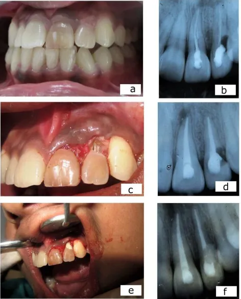
A 23-year-old female patient was referred to the Department of Conservative Dentistry and Endodontics with a complaint of discoloured upper left front tooth. The tooth was asymptomatic except bleeding from gingiva and the patient gave a history of a traumatic injury in front region 12 years back followed by endodontic treatment 2 years later(figure b). Medical history was non-contributory, and an intraoral examination revealed discoloured #9 and #10 and also the distal half of cervical one third #10 had pinkish discolouration(figure a). Periodontal probing depths were physiological (<3 mm) at all sites except for the distal surface of left lateral incisor in which copious bleeding and periodontal pockets (6 mm) was found , Periapical radiograph of tooth revealed previous endodontic treatment of tooth #9 and #10, presence of periapical radiolucency in
relation to tooth #9,tooth # 10 revealed presence of well-demarcated area of radiolucency at the cementoenamel junction invading the coronal dentin, with extension into the radicular dentin in its distal aspect and discontinuity in the lamina dura.A provisional diagnosis of apical inflammatory resorption and ICR was made associated with #10. Endodontic retreatment followed by surgery and restoration of the defect was planned.
Informed consent was obtained fromthepatient. After oral prophylaxis, under local anesthesia of 2% lidocaine with 1:100,000 adrenaline (LOX 2%; Neon Laboratories Ltd, Mumbai, India). A rubber dam was placed and access to the pulp chamber was performed using Endo access bur no. A0164 and slow-speed diamond KGS 3203 (Dentsply Maillefer) in tooth #9 and #10. Gutta-percha was removed using rotary files. Working length was established with the use of an apex locator (Root ZX, J. Mortina Inc, USA) and confirmed by a radiograph. The canal was prepared to size 60 by using K-files. Intracanal irrigation was performed with 2% chlorhexidine and sterile saline alternatively and a final rinse with 17% EDTA. The canal system was dried with absorbent paper points and obturated by the warm vertical condensation technique with gutta-percha and AH plus sealer (Dentsply Maillefer) (figure d). The entrance filling for coronal seal was given with GIC (Fuji II, GC Corporation, Tokyo, Japan).
Pag
e
135
Pag
e
135
Pag
e
13
5
Pag
e
135
Pag
e
135
Pag
e
135
Pag
e
135
Pag
e
135
Pag
e
135
Pag
e
135
Pag
e
135
Pag
e
135
Pag
e
135
Pag
e
135
Pag
e
135
Pag
e
135
Pag
e
135
Pag
e
135
Pag
e
135
Pag
e
135
Pag
e
135
ethylene diamine tetraacetic acid (EDTA) (Prime Dental product Limited, India)(7) because of property of organic tissue fixation and antibacterial efficacy of NaOCl and some regenerative potential of EDTA -1 from human dentin (to
improve clinical outcomes)(8). After isolation of the
surgical site, Biodentine capsule (Septodont, Saint-Maurdes Fosses, France) was mixed according to manufacturer instructions to creamy consistency. Defect was filled with the mix and any excess material was stripped off with a curette in the same
session. Once the material had set, the hardened material was shaped with a bur to a smooth finish (figure e). The flap was repositioned without tension and sutured interproximally with nonabsorbable sutures (Silk; Dogsan, Trabzon, Turkey).Immediate post-surgical radiograph was taken to evaluate approximation of biodentine to the defect (figure f). Postoperative medications and instructions are given. One week after the surgery, the sutures were removed. The healing was uneventful and the patient was asymptomatic.
Figure a - clinical photograph showing discoloured tooth #9 and 10 and pinkish discoloration in cervical aspect of tooth # 10.
Figure b – preoperative radiograph showing inadequate obturation and presence of a radiolucent lesion in cervical aspect of tooth #10.
Figure c – surgical exploration confirming the presence of invasive cervical root resorption in tooth # 10.
Figure d – postobturation radiograph showing endodontic retreatment in tooth # 9 and 10.
Figure e – restoration of resorptive defect with Biodentine.
Figure f –postoperative radiograph showing resorptive defect restored with Biodentine (radiopaque).
Pag
e
136
Pag
e
136
Pag
e
136
Pag
e
136
Pag
e
136
Pag
e
136
Pag
e
136
Pag
e
136
Pag
e
136
Pag
e
136
Pag
e
136
Pag
e
136
Pag
e
136
Pag
e
136
Pag
e
136
Pag
e
136
Pag
e
136
Pag
e
136
Pag
e
136
Pag
e
136
Pag
e
136
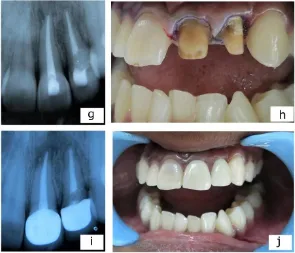
Figure g – 6 months post operative radiograph.
Figure h – full crown preparation done in tooth # 9 and 10.
Figure i – 12 months follow up radiograph showing absence of any recurrence of resorption.
Figure j – 12 months follow up clinical photograph showing complete gingival healing and no sign of loss of clinical attachment concluding favourable tissue response.
DISCUSSION
The precise aetiology of the cervical resorption is still unknown and many related factors have been proposed. It has been speculated that it is the result of local damage or alteration of the cervical aspect of the root surface, rendering it susceptible to multinuclear cells with resorptive-clastic activity during an inflammatory response of the periodontal ligament to traumatic or bacterial stimulus(9). External cervical resorption (ECR) in this case may have been induced by trauma to the tooth before 12 years and inadequate endodontic treatment would have served as source of chronic irritation leading to progression of the resorptive process.
Heithersay (2,4)divides ICR into four classes according to the degree of damage to the mineralized tissues.
Class 1 - a small invasive resorptive lesion near the cervical area with shallow penetration into dentin,
Class 2 - a well-defined resorptive lesion close to the pulp chamber with little or no extension into the
radicular dentin,
Class 3 - a resorptive defect involving the coronal third of the root,
Class 4 - a resorptive defect extending beyond the root’s cervical third.
In this case, the resorptive defect appeared to arise close to the epithelial attachment and showed extension into the coronal third of the root, thus it could be assessed as class 3 according to this classification.
Although it might be possible to detect resorptive lesions located on the proximal (mesial and distal) aspects of the tooth, it is much more challenging to identify extent of defects on the labial or palatal aspects of a tooth by using conventional radiographic techniques. The true nature of the defect can only be assessed with imaging modality like CBCT(10).
Pag
e
137
Pag
e
137
Pag
e
137
Pag
e
137
Pag
e
137
Pag
e
137
Pag
e
137
Pag
e
137
Pag
e
137
Pag
e
137
Pag
e
137
Pag
e
137
Pag
e
137
Pag
e
137
Pag
e
137
Pag
e
137
Pag
e
137
Pag
e
137
Pag
e
137
Pag
e
137
Pag
e
137
of a suitable filling material so that the tooth may be healthy and aesthetically retained. In this case, after endodontic retreatment, resorption lacuna was exposed surgically and restored using Biodentine.
Several new calcium silicate–based materials have recently been developed, aiming to improve some MTA drawbacks such as its difficult handling property(11), MTA is not a hard material, it could be partially scraped off during mechanical cleaning of the root surface, The development of subgingival plaque could be promoted as a result of the rough surface of MTA, is not cost effective , has ability todiscolour dental tissue because of the presence of oxides and long setting time.
Biodentine, a new tricalcium silicate–based cement is amongst these materials and is claimed to be used as a dentine restorative material in addition to endodontic indications similar tothose of MTA. B Biodentine when tested according to the ISO standard with the Gilmore needle, the working time is over 1 minute and the setting time is between 9 and 12 minutes. This represents a great improvement compared to the other calcium silicate dental materials (ProRoot® MTA), which set in more than 2 hours.
The rise of the mechanical strength is also expected to be rapid as the maximum of the exothermic peak is observed after 30 min(12). A specific feature of Biodentine™ is its capacity to continue improving with time over several days until reaching 300 MPa after one month. This value becomes quite stable and is in the range of the compressive strength of natural dentine (297 MPa). This maturation process can be related to the decrease of porosity with time. This demonstrates the superiority of Biodentine™ for building in short time (9-12 min) sufficient mechanical resistance to be used as a dentine substitute, compatible with dental restorations.
The consistency of Biodentine is similar to that of phosphate cement. The material can be applied directly in the restorative cavity with a spatula as a bulk dentin substitute without any conditioning treatment(12,8).Compared with Biodentine, MTA placement was more time consuming and technically difficult.
It is interesting to note that Biodentine has the capacity to develop watertight interfaces both with
dental structures and with adhesive systems.Studies on the biological effects of Biodentine on immortalized murine pulp cells concluded that Biodentine shows apatite formation after immersion in phosphate solution indicative of its bioactivity. The deposition of apatite structures might increase the marginal sealing of the material(13).
Biodentine cement is part of a new approach seeking to simplify clinical procedures. A modified powder composition, the addition of setting accelerators and softeners, and a new pre dosed capsule formulation for use in a mixing device, largely improved the physical properties of this material making it much more user-friendly(14).
CONCLUSION
This case report presents a favourable clinical outcome when Biodentine was used for treating external cervical resorption. However, further studies are necessary to provide more information about the use of Biodentine for the treatment of resorptive defects.
REFERENCES:
1. Patel S, Ford TP. Is the resorption external or internal? Dent Update. 2007 May;34(4):218– 20, 222, 224–6, 229.
2. Heithersay GS. Treatment of invasive cervical resorption: an analysis of results using topical application of trichloracetic acid, curettage, and restoration. Quintessence Int. 1999 Feb;30(2):96–110.
3. Tronstad L. Root resorption--etiology, terminology and clinical manifestations. Endod Dent Traumatol. 1988 Dec;4(6):241– 52.
4. Heithersay GS. Clinical, radiologic, and histopathologic features of invasive cervical resorption. Quintessence Int. 1999 Jan;30(1):27–37.
5. Heithersay GS. Invasive cervical resorption. Endod Top. Munksgaard International Publishers; 2004 Mar;7(1):73–92.
6. Yilmaz HG, Kalender A, Cengiz E. Use of mineral trioxide aggregate in the treatment of invasive cervical resorption: a case report. J Endod. 2010 Jan;36(1):160–3.
Pag
e
138
Pag
e
138
Pag
e
138
Pag
e
138
Pag
e
138
Pag
e
138
Pag
e
138
Pag
e
138
Pag
e
138
Pag
e
138
Pag
e
138
Pag
e
138
Pag
e
138
Pag
e
138
Pag
e
138
Pag
e
138
Pag
e
138
Pag
e
138
Pag
e
138
Pag
e
138
Pag
e
138
aggregate: a case report. Case Rep Med. 2013;2013:139801.
8. Laurent P, Camps J, About I. Biodentine(TM) induces TGF-β1 release from human pulp cells and early dental pulp mineralization. Int Endod J. 2012 May;45(5):439–48.
9. Gold SI, Hasselgren G. Peripheral inflammatory root resorption. A review of the literature with case reports. J Clin Periodontol. 1992 Sep;19(8):523–34.
10.Estevez R, Aranguren J, Escorial A, de Gregorio C, De La Torre F, Vera J, et al. Invasive Cervical Resorption Class III in a Maxillary Central Incisor: Diagnosis and Follow-up by Means of Cone-Beam Computed Tomography. J Endod. 2010 Dec;36(12):2012–4.
11.Johnson BR. Considerations in the selection of a root-end filling material. Oral Surg Oral
Med Oral Pathol Oral Radiol Endod. 1999 Apr;87(4):398–404.
12.Koubi G, Colon P, Franquin J-C, Hartmann A, Richard G, Faure M-O, et al. Clinical evaluation of the performance and safety of a new dentine substitute, Biodentine, in the restoration of posterior teeth - a prospective study. Clin Oral Investig 2013 Jan 14;17(1):243–9.
13.Zanini M, Sautier JM, Berdal A, Simon S. Biodentine induces immortalized murine pulp cell differentiation into odontoblast-like cells and stimulates biomineralization. J Endod. 2012 Sep;38(9):1220–6.