RVC OPEN ACCESS REPOSITORY – COPYRIGHT NOTICE
This is the peer reviewed version of: Bristow, P., Sargent, J., Luis Fuentes, V. and Brockman, D. (2017), Outcome of bioprosthetic valve replacement in dogs with tricuspid valve dysplasia. J Small Anim Pract, 58: 205–210. doi:10.1111/jsap.12630
which has been published in final form at http://dx.doi.org/10.1111/jsap.12630.
This article may be used for non-commercial purposes in accordance with Wiley Terms and Conditions for Self-Archiving.
The full details of the published version of the article are as follows:
TITLE: Outcome of bioprosthetic valve replacement in dogs with tricuspid valve dysplasia AUTHORS: Bristow, P., Sargent, J., Luis Fuentes, V. and Brockman, D.
JOURNAL TITLE: Journal of Small Animal Practice PUBLISHER: Wiley
Outcome of bioprosthetic valve replacement in dogs with tricuspid valve 1
dysplasia 2
3
Objectives: to describe the short term and long term outcome in dogs with tricuspid 4
valve dysplasia (TVD) undergoing tricuspid valve replacement under 5
cardiopulmonary bypass (CPB). 6
Methods: data were collected from the hospital records of all dogs that had 7
undergone tricuspid valve replacement under cardiopulmonary bypass between 2006-8
2012. Dogs were considered candidates for TV replacement if they had severe 9
tricuspid valve regurgitation associated with clinical signs of cardiac compromise. 10
Results: 9 dogs of 6 different breeds were presented. Median age was 13 months 11
(range 7-61 months), median weight 26.5kg (range 9.7-59 kg). Eight bovine 12
pericardial valves and 1 porcine aortic valve designed for use in the mitral position in 13
man were used. One non-fatal intra-operative complication occurred. Complications 14
during hospitalisation occurred in 6 dogs, 4 of which were fatal. Of the 5 dogs 15
discharged, one presented dead due to haemothorax after minor trauma 7 days later. 16
The 4 remaining dogs survived a median of 533 days; all of these dogs received a 17
bovine pericardial valve. 18
Clinical significance: based on our results, TVR with bovine or porcine prosthetic 19
valves is associated with a high incidence of complications. Until better techniques 20
are devised for controlling the canine coagulation system or less thrombogenic valve 21
materials are developed, bioprosthetic valve replacement using this protocol remains a 22
high risk treatment in dogs. 23
Introduction 25
Tricuspid valve dysplasia is an uncommon congenital malformation in small animals, 26
accounting for approximately 3% of congenital cardiac malformations in dogs 27
(Oliveira et al. 2011). It is more common in larger dogs, with Labrador Retrievers, 28
English Bulldogs and Golden Retrievers amongst others predisposed (Famula et al. 29
2005, Oliveira et al. 2011). A spectrum of valvular lesions are possible with the most 30
common being thickened, immobile septal leaflets that are effectively tethered to the 31
ventricular septum (Liu & Tilley 1976). The resulting valvular dysfunction leads to 32
progressive right atrial and ventricular volume overload, with chamber dilatation and 33
dilatation of the tricuspid annulus, which further exacerbates the valvular 34
incompetence. Treatment options for canine tricuspid valve dysplasia typically consist 35
of medical therapy for right sided heart failure (Adin 2008). In the human literature 36
both valve repair and valve replacement are reported with the decision based on the 37
specific valvular morphology and whether repair is feasible. In man, both techniques 38
have advantages and disadvantages. There is one report describing the surgical 39
treatment of tricuspid valve dysplasia in the veterinary literature to date (Arai et al. 40
2011) which documents the outcome of bioprosthetic valve implantation, under 41
conditions of cardiopulmonary bypass (CPB) in 12 dogs. In that study, dogs were 42
considered candidates for tricuspid valve replacement if they had severe tricuspid 43
valve regurgitation associated with clinical signs of cardiac compromise such as 44
severe exercise intolerance and ascites, and required on-going medical therapy (Arai 45
et al). Ten of the dogs survived surgery with a further two euthanatised at 10 and 13 46
months post-operatively due to inflammatory pannus formation and consequent 47
failure of the bioprosthesis. The purpose of the study reported here is to describe the 48
50
Materials and Methods 51
Similar to Arai et al.’s description (2011), tricuspid valve replacement was undertaken in 52
dogs that had severe tricuspid valve incompetence associated with right heart failure and 53
whose owners fully accepted the risks associated with this treatment, along with the 54
financial obligations associated with surgery. Written client consent was obtained from all 55
owners. Data were collected from the medical records of all dogs that had undergone 56
tricuspid valve replacement under CPB at the RVC between 2006 and 2012. Data gathered 57
included: signalment, clinical signs, previous and current medication, echocardiographic 58
findings, duration of anaesthesia, CPB and cross clamp time, type and size of valve used, 59
pre- and post-operative complications and time to discharge. Follow up data were obtained 60
from the medical records for subsequent visits to our referral centre, and long term 61
outcome was obtained from either the medical record if the patient was known to be 62
deceased, including post mortem data if applicable, or by referring veterinarian or owner 63
contact. Minor complications were defined as those requiring no surgical intervention; 64
major complications were those requiring surgical intervention or resulting in death. 65
The technique for tricuspid valve replacement has been previously reported by Arai 66
and others (Arai et al. 2011). The protocols for anaesthesia and cardiopulmonary 67
bypass used in this study, have also been reported previously (Griffiths et al. 2005, 68
Orton et al. 2001). All dogs were administered peri-operative antibiotics (cefuroxime 69
(Zinacef; GlaxoSmithKline) n=8, imipenem (Primaxin; Merck Sharp & Dohme Ltd), 70
n=1). Briefly, a right fifth intercostal thoracotomy was performed. The pericardium 71
was opened and pericardial basket sutures placed. Venous drainage was achieved 72
purse-string sutures in the adjacent right atrial myocardium. The arterial limb of the 74
circuit was completed with arterial cannula in the right external carotid. 75
Cardiopulmonary bypass was initiated and the dogs were cooled to an oesophageal 76
temperature of 28º C. Rummel tourniquets of umbilical tape were used to form a seal 77
around the intracaval part of the venous cannulas and the azygous rummel was 78
tightened to stop flow through the azygous vein. An 18g cardioplegia cannula was 79
inserted into the aortic root through a horizontal mattress suture of 5-0 polypropylene 80
(Prolene; Ehicon) with expanded polytetrofluoroethylene (ePTFE) pledgets. 81
Following aortic cross-clamping, cold (4º C) cardioplegia solution (Cardioplegia 82
infusion; Martindale), combined with blood from the bypass circuit, was infused into 83
the aortic root. Cardioplegia was delivered at 20 minute intervals or whenever 84
mechanical cardiac muscular activity was observed. 85
The right atrial incision was made along a line parallel with the atrioventricular 86
groove and equidistant from it and the dorsal pericardial reflection of the right atrium. 87
Stay sutures of 3-0 polyglactin 910 (Vicryl; Ethicon) were placed around the atrial 88
incision to maintain exposure of the tricuspid valve orifice. The tricuspid valve was 89
inspected and the septal leaflet excised. Interrupted mattress sutures of 2-0 braided 90
polyester with 7mm x 3mm PTFE pledgets (Ti-Cron Davis and Geck) were placed 91
around the tricuspid annulus such that the edges of the pledgets were closely 92
approximated on the ventricular side of the annulus, (Figure 1). The mural valve 93
leaflet was “gathered” or “reefed” to preserve chordal attatchments but prevent the 94
valve leaflet impinging on the artificial valve. Once all the sutures had been placed 95
around the annulus, a valve “sizer” was gently inserted into the annulus so that the 96
correct valve size could be selected. The pre-placed sutures were then passed through 97
(Figure 2). The valve holding apparatus was removed, the heart was de-aired by 99
allowing it to fill with blood from the azygous vein. The atriotomy incision was 100
closed using 4-0 polypropylene (Prolene; Ethicon) with ePTFE pledgets in a 101
continuous mattress suture oversewn by a simple continuous suture. Two suture 102
strands were used, one starting from each end of the atriotomy and the sutures were 103
tied in the middle of the incision following a final de-airing of the atrium. 104
During atriotomy closure the dogs were warmed to an oesophageal temperature of 105
370C. At the end of atriotomy closure, the aortic cross clamp was removed and the 106
myocardium allowed to re-perfuse. If normal sinus rhythm did not resume, 107
ventricular fibrilliation was managed by direct internal electrical defibrillation (20 – 108
50J) and asystole was managed by the placement of temporary epicardial pacing leads 109
(Ethicon temporary pacing leads (2-0)), and pacing begun at 100 beats per minute. 110
The dogs were weaned from bypass, a thoracostomy tube was placed and the 111
thoracotomy closed in a routine fashion. Ventilatory support was provided using a 112
mechanical ventilator that provided inspiratory pressure support (2 to 8 cm H2O)
113
along with supplemental oxygen. The level of ventilatory support and supplemental 114
oxygen required was determined by results of arterial blood gas analysis. The dogs 115
were recovered from anaesthesia in the intensive care unit where their therapy was 116
adjusted according to perceived needs based on changes in arterial blood gas 117
measurements, blood pressure, urine production and fluid retrieved from the chest 118
drain. Once the thoracostomy tube had been removed and all post-operative bleeding 119
had stopped, heparin (100 U kg-1 SC q 8h) was administered in all but one dog (which
120
received aspirin alone). Warfarin (0.1 mg kg-1 PO q 24hrs), was initiated the day after 121
heparin was started and was continued for three months after valve implantation. 122
which stage aspirin was started and continued for the remainder of the dogs life. The 124
dose of warfarin was adjusted according to changes in the measured prothrombin time 125
and subsequent calculation of the international normalized ratio (INR) with the goal 126
of maintaining the INR between 2.5 and 3.5. The INR was calculated 72 hours after 127
initiating warfarin and checked weekly to 4-6 weeks depending on the result of the 128
INR at each check. 129
Dogs remained in the ICU for a minimum of five days. Unless complications were 130
encountered, echocardiography was repeated at 48 hours post-operatively and on 131
alternate days thereafter for the remainder of their hospitalisation. 132
133
Results 134
Nine dogs met the inclusion criteria with their owners electing surgery. A variety of 135
breeds were represented, with Labrador Retrievers (n=3) being the most common, 136
followed by Golden Retrievers (n=2) and one each of Dogue de Bordeaux, Rhodesian 137
Ridgeback, Rottweiler and Bassett Hound, (Table 1). 138
Six males, (two neutered) and three females, (all entire) were treated. Median age at 139
surgery was 13 months (range 7-61 months). Median weight was 26.5kg (range 9.7-140
59 kg), (Table 1). Six dogs had a history of CHF prior to surgery and three had atrial 141
fibrillation. In one dog, electrical cardioconversion was attempted prior to surgery but 142
was unsuccessful. A variety of clinical signs were present including exercise 143
intolerance, polyuria/polydipsia, distended abdomen, lethargy, stunted growth, 144
dyspnoea and cachexia. All dogs apart from one were receiving a combination of 145
Frusol; Rosemont), (n=6), pimobendan (Vetmedin; Boehringer Ingelheim), (n=3), 147
enalapril (Enacard; Merial), (n=8), digoxin (Lanoxin; Asper Pharma trading), (n=3), 148
spironolactone (Prilactone; Ceva), (n=1). 149
The grade of heart murmur was recorded in 6 dogs pre-operatively, and was a grade 150
V/VI in five and III/VI in one. On echocardiographic examination no dogs had 151
evidence of apical valve displacement and all dogs had tethering of the septal valve 152
leaflet to the septal wall. The free wall leaflets varied in appearance, ranging from thin 153
and tethered to very thick, with variable chordae tendinae attachments. Eight dogs had 154
tricuspid regurgitation, and the remaining dog had tricuspid stenosis. 155
Cross clamp and total bypass times were available in 8 dogs with a median of 65 156
minutes (range 45-90) and 98.5 minutes (range 65-120) respectively. One dog had a 157
patent foramen ovale closed during the procedure. Eight bovine pericardial valves 158
were used (27-33mm sizes), (Perimount Plus; Carpentier-Edwards) and one 25 mm 159
porcine aortic valve prosthesis (Baxter). 160
One intra-operative complication occurred: a tear in the aorta at the insertion site of 161
the cardioplegia cannula which was successfully repaired with sutures. All dogs 162
survived surgery but six dogs experienced complications during hospitalisation, and 163
four of these were fatal. Of the minor complications, one dog developed partial 164
tongue necrosis, minor wound dehiscence and a supraventricular tachycardia, all of 165
which resolved. The other dog developed a pneumothorax after thoracostomy tube 166
removal which was successfully managed by a period of continuous pleural drainage. 167
This dog also developed a large right atrial thrombus but remained stable with a good 168
cardiac output and was discharged 29 days post-operatively. Of the dogs experiencing 169
the morning of planned hospital discharge (day five post-operatively). He 171
subsequently became acutely hypotensive with low output heart failure after an 172
uneventful initial recovery, presumed to be due to a thrombus on the valve. The cause 173
of the neurological signs was thought likely due to a transient hypoxia either due to 174
low output heart failure or a pulmonary embolus. This dog was treated with a 175
thrombolytic agent (Tenecteplase (TNKase®; Genentech)) at a dose according to the 176
recommendations in people, but developed profuse haemorrhagic diarrhoea and was 177
euthanatised. Post mortem evaluation confirmed the presence of thrombus on this 178
dog’s valve as a cause of acute valve failure (Figure 3). This dog was the only case 179
that had a porcine aortic valve implanted, and was also the only dog to receive just 180
aspirin rather than heparin and warfarin as well. The second dog developed 181
hypotension, hypoxia and oliguria approximately 12 hours post-operatively. Despite 182
aggressive supportive care this dog continued to deteriorate and was euthanatised at 183
approximately 20 hours post-operatively. The third dog initially recovered well but 184
remained in the hospital while warfarin treatment was stabilised. He became pyrexic 185
on the 8th post-operative day and on day 11, a positive blood culture confirmed 186
highly resistant strains of Enterobacter cloacae and Escherichia coli. This dog 187
experienced a suspected brain stem haemorrhage, with the loss of brain stem auditory 188
evoked responses on day 14 and was euthanatised. The fourth dog also made a good 189
recovery initially but became pyrexic on the fourth post-operative day and died from a 190
cardiorespiratory arrest. Again, a multi-resistant Enterobacter cloacae and 191
Acinetobacter baumanii were cultured from ante mortem blood samples. 192
Five dogs were discharged from the hospital. One dog collapsed after a minor fall at 193
home and was returned to the hospital seven days after discharge, and was dead on 194
haemorrhage, likely due to minor trauma in conjunction with the anti-coagulant 196
medications. Despite this fatal haemorrhage, this dog had thrombus covering his 197
valve, (Figure 3). Of the four remaining dogs, one dog had a low volume pleural 198
effusion at three months post-operatively at which stage he was started on furosemide. 199
He remained well for the following month, and at 4 months post-operatively he had 200
no evidence of a thrombus or micro clots, and had only mild tricuspid regurgitation. 201
At 8 months post-operatively he was presented in congestive heart failure and atrial 202
fibrillation; a very large thrombus was found on the valve causing valvular stenosis 203
and the dog was euthanatised. The second dog had an echocardiogram performed four 204
months post-operatively, which showed improved right ventricular function and a 205
reduction in his heart murmur from a grade IV/VI to a grade I/VI, but was 206
euthanatised due to metastatic osteosarcoma at 246 days after surgery. Revision 207
surgery was attempted in the third dog 12 months post-operatively, but she was 208
euthanatised on the table when it became clear that explanting the valve would be 209
impossible due to extensive inflammatory tissue engulfing the prosthesis. 210
Inflammatory pannus was confirmed histologically at post mortem examination 211
(Figure 4). The final dog collapsed and died 1277 days post-operatively whilst 212
exercising. A post mortem examination was declined but three months prior to this a 213
repeat echocardiogram of the valve showed no abnormalities, (Table 1). 214
Discussion 215
In the group of dogs undergoing cardiopulmonary bypass for tricuspid valve 216
replacement in the study reported here, only 5/9 dogs survived to discharge. Of the 217
five dogs that died in the short term, three died because of problems associated with 218
Two dogs developed pyrexia with positive blood cultures, and it is assumed they were 220
septicaemic, several days after apparently uneventful recovery. Of the four dogs that 221
survived in the long term, two died as a result of stenosis of the valve with the 222
presence of fibrous tissue (inflammatory pannus/organized thrombus) confirmed 223
histopathologically, the cause of one death was unknown and one death (euthanasia 224
because of osteosarcoma) was unrelated to cardiac disease. 225
There is only one other report in the veterinary literature describing tricuspid valve 226
replacement in dogs (Arai et al. 2011). The mortality rate in the study reported here 227
was higher in the short term (n=5/9) when compared to Arai et al. 2011 (n=2/12). The 228
reason for this difference is unknown; the surgical technique including cannulation 229
methods are identical between both centers, indeed, the surgery, perfusion and post 230
operative care team from Colorado State University performed the first tricuspid 231
valve replacement at the Royal Veterinary College (RVC), alongside the RVC team. 232
These nine dogs, along with 12 dogs that underwent open patch grafting of the right 233
ventricular outflow tract to treat pulmonic stenosis and double chambered right 234
ventricle (unpublished data), represent the first 21 dogs operated on at the RVC under 235
cardiopulmonary bypass and so it would be reasonable to expect a higher incidence of 236
technical failures initially, but similarly, it would be expected that these would reduce 237
as familiarity with the techniques developed. 238
Most of the deaths in the dogs in our study were related to problems with blood 239
clotting (inadequate haemostasis and thrombogenic complications), despite our 240
attempts to use the anti-coagulation therapy previously reported, which consisted of 241
heparin and warfarin once post-operative bleeding had ceased. The only difference 242
(2011), was that warfarin therapy was started the day following heparin initiation in 244
our population, compared with the second post-operative day in the study reported by 245
Arai et al. (2011). One of the dogs in our study only received antiplatelet therapy 246
(aspirin) following immediate post-operative heparin therapy, based on the 247
recommendation of an experienced human cardiac surgeon; and this was the dog that 248
died as a result of acute valve failure secondary to thrombus formation. Although only 249
one case, it would appear that aspirin alone is not an effective strategy in dogs, despite 250
its success in humans. This was also the only dog in our paper to have a porcine aortic 251
valve implanted. One of Arai et al.’s (2011) conclusions was that inflammatory 252
pannus was more likely with implantation of a bovine pericardial valve (2/4 253
developed this in their cases), as opposed to a porcine aortic valve (0/5 developed 254
this), however because the only case that received a porcine valve was also the only 255
case treated with aspirin alone, the finding of tricuspid valve thrombus on post 256
mortem should be interpreted cautiously. In contrast with our findings, humans appear 257
to have a relatively low risk of death or embolic complications in the first three 258
months following surgery for aortic valve bioprosthesis replacement (Brennan et al. 259
2012). This study showed that the combination of aspirin and warfarin relative to 260
aspirin alone had a lower adjusted risk of death and embolic events, however this 261
group of patients had a higher risk of bleeding (Brennan et al. 2012). A meta-analysis 262
from 2001 on humans with prosthetic heart valves, concluded that adding low dose 263
aspirin to warfarin decreases the risk of embolism or death, with a slightly increased 264
risk of bleeding, and concluded that there was a favorable risk to benefit ratio with 265
this protocol (Massel & Little 2001). Even in human medicine, controlling the balance 266
the evidence of the dogs reported here, much work is needed before we can 268
recommend the use of valves that require even short-term anticoagulation in dogs. 269
The reason tissue valves were chosen as the prosthesis for these dogs was because 270
human patients with tissue valves do not require life-long anticoagulant therapy once 271
the exposed elements of the valve are coated with native endothelium, (Bloomfield 272
2002). In addition, Orton et al. (2005) concluded that long term anticoagulant therapy 273
was difficult to monitor and control in a report of a series of dogs that underwent 274
mitral valve replacement using a bi-leaflet mechanical valve; with thrombus-related 275
valve failure seen as a frequent event (Orton et al. 2005). Again, it is not clear why 276
our results differ from those of Arai et al. (2011) as the variation in anticoagulant 277
therapy (with the exception of one dog) is minor and would have been more likely to 278
reduce coagulation related problems. The group at Colorado State have published 279
several reports on the use of warfarin in dogs, (Arai et al. 2011, Monnet & Morgan 280
2005, Orton et al. 2005), so we conclude that some of the complications we saw 281
associated with anticoagulation were due to our relative inexperience, but also that 282
this remains problematic even in the hands of those more experienced in its use. 283
Two dogs in the study reported here died of septic complications, one dog four days 284
after surgery and the other dog ten days after surgery. Both of these dogs had 285
recovered uneventfully initially, having received cefuroxime (Zinacef; 286
GlaxoSmithKline), during the perioperative period. In both dogs, multi resistant 287
enterobacteriaciae were involved in the infection. It is assumed, therefore, that these 288
were nosocomial infections that gained access to the body through either the 289
intravenous access sites, chest drain or urinary catheter. Whilst we endeavored not to 290
hours recovery period necessitates intensive monitoring and such “instrumentation” is 292
essential. Clearly, in any busy hospital, it is advisable to remove any instrumentation 293
as soon as it is reasonable to do so, to eliminate or reduce the risk of ascending 294
infection. Imipenem was used in the case subsequently to these two cases for 48 295
hours, based on the above dogs’ culture and sensitivity and the presumption that these 296
were hospital acquired. We have subsequently reverted back to the protocol of using 297
cefuroxime and now de-instrument dogs sooner if they are stable. 298
The reasons for the poorer outcome in the study reported here remain unclear. With 299
so many variables (surgery team, anaesthesia team, cross clamp time, bypass time, 300
total surgery time, valve type used, weight, etc) that could affect outcome, a larger 301
number of dogs undergoing this procedure would have to be studied. Based on the 302
results reported here, we have to conclude that at least in our hands, bioprosthetic 303
tricuspid valve replacement in dogs has poor results with a high short term mortality 304
rate and a short survival time postoperatively. 305
No conflicts of interest have been declared 306
References 308
Adin D.B. (2008) Tricuspid valve dysplasia. In: Kirk's Current Veterinary Therapy. 309
14th edn. Eds J.D. Bonagura and D.C. Twedt. Saunders Elsevier, Missouri. pp 310
762-765 311
Arai, S. et al., 2011. Bioprosthesis valve replacement in dogs with congenital 312
tricuspid valve dysplasia: Technique and outcome. Journal of Veterinary 313
Cardiology, 13(2), pp.91–99. 314
Attenhofer Jost, C.H. et al., 2007. Ebstein's anomaly. Circulation, 115(2), pp.277– 315
285. 316
Bloomfield, P., 2002. Choice of heart valve prosthesis. Heart, 87(6), pp.583–589. 317
Brennan, J.M. et al., 2012. Early Anticoagulation of Bioprosthetic Aortic Valves in 318
Older Patients: Results From the Society of Thoracic Surgeons Adult Cardiac 319
Surgery National Database. Journal of the American College of Cardiology, 320
60(11), pp.971–977. 321
Famula, T.R. et al., 2005. Evaluation of the genetic basis of tricuspid valve dysplasia 322
in Labrador Retrievers. Am J Vet Research, 63(6), pp.816–820. 323
Funayama, M. et al., 2015. Successful implantation of autologous valved conduits 324
with self-expanding stent (stent-biovalve) within the pulmonary artery in beagle 325
dogs. Journal of Veterinary Cardiology, 17(1), pp.54–61. 326
Griffiths, L.G., Orton, E.C. & Boon, J.A., 2005. Evaluation of techniques and 327
outcomes of mitral valve repair in dogs. Journal of the American Veterinary 328
Medical Association, 224(12), pp.1941–1945. 329
Liu, SK., & Tilley LP., 1976. Dysplasia of the tricuspid valve in the dog and cat. 330
Journal of the American Veterinary Medical Association, 169(6), pp.623–630. 331
Massel, D. & Little, S.H., 2001. Risks and benefits of adding anti-platelet therapy to 332
warfarin among patients with prosthetic heart valves: a meta-analysis. Journal of 333
the American College of Cardiology, 37(2), pp.569–578. 334
Monnet, E. & Morgan, M.R., 2005. Effect of three loading doses of warfarin on the 335
international normalized ratio for dogs. Am J Vet Research, 61(1), pp.48–50. 336
Nakayama, Y. et al., 2013. Abstract 13742: In Body Tissue-engineered Heart Valve 337
(Biovalve) Architecture Based on 3D Printer Molding. Circulation, 128(22 338
Supplement), p.A13742. 339
Oliveira, P. et al., 2011. Retrospective review of congenital heart disease in 976 dogs. 340
Journal of Veterinary Internal Medicine, 25(3), pp.477–483. 341
Orton, E.C. et al., 2001. Open surgical repair of tetralogy of Fallot in dogs. Journal of 342
the American Veterinary Medical Association, 219(8), pp.1089–93– 1073. 343
Journal of the American Veterinary Medical Association, 226(9), pp.1508–1511. 345
Uechi M., Mizukoshi T., Mizuno T., et al. 2012. Mitral valve repair under 346
cardiopulmonary bypass in small-breed dogs: 48 cases (2006-2009). Journal of 347
Veterinary Cardiology 2012;14:185-192 348
349
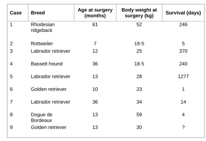
Table 1: Signalment and survival details 351
Case Breed Age at surgery
(months)
Body weight at
surgery (kg) Survival (days)
1 Rhodesian
ridgeback
61 52 246
2 Rottweiler 7 19·5 5
3 Labrador retriever 12 25 370
4 Bassett hound 36 18·5 240
5 Labrador retriever 13 28 1277
6 Golden retriever 10 23 1
7 Labrador retriever 36 34 14
8 Dogue de
Bordeaux
13 59 4
9 Golden retriever 13 30 ?
352
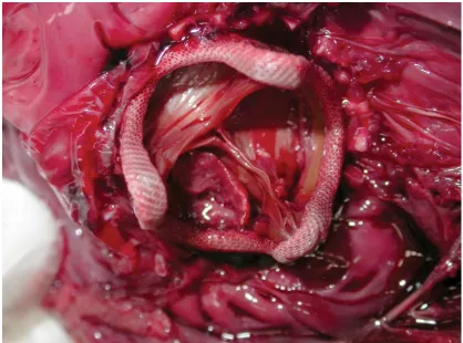
Figure 1: Sutures of 2-0 TiCron placed in the tricuspid annulus with pledgets on the 354
ventricular side 355
356
Figure 2: Prosthetic valve mounted on handle, after preplaced sutures have been 357
passed through suturing ring 358
359
Figure 3: Post-mortem picture of thrombus on valve (day four postoperatively, case 8) 361