Perspective
5-Hydroxymethylcytosine (5hmC), or How to Identify
Your Favorite Cell
Szilvia Ecsedi1, Jesús Rafael Rodríguez-Aguilera2ID and Héctor Hernandez-Vargas3,*ID
1 Institute of Biology Valrose (iBV), The National Center for Scientific Research (CNRS)—National Institute of
Health and Medical Research (Inserm), UniversitéCôte d’Azur, 06108 Nice, France; szilvia.ecsedi@unice.fr 2 Laboratory 305-Sur, Department of Cellular Biology and Development, Institute of Cellular Physiology,
National Autonomous University of Mexico (UNAM), Coyoacán, 04510 Ciudad de México, Mexico; jesusr_rodagu@comunidad.unam.mx
3 Cancer Research Centre of Lyon (CRCL), Inserm U 1052, CNRS UMR 5286, Centre Léon Bérard,
Universitéde Lyon, 28 rue Laennec, 69373 Lyon CEDEX 08, France * Correspondence: hector.hernandez@inserm.fr; Tel.: +33-046-985-6186
Received: 20 December 2017; Accepted: 23 January 2018; Published: 30 January 2018
Abstract: Recently described as the sixth base of the DNA macromolecule, the precise role of 5-hydroxymethylcytosine (5hmC) is the subject of debate. Early studies indicate that it is functionally distinct from cytosine DNA methylation (5mC), and there is evidence for 5hmC being a stable derivate of 5mC, rather than just an intermediate of demethylation. Moreover, 5hmC events correlate in time and space with key differentiation steps in mammalian cells. Such events span the three embryonic germ layers and multiple progenitor cell subtypes, suggesting a general mechanism. Because of the growing understanding of the role of progenitor cells in disease origin, we attempted to provide a detailed summary on the currently available literature supporting 5hmC as a key player in adult progenitor cell differentiation. This summary consolidates the emerging role for 5hmC in defining cellular fate.
Keywords:DNA methylation; progenitor cells; differentiation; 5mC; 5hmC
1. Introduction
Like evidence in a crime scene, cytosine DNA methylation (5mC) can reconstitute diverse information about the origin and context of the host cell [1]. While sex can be easily identified from 5mC data, algorithms designed in recent years use 5mC to also extract information about the major tissue types in which a given cell resides and the age of the donor [2]. Even in complex samples such as blood, DNA methylation can be used to infer the proportion of major cell subpopulations [3], illustrating the tissue- and cell-specificity of 5mC. More recently, several studies showed that, at the level of progenitor cell differentiation, it is the loss of 5mC at particular intragenic regions that precedes a switch in cell identity, as we will show throughout this study. The improved knowledge of how this DNA demethylation occurs is leading to a redefinition of DNA methylation dynamics.
Presence of 5-hydroxymethylcytosine (5hmC) in the genome was suggested in 1972, in rat’s brain and liver DNA [4]. However, it was not until 2009 when 5hmC was identified as an unusual nucleotide when comparing the abundance of 5mC in Purkinje and granule cells [5]. In parallel, Tet Methylcytosine Dioxygenase 1 (TET1) was first described as an enzyme able to convert 5mC to 5hmC by oxidation [6]. Increasing the complexity of this field, additional oxidized derivatives of 5mC (i.e., 5-carboxylcytosine (5caC) and 5-formylcytosine (5fC) [7] and two new members of the TET family (i.e., TET2 and TET3) have been described [8,9]. Although with a slightly distinct chemical construction, all three TET proteins are considered as 5hmC “writers” [8,9], while all three oxidized
Epigenomes 2018,2, 3 2 of 20
derivatives of 5mC were initially defined as intermediaries in the active process of DNA demethylation (Figure1).
Epigenomes 2018, 2, x FOR PEER REVIEW 2 of 20
derivatives of 5mC were initially defined as intermediaries in the active process of DNA demethylation (Figure 1).
Ever since the discovery of the “sixth base”, strenuous efforts have been made to characterize the precise role of 5hmC in development. Such roles are becoming more evident as we learn about 5hmC specific genomic localization, its relative stability, and its recognition by other proteins. Based on the available evidence, we illustrate here how 5hmC is consistently found as a dynamic mark during the establishment of cell identity in multiple model systems.
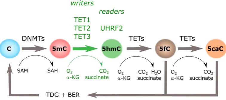
Figure 1. Cytosine methylation cycle. Cytosine DNA methylation (5mC) groups introduced by DNA Methyl-Transferases (DNMTs) can be iteratively oxidized to hydroxymethylcytosine (5hmC), 5-formylcytosine (5fC), and 5-carboxylcytosine (5caC) by the action of Tet Methylcytosine Dioxygenases (TETs). Three “writers” (i.e., TET1, TET2, and TET3), and at least one “reader” (i.e., UHRF2), have been identified for 5hmC. α-KG: α-ketoglutarate; BER: base excision repair; SAH: S -adenosylhomocysteine; SAM: S-adenosylmethionine; TDG: thymine DNA glycosylase.
2. 5-Hydroxymethylcytosine: “Localized, Stable and Readable”
Mapping 5hmC location represented a first step in deciphering its function. Early methods such as immunofluorescence (IF) and mass spectrometry (MS) allowed researchers to assess global levels of 5hmC, and high-performance liquid chromatography-mass spectrometry (HPLC-MS) is still the most accurate method for 5hmC global detection [10]. However, the development of locus-specific methods has played a pivotal role in our comprehension of the functional role of 5hmC. In general, affinity-based enrichment methods are widely used and can be easily combined with genome-wide profiling. Affinity-based applications can be divided into antibody and chemical capture-based methods, with both approaches providing highly reproducible results [11], and the vast majority of information on the distribution of 5hmC has been generated by antibody-based methods as 5hmC immunoprecipitation (hMeDIP) [12]. Regarding chemical labelling enrichment technics, those based on β-glycosyltransferase (βGT)-catalyzed 5hmC glycosylation such as glycosylation labelled sequencing (hMe-Seal), glucosylation, periodate oxidation and biotinylation (GLIB), and J-binding protein 1 sequencing (JBP-1) have also been extensively used; however, they are claimed to exhibit relatively low resolution compared to the antibody-based methods [11,13]. More recently, oxidative bisulfite sequencing (oxBS-Seq) [14] and TET assisted bisulfite sequencing (TAB-Seq) [15] were published and, starting now, they will be widely used methods in order to provide single resolution maps of 5hmC. However, both techniques require excessive chemical treatments and therefore can cause DNA damage. Furthermore, oxBS-seq and TAB-Seq require at least 30x sequencing depths to provide reliable, quantitative results.
Globally, 5hmC was shown by IF to be enriched in the euchromatin compartment [16]. Although exposed and accessible to anti-5hmC antibodies in euchromatin, the presence of 5hmC in heterochromatin seems to be hindered by binding proteins under non-denaturizing IF conditions [17]. Novel chemical methods allowed researchers to precisely map the genomic distribution of 5hmC
Figure 1. Cytosine methylation cycle. Cytosine DNA methylation (5mC) groups introduced by DNA Methyl-Transferases (DNMTs) can be iteratively oxidized to 5-hydroxymethylcytosine (5hmC), 5-formylcytosine (5fC), and 5-carboxylcytosine (5caC) by the action of Tet Methylcytosine Dioxygenases (TETs). Three “writers” (i.e., TET1, TET2, and TET3), and at least one “reader” (i.e., UHRF2), have been identified for 5hmC.α-KG:α-ketoglutarate; BER: base excision repair; SAH:S-adenosylhomocysteine; SAM:S-adenosylmethionine; TDG: thymine DNA glycosylase.
Ever since the discovery of the “sixth base”, strenuous efforts have been made to characterize the precise role of 5hmC in development. Such roles are becoming more evident as we learn about 5hmC specific genomic localization, its relative stability, and its recognition by other proteins. Based on the available evidence, we illustrate here how 5hmC is consistently found as a dynamic mark during the establishment of cell identity in multiple model systems.
2. 5-Hydroxymethylcytosine: “Localized, Stable and Readable”
Globally, 5hmC was shown by IF to be enriched in the euchromatin compartment [16]. Although exposed and accessible to anti-5hmC antibodies in euchromatin, the presence of 5hmC in heterochromatin seems to be hindered by binding proteins under non-denaturizing IF conditions [17]. Novel chemical methods allowed researchers to precisely map the genomic distribution of 5hmC and distinguish it from other cytosine variants. Immunoprecipitation with 5hmC antibodies followed by sequencing (hMeDIP-Seq) provided the first genome-wide map of 5hmC distribution, and confirmed its association with active transcription, CpG islands, 50regions of LINE1, CTCF, and pluripotency transcription factor (TF) binding sites and, in general, regions marked by 5mC decrease and active chromatin marks [16]. Upon differentiation from embryonic stem cells (ESCs) into embryonic bodies, the levels of 5hmC decreased. As will be discussed below, with the exception of neurogenesis, this distribution and global decrease of 5hmC is consistent across many models of differentiation. Also in ESCs, 5hmC was described in association with bivalent promoters (i.e., simultaneously occupied by histone marks H3K4me3 and H3K27me3). In this sense, it was recently shown that TET enzymes block aberrant hypermethylation in this type of promoter, ensuring robust lineage-specific transcription upon differentiation [18]. Another type of “bivalency” has been recently described at later stages of differentiation, in which genes simultaneously display promoter 5mC and gene body 5hmC at developmentally important genes [19]. Finally, Ficz et al. also described 5hmC association with non-CpG methylation resulting in strand bias that increased as a response of TET1/2 silencing. Interestingly, whenever 5hmC and 5mC occurred in parallel, gene expression was higher compared to regions in which 5mC occurred alone [16].
Growing evidence confirms the distribution and dynamics of 5hmC during embryonic cell development, outlining that it is functionally distinct from 5mC. However, the relatively low levels of 5hmC in the human genome, as well as the presence of other oxidized cytosine variants (5fC and 5caC), raised fundamental questions regarding the stability of the novel mark. Indirectly, genome-wide acquisition of 5hmC during neurogenesis in the absence of a major 5mC drop suggested that rather than an intermediate of demethylation, 5hmC serves as a stable signal on its own [20,21]. In addition, direct evidence of 5hmC stability in different cell types was obtained from ultrasensitive analytical liquid chromatography-tandem mass spectrometry (LC-MS/MS), showing no change in the levels of 5hmC over cell cycle phases [22]. Moreover, data suggested that 5mC oxidation does not occur on the nascent strand but must be formed in the double-stranded DNA produced immediately after replication [22]. Although several fundamental questions remain to be explained regarding TET binding and interaction affinities, a single, remarkable study described 5hmC-DNA interactions as much more stable and less prone than 5mC-DNA to further oxidation [23]. Favoring the idea that 5hmC is a persistent mark, compared to 5fC and 5caC, 5hmC is a poor substrate for TETs, thus protecting 5hmC from further oxidation [23,24] (reviewed in [25]). In a similar way, 5fC and 5caC are more efficiently recognized by thymine-DNA glycosylase (TDG) for removal and subsequent base excision repair (BER) leading to demethylation [26] (Figure1). Together, higher affinities of TETs and TDG for 5fC and 5caC ensure a relative abundance and recognition of 5hmC in the genome.
Epigenomes 2018,2, 3 4 of 20
Interesting New Gene (RING) finger-associated (SRA) domain of UHRF2 specifically interacts with 5hmC [29]. This discovery was subsequently supported by a structural and biochemical study [30], which demonstrated that the UHRF2-SRA domain, unlike the equivalent domain in UHRF1, does not preferentially bind to hemi-methylated DNA. Instead, it binds to fully hydroxymethylated and hemi-hydroxymethylated DNA 3.2- and 1.5-fold more tightly than it does to hemi-methylated DNA, respectively. In addition, the Chromatin target of Prmt1 (CHTOP)-methylosome complex, which methylates H4R3, has been recently described as a novel molecular interactor of 5hmC in a study conducted in gliomas [31,32].
Interestingly, 5fC and 5caC seem to be associated with numerous molecular readers [28,33–35]. Of note, these methylated variations have been shown to cause pause at the transcription rate generated by RNA Polymerase II (Pol II) binding. No similar mechanism has been detected for 5hmC so far, which is in line with the previously described phenomenon that 5hmC is a hallmark of active gene expression.
3. Specific 5-Hydroxymethylcytosine Enrichment Identifies Blood Cell Subtypes
In agreement with its relative stability and readability, recent studies provide ample evidence of the importance of the “sixth base” during differentiation and lineage commitment of several adult cell types. Given the existence of relatively easy approaches to perform in vitro differentiation and selection of well-characterized types of hematopoietic cells, substantial data has been accumulated to favor the role of 5hmC in transcriptional programming of myeloid and lymphoid differentiation (Table1).
Table 1.5-hydroxymethylcytosine (5hmC) and blood cell subtypes.
Cell Type Organism Model System 5hmC Technique 5hmC Enrichment 5hmC Writer Authors
Myeloid & Lymphoid
HSCs Hs umbilical cord CD34+
cells RRHP + NGS
exons, promoters,
enhancers no data Tekpli et al., 2016
HSCs Mm Tet2 and Dnmt3aknockout mice CMS-IP + sequencing
promoters and gene bodies of highly expressed genes
Dnmt3a, Tet2 Zhang et al., 2016
Myeloid
Macrophages,
Osteoclasts Hs in vitro differentiation OxBS + HM450
enhancers, 5hmC changes at promoters and gene bodies
TET2, TDG, AID Garcia-Gomez et al., 2017
Macrophages Hs monocyte elutriation and
differentiation OxBS + sequencing
Demethylation at nucleosome-free loci that gain active enhancer marks
TET2 Wallner et al., 2016
Dendritic
cells Hs in vitro differentiation
hMedIP (and glycosylation-sensitive
restriction) + qPCR
candidate gene promoters TET2 Klug et al., 2013
Mast cells Mm Tet2 knockout mice +
in vitro differentiation GLIB-Seq
TSS distal (enhancers),
gene bodies Tet2
Montagner et al., 2016
Erythrocytes Hs in vitro culture of HSCs hMe-Seal; TAB-Seq
TF binding sites, gene bodies of overexpressed genes
TET2, TET3 Madzo et al., 2014
Erythrocytes Hs cultured cord blood
CD34+ cells LC-MS global loss of 5hmC TET2, TET3 Yan et al., 2017
Lymphoid
B cells Hs in vitro activation of
human naïve B cells hMe-Seal
enhancers, gene bodies. B cell identity genes lose whereas Plasma Cell identity genes gain 5hmC
no data Caron et al., 2015
B cells Mm mice deficient for Tet2
and Tet3 in early B cells CMS-IP + sequencing
B cell enhancers, chromatin-accessible regions
Tet2, Tet3 Lio et al., 2016
T cells Mm
purified naïve and in vitro differentiated T cell subtypes
CMS-IP + sequencing
gene bodies of highly expressed genes, active tissue-specific enhancers
no data Tsagaratou et al., 2014
T cells Mm sorted mouse cell
subpopulations
βGT catalyzed restriction enzyme protection + qPCR; OxBS
+ qPCR
candidate gene approach
Table 1.Cont.
Cell Type Organism Model System 5hmC Technique 5hmC Enrichment 5hmC Writer Authors
Tfh cells Mm
in vivo-generated Tfh cells from Bcl6-RFP reporter mice
hMedIP-Seq
5hmC loss in Tfh cells at Bcl6 binding sites compared to naïve T cells
Tet1 Liu et al., 2016
Tfh cells Hs human PBMCs, and
lupus prone mice
DNA dot plot, MedIP-Seq
global 5hmC gain, no
genome context data TET2, TET3 Wu et al., 2016
CD4 T cells Hs human CD4+ T cell
differentiation ex vivo
Methyl/Hydroxymethyl sensitive qPCR
(EpiMark)
gene body enrichment and TSS depletion, actively transcribed genes
TET1 Nestor et al., 2016
CD4 T cells Mm mouse T cell subtypes hMedIP + sequencing
introns and intergenic regions, lineage-specific TFs
Tet2 Ichiyama et al., 2015
CD8 T cells Mm
mouse CD8+ T cells with low or high HIF signalling
hMedIP + qPCR overall reduction of
5hmC Tet2 Tyrakis et al., 2016
iNKT cells Mm Tet2-Tet3 DKO mice CMS-IP + sequencing
chromatin accessible-regions associated with iNK T cell development
Tet2, Tet3 Tsagaratou et al., 2017
Treg cells Mm H2S-deficient (Cbs−/−)
mice dot blot; hMedIP + qPCR
Foxp3promoter and
CNS2 Tet1, Tet2 Yang et al., 2015
Treg cells Mm Tet2-Tet3 DKO mice OxBS + sequencing Foxp3CNS1 and CNS2 Tet2, Tet3 Yue et al., 2016
Hs: Homo Sapiens;Mm: Mus Musculus; HSC: Hematopoietic stem cells; iNKT cells: Invariant natural killer T cells; Peripheral blood mononuclear cells (PBMCs); HIF: Hypoxia-inducible factors; DKO: Double knock-out; RRHP: reduced representation of hydroxymethylation profiling; NGS: next-generation sequencing; CMS-IP: cytosine-5-methylenesulphonate immunoprecipitation; OxBS: oxidative bisulfite; HM450: Infinium 450K bead arrays; hMeDIP: 5hmC immunoprecipitation; hMeDIP-Seq: hMeDIP Sequencing; qPCR: Quantitative polymerase chain reaction; GLIB: glycosylation, periodate oxidation, and biotinylation; GLIB-Seq: GLIB-Sequencing; hMe-Seal:
βGT-catalyzed 5hmC glycosylation labelled sequencing; TAB-Seq: Tet-assisted bisulfite sequencing; LC-MS: liquid chromatography mass spectrometry; TSS: transcription start site; TF: Transcription factor; CNS: Conserved non-coding sequences; TDG: thymine DNA glycosylase, AID: activation-induced deaminase.
Globally, a drop in 5hmC levels has been described during differentiation from hematopoietic stem cells (HSCs) [36], except for one study showing overall 5hmC increase (without genome context data) [37]. Despite this global reduction, 5hmC remains enriched at particular loci, matching the distribution shown by earlier studies (i.e., promoters and gene bodies). In addition, 5hmC enrichment has been described in enhancers [19,36,38–41] and lineage-specific TF binding sites [42,43]. In line with this, 5hmC changes are overrepresented in highly transcribed genes. For example, chromatin-accessible-regions are associated with T cell [36,44] and induced natural killers (iNK) T cell development [45], Treg FoxP3 activation [46,47], B cell identity genes [39,41], or Bcl6 binding sites in T follicular helper (Tfh) cells [48] (Figure 2and Table1). During mast cell development, it was shown that Tet2 knockout caused differential 5hmC levels in genes enriched with leukocyte differentiation and proliferation, malignant transformation, and mast cell-related processes [40]. Hypo-hydroxymethylated loci overlapped with enhancer regions, suggesting that 5hmC had a role in transcriptional programming [40].
Epigenomes 2018,2, 3 6 of 20
Epigenomes 2018, 2, x FOR PEER REVIEW 6 of 20
developmental changes upon Tet2 knockout was only possible by the re-expression of Tet2 independently of its catalytic activity, which assumes that Tet2 possibly interacts with other complexes (something similar has been shown between TET1 and the SIN3A co-repressor complex [53]).
Figure 2. 5-Hydroxymethylcytosine dynamics during blood cell differentiation. Evidence for dynamic changes in 5mC (gray lines) or 5hmC (red lines) at the indicated loci, as associated with immune lineage commitment. Dashed lines represent additional evidence. Corresponding PubMed PMIDs for each study are shown in red. TSDRs: Treg-specific demethylated regions. Th0: naïve T helper cells.
3.1. Myeloid Lineage
In addition to mast cells, specific redistribution of 5hmC has been described during differentiation of monocytes into macrophages or into dendritic cells [50,51]. Moreover, a unique role of 5hmC in red blood cell development seems well established according to a study that provided a comprehensive view of 5hmC distribution at stem/early progenitors, primitive erythroid progenitors, basophilic, polychromatic, orthochromatic, and reticulocytes [42].
Initially, by mass spectrometry measurements, using both in vitro as well as in vivomodels, the authors described a general increase of 5hmC during commitment to the erythroid lineage, followed by remarkable decrease during the subsequent steps of differentiation [42]. In contrast to the dynamic fluctuation of 5hmC, 5mC exhibited modest decrease. Besides negligible levels of TET1 and TET3, TET2 seemed to be involved in the differentiation process, being the highest at the time point of specific erythroid lineage commitment. As for other cell types, in parallel with the overall decrease, elevated levels of 5hmC were detected at particular loci [42]. As erythropoiesis is a replication
Figure 2.5-Hydroxymethylcytosine dynamics during blood cell differentiation. Evidence for dynamic changes in 5mC (gray lines) or 5hmC (red lines) at the indicated loci, as associated with immune lineage commitment. Dashed lines represent additional evidence. Corresponding PubMed PMIDs for each study are shown in red. TSDRs: Treg-specific demethylated regions. Th0: naïve T helper cells.
3.1. Myeloid Lineage
In addition to mast cells, specific redistribution of 5hmC has been described during differentiation of monocytes into macrophages or into dendritic cells [50,51]. Moreover, a unique role of 5hmC in red blood cell development seems well established according to a study that provided a comprehensive view of 5hmC distribution at stem/early progenitors, primitive erythroid progenitors, basophilic, polychromatic, orthochromatic, and reticulocytes [42].
3.2. Lymphoid Lineage
On a pioneer study of 5hmC and T-cell lineage commitment, deep sequencing was performed on purified samples (double positive, CD4 single positive, CD8 single positive, naïve CD4, naïve CD8, and CD4-differentiated Th1 and Th2 T helper cells) enriched by cytosine-5-methylenesulfonate-based immunoprecipitation [36]. In all cell types, 5hmC was found to be highest at promoters and intragenic regions, without enrichment for transcription start sites (TSS). On average, naive T-cells showed higher global 5hmC compared to the previous stage of differentiation, followed by a drop at the later stages (Th1 and Th2). As the reduction of 5hmC was replication-dependent, this initially suggested that 5hmC had been passively diluted in a cell cycle manner. However, in contrast to 5mC, gene body 5hmC exhibited significant positive correlation with gene expression accompanied with Pol II occupancy and enrichment for active histone marks. Moreover, 5hmC was suggested to mark long-range interactions between enhancers and other regulatory regions at the transition points of T-cell development. More recently, using Tet2-Tet3 double knock-out (DKO) mice, the same group showed that TET proteins are essential for maturation of invariant natural killer T cells (iNKT), and its absence generates a T cell antigen receptor (TCR)-mediated expansion of these cells and a lethal lymphocytic infiltrate in lung and liver [45]. Transcriptomic evaluation identified a characteristic profile of natural killer NKT17 cells (expressing RAR-related orphan receptorγ(RORγt) and Interleukin 17) and genes related with malignant transformation.
Of note, studies supporting a role of 5mC/5hmC in immune cell development have been performed in both mouse and human cells (Table1). For example, analogous DNA methylation dynamics are observed during differentiation of human naive B cells into plasma cells [41]. Most differentially methylated loci identified in this process were demethylated and enriched for enhancers, with demethylation becoming particularly pronounced at terminal stages of development. Initial assessment by selective chemical labeling followed by dot blot showed global loss of 5hmC during differentiation, accompanied by decrease in TETs expression. However, 5hmC was found to be enriched at specific DNA regions, particularly at B-cell identity genes with enhancer chromatin marks (i.e., H3K4me1 and H3K27ac) (Figure2). As mentioned above, 5hmC at gene bodies correlated with gene expression.
Therefore, 5hmC events take place at all stages of lymphoid cell differentiation, from HSCs to highly specialized cell types (Figure2). For example, 5hmC seems to define the transition to the lymphoid lineage, the choice between B and T cell types, and between CD4 and CD8 T cells [36]. Furthermore, 5hmC enrichment at specific loci has been described for each of the well specialized fates of naïve T helper (Th0) cells (i.e., Th1, Th2, Th17, Thf, and Treg). Indeed, studies of Treg cell differentiation represent a good example of what has been done in other systems in terms of linking 5hmC to cell identity. Defined as the master TF of Treg programming almost 15 years ago [54],Foxp3 was later shown to be demethylated in particular loci called Treg-specific demethylated regions (TSDRs) (Figure2). TSDRs were described as the most reliable criterion for natural Treg identification long before the first 5hmC studies became available [55,56]. In addition, TSDRs were suggested as a requirement for lineage stabilization [55–58] and a way to distinguish Tregs from different origins (i.e., thymic vs. periphery vs. in vitro) [56–60]. While TSDRs were associated with intragenic conserved non-coding sequences (CNS) in theFoxp3loci displaying a particular chromatin landscape [57], additional TSDRs were found in mice in other immune-related genes as well (i.e.,Ctla4,Il2ra,Ikzf4, andTnfrsf18) [61]. After several additional studies highlighted that CNS2 is specifically demethylated in Tregs [62,63], subsequent work demonstrated that TET proteins were necessary forFoxp35hmC enrichment [46,47]. More recently, one additional CNS loci (CNS0) has been described as a FoxP3 super-enhancer and a binding site for the chromatin organizer Satb1 [64].
Epigenomes 2018,2, 3 8 of 20
Indeed, the frequency of frameshift and nonsense mutations inTET2is estimated to be as high as 45% in patients with chronic myelomonocytic leukemia [66]. Although this places TET mutations as driver events in leukemia, it is worth mentioning that TET2 mutations alone have proven insufficient to cause cancer [65]. Future studies aimed at elucidating a more comprehensive role of TET mutations in the 5mC-5hmC balance may shed light on the dynamic interplay between genetic and epigenetic machinery in disease development, rather than considering mutations as master regulators.
4. 5-Hydroxymethylcytosine Is Redistributed during Terminal Differentiation from All Germ Layers
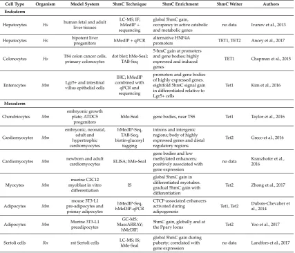
In addition to most blood cell subtypes, 5hmC has been associated with differentiation into other cell types from mesodermal origin. Moreover, examples also exist for the endodermal and ectodermal germ layers, as described below (Table2).
Table 2.5-hydroxymethylcytosine and non-blood cell subtypes.
Cell Type Organism Model System 5hmC Technique 5hmC Enrichment 5hmC Writer Authors
Endoderm
Hepatocytes Hs human fetal and adult
liver tissues
LC-MS; IF; hMedIP + sequencing
global 5hmC gain, occupancy in active catabolic and metabolic genes
no data Ivanov et al., 2013
Hepatocytes Hs bipotent liverprogenitors hMedIP + qPCR promotersalternative HNF4A TET1, TET2 Ancey et al., 2017
Colonocytes Hs T84 colon cancer cells,
primary colonocytes
dot blot; hMe-Seal; TAB-Seq
5-hmC gain at promoters and gene bodies; highly expressed and induced genes
TET1 Chapman et al., 2015
Enterocytes Mm villus epithelial cellsLgr5+ and intestinal
IHC; hMedIP combined with
qPCR and sequencing
promoters and gene bodies of highly expressed genes. eightfold 5hmC signal gain in differentiated relative to Lgr5+ cells
Tet1 Kim et al., 2016
Mesoderm
Chondriocytes Mm
embryonic growth plate; ATDC5
progenitors
hMe-Seal gene bodies, near TSS Tet1 Taylor et al., 2016
Cardiomyocytes Mm embryonic, neonatal, adult and hypertrophic cardiomyocytes hMedIP-Seq, TAB-Seq, biotin-glucosyl tagging
introns and intergenic regions; body of highly expressed genes and distal regulatory regions
Tet2 Greco et al., 2016
Cardiomyocytes Mm newborn and adult
cardiomyocytes ELISA; hMe-Seal
gene bodies and low methylated enhancers; positively associated with gene expression
no data Kranzhofer et al., 2016
Myocytes Mm
murine C2C12 myoblast in vitro
differentiation
IS
global 5hmC gain in differentiated myotubes. gradual 5hmC gain with differentiation
Tet2 Zhong et al., 2017
Adipocytes Mm mouse 3T3-L1 pre-adipocytes and primay adipocytes hMedIP-Seq, hMeDIP-qPCR CTCF-associated enhancers activated during adipogenesis
Tet1, Tet2 Dubois-Chevalier et al., 2014
Adipocytes Mm Murine 3T3-L1
preadipocytes
GC-MS; MassARRAY;
hMeDIP,
5hmC gain, globally and at
the Pparγlocus Tet2 Yoo et al., 2017
Sertoli cells Rn rat Sertoli cells LC-MS; IS;hMe-Seal
global 5hmC gain during puberty; correlated with gene expression
Table 2.Cont.
Cell Type Organism Model System 5hmC Technique 5hmC Enrichment 5hmC Writer Authors
Ectoderm
Neurons Mm
mouse Nestin+/Sox2+ adult neural stem
cells (aNSC)
dot blot; IS; hMe-Seal; LC-MS
global gain; positive correlation with
transcriptional upregulation
Tet2 Li et al., 2017
Neurons Hs human fetal brain
samples
OxBS combined + HM450
CpG island shores and shelves, flanking TSS and gene bodies
no data Spiers et al., 2017
Neurons Mm mouse cerebellumand hippocampus dot blot; hMe-Seal;IS; hMeDIP-qPCR
developmentally activated gene bodies; repetitive loci; 60% of DhMRs intragenic and enriched in exons
MeCP2, no TET
data Szulwach et al., 2011
Neurons Mm
transgenic mice with dual reporter for neural progenitors
and progeny
IS; LC-MS; hMe-Seal labelled
NimbleGen 2.1M arrays; TAB-Seq
global 5hmC gain; promoters, gene bodies, intragenic regions, during neurogenesis. associated with higher transcription
Tet2, Tet3 Hahn et al., 2013
Retina Dr zebrafish retinal
neurogenesis
IS; ELISA; hMe-Seal enzyme
protection + sequencing
body of one candidate gene Tet2, Tet3 Seritrakul et al., 2017
Rn: Rattus norvegicus;Dr: Danio rerio;ATDC5: chondroprogenitor cell line; TAB-Seq: Tet-assisted bisulfite sequencing; GC-MS: Gas chromatography mass spectrometry; IF: immunofluorescence; IHC: Immunohistochemistry; IS: Immunostaining; ELISA: Enzyme-linked immunosorbent assay.
4.1. Endoderm
Endoderm gives rise to many of the cell types that form the gastrointestinal tract and related organs. The adult liver is relatively rich in 5hmC content, and global redistributions of 5mC [67–69] and 5hmC [70] have been described during the fetal to adult liver transition. Specifically, liquid chromatography mass spectrometry (LC-MS) estimations indicate that 5hmC increases from 0.125% to 1% of the total cytosine content. In adult livers, such 5hmC occupancy is overrepresented at genes involved in active catabolic and metabolic processes [71] (Figure3). While these studies suggest a role of 5hmC in liver physiology, a recent report described a specific 5hmC event during hepatocyte differentiation [72]. TET-dependent 5hmC activation preceded activation of the HNF4A P1 promoter, a master TF of hepatocyte identity (Figure3). In the same model of hepatocyte differentiation from a bipotent progenitor, we recently profiled 5mC/5hmC using oxidative bisulfite (OxBS) coupled to HumanMethylationEPIC arrays (Illumina, San Diego, CA, USA), and noticed that this process involves 5mC reduction and 5hmC gain partially associated with the acquisition of hepatocyte expression profile (unpublished results). Of note, liver tumor development has been associated with reduced TET activity and 5hmC in human samples [73], as well as in mouse [71] and in vitro models, suggesting 5hmC could be used as a biomarker of hepatocarcinogenesis [74].
Epigenomes 2018,2, 3 10 of 20
Epigenomes 2018, 2, x FOR PEER REVIEW 10 of 20
this process [75,76]. This contrasts with the pattern shown for blood cell subtypes, at least at earlier stages of blood cell differentiation (i.e., from HSCs) [19], in which there is a more general presence of 5hmC followed by a global drop that respects lineage-related loci, as described above. Interestingly, both in blood cells and endoderm differentiation, 5hmC redistribution was accompanied by a drop in 5mC (compare 5mC kinetics in Figures 2 and 3). Also, in both cases, loss of gene expression in committed cells correlated with a significant decrease in gene body 5hmC levels.
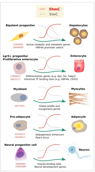
Figure 3. 5-Hydroxymethylcytosine dynamics during cell differentiation. Evidence for dynamic changes in 5mC (gray lines) or 5hmC (red lines) at the indicated loci, as associated with lineage commitment. Dashed lines represent additional evidence. Corresponding PubMed PMIDs for each study are shown in red.
Figure 3. 5-Hydroxymethylcytosine dynamics during cell differentiation. Evidence for dynamic changes in 5mC (gray lines) or 5hmC (red lines) at the indicated loci, as associated with lineage commitment. Dashed lines represent additional evidence. Corresponding PubMed PMIDs for each study are shown in red.
4.2. Mesoderm (Other Than Blood Cells)
the first stage of chondrogenic differentiation, followed by decrease at a later stage [77]. Using β-Glucosyltransferase-based chemical labelling and sequencing, the authors benefited from the previously established ATDC5 chondroprogenitor model system and found 5hmC enrichment near TSSs. In a similar way to what was described in immune cell subtypes, the increase in 5hmC was even more evident in gene bodies of key chondrogenic lineage-specific factors (Sox9, 5, and 6, Runx2, and Col2a1) and was associated with transcriptional activation.
Global and localized enrichment of 5hmC has also been described for myocyte [78] and cardiomyocyte development [79,80]. 5hmC enrichment was mainly found at gene bodies and distal regulatory regions, marking active genes enriched for Pol II occupancy, as well as active histone marks. The transition from embryonic into adult heart development was characterized by 5hmC peaks in genes related to heart development, such as actin-filament-based process and regulation of heart contraction. In addition, Tet2 has been implicated in both cardiomyocyte and myocyte development. Tet2 knockout resulted in 5hmC depletion at specific cardiomyocyte gene loci [80]. In a similar way, knockdown of Tet2, but not Tet1, significantly reduced myocyte gene expression (myogenin, Myf6, and myomaker) and impaired myoblast differentiation [78].
At least two studies have shown 5hmC enrichment during adipocyte development, using murine 3T3-L1 preadipocytes as a model (Figure3). In the first study, the chromatin insulator CTCF was found to interact with TET enzymes to promote 5hmC deposition at adipogenic transcriptional enhancers [81]. In the second study, also TET proteins (and particularly TET2) were shown to be required for adipogenesis by increasing 5hmC at the Pparγlocus and inducing Pparγgene expression [82].
Finally, a recent study has shown that 5hmC is also redistributed in Sertoli cells, mesodermal cells that are required for spermatogenesis. LC-MS evaluation of primary isolates of rat Sertoli cells from juvenile (7 days) and adult animals showed a puberty-associated reduction of 5mC and increase of 5hmC [83]. 5hmC peaks gained during maturation were related to cell transport, catabolic process, mitochondrion, and endocytosis, which go hand in hand with the fact that adult Sertoli cells have a diverse and highly active metabolism with an abundance of mitochondria.
Similar to blood cell types, these studies relate the differentiation process to an enrichment of 5hmC, suggesting that a general 5hmC shift in genomic distribution could be associated with the upregulation of genes that define cell state.
4.3. Ectoderm
At the time of 5hmC rediscovery, using a wide range of biochemical methods, the newly identified 5-hydroxymethyl-20-deoxycytidine was shown to constitute 0.6% of total nucleotides in Purkinje cells [5]. Being highly abundant in brain, 5hmC was immediately suggested to have a role in the genomic control of neural functions. Therefore, together with ESCs, neurons of various types, anatomical locations, and levels of differentiation have been some of the best-studied cell types in terms of 5mC/5hmC content, distribution, and function. We refer the readers to specialized reviews in 5hmC and brain function [84,85], and only outline here some of the major findings in the context of dynamic models of neurogenesis.
Epigenomes 2018,2, 3 12 of 20
genes and pathways related to neural programs, many of which are developmentally regulated [90]. Specific 5hmC enrichment has been observed at sequences adjacent to p300 sites in genes critical for neuronal differentiation [21], or Notch and Wnt pathway genes in retinal neurogenesis [91].
Another fact that increased the interest for 5hmC was the possibility of methylcytosine binding protein 2 (MeCP2) as a 5hmC reader [53], as this protein was previously associated with neurodevelopmental disorders [92] (reviewed in [93]). Indeed, it was recently shown that distribution of 5hmC in CG and non-CpG dinucleotides is distinct and reflects the binding specificity and genome occupancy of MeCP2 [86]. In addition, loss of MeCP2 led to specific reduction of 5hmC signals in dynamically hydroxymethylated intragenic non-CpGs involved in gene activation [20]. However, while the role of MeCP2 as a reader needs to be further validated [28], other candidate readers have been suggested in the context of neurogenesis and adult brain [29,94].
Therefore, dynamic models of neurogenesis are consistent with an important role of 5hmC, not only during differentiation, but also later in adult neuron life. Contrary to other tissues, 5mC depletion does not necessarily precede 5hmC acquisition [21] (Figure3). Moreover, the high levels of 5hmC in adult neurons (and capacity to further increase), and the specific distribution across cell types and brain regions, indicate an important functional role in addition to preserving cell identity. Recent studies showing that demethylation occurs rapidly after neuronal or behavioral stimulation [95–97], and that 5hmC is dynamically regulated during aging [20], are helping to elucidate such role.
5. Perspectives and Concluding Remarks
Cell identity is acquired through the sequential and combinatorial activity of TFs on lineage-specific enhancers [98]. A global picture is emerging in which, in joint action with TFs and enhancers, TET-catalyzed 5hmC marks identity genes at key differentiation checkpoints. In most cases, global 5hmC seems to be progressively reduced from progenitor to differentiated state, but remains (or is acquired) at relevant lineage-specific loci or at genes related to adult cell function. As illustrated here, examples exist for all embryonic layers, and both epithelial and mesenchymal phenotypes, suggesting a general mechanism. In a time when cell subtypes are being systematically redefined [99], 5hmC promises to be an exquisite marker of cell identity. Moreover, as has been shown for the FoxP3 CNS loci in Treg cells, 5hmC may be able not only to identify a cell subtype, but also its origin, something that gene expression alone cannot do [57,62,63].
Although proliferation status seems to be critical for global 5hmC [22], this does not explain the specificity of 5hmC occupancy during differentiation. Recent studies are shedding light into such mechanisms. For example, interaction between TET proteins and lineage-specific TFs such as the FOXA family have been described [72,89,100]. Although this is better supported at the level of pioneer factors, it is feasible that other TFs involved in lineage commitment are also able to recruit TETs to specific genomic loci. While several TFs are involved in DNA demethylation by interacting with TET proteins (e.g., Nanog during establishment of pluripotency [101], the hematopoiesis-related RUNX1 [102] and EBF1 [103], PU.1 in osteoclast differentiation [104], and PPARγin adipogenesis [105]), screening systems suggest that many others may share this property [106]. Alternatively, enhancer elements that distinguish cell types may be specifically targeted by TETs based on their chromatin conformation and histone mark profile. Indeed, based on their abundance in gene bodies of active genes and poised enhancers, TETs have been suggested as general transcriptional activators that recruit other proteins to demethylate enhancers [25].
5hmC in maintaining cell phenotype is liver fibrosis. In this pathology, chronic inflammatory injury triggers an adipogenic-to-fibrogenic phenotype switch in hepatic stellate cells, eventually leading to cirrhosis [110]. During different stages of chronic liver injury, 5hmC was found reduced, while hepatic stellate cells became transdifferentiated [111]. Consistent with these data, global 5hmC was shown to be reduced in a rat model of CCl4-induced cirrhosis [112]. It is important to note the recent availability of liver methylome and hydroxymethylome data on Wistar and Sprague-Dawley rats, as well as the genome-wide distribution of enhancer related chromatin marks (i.e., H3K4me1 and H3K27ac), which could represent a baseline reference for future studies of liver pathology [113].
An obvious context in which 5hmC occupancy is expected to be dysregulated is malignant disease (specialized reviews on cancer and TET-dependent 5hmC have been recently published [114–118]). Indeed, while Tet deletions/mutations have been shown to predispose to blood malignancies, cancer was initially associated with a global loss of 5hmC in various tissues [65,66]. Although such reports revealing 5hmC decrease in cancer lacked genome-wide or base-level resolution, studies consistently show that 5hmC profiles are able to distinguish malignant tissues, even under cell culture conditions [119]. If 5hmC does indeed preserve cellular phenotypes, it would be expected to have a protective role against cancer. In line with this, it was suggested that 5hmC is a hallmark from the early intestinal development that prevents neoplastic DNA hypermethylation events [120]. In addition, 5hmC was recently shown to correlate with lower regional CpG > T mutation frequency in cancers from various tissues (i.e., brain, kidney, and blood), also suggesting a protective role [121].
Finally, it has been suggested for years that DNA methylation may represent an interphase between environment and disease. The debate has been centered around which genomic locations are more dynamic or sensitive to external factors, and high interest has been placed on DNA methylation fluctuations at enhancer elements. Nowadays, emerging work suggests an important role of the oxidized forms of 5m—in particular, of 5hmC and the enzymes that establish it. For example, TETs are able to control differentiation into immune cell subtypes in response to specific inflammatory signals and are sensors of diverse metabolic processes. Another interesting example could be the critical role of oxygen concentration (as a co-factor) that influences TET activity and hence regulating cellular differentiation and fate in embryogenesis in which there are graded levels of molecular oxygen [122]. In a similar way, asα-ketoglutarate-dependent dioxygenases, the activity of TETs can be inhibited by 2-hydroxyglutarate in pathological conditions such as aberrant liver progenitor differentiation in presence of IDH1/2 mutations [123], or CD8+T cell fate under hypoxia [49]. Moreover, TET activity has been shown to be influenced by the availability of vitamins C [46,124,125] and A [125], and hydrogen sulphide [47].
In conclusion, the data sets presented throughout this study lead us to suggest that previously published studies that lack discriminated detection of 5mC and 5hmC (such as the widely used bisulphite conversion) have to be carefully revisited. Furthermore, approaches that are able to distinguish between DNA methylation intermediates should be superior in the future. Evidence points towards 5hmC (rather than 5mC) as a more likely interphase between the environment and dynamic gene expression changes, and most importantly, it seems to regulate the directors of cell fate. Therefore, its distribution in the genome could be used as a “fingerprint” to clearly indicate cell identity. This new potential of the 5hmC not only opens the door to deeper studies of cell differentiation and tissue development, but could also provide a useful tool for early detection and prevention of several conditions associated with the loss of cellular identity such as cancer.
Epigenomes 2018,2, 3 14 of 20
Author Contributions:S.E., J.R.R.-A. and H.H.-V. wrote jointly this manuscript.
Conflicts of Interest:The authors declare no conflict of interest.
References
1. Schübeler, D. Function and information content of DNA methylation.Nature2015,517, 321–326. [CrossRef]
[PubMed]
2. Horvath, S. DNA methylation age of human tissues and cell types.Genome Biol.2013,14, R115. [CrossRef]
[PubMed]
3. Houseman, E.A.; Accomando, W.P.; Koestler, D.C.; Christensen, B.C.; Marsit, C.J.; Nelson, H.H.; Wiencke, J.K.; Kelsey, K.T. DNA methylation arrays as surrogate measures of cell mixture distribution. BMC Bioinform. 2012,13, 86. [CrossRef] [PubMed]
4. Penn, N.W.; Suwalski, R.; O’Riley, C.; Bojanowski, K.; Yura, R. The presence of 5-hydroxymethylcytosine in animal deoxyribonucleic acid.Biochem. J.1972,126, 781–790. [CrossRef] [PubMed]
5. Kriaucionis, S.; Heintz, N. The nuclear DNA base 5-hydroxymethylcytosine is present in Purkinje neurons and the brain.Science2009,324, 929–930. [CrossRef] [PubMed]
6. Tahiliani, M.; Koh, K.P.; Shen, Y.; Pastor, W.A.; Bandukwala, H.; Brudno, Y.; Agarwal, S.; Iyer, L.M.; Liu, D.R.; Aravind, L.; et al. Conversion of 5-methylcytosine to 5-hydroxymethylcytosine in mammalian DNA by MLL partner TET1.Science2009,324, 930–935. [CrossRef] [PubMed]
7. Ito, S.; Shen, L.; Dai, Q.; Wu, S.C.; Collins, L.B.; Swenberg, J.A.; He, C.; Zhang, Y. Tet proteins can convert 5-methylcytosine to 5-formylcytosine and 5-carboxylcytosine. Science2011,333, 1300–1303. [CrossRef]
[PubMed]
8. Koh, K.P.; Yabuuchi, A.; Rao, S.; Huang, Y.; Cunniff, K.; Nardone, J.; Laiho, A.; Tahiliani, M.; Sommer, C.A.; Mostoslavsky, G.; et al. Tet1 and Tet2 regulate 5-hydroxymethylcytosine production and cell lineage specification in mouse embryonic stem cells.Cell Stem Cell2011,8, 200–213. [CrossRef] [PubMed]
9. Huang, Y.; Chavez, L.; Chang, X.; Wang, X.; Pastor, W.A.; Kang, J.; Zepeda-Martínez, J.A.; Pape, U.J.; Jacobsen, S.E.; Peters, B.; et al. Distinct roles of the methylcytosine oxidases Tet1 and Tet2 in mouse embryonic stem cells.Proc. Natl. Acad. Sci. USA2014,111, 1361–1366. [CrossRef] [PubMed]
10. Qing, Y.; Tian, Z.; Bi, Y.; Wang, Y.; Long, J.; Song, C.-X.; Diao, J. Quantitation and mapping of the epigenetic marker 5-hydroxymethylcytosine.BioEssays News Rev. Mol. Cell. Dev. Biol.2017,39. [CrossRef] [PubMed] 11. Thomson, J.P.; Hunter, J.M.; Nestor, C.E.; Dunican, D.S.; Terranova, R.; Moggs, J.G.; Meehan, R.R.
Comparative analysis of affinity-based 5-hydroxymethylation enrichment techniques. Nucleic Acids Res. 2013,41, e206. [CrossRef] [PubMed]
12. Song, C.-X.; Yi, C.; He, C. Mapping recently identified nucleotide variants in the genome and transcriptome. Nat. Biotechnol.2012,30, 1107–1116. [CrossRef] [PubMed]
13. Rivera, C.M.; Ren, B. Mapping human epigenomes.Cell2013,155, 39–55. [CrossRef] [PubMed]
14. Booth, M.J.; Branco, M.R.; Ficz, G.; Oxley, D.; Krueger, F.; Reik, W.; Balasubramanian, S. Quantitative sequencing of 5-methylcytosine and 5-hydroxymethylcytosine at single-base resolution.Science2012,336, 934–937. [CrossRef] [PubMed]
15. Yu, M.; Hon, G.C.; Szulwach, K.E.; Song, C.-X.; Zhang, L.; Kim, A.; Li, X.; Dai, Q.; Shen, Y.; Park, B.; et al. Base-resolution analysis of 5-hydroxymethylcytosine in the mammalian genome.Cell2012,149, 1368–1380.
[CrossRef] [PubMed]
16. Ficz, G.; Branco, M.R.; Seisenberger, S.; Santos, F.; Krueger, F.; Hore, T.A.; Marques, C.J.; Andrews, S.; Reik, W. Dynamic regulation of 5-hydroxymethylcytosine in mouse ES cells and during differentiation.Nature2011, 473, 398–402. [CrossRef] [PubMed]
17. Zhong, S.; Li, Z.; Jiang, T.; Li, X.; Wang, H. Immunofluorescence imaging strategy for evaluation of the accessibility of DNA 5-hydroxymethylcytosine in chromatins.Anal. Chem.2017,89, 5702–5706. [CrossRef]
[PubMed]
19. Tekpli, X.; Urbanucci, A.; Hashim, A.; Vågbø, C.B.; Lyle, R.; Kringen, M.K.; Staff, A.C.; Dybedal, I.; Mills, I.G.; Klungland, A.; et al. Changes of 5-hydroxymethylcytosine distribution during myeloid and lymphoid differentiation of CD34+cells.Epigenet. Chromatin2016,9, 21. [CrossRef] [PubMed]
20. Szulwach, K.E.; Li, X.; Li, Y.; Song, C.-X.; Wu, H.; Dai, Q.; Irier, H.; Upadhyay, A.K.; Gearing, M.; Levey, A.I.; et al. 5-hmC–mediated epigenetic dynamics during postnatal neurodevelopment and aging.Nat. Neurosci. 2011,14, 1607–1616. [CrossRef] [PubMed]
21. Hahn, M.A.; Qiu, R.; Wu, X.; Li, A.X.; Zhang, H.; Wang, J.; Jui, J.; Jin, S.-G.; Jiang, Y.; Pfeifer, G.P.; et al. Dynamics of 5-hydroxymethylcytosine and chromatin marks in mammalian neurogenesis.Cell Rep.2013,3, 291–300. [CrossRef] [PubMed]
22. Bachman, M.; Uribe-Lewis, S.; Yang, X.; Williams, M.; Murrell, A.; Balasubramanian, S. 5-Hydroxymethylcytosine is a predominantly stable DNA modification.Nat. Chem.2014,6, 1049–1055. [CrossRef] [PubMed]
23. Hu, L.; Lu, J.; Cheng, J.; Rao, Q.; Li, Z.; Hou, H.; Lou, Z.; Zhang, L.; Li, W.; Gong, W.; et al. Structural insight into substrate preference for TET-mediated oxidation.Nature2015,527, 118–122. [CrossRef] [PubMed] 24. He, Y.-F.; Li, B.-Z.; Li, Z.; Liu, P.; Wang, Y.; Tang, Q.; Ding, J.; Jia, Y.; Chen, Z.; Li, L.; et al. Tet-mediated
formation of 5-carboxylcytosine and its excision by TDG in mammalian DNA.Science2011,333, 1303–1307.
[CrossRef] [PubMed]
25. Szyf, M. The elusive role of 50-hydroxymethylcytosine.Epigenomics2016,8, 1539–1551. [CrossRef] [PubMed] 26. Hashimoto, H.; Hong, S.; Bhagwat, A.S.; Zhang, X.; Cheng, X. Excision of 5-hydroxymethyluracil and 5-carboxylcytosine by the thymine DNA glycosylase domain: its structural basis and implications for active DNA demethylation.Nucleic Acids Res.2012,40, 10203–10214. [CrossRef] [PubMed]
27. Juan, D.; Perner, J.; Carrillo de Santa Pau, E.; Marsili, S.; Ochoa, D.; Chung, H.-R.; Vingron, M.; Rico, D.; Valencia, A. Epigenomic co-localization and co-evolution reveal a key role for 5hmC as a communication hub in the chromatin network of ESCs.Cell Rep.2016,14, 1246–1257. [CrossRef] [PubMed]
28. Song, J.; Pfeifer, G.P. Are there specific readers of oxidized 5-methylcytosine bases?BioEssays News Rev. Mol. Cell. Dev. Biol.2016,38, 1038–1047. [CrossRef] [PubMed]
29. Spruijt, C.G.; Gnerlich, F.; Smits, A.H.; Pfaffeneder, T.; Jansen, P.W.T.C.; Bauer, C.; Münzel, M.; Wagner, M.; Müller, M.; Khan, F.; et al. Dynamic readers for 5-(hydroxy)methylcytosine and its oxidized derivatives.Cell 2013,152, 1146–1159. [CrossRef] [PubMed]
30. Zhou, T.; Xiong, J.; Wang, M.; Yang, N.; Wong, J.; Zhu, B.; Xu, R.-M. Structural basis for hydroxymethylcytosine recognition by the SRA domain of UHRF2.Mol. Cell2014,54, 879–886. [CrossRef]
[PubMed]
31. Fanis, P.; Gillemans, N.; Aghajanirefah, A.; Pourfarzad, F.; Demmers, J.; Esteghamat, F.; Vadlamudi, R.K.; Grosveld, F.; Philipsen, S.; van Dijk, T.B. Five friends of methylated chromatin target of protein-arginine-methyltransferase[prmt]-1 (chtop), a complex linking arginine methylation to desumoylation. Mol. Cell. Proteom. MCP2012,11, 1263–1273. [CrossRef] [PubMed]
32. Takai, H.; Masuda, K.; Sato, T.; Sakaguchi, Y.; Suzuki, T.; Suzuki, T.; Koyama-Nasu, R.; Nasu-Nishimura, Y.; Katou, Y.; Ogawa, H.; et al. 5-Hydroxymethylcytosine plays a critical role in glioblastomagenesis by recruiting the CHTOP-methylosome complex.Cell Rep.2014,9, 48–60. [CrossRef] [PubMed]
33. Hashimoto, H.; Olanrewaju, Y.O.; Zheng, Y.; Wilson, G.G.; Zhang, X.; Cheng, X. Wilms tumor protein recognizes 5-carboxylcytosine within a specific DNA sequence.Genes Dev.2014,28, 2304–2313. [CrossRef]
[PubMed]
34. Wang, L.; Zhou, Y.; Xu, L.; Xiao, R.; Lu, X.; Chen, L.; Chong, J.; Li, H.; He, C.; Fu, X.-D.; et al. Molecular basis for 5-carboxycytosine recognition by RNA polymerase II elongation complex.Nature2015,523, 621–625.
[CrossRef] [PubMed]
35. Jin, S.-G.; Zhang, Z.-M.; Dunwell, T.L.; Harter, M.R.; Wu, X.; Johnson, J.; Li, Z.; Liu, J.; Szabó, P.E.; Lu, Q.; et al. Tet3 Reads 5-Carboxylcytosine through Its CXXC Domain and Is a Potential Guardian against Neurodegeneration.Cell Rep.2016,14, 493–505. [CrossRef] [PubMed]
36. Tsagaratou, A.; Äijö, T.; Lio, C.-W.J.; Yue, X.; Huang, Y.; Jacobsen, S.E.; Lähdesmäki, H.; Rao, A. Dissecting the dynamic changes of 5-hydroxymethylcytosine in T-cell development and differentiation.Proc. Natl. Acad. Sci. USA2014,111, E3306–E3315. [CrossRef] [PubMed]
Epigenomes 2018,2, 3 16 of 20
38. Garcia-Gomez, A.; Li, T.; Kerick, M.; Català-Moll, F.; Comet, N.R.; Rodríguez-Ubreva, J.; de la Rica, L.; Branco, M.R.; Martín, J.; Ballestar, E. TET2- and TDG-mediated changes are required for the acquisition of distinct histone modifications in divergent terminal differentiation of myeloid cells.Nucleic Acids Res.2017, 45, 10002–10017. [CrossRef] [PubMed]
39. Lio, C.-W.; Zhang, J.; González-Avalos, E.; Hogan, P.G.; Chang, X.; Rao, A. Tet2 and Tet3 cooperate with B-lineage transcription factors to regulate DNA modification and chromatin accessibility.eLife2016,5, e18290.
[CrossRef] [PubMed]
40. Montagner, S.; Leoni, C.; Emming, S.; Chiara, G.D.; Balestrieri, C.; Barozzi, I.; Piccolo, V.; Togher, S.; Ko, M.; Rao, A.; et al. TET2 Regulates Mast Cell Differentiation and Proliferation through Catalytic and Non-catalytic Activities.Cell Rep.2016,15, 1566–1579. [CrossRef] [PubMed]
41. Caron, G.; Hussein, M.; Kulis, M.; Delaloy, C.; Chatonnet, F.; Pignarre, A.; Avner, S.; Lemarié, M.; Mahé, E.A.; Verdaguer-Dot, N.; et al. Cell-Cycle-Dependent Reconfiguration of the DNA Methylome during Terminal Differentiation of Human B Cells into Plasma Cells.Cell Rep.2015,13, 1059–1071. [CrossRef] [PubMed] 42. Madzo, J.; Liu, H.; Rodriguez, A.; Vasanthakumar, A.; Sundaravel, S.; Caces, D.B.D.; Looney, T.J.; Zhang, L.;
Lepore, J.B.; Macrae, T.; et al. Hydroxymethylation at Gene Regulatory Regions Directs Stem/Early Progenitor Cell Commitment during Erythropoiesis.Cell Rep.2014,6, 231–244. [CrossRef] [PubMed] 43. Ichiyama, K.; Chen, T.; Wang, X.; Yan, X.; Kim, B.-S.; Tanaka, S.; Ndiaye-Lobry, D.; Deng, Y.; Zou, Y.; Zheng, P.;
et al. The methylcytosine dioxygenase Tet2 promotes DNA demethylation and activation of cytokine gene expression in T cells.Immunity2015,42, 613–626. [CrossRef] [PubMed]
44. Nestor, C.E.; Lentini, A.; Hägg Nilsson, C.; Gawel, D.R.; Gustafsson, M.; Mattson, L.; Wang, H.; Rundquist, O.; Meehan, R.R.; Klocke, B.; et al. 5-Hydroxymethylcytosine Remodeling Precedes Lineage Specification during Differentiation of Human CD4+ T Cells.Cell Rep.2016,16, 559–570. [CrossRef] [PubMed]
45. Tsagaratou, A.; González-Avalos, E.; Rautio, S.; Scott-Browne, J.P.; Togher, S.; Pastor, W.A.; Rothenberg, E.V.; Chavez, L.; Lähdesmäki, H.; Rao, A. TET proteins regulate the lineage specification and TCR-mediated expansion of iNKT cells.Nat. Immunol.2017,18, 45–53. [CrossRef] [PubMed]
46. Yue, X.; Trifari, S.; Äijö, T.; Tsagaratou, A.; Pastor, W.A.; Zepeda-Martínez, J.A.; Lio, C.-W.J.; Li, X.; Huang, Y.; Vijayanand, P.; et al. Control of Foxp3 stability through modulation of TET activity.J. Exp. Med.2016,213, 377–397. [CrossRef] [PubMed]
47. Yang, R.; Qu, C.; Zhou, Y.; Konkel, J.; Shi, S.; Liu, Y.; Chen, C.; Liu, S.; Liu, D.; Chen, Y.; et al. Hydrogen sulfide promotes Tet1- and Tet2-mediated Foxp3 demethylation to drive regulatory T cell differentiation and Maintain Immune Homeostasis.Immunity2015,43, 251–263. [CrossRef] [PubMed]
48. Liu, X.; Lu, H.; Chen, T.; Nallaparaju, K.C.; Yan, X.; Tanaka, S.; Ichiyama, K.; Zhang, X.; Zhang, L.; Wen, X.; et al. Genome-wide analysis identifies Bcl6-controlled regulatory networks during T follicular helper cell differentiation.Cell Rep.2016,14, 1735–1747. [CrossRef] [PubMed]
49. Tyrakis, P.A.; Palazon, A.; Macias, D.; Lee, K.L.; Phan, A.T.; Veliça, P.; You, J.; Chia, G.S.; Sim, J.; Doedens, A.; et al. The immunometabolite S-2-hydroxyglutarate regulates CD8+ T-lymphocyte fate. Nature2016,540, 236–241. [CrossRef] [PubMed]
50. Wallner, S.; Schröder, C.; Leitão, E.; Berulava, T.; Haak, C.; Beißer, D.; Rahmann, S.; Richter, A.S.; Manke, T.; Bönisch, U.; et al. Epigenetic dynamics of monocyte-to-macrophage differentiation.Epigenet. Chromatin2016, 9, 33. [CrossRef] [PubMed]
51. Klug, M.; Schmidhofer, S.; Gebhard, C.; Andreesen, R.; Rehli, M. 5-Hydroxymethylcytosine is an essential intermediate of active DNA demethylation processes in primary human monocytes.Genome Biol.2013,14, R46. [CrossRef] [PubMed]
52. Yan, H.; Wang, Y.; Qu, X.; Li, J.; Hale, J.; Huang, Y.; An, C.; Papoin, J.; Guo, X.; Chen, L.; et al. Distinct roles for TET family proteins in regulating human erythropoiesis.Blood2017,129, 2002–2012. [CrossRef] [PubMed] 53. Williams, K.; Christensen, J.; Pedersen, M.T.; Johansen, J.V.; Cloos, P.A.C.; Rappsilber, J.; Helin, K. TET1 and
hydroxymethylcytosine in transcription and DNA methylation fidelity.Nature2011,473, 343–348. [CrossRef]
[PubMed]
54. Fontenot, J.D.; Gavin, M.A.; Rudensky, A.Y. Foxp3 programs the development and function of CD4+CD25+ regulatory T cells.Nat. Immunol.2003,4, 330–336. [CrossRef] [PubMed]
56. Floess, S.; Freyer, J.; Siewert, C.; Baron, U.; Olek, S.; Polansky, J.; Schlawe, K.; Chang, H.-D.; Bopp, T.; Schmitt, E.; et al. Epigenetic control of the foxp3 locus in regulatory T cells.PLoS Biol.2007,5, e38. [CrossRef]
[PubMed]
57. Zheng, Y.; Josefowicz, S.; Chaudhry, A.; Peng, X.P.; Forbush, K.; Rudensky, A.Y. Role of conserved non-coding DNA elements in the Foxp3 gene in regulatory T-cell fate.Nature2010,463, 808–812. [CrossRef] [PubMed] 58. Toker, A.; Engelbert, D.; Garg, G.; Polansky, J.K.; Floess, S.; Miyao, T.; Baron, U.; Düber, S.; Geffers, R.;
Giehr, P.; et al. Active demethylation of the Foxp3 locus leads to the generation of stable regulatory T cells within the thymus.J. Immunol.2013,190, 3180–3188. [CrossRef] [PubMed]
59. Kim, H.-P.; Leonard, W.J. CREB/ATF-dependent T cell receptor-induced Foxp3 gene expression: A role for DNA methylation.J. Exp. Med.2007,204, 1543–1551. [CrossRef] [PubMed]
60. Polansky, J.K.; Kretschmer, K.; Freyer, J.; Floess, S.; Garbe, A.; Baron, U.; Olek, S.; Hamann, A.; von Boehmer, H.; Huehn, J. DNA methylation controls Foxp3 gene expression. Eur. J. Immunol. 2008, 38, 1654–1663. [CrossRef] [PubMed]
61. Ohkura, N.; Hamaguchi, M.; Morikawa, H.; Sugimura, K.; Tanaka, A.; Ito, Y.; Osaki, M.; Tanaka, Y.; Yamashita, R.; Nakano, N.; et al. T cell receptor stimulation-induced epigenetic changes and Foxp3 expression are independent and complementary events required for Treg cell development.Immunity2012,37, 785–799.
[CrossRef] [PubMed]
62. Li, X.; Liang, Y.; LeBlanc, M.; Benner, C.; Zheng, Y. Function of a Foxp3 cis-element in protecting regulatory T cell identity.Cell2014,158, 734–748. [CrossRef] [PubMed]
63. Feng, Y.; Arvey, A.; Chinen, T.; van der Veeken, J.; Gasteiger, G.; Rudensky, A.Y. Control of the inheritance of regulatory T cell identity by a cis element in the Foxp3 locus.Cell2014,158, 749–763. [CrossRef] [PubMed] 64. Kitagawa, Y.; Ohkura, N.; Kidani, Y.; Vandenbon, A.; Hirota, K.; Kawakami, R.; Yasuda, K.; Motooka, D.;
Nakamura, S.; Kondo, M.; et al. Guidance of regulatory T cell development by Satb1-dependent super-enhancer establishment.Nat. Immunol.2017,18, 173–183. [CrossRef] [PubMed]
65. Chiba, S. Dysregulation of TET2 in hematologic malignancies.Int. J. Hematol.2017,105, 17–22. [CrossRef]
[PubMed]
66. Patnaik, M.M.; Zahid, M.F.; Lasho, T.L.; Finke, C.; Ketterling, R.L.; Gangat, N.; Robertson, K.D.; Hanson, C.A.; Tefferi, A. Number and type of TET2 mutations in chronic myelomonocytic leukemia and their clinical relevance.Blood Cancer J.2016,6, e472. [CrossRef] [PubMed]
67. Bonder, M.J.; Kasela, S.; Kals, M.; Tamm, R.; Lokk, K.; Barragan, I.; Buurman, W.A.; Deelen, P.; Greve, J.-W.; Ivanov, M.; et al. Genetic and epigenetic regulation of gene expression in fetal and adult human livers. BMC Genom.2014,15, 860. [CrossRef] [PubMed]
68. Huse, S.M.; Gruppuso, P.A.; Boekelheide, K.; Sanders, J.A. Patterns of gene expression and DNA methylation in human fetal and adult liver.BMC Genom.2015,16. [CrossRef] [PubMed]
69. Wilson, A.A.; Ying, L.; Liesa, M.; Segeritz, C.-P.; Mills, J.A.; Shen, S.S.; Jean, J.; Lonza, G.C.; Liberti, D.C.; Lang, A.H.; et al. Emergence of a stage-dependent human liver disease signature with directed differentiation of alpha-1 antitrypsin-deficient iPS cells.Stem Cell Rep.2015,4, 873–885. [CrossRef] [PubMed]
70. Ivanov, M.; Kals, M.; Kacevska, M.; Barragan, I.; Kasuga, K.; Rane, A.; Metspalu, A.; Milani, L.; Ingelman-Sundberg, M. Ontogeny, distribution and potential roles of 5-hydroxymethylcytosine in human liver function.Genome Biol.2013,14. [CrossRef] [PubMed]
71. Thomson, J.P.; Ottaviano, R.; Unterberger, E.B.; Lempiäinen, H.; Muller, A.; Terranova, R.; Illingworth, R.S.; Webb, S.; Kerr, A.R.W.; Lyall, M.J.; et al. Loss of Tet1-associated 5-hydroxymethylcytosine is concomitant with aberrant promoter hypermethylation in liver cancer.Cancer Res.2016,76, 3097–3108. [CrossRef] [PubMed] 72. Ancey, P.-B.; Ecsedi, S.; Lambert, M.-P.; Talukdar, F.R.; Cros, M.-P.; Glaise, D.; Narvaez, D.M.; Chauvet, V.;
Herceg, Z.; Corlu, A.; et al. TET-Catalyzed 5-Hydroxymethylation Precedes HNF4A Promoter Choice during Differentiation of Bipotent Liver Progenitors.Stem Cell Rep.2017,9, 264–278. [CrossRef] [PubMed] 73. Yang, H.; Liu, Y.; Bai, F.; Zhang, J.-Y.; Ma, S.-H.; Liu, J.; Xu, Z.-D.; Zhu, H.-G.; Ling, Z.-Q.; Ye, D.; et al.
Tumor development is associated with decrease of TET gene expression and 5-methylcytosine hydroxylation. Oncogene2013,32, 663–669. [CrossRef] [PubMed]
Epigenomes 2018,2, 3 18 of 20
75. Kim, R.; Sheaffer, K.L.; Choi, I.; Won, K.-J.; Kaestner, K.H. Epigenetic regulation of intestinal stem cells by Tet1-mediated DNA hydroxymethylation.Genes Dev.2016,30, 2433–2442. [CrossRef] [PubMed]
76. Chapman, C.G.; Mariani, C.J.; Wu, F.; Meckel, K.; Butun, F.; Chuang, A.; Madzo, J.; Bissonette, M.B.; Kwon, J.H.; Godley, L.A. TET-catalyzed 5-hydroxymethylcytosine regulates gene expression in differentiating colonocytes and colon cancer.Sci. Rep.2015,5, 17568. [CrossRef] [PubMed]
77. Taylor, S.E.B.; Li, Y.H.; Smeriglio, P.; Rath, M.; Wong, W.H.; Bhutani, N. Stable 5-hydroxymethylcytosine (5hmC) acquisition marks gene activation during chondrogenic differentiation.J. Bone Miner. Res. Off. J. Am. Soc. Bone Miner. Res.2016,31, 524–534. [CrossRef] [PubMed]
78. Zhong, X.; Wang, Q.-Q.; Li, J.-W.; Zhang, Y.-M.; An, X.-R.; Hou, J. Ten-eleven translocation-2 (Tet2) is involved in myogenic differentiation of skeletal myoblast cells in vitro.Sci. Rep.2017,7, 43539. [CrossRef] [PubMed] 79. Kranzhöfer, D.K.; Gilsbach, R.; Grüning, B.A.; Backofen, R.; Nührenberg, T.G.; Hein, L.
50-hydroxymethylcytosine precedes loss of CpG methylation in enhancers and genes undergoing activation in cardiomyocyte maturation.PLoS ONE2016,11, e0166575. [CrossRef] [PubMed]
80. Greco, C.M.; Kunderfranco, P.; Rubino, M.; Larcher, V.; Carullo, P.; Anselmo, A.; Kurz, K.; Carell, T.; Angius, A.; Latronico, M.V.G.; et al. DNA hydroxymethylation controls cardiomyocyte gene expression in development and hypertrophy.Nat. Commun.2016,7. [CrossRef] [PubMed]
81. Dubois-Chevalier, J.; Oger, F.; Dehondt, H.; Firmin, F.F.; Gheeraert, C.; Staels, B.; Lefebvre, P.; Eeckhoute, J. A dynamic CTCF chromatin binding landscape promotes DNA hydroxymethylation and transcriptional induction of adipocyte differentiation.Nucleic Acids Res.2014,42, 10943–10959. [CrossRef] [PubMed] 82. Yoo, Y.; Park, J.H.; Weigel, C.; Liesenfeld, D.B.; Weichenhan, D.; Plass, C.; Seo, D.-G.; Lindroth, A.M.;
Park, Y.J. TET-mediated hydroxymethylcytosine at the Pparγlocus is required for initiation of adipogenic differentiation.Int. J. Obes. (2005)2017,41, 652–659. [CrossRef] [PubMed]
83. Landfors, M.; Johansen, J.; Aronsen, J.M.; Vågbø, C.B.; Doré, L.C.; He, C.; Sjaastad, I.; Sætrom, P.; Fedorcsák, P.; Dahl, J.A.; et al. Genome-wide profiling of DNA 5-hydroxymethylcytosine during rat Sertoli cell maturation. Cell Discov.2017,3, 17013. [CrossRef] [PubMed]
84. Sun, W.; Zang, L.; Shu, Q.; Li, X. From development to diseases: the role of 5hmC in brain.Genomics2014, 104, 347–351. [CrossRef] [PubMed]
85. Wen, L.; Tang, F. Genomic distribution and possible functions of DNA hydroxymethylation in the brain. Genomics2014,104, 341–346. [CrossRef] [PubMed]
86. Mellén, M.; Ayata, P.; Heintz, N. 5-hydroxymethylcytosine accumulation in postmitotic neurons results in functional demethylation of expressed genes.Proc. Natl. Acad. Sci. USA2017,114, E7812–E7821. [CrossRef]
[PubMed]
87. Zhang, R.-R.; Cui, Q.-Y.; Murai, K.; Lim, Y.C.; Smith, Z.D.; Jin, S.; Ye, P.; Rosa, L.; Lee, Y.K.; Wu, H.-P.; et al. Tet1 regulates adult hippocampal neurogenesis and cognition.Cell Stem Cell2013,13, 237–245. [CrossRef]
[PubMed]
88. Rudenko, A.; Dawlaty, M.M.; Seo, J.; Cheng, A.W.; Meng, J.; Le, T.; Faull, K.F.; Jaenisch, R.; Tsai, L.-H. Tet1 is critical for neuronal activity-regulated gene expression and memory extinction.Neuron2013,79, 1109–1122.
[CrossRef] [PubMed]
89. Li, X.; Yao, B.; Chen, L.; Kang, Y.; Li, Y.; Cheng, Y.; Li, L.; Lin, L.; Wang, Z.; Wang, M.; et al. Ten-eleven translocation 2 interacts with forkhead box O3 and regulates adult neurogenesis.Nat. Commun. 2017,8, 15903. [CrossRef] [PubMed]
90. Spiers, H.; Hannon, E.; Schalkwyk, L.C.; Bray, N.J.; Mill, J. 5-hydroxymethylcytosine is highly dynamic across human fetal brain development.BMC Genom.2017,18, 738. [CrossRef] [PubMed]
91. Seritrakul, P.; Gross, J.M. Tet-mediated DNA hydroxymethylation regulates retinal neurogenesis by modulating cell-extrinsic signaling pathways.PLoS Genet.2017,13, e1006987. [CrossRef] [PubMed] 92. Amir, R.E.; Van den Veyver, I.B.; Wan, M.; Tran, C.Q.; Francke, U.; Zoghbi, H.Y. Rett syndrome is caused
by mutations in X-linked MECP2, encoding methyl-CpG-binding protein 2.Nat. Genet.1999,23, 185–188.
[CrossRef] [PubMed]
93. Qiu, Z. Deciphering MECP2-associated disorders: disrupted circuits and the hope for repair.Curr. Opin. Neurobiol. 2017,48, 30–36. [CrossRef] [PubMed]
95. Martinowich, K.; Hattori, D.; Wu, H.; Fouse, S.; He, F.; Hu, Y.; Fan, G.; Sun, Y.E. DNA methylation-related chromatin remodeling in activity-dependent BDNF gene regulation.Science2003,302, 890–893. [CrossRef]
[PubMed]
96. Ma, D.K.; Jang, M.-H.; Guo, J.U.; Kitabatake, Y.; Chang, M.-L.; Pow-Anpongkul, N.; Flavell, R.A.; Lu, B.; Ming, G.-L.; Song, H. Neuronal activity-induced Gadd45b promotes epigenetic DNA demethylation and adult neurogenesis.Science2009,323, 1074–1077. [CrossRef] [PubMed]
97. Chen, W.G.; Chang, Q.; Lin, Y.; Meissner, A.; West, A.E.; Griffith, E.C.; Jaenisch, R.; Greenberg, M.E. Derepression of BDNF transcription involves calcium-dependent phosphorylation of MeCP2.Science2003, 302, 885–889. [CrossRef] [PubMed]
98. Adam, R.C.; Fuchs, E. The Yin and Yang of chromatin dynamics in stem cell fate selection.Trends Genet. TIG 2016,32, 89–100. [CrossRef] [PubMed]
99. Regev, A.; Teichmann, S.A.; Lander, E.S.; Amit, I.; Benoist, C.; Birney, E.; Bodenmiller, B.; Campbell, P.; Carninci, P.; Clatworthy, M.; et al. Human Cell Atlas Meeting Participants The Human Cell Atlas.eLife2017,
6. [CrossRef]
100. Yang, Y.A.; Zhao, J.C.; Fong, K.-W.; Kim, J.; Li, S.; Song, C.; Song, B.; Zheng, B.; He, C.; Yu, J. FOXA1 potentiates lineage-specific enhancer activation through modulating TET1 expression and function.Nucleic Acids Res.2016,44, 8153–8164. [CrossRef] [PubMed]
101. Costa, Y.; Ding, J.; Theunissen, T.W.; Faiola, F.; Hore, T.A.; Shliaha, P.V.; Fidalgo, M.; Saunders, A.; Lawrence, M.; Dietmann, S.; et al. NANOG-dependent function of TET1 and TET2 in establishment of pluripotency.Nature2013,495, 370–374. [CrossRef] [PubMed]
102. Suzuki, T.; Shimizu, Y.; Furuhata, E.; Maeda, S.; Kishima, M.; Nishimura, H.; Enomoto, S.; Hayashizaki, Y.; Suzuki, H. RUNX1 regulates site specificity of DNA demethylation by recruitment of DNA demethylation machineries in hematopoietic cells.Blood Adv.2017,1. [CrossRef] [PubMed]
103. Guilhamon, P.; Eskandarpour, M.; Halai, D.; Wilson, G.A.; Feber, A.; Teschendorff, A.E.; Gomez, V.; Hergovich, A.; Tirabosco, R.; Fernanda, A.M.; et al. Meta-analysis of IDH-mutant cancers identifies EBF1 as an interaction partner for TET2.Nat. Commun.2013,4, 2166. [CrossRef] [PubMed]
104. de la Rica, L.; Rodríguez-Ubreva, J.; García, M.; Islam, A.B.M.M.K.; Urquiza, J.M.; Hernando, H.; Christensen, J.; Helin, K.; Gómez-Vaquero, C.; Ballestar, E. PU.1 target genes undergo Tet2-coupled demethylation and DNMT3b-mediated methylation in monocyte-to-osteoclast differentiation.Genome Biol.2013,14, R99. [CrossRef]
[PubMed]
105. Fujiki, K.; Shinoda, A.; Kano, F.; Sato, R.; Shirahige, K.; Murata, M. PPARγ-induced PARylation promotes local DNA demethylation by production of 5-hydroxymethylcytosine.Nat. Commun.2013,4, 2262. [CrossRef]
[PubMed]
106. Suzuki, T.; Maeda, S.; Furuhata, E.; Shimizu, Y.; Nishimura, H.; Kishima, M.; Suzuki, H. A screening system to identify transcription factors that induce binding site-directed DNA demethylation.Epigenet. Chromatin
2017,10. [CrossRef] [PubMed]
107. Liang, J.; Yang, F.; Zhao, L.; Bi, C.; Cai, B. Physiological and pathological implications of 5-hydroxymethylcytosine in diseases.Oncotarget2016,7, 48813–48831. [CrossRef] [PubMed]
108. Sherwani, S.I.; Khan, H.A. Role of 5-hydroxymethylcytosine in neurodegeneration.Gene2015,570, 17–24.
[CrossRef] [PubMed]
109. Cadena-del-Castillo, C.; Valdes-Quezada, C.; Carmona-Aldana, F.; Arias, C.; Bermúdez-Rattoni, F.; Recillas-Targa, F. Age-dependent increment of hydroxymethylation in the brain cortex in the triple-transgenic mouse model of Alzheimer’s disease.J. Alzheimer’s Dis. JAD2014,41, 845–854. [CrossRef]
110. Tsuchida, T.; Friedman, S.L. Mechanisms of hepatic stellate cell activation.Nat. Rev. Gastroenterol. Hepatol. 2017,14, 397–411. [CrossRef] [PubMed]
111. Page, A.; Paoli, P.; Moran Salvador, E.; White, S.; French, J.; Mann, J. Hepatic stellate cell transdifferentiation involves genome-wide remodeling of the DNA methylation landscape. J. Hepatol. 2016, 64, 661–673.
[CrossRef] [PubMed]