C A S E R E P O R T
Open Access
Rapid regression of calciphylaxis in a
hemodialysis patient after intensive
management of disturbance of calcium and
phosphate metabolism: a case report with
literature review
Yuuki Mima, Yukihiro Wada
*, Yasuto Shikida, Toma Hamada, Nobuhiro Kanazawa, Ayana Iida,
Motonori Sugiyama and Takanori Shibata
Abstract
Background:Calciphylaxis, a multifactorial cutaneous vascular disease, is a rare but therapy-resistant and life-threatening disorder that usually occurs in patients with end-stage kidney disease (ESKD). Although there have been many reports regarding calciphylaxis, its pathophysiology is not fully understood and strong evidence for its treatment is lacking. Case presentation:A 63-year-old woman with a 20-year history of hemodialysis (HD) for ESKD of unknown etiology was admitted because of severe painful cutaneous ulcers on the right lower leg. She underwent artificial heart valve replacement surgery for infective endocarditis 13 years earlier, following which warfarin was prescribed. Laboratory findings on admission showed elevation of the calcium (Ca)-phosphorus (P) product due to hyperphosphatemia, and parathyroid hormone (PTH) levels were above the target range. Additionally, the efficiency of HD, based on Kt/V urea, was reduced because of vascular access (VA) failure. Furthermore, there was no evidence of inflammation, and the ankle-brachial index was within the normal range. Skin biopsy specimens showed thrombosis of the small vessels and marked Ca deposition in the media of the arterioles, which were compatible with a diagnosis of calciphylaxis. Hence, in addition to surgical repair of VA failure and prolongation of HD treatment time, we discontinued administration of the synthetic vitamin D3, calcitriol, and switched treatment from cinacalcet to etelcalcetide, which is a novel peptide agonist of the Ca-sensing receptor. Consequently, serum Ca, P, and Ca-P product levels decreased immediately, although her PTH levels remained high. Her painful severe skin ulcers regressed completely within 3 months.
Conclusion: In this patient, although the influence of oral warfarin therapy and secondary hyperparathyroidism, which are known risk factors for calciphylaxis, was not removed, her ulcers appeared to respond to the optimization of disturbed Ca-P metabolism. Our experience suggests that intensive management of serum Ca and P levels is essential to prevent the development of calciphylaxis. Hypercalcemia and hyperphosphatemia due to excess vitamin D receptor activators could be risk factors for calciphylaxis.
Keywords:Calciphylaxis, Calcium-phosphorus metabolism, Secondary hyperparathyroidism, Calcitriol, Etelcalcetide
© The Author(s). 2019Open AccessThis article is distributed under the terms of the Creative Commons Attribution 4.0 International License (http://creativecommons.org/licenses/by/4.0/), which permits unrestricted use, distribution, and reproduction in any medium, provided you give appropriate credit to the original author(s) and the source, provide a link to the Creative Commons license, and indicate if changes were made. The Creative Commons Public Domain Dedication waiver (http://creativecommons.org/publicdomain/zero/1.0/) applies to the data made available in this article, unless otherwise stated. * Correspondence:yukihiro@med.showa-u.ac.jp
Background
Calciphylaxis, recognized as uremic small artery disease with calcification of the media and intimal hyperplasia, is a rare but therapy-resistant and life-threatening cutaneous vascular disease that usually occurs in patients with chronic kidney disease (CKD), predominantly in those with end-stage kidney disease (ESKD) who are undergoing dialysis [1–7]. Clinically, calciphylaxis is characterized by chronic, painful, and non-healing wounds [1, 3]. Its esti-mated annual incidence is 35 cases per 10,000 patients
undergoing hemodialysis (HD) in the USA [5], 4 per
10,000 in Germany [2], and less than 1 per 10,000 in Japan [6]. Approximately 50% of patients with calciphylaxis are bedridden or wheelchair-bound, and more than 70% re-quire hospitalization because of severe ulcers [8]. More-over, the 1-year mortality rate is estimated as 45–80% [7].
The pathophysiology of calciphylaxis is complex, and it is believed to have several causative factors. Previous histological analysis of the skin lesions of patients with calciphylaxis revealed that calcified narrow microvessels lead to ischemia, microthrombosis, and occlusion of ves-sels with endothelial injury, which eventually results in extensive areas of infarction in the skin [9, 10]. Further-more, the development of microvascular calcifications is attributed to a cell-mediated process that regulates the balance between promoters and inhibitors of calcifica-tion [10, 11]. Epidemiological studies have shown that the possible risk factors for calciphylaxis include renal insufficiency, obesity, therapy with vitamin K antago-nists, calcium-based phosphate binders, vitamin D re-ceptor activators (VDRA) and corticosteroids, secondary hyperparathyroidism (SHPT), hypercoagulable states, and female sex [7, 12, 13] (Table 1). However, crucial risk factors for the onset and progression of calciphylaxis have not been defined yet. Similarly, no definitive treat-ment has been established. The clinical importance of multiple interventions for the management of calciphy-laxis has been proposed [12], and sodium thiosulfate (STS) with antioxidant and calcium-chelating effects has received focus as a viable first-line treatment [14]. How-ever, the overall quality of evidence regarding its utility is poor, and there is no published data from randomized controlled trials.
Herein, we present a case of calciphylaxis in an HD patient whose severe skin ulcers drastically regressed in a short period of time, in parallel with decrease in serum calcium (Ca), phosphorus (P), and Ca-P product levels, which provides significant clues to determining the es-sence of pathogenesis and treatment strategies for calciphylaxis.
Case presentation
A 63-year-old woman was admitted for severe painful cutaneous ulcers on her right lower leg. She had
commenced HD for ESKD of unknown etiology when she was 43 years old. She underwent artificial heart valve replacement surgery for infective endocarditis at the age of 50 years, following which she was on warfarin therapy. Furthermore, she developed secondary hyperparathyr-oidism (SHPT) as a complication. Ultrasonography showed enlarged parathyroid glands in the neck. The major diameter of her left upper parathyroid gland, which was the maximally enlarged gland, was 17 mm. She did not have a history of alcohol consumption or smoking. Her family medical history was unremarkable. Three months prior to the current admission, cutaneous ulcers developed rapidly on the peroneal aspect of her right lower leg. The skin ulcers worsened despite sup-portive wound management, such as dressing, anti-biotic therapy, and analgesics. At that time, her corrected serum Ca level was 9.8 mg/dL, with a P level of 8.5 mg/dL and intact parathyroid hormone (i-PTH) level of 176 pg/mL, indicating the significant elevation of Ca-P product levels. The efficiency of HD performed thrice weekly for 3 h each time, assessed as dialysate clearance of urea (Kt/V urea), was 1.61, which was within the target range for HD patients. She had been treated with calcium carbonate (3000 mg/day), the phosphate binder bixalomer (1500 mg/day), the calcimi-metic cinacalcet (25 mg/day), and intermittent intraven-ous doses of the synthetic vitamin D3 calcitriol (1.5μg/ week). Furthermore, just before her admission, a high-pitched bruit and poor blood flow due to vessel stenosis were confirmed at her vascular access (VA) site. She was finally transferred to our institution for further evaluation and treatment.
Table 1Representative risk factors for calciphylaxis based on previous reports [7,12,13]
Renal insufficiency
Hypercalcemia
Hyperphosphatemia
Hyperparathyroidism (both primary and secondary)
Over-suppression of i-PTH with adynamic bone disease
Vitamin K deficiency
Female sex
Obesity
Diabetes mellitus
Hypoalbuminemia
Rapid weight loss
Thrombophilia (e.g., antithrombin III deficiency, protein C deficiency, protein S deficiency, or lupus anticoagulant)
Skin trauma (e.g., from subcutaneous injections)
Medications (e.g., warfarin, VDRA, calcium carbonate, corticosteroids, and recombinant PTH)
On admission, her height was 150.7 cm, weight was
38.7 kg, and body mass index was 17.0 kg/m2. Her
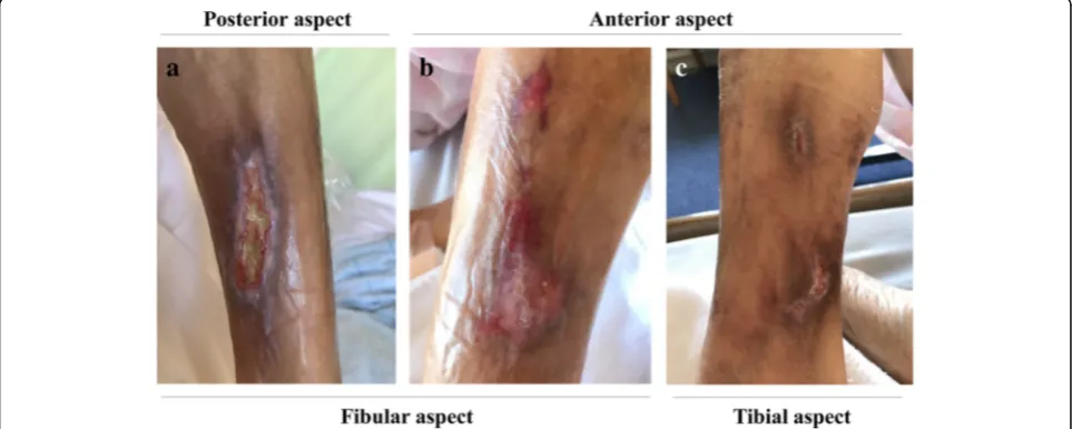
temperature was 36.5 °C, pulse rate was 68 beats per mi-nute, and blood pressure was 144/71 mmHg. Physical examination revealed a systolic heart murmur at Erb’s point and painful leg ulcers (Fig.1a–c). Laboratory tests
revealed the following results: white blood cell count, 6400/mm3; erythrocyte count, 297 × 104/μL; hemoglobin, 8.5 g/dL; hematocrit, 24.7%; platelet count, 16.6 × 10⁴/ mm3; prothrombin time-international normalized ratio, 1.59; activated partial thromboplastin time, 35.2 s; fibrin, 384 mg/dL; D-dimer, 1.9μg/mL; albumin, 3.3 g/dL; blood
urea nitrogen, 93.8 mg/dL; creatinine, 11.0 mg/dL;
adjusted-Ca, 10.1 mg/dL; P, 6.2 mg/dL; i-PTH, 392 pg/ mL; alkaline phosphatase, 312 IU/L; hemoglobin A1c, 5.0%; and C-reactive protein, 0.2 mg/dL. Bone turnover markers showed the following results: undercarboxylated osteocalcin, 220.1 ng/mL (reference range < 4.5 ng/mL) and tartrate-resistant acid phosphatase type 5b, 871 mU/ dL (reference range 170–590 mU/dL). Her Kt/V urea was 1.35, which was lower than her previous values. Anti-nuclear antibody, anti-cardiolipin antibody, and
lupus anticoagulant were not detected. Likewise,
anti-neutrophil cytoplasmic antibodies and cryoglobulin were absent. Levels of protein C and protein S were within the normal range. In addition, her ankle-brachial index was within the normal range, suggesting the ab-sence of peripheral arterial disease. Histological examin-ation of a hematoxylin and eosin-stained skin biopsy specimen from the relatively normal tissue around the ulcers in her right lower leg showed edematous thicken-ing of the vessel wall and thrombosis of small vessels (Fig. 2a, b). Von Kossa staining showed marked Ca de-position in the media of the arterioles (Fig.2c, d). X-rays
of the patient’s right lower leg revealed subcutaneous extravascular calcifications and a calcified artery (Fig.3a, b). Based on these findings, the patient was diagnosed as having calciphylaxis.
The patient’s clinical course is shown in Fig.4. As part of her treatment, the VA failure was surgically repaired and HD treatment time was prolonged, which restored the Kt/V urea (1.43) and allowed for P removal. Next, we discontinued administration of intravenous calcitriol and temporally stopped prescription of calcium carbon-ate. Additionally, treatment with oral cinacalcet was switched to intravenous etelcalcetide (15μg/week) in order to enhance treatment adherence. Although war-farin is a risk factor for calciphylaxis, we did not discon-tinue warfarin because anticoagulation therapy following artificial heart valve replacement surgery was considered essential. Administration of STS was ceased immediately because it caused severe nausea. Consequently, serum Ca, P, and Ca-P product levels decreased immediately, although PTH levels remained high (Fig. 4). With this therapy, her painful and severe skin ulcers dramatically regressed in only 3 months (Fig. 5a, c). Currently, her calciphylaxis is completely cured, and we are considering the next treatment strategy for SHPT. Surgical interven-tion such as parathyroidectomy was considered earlier, but such intervention was not feasible because surgery under general anesthesia was impossible due to her poor cardiac status. Restarting a low dose of calcium carbon-ate or VDRA, which will enable an increase in the dose of etelcacetide, will be considered as the next option.
Discussion
With reference to previous reports [7, 12, 13], the risk factors (Table 1) for calciphylaxis identified in the
present case were mainly warfarin therapy, development of SHPT as a complication, decline in Kt/V urea due to VA failure, disturbed Ca-P metabolism, use of calcium carbonate as a phosphate binder, and inappropriate ad-ministration of VDRA.
In terms of warfarin therapy, we decided not to switch warfarin to non-vitamin K-dependent oral anticoagu-lants (NOAC) because of the lack of a clear benefit of
NOAC in patients with CKD [15]. Additionally, NOAC
is a non-insurance medication for dialysis patients in Japan. Regarding the mechanism by which warfarin is a risk factor for calciphylaxis, Danziger indicated that en-dogenous inhibitors of vascular calcification (VC), such as matrix Gla protein (MGP), are activated by vitamin K-dependent mechanisms [16]. A recent report also im-plied that CKD patients on warfarin therapy may not be able to inhibit VC due to a reduction in the activation of
such proteins [17]. Thus, careful assessment of the
Fig. 2Pathological findings of a skin biopsy specimen obtained from the right lower leg at admission.a,bEdematous thickening of the vessel wall and thrombosis in a small vessel were observed (hematoxylin and eosin staining, original magnification × 40).c,dMarked calcium deposition was observed in the media of the arterioles (Von Kossa staining, original magnification × 40)
therapeutic indications for warfarin is needed in patients with CKD who have a predisposition to VC.
SHPT is known to play a pivotal role in the development of calciphylaxis [7]. However, a recent cohort study demon-strated that approximately 45% of dialysis-dependent pa-tients with calciphylaxis have relatively low levels of i-PTH
[2]. A case-control study by Hayashi et al. indicated that the elevation of i-PTH was not a significant risk factor for calciphylaxis [13]. Moreover, Karmegam et al. reported an intriguing case in which calciphylaxis developed after sub-total parathyroidectomy for severe SHPT [18]. Taken to-gether, these previous reports temper the significance of Fig. 4Clinical course and treatment of the patient. Adj-Ca, adjusted calcium; CaCO3, calcium carbonate; Ca × P, calcium-phosphorus product; P, phosphorus; i-PTH, intact parathyroid hormone
SHPT as a risk factor for the progression of calciphylaxis, which is compatible with our patient’s clinical course in which calciphylaxis improved rapidly although i-PTH levels
remained high. As pointed out previously [19],
over-suppression of i-PTH, leading to adynamic bones (low bone turnover), can exacerbate extraskeletal Ca deposition, which may conversely aggravate calciphylaxis.
Calciphylaxis is also known by the alias calcific uremic arteriolopathy [2], since the accumulation of uremic toxins is believed to be involved in the pathogenesis of this condition [20]. Previous reports indicated that the uremic condition accelerated VC via activation of the re-ceptor activator of NF-κB ligand [21,22], and that inten-sifying dialysis by increasing its duration or frequency was critical for preventing the progression of calciphy-laxis [20]. However, non-uremic calciphylaxis has re-cently been reported [1, 3, 7, 22], demonstrating that causative factors other than the uremic condition could be central to the development of calciphylaxis. Actually, Kt/V urea in our patient decreased just before her hospitalization, indicating that the state of under-dialysis that causes accumulation of uremic toxins was transient. Hence, we speculate that the favorable outcome in the present case could be attributable to the elimination of more crucial causative factors rather than uremic toxins.
Generally, appropriate management of Ca-P metabol-ism is recognized to be important in the treatment of calciphylaxis. A recent review [23] by Brandenburg et al. described that misdirected Ca-P metabolism is a key fac-tor contributing to the development of calciphylaxis. In other words, natural mineralization methods are dis-turbed and pro-calcifying activities are present in the vascular walls of patients with calciphylaxis [23]. Nigwe-kar et al. demonstrated the rationale of this theory by detecting significant stimulation of the bone morpho-genetic protein pathway in the small vessels of skin sam-ples from patients with calciphylaxis [11]. This suggests that a treatment strategy to increase P removal and lessen the Ca burden is indispensable to prevent the pro-gression of calciphylaxis. Indeed, in the present case, ap-propriate management of Ca-P metabolism led to regression of the ulcers. Immediate repair of the VA fail-ure and prolongation of HD treatment time was effective for greater P removal. Discontinuation of intravenous calcitriol administration was also effective and reason-able to avoid hyperphosphatemia, hypercalcemia, and elevation of the Ca-P product. Furthermore, switching from oral cinacalcet to intravenous etelcalcetide, a novel peptide agonist of the Ca-sensing receptor (CaSR), might also have provided some benefit to the patient.
No definitive evidence exists regarding the influence of VDRA on VC. However, a previous report by Bas et al. showed that calcitriol treatment induced time-dependent VC in rats without renal dysfunction, with the VC
regressing rapidly with decreasing aortic Ca-P deposition after the withdrawal of calcitriol [24]. In the present case, discontinuation of intravenous calcitriol administration re-sulted in alleviation of her ulcers in a short period of time, which indicated that induction of significant elevation of the Ca-P product by excessive VDRA administration is definitely harmful. Future studies are required to elucidate the role of VDRA in the development of calciphylaxis.
To the best of our knowledge, this is the first case of calciphylaxis in which the CaSR agonist was switched from cinacalcet to etelcalcetide in order to enhance the treatment for mineral and bone disorders (MBD). Ac-cording to recent reports [25–27], etelcalcetide signifi-cantly improved treatment adherence and reduced adverse gastrointestinal effects in ESKD patients with SHPT. Furthermore, several clinical trials and observa-tional studies have indicated that etelcalcetide is more potent than cinacalcet in reducing i-PTH levels, with the maintenance of Ca and P levels within the target range [26–28]. Thus, etelcalcetide is currently expected to be a promising agent for CKD-MBD. Unfortunately, adminis-tration of etelcalcetide did not reduce i-PTH levels in the present case, which was considered to attribute that i-PTH level just increased in response to hypocalcemia due to discontinuation of VDRA and calcium carbonate. Hence, the contribution of intravenous etelcalcetide therapy to a decrease in serum Ca and P levels appeared to be minor in this case when considering the pharma-cological effects of etelcalcetide. However, we presumed that its administration might have partially contributed to the regression of calciphylaxis, as is seen in cases effectively treated with cinacalcet [29–32]. In addition, several basic studies revealed the direct therapeutic ef-fect of calcimimetics on VC [33–35]. Mendoza et al. demonstrated the inhibitory effects of calcimimetic AMG 641 on VC in vivo and in vitro via activation of MGP [35]. These reports suggested the potential efficacy of etelcalcetide for the treatment of calciphylaxis. Actu-ally, our patient showed poor medical adherence and re-sistance to increasing the dosage of cinacalcet because of nausea. Therefore, we speculate that treatment with cinacalcet alone without switching to etelcalcetide might have been slightly effective in the treatment of calciphy-laxis in the present case. Accumulation of similar cases and further analysis will be necessary to elucidate the therapeutic effects of etelcalcetide on the progression of calciphylaxis.
Literature review
There are only few reported cases of calciphylaxis in which a CaSR agonist, such as cinacalcet and
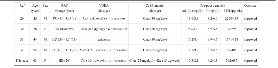
etelcalce-tide, was administered. Table 2 shows a summary of
Almost all the cases had severe SHPT and remarkable disturbance of Ca and P metabolism. Of note, four of the five cases were receiving inappropriate VDRA ther-apy despite the metabolic disturbance. Eventually, all cases showed regression of calciphylaxis after optimizing hypercalcemia and hyperphosphatemia with the admin-istration of a CaSR agonist and cessation of inappropri-ate VDRA therapy. Besides the present case, another case [32] also achieved a favorable outcome without sup-pressing i-PTH levels.
Conclusion
We successfully treated calciphylaxis by optimization of disturbed serum Ca and P levels without removing the influence of warfarin therapy or severe SHPT. Therefore, we suggest that intensive management of Ca-P metabol-ism is essential to prevent the development of calciphy-laxis. Hypercalcemia or hyperphosphatemia due to excess VDRA therapy could be an aggravating factor for calciphylaxis in CKD patients on HD. Administration of
etelcalcetide, which enhances treatment adherence
might contribute to the healing of the ulcers in patients with calciphylaxis.
Abbreviations
Ca:Calcium; CaSR: Ca-sensing receptor; CKD: Chronic kidney disease; ESKD: End-stage kidney disease; HD: Hemodialysis; i-PTH: Intact parathyroid hormone; MBD: Mineral and bone disorders; MGP: Matrix Gla protein; NOAC: Non-vitamin K-dependent oral anticoagulants; P: Phosphorus; SHPT: Secondary hyperparathyroidism; STS: Sodium thiosulfate; VA: Vascular access; VC: Vascular calcification; VDRA: Vitamin D receptor activators
Acknowledgements
None applicable.
Funding
T. Shibata has received research funding from Kyowa Hakko Kirin Co., Ltd., Ono Pharmaceutical Co., Ltd. and Astellas Pharma Inc., although the funders played no role in study design and preparation of the manuscript.
Availability of data and materials
The data and materials relevant to this case presentation are all included in the published manuscript.
Authors’contributions
All the authors have approved the manuscript and agreed to the submission to this journal. YW is responsible for the manuscript. YW, YM, YS, TH, and TS participated in the study conception and design, acquisition of data, interpretation of data, drafting or revision of the manuscript, and approval of the final version of the manuscript. YM, YW, YS, TH, NK, AI, and MS provided medical care for the patient.
Ethics approval and consent to participate
According to the Ethical Guidelines for Medical and Health Research involving Human Subjects in Japan, ethics approval is not necessary for case reports.
Consent for publication
Written informed consent was obtained from the patient for the publication of this case report and any accompanying test results.
Competing interests
The authors declare that they have no competing interests.
Publisher’s Note
Springer Nature remains neutral with regard to jurisdictional claims in published maps and institutional affiliations.
Received: 4 December 2018 Accepted: 18 April 2019
References
1. Nigwekar SU, Thadhani R, Brandenburg VM. Calciphylaxis. N Engl J Med. 2018;378:1704–14.
2. Brandenburg VM, Kramann R, Rothe H, Kaesler N, Korbiel J, Specht P, et al. Calcific uraemic arteriolopathy (calciphylaxis): data from a large nationwide registry. Nephrol Dial Transplant. 2018;32:126–32.
3. Garcia-Lozano JA, Ocampo-Candiani J, Martinez-Cabriales SA, Garza-Rodriguez V. An update on calciphylaxis. Am J Clin Dermatol. 2018;19:599– 608.
4. Devey S, Valois A, Cazajous G, Beaume J, Abed S, Okhremchuk I, et al. Calciphylaxis in hemodialysis patients: 8 cases treated with sodium thiosulfate. Ann Dermatol Venereol. 2018;145:288–92.
5. Nigwekar SU, Zhao S, Wenger J, Hymes JL, Maddux FW, Thadhani RI, et al. A nationally representative study of calcific uremic Arteriolopathy risk factors. J Am Soc Nephrol. 2016;27:3421–9.
6. Hayashi M. Calciphylaxis: diagnosis and clinical features. Clin Exp Nephrol. 2013;17:498–503.
7. Nigwekar SU, Kroshinsky D, Nazarian RM, Goverman J, Malhotra R, Jackson VA, et al. Calciphylaxis: risk factors, diagnosis, and treatment. Am J Kidney Dis. 2015;66:133–46.
8. Weenig RH, Sewell LD, Davis MD, McCarthy JT, Pittelkow MR. Calciphylaxis: natural history, risk factor analysis, and outcome. J Am Acad Dermatol. 2007; 56:569–79.
Table 2Clinical features of previously reported cases of calciphylaxis treated by calcium-sensing receptor agonist
9. Chen TY, Lehman JS, Gibson LE, Lohse CM, El-Azhary RA. Histopathology of calciphylaxis: cohort study with clinical correlations. Am J Dermatopathol. 2017;39:795–802.
10. Kramann R, Brandenburg VM, Schurgers LJ, Ketteler M, Westphal S, Leisten I, et al. Novel insights into osteogenesis and matrix remodelling associated with calcific uraemic arteriolopathy. Nephrol Dial Transplant. 2013;28:856–68. 11. Nigwekar SU, Jiramongkolchai P, Wunderer F, Bloch E, Ichinose R, Nazarian RM,
et al. Increased bone morphogenetic protein signaling in the cutaneous vasculature of patients with calciphylaxis. Am J Nephrol. 2017;46:429–38. 12. Vedvyas C, Winterfield LS, Vleugels RA. Calciphylaxis: a systematic review of
existing and emerging therapies. J Am Acad Dermatol. 2012;67:e253–60. 13. Hayashi M, Takamatsu I, Kanno Y, Yoshida T, Abe T, Sato Y. A case-control
study of calciphylaxis in Japanese end-stage renal disease patients. Nephrol Dial Transplant. 2012;27:1580–4.
14. Cohen GF, Vyas NS. Sodium thiosulfate in the treatment of calciphylaxis. J Clin Aesthet Dermatol. 2013;6:41–4.
15. Feldberg J, Patel P, Farrell A, Sivarajahkumar S, Cameron K, Ma J, et al. A systematic review of direct oral anticoagulant use in chronic kidney disease and dialysis patients with atrial fibrillation. Nephrol Dial Transplant. 2018; [Epub ahead of print].
16. Danziger J. Vitamin K-dependent proteins, warfarin, and vascular calcification. Clin J Am Soc Nephrol. 2008;3:1504–10.
17. Christiadi D, Singer RF. Calciphylaxis in a dialysis patient successfully treated with high-dose vitamin K supplementation. Clin Kidney J. 2018;11:528–9. 18. Karmegam S, Shetty A. Calciphylaxis after parathyroidectomy. Hemodial Int.
2018;21(Suppl 2):S62–6.
19. Mawad HW, Sawaya BP, Sarin R, Malluche HH. Calcific uremic arteriolopathy in association with low turnover uremic bone disease. Clin Nephrol. 1999;52: 160–6.
20. Baldwin C, Farah M, Leung M, Taylor P, Werb R, Kiaii M, et al. Multi-intervention management of calciphylaxis: a report of 7 cases. Am J Kidney Dis. 2011;58:988–91.
21. Byon CH, Chen Y. Molecular mechanisms of vascular calcification in chronic kidney disease: the link between bone and the vasculature. Curr Osteoporos Rep. 2015;13:206–15.
22. Nigwekar SU, Wolf M, Sterns RH, Hix JK. Calciphylaxis from nonuremic causes: a systematic review. Clin J Am Soc Nephrol. 2008;3:1139–43. 23. Brandenburg VM, Sinha S. Calciphylaxis: another piece of the puzzle. Am J
Nephrol. 2017;46:427–8.
24. Bas A, Lopez I, Perez J, Rodriguez M, Aguilera-Tejero E. Reversibility of calcitriol-induced medial artery calcification in rats with intact renal function. J Bone Miner Res. 2006;21:484–90.
25. Block GA, Bushinsky DA, Cheng S, Cunningham J, Dehmel B, Drueke TB, et al. Effect of etelcalcetide vs cinacalcet on serum parathyroid hormone in patients receiving hemodialysis with secondary hyperparathyroidism: a randomized clinical trial. JAMA. 2017;317:156–64.
26. Shigematsu T, Fukagawa M, Yokoyama K, Akiba T, Fujii A, Odani M, et al. Long-term effects of etelcalcetide as intravenous calcimimetic therapy in hemodialysis patients with secondary hyperparathyroidism. Clin Exp Nephrol. 2018;22:426–36.
27. Eidman KE, Wetmore JB. Treatment of secondary hyperparathyroidism: how do cinacalcet and etelcalcetide differ? Semin Dial. 2018;31:440–4. 28. Yoshimura K, Funakoshi Y, Terawaki H. Dramatic regression of parathyroid
gland swelling after conversion of calcimimetic medication from cinacalcet to etelcalcetide. Ther Apher Dial. 2018;22:553–4.
29. Velasco N, MacGregor MS, Innes A, MacKay IG. Successful treatment of calciphylaxis with cinacalcet-an alternative to parathyroidectomy? Nephrol Dial Transplant. 2006;21:1999–2004.
30. Mohammed IA, Sekar V, Bubtana AJ, Mitra S, Hutchison AJ. Proximal calciphylaxis treated with calcimimetic‘cinacalcet’. Nephrol Dial Transplant. 2008;23:387–9.
31. Sharma A, Burkitt-Wright E, Rustom R. Cinacalcet as an adjunct in the successful treatment of calciphylaxis. Br J Dermatol. 2006;155:1295–7. 32. Arai N, Mizobuchi M, Wada Y, Inoue T, Katou N, Shibata T, et al. A case of
calciphylaxis effectively treated using cinacalcet. J Japanese Soc Dial Ther. 2013;46:955–61.
33. Lopez I, Aguilera-Tejero E, Mendoza FJ, Almaden Y, Perez J, Martin D, et al. Calcimimetic R-568 decreases extraosseous calcifications in uremic rats treated with calcitriol. J Am Soc Nephrol. 2006;17:795–804.
34. Ivanovski O, Nikolov IG, Joki N, Caudrillier A, Phan O, Mentaverri R, et al. The calcimimetic R-568 retards uremia-enhanced vascular calcification and
atherosclerosis in apolipoprotein E deficient (apoE−/−) mice. Atherosclerosis. 2009;205:55–62.
![Table 1 Representative risk factors for calciphylaxis based onprevious reports [7, 12, 13]](https://thumb-us.123doks.com/thumbv2/123dok_us/780614.1575067/2.595.304.541.107.336/table-representative-risk-factors-calciphylaxis-based-onprevious-reports.webp)