R E S E A R C H
Open Access
Protocol for the detection and nutritional
management of high-output stomas
Jose J Arenas Villafranca
1,2*, Cristobal López-Rodríguez
2, Jimena Abilés
2, Robin Rivera
3,
Norberto Gándara Adán
4and Pilar Utrilla Navarro
5Abstract
Introduction:An issue of recent research interest is excessive stoma output and its relation to electrolyte abnormalities. Some studies have identified this as a precursor of dehydration and renal dysfunction. A prospective study was performed of the complications associated with high-output stomas, to identify their causes, consequences and management.
Materials and methods:This study was carried out by a multidisciplinary team of surgeons, gastroenterologists, nutritionists and hospital pharmacists. High-output stoma (HOS) was defined as output≥1500 ml for two consecutive days. The subjects included in the study population, 43 patients with a new permanent or temporary stoma, were classified according to the time of HOS onset as early HOS (<3 weeks after initial surgery) or late HOS (≥3 weeks after surgery). Circumstances permitting, a specific protocol for response to HOS was applied. Each patient was followed up until the fourth month after surgery.
Results:Early HOS was observed in 7 (16 %) of the sample population of 43 hospital patients, and late HOS, in 6 of the 37 (16 %) non-early HOS population. By type of stoma, nearly all HOS cases affected ileostomy, rather than colostomy, patients. The patients with early HOS remained in hospital for 18 days post surgery, significantly longer than those w ith no HOS (12 days). The protocol was applied to the majority of EHOS patients and achieved 100 % effectiveness. 50 % of readmissions were due to altered electrolyte balance. Hypomagnesaemia was observed in 33 % of the late HOS patients.
Conclusion:The protocol developed at our hospital for the detection and management of HOS effectively addresses possible long-term complications arising from poor nutritional status and chronic electrolyte alteration.
Keywords:High-output stomas, Hypomagnesaemia, Ileostomy, Readmission, Protocol
Introduction
The most common causes of intestinal resection in adults are aggressive secondary surgery for vascular and neoplas-tic disease, and the sequelae of Crohn’s disease with poor pharmacological control [1]. Many of these resections re-quire the construction of a stoma, a surgically-created opening in the abdomen for the discharge of faecal contents, to protect a fragile anastomosis, to manage incontinence or, in the case of certain temporary ostomies, to alleviate symptoms [2].
However, creating a stoma can, in itself, provoke peri-operative and postperi-operative complications, with an esti-mated incidence of 20–60 % [3, 4]. Local complications that have commonly been reported include peristomal skin damage, infections, parastomal hernia, stenosis, re-traction, prolapse and peristomal varices, which have been associated with high rates of morbidity and mortality [5].
An issue of recent research interest is excessive output from the stoma and its relation to electrolyte abnormal-ities. Some studies have identified this complication as a precursor of dehydration and renal dysfunction, with an estimated incidence of 1–17 %. It is believed to be the cause of 4–43 % of hospital readmissions [3, 6].
High output stomas (HOS) are normally managed by identifying the cause and by treatment consisting of the
* Correspondence:[email protected] 1
Clinical Medicine and Health Care PhD programme, University of Granada, Granada, Spain
2
Pharmacy and Nutrition Service, A7, km. 187, Hospital Costa del Sol, Marbella (Málaga) 29603, Spain
Full list of author information is available at the end of the article
oral or/and intravenous replacement of water and electrolytes, antisecretory and antidiarrheal medication, nutritional and psychological support [7]. However, there is little information about HOS and most studies report-ing rates of complications and mortality followreport-ing the creation of a stoma do not inform of the incidence, causes and management of HOS.
Hospital readmissions by patients who have undergone colorectal surgery are frequent and costly [8], and HOS are known to delay patient recovery after surgery. Further-more, HOS constitute a frequent complication in ileos-tomy patients and are poorly identified; in consequence, the clinical approach adopted is often inadequate. In this prospective study, we examine HOS and identify their possible causes, consequences and management.
Materials and methods
A prospective, analytical study of a cohort of all patients who underwent surgery resulting in a stoma was carried out. Patients of both sexes, aged 18 years and over, were included. Patients who had undergone previous ostomies were excluded, as were patients who, after surgery, required intensive care for more than 7 days, and those who did not give written informed consent.
Assuming a prevalence of HOS in the overall population of 16 % [7], a sample with 50 patients would achieve a statistical power of over 80 %, for alpha = 0.05. Therefore, 50 patients were recruited after their surgery. All subjects gave their written informed consent. The study was ap-proved by the Hospital’s Ethics Committee, and adhered in all respects and at all times to the Helsinki Declaration.
HOS was defined as ostomy output ≥1500ml for two consecutive days [7, 9, 10]. The patients were classified as early or late according to the time of onset of HOS [7]:
Early HOS(EHOS):Patients who developed HOS in the postoperative phase (<3 weeks after stoma formation), normally before hospital discharge. Late HOS(LHOS):Patients who developed HOS≥3 weeks after stoma formation.
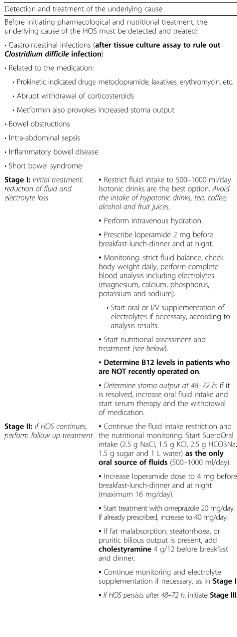
A multidisciplinary team of surgeons, gastroenterolo-gists, nutritionists and hospital pharmacists was estab-lished, and a protocol for response to HOS was designed [11] (Table 1).
Stoma output was measured for all patients included in the study, carried out over a postoperative follow-up period of four months. Any readmissions needed during this period were recorded.
The descriptive analysis was performed using mea-sures of position and central tendency (median and interquartile range) for quantitative variables and the frequency distribution for qualitative variables. Variables were compared by the chi-square test (or Fisher’s exact
Table 1Treatment protocol for high output stomas Detection and treatment of the underlying cause
Before initiating pharmacological and nutritional treatment, the underlying cause of the HOS must be detected and treated: •Gastrointestinal infections (after tissue culture assay to rule out Clostridium difficileinfection)
•Related to the medication:
•Prokinetic indicated drugs: metoclopramide, laxatives, erythromycin, etc. •Abrupt withdrawal of corticosteroids
•Metformin also provokes increased stoma output •Bowel obstructions
•Intra-abdominal sepsis •Inflammatory bowel disease •Short bowel syndrome Stage I:Initial treatment: reduction of fluid and electrolyte loss
▪Restrict fluid intake to 500–1000 ml/day. Isotonic drinks are the best option.Avoid the intake of hypotonic drinks, tea, coffee, alcohol and fruit juices.
▪Perform intravenous hydration.
▪Prescribe loperamide 2 mg before breakfast-lunch-dinner and at night.
▪Monitoring: strict fluid balance, check body weight daily, perform complete blood analysis including electrolytes (magnesium, calcium, phosphorus, potassium and sodium).
•Start oral or I/V supplementation of electrolytes if necessary, according to analysis results.
▪Start nutritional assessment and treatment(see below).
▪Determine B12 levels in patients who are NOT recently operated on.
▪Determine stoma output at 48–72 h: if it is resolved, increase oral fluid intake and start serum therapy and the withdrawal of medication.
Stage II:If HOS continues, perform follow up treatment
▪Continue the fluid intake restriction and the nutritional monitoring. Start SueroOral intake (2.5 g NaCl, 1.5 g KCl, 2.5 g HCO3Na, 1.5 g sugar and 1 L water)as the only oral source of fluids(500–1000 ml/day).
▪Increase loperamide dose to 4 mg before breakfast-lunch-dinner and at night (maximum 16 mg/day).
▪Start treatment with omeprazole 20 mg/day. If already prescribed, increase to 40 mg/day.
▪If fat malabsorption, steatorrhoea, or pruritic bilious output is present, add cholestyramine4 g/12 before breakfast and dinner.
▪Continue monitoring and electrolyte supplementation if necessary, as inStage I.
test if fewer than 5 observations were expected) for the qualitative variables and the Mann–Whitney U test for the quantitative ones. In all analyses, the level of statis-tical significance was set at p < 0.05.
Results
Of the 50 patients recruited at the outset, 7 were lost to follow up. Hence, the statistical analysis was performed on a final sample of 43 patients; of these, 63 % were male, with a median age of 66 years (range 58–73).
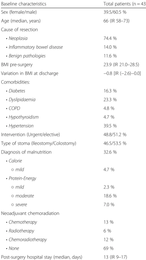
Colorectal cancer was the most common indication for the formation of a stoma. With respect to the type of surgical intervention, a small majority of cases required a colostomy, and an ileostomy was created in the other cases. Almost half of the interventions were performed as an urgent procedure. Table 2 describes the general characteristics of the study population.
High-output stoma
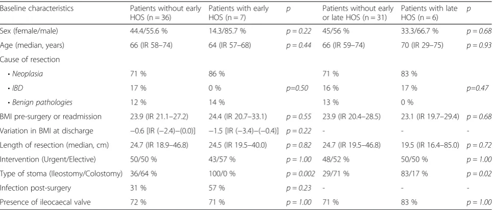
16 % of patients had EHOS and 14 % had LHOS. Table 3 shows the characteristics of the patients with EHOS, LHOS and no HOS. The median period elapsed from surgery to the appearance of EHOS was eight days. The underlying cause of high output was prokinetic agents (14 %), infection (43 %) and unknown (43 %).
The protocol was applied to five of the seven EHOS patients. In one case, it was not applied due to spontan-eous resolution and in the other, because the patient was lost to follow up. In four of the five to whom it was ap-plied, the HOS was resolved in Phase I protocol, and in
the remaining one it was resolved in Phase II (Table 1). These patients received pharmacological support and nutritional recommendations for the control of their HOS, according to the standard protocol [11].
The patients who had postoperative EHOS remained in hospital for 18 days, a significantly longer period than those who did not suffer this complication (12 days) (U= 53.00; p = 0.02). With respect to the outcome variables, al-though the difference was not significant, there was a higher rate of infections among the patients with EHOS (57.1 % vs. 31.4 %;χ2= 1.68; p = 0.23). The following vari-ables presented no statistical differences between patients with EHOS and no HOS: age (U= 102.00; p = 0.45), sex (χ2= 2.23; p = 0.22), reason for surgical intervention (χ2= 1.40; p = 0.50), emergency surgery (χ2= 0.12, p = 1.00),
Table 1Treatment protocol for high output stomas(Continued)
Stage III.If HOS persists, evaluate treatment and case management
▪Supplement with hydro- and lipid-soluble oral vitamins.
▪Maintain loperamide and addcodeine 15–60 mg. before breakfast-lunch-dinner. Contraindicated if the patient has CrCl <15 ml/min.
▪If fat malabsorption persists, increase cholestyramine dose to 4 g before breakfast-lunch-dinner.
▪If HOS > 2000 mlafter two weeks: add octreotide 200 mcg/day for 3–5 days. If no improvement is obtained, suspend this treatment.
▪Monitor fluid intake. Specific nutritional treatment
▪Avoid fluid intake during meals.
▪It may be advisable to temporarily increase the salt content of foods in order to promote fluid reabsorption.
▪Little is known about the use of soluble fibre. Insoluble fibre is contraindicated because of the risk of bowel obstruction.
▪The effect of antidiarrhoeal microorganisms on HOS is unknown.
Table 2General characteristics of the population
Baseline characteristics Total patients (n = 43)
Sex (female/male) 39.5/60.5 %
Age (median, years) 66 (IR 58–73)
Cause of resection
•Neoplasia 74.4 %
•Inflammatory bowel disease 14.0 % •Benign pathologies 11.6 %
BMI pre-surgery 23.9 (IR 21.0–28.5)
Variation in BMI at discharge −0.8 [IR (−2.6)−0.0]
Comorbidities:
•Diabetes 16.3 %
•Dyslipidaemia 23.3 %
•COPD 4.8 %
•Hypothyroidism 4.7 %
•Hypertension 39.5 %
Intervention (Urgent/elective) 48.8/51.2 %
Type of stoma (Ileostomy/Colostomy) 46.5/53.5 %
Diagnosis of malnutrition 32.6 % •Calorie
○mild 4.7 %
•Protein-Energy
○mild 2.3 %
○moderate 18.6 %
○severe 7.0 %
Neoadjuvant chemoradiation
•Chemotherapy 13 %
•Radiotherapy 6 %
•Chemoradiotherapy 12 %
•None 69 %
Post-surgery hospital stay (median, days) 13 (IR 9–17)
absence of ileocaecal valve (χ2= 0.00; p = 1.00), or malnu-trition (χ2= 0.01, p = 1.00). Regarding the type of ostomy, all of the EHOS and 83 % of the LHOS cases affected pa-tients with ileostomy (χ2 = 9.62; p = 0.002 and χ2 = 6.30; p = 0.02, respectively). Thus, the location of the stoma is of evident significance.
The median period from surgery to the appearance of LHOS was 25 days. The protocol was applied to 33 % of LHOS patients (all resolved in Phase I). The rest did not receive protocol care because they were readmitted for non-general surgery services (Internal Medicine and Emergency). The underlying cause of the LHOS was not detected in any of these cases. 50 % of these patients were given pharmacological support and nutritional rec-ommendations by clinicians to control the high output at discharge. However, 17 % were given erroneous rec-ommendations based on increasing fluid intake, and no pharmacological control was recommended to control the high output.
Readmission
28 % of the stoma patients were subsequently readmitted to hospital, with a mean weight loss from discharge to readmission of 5.2 ± 2.3 kg. In 50 % of the patients who were readmitted for LHOS, the principal cause was altered electrolyte balance and dehydration. Electrolyte abnormalities were detected in 100 % of these patients on readmission. Magnesium levels were monitored in three of the six LHOS, and hypomagnesaemia was observed in two patients.
No differences were found between LHOS and no HOS patients regarding the following variables related to readmis-sion: age (U= 91.00; p = 0.93), gender (χ2= 0.29; p = 0.68), cause of surgical intervention (χ2= 0.88; p = 0.47), urgent
surgery (χ2= 0.01; p = 1.00), absence of ileocaecal valve (χ2= 0.39; p = 1.00), neoadjuvant chemotherapy (χ2= 3.18; p = 0.11), neoadjuvant radiotherapy (χ2= 1.41; p = 0.27), BMI at discharge U= 75.00, p = 0.68), or malnutrition (χ2= 1.22, p = 0.58).
Discussion
The need to construct a stoma often arises in clinical practice [3]. According to statistical records, 1.5 per thousand of the Spanish population (i.e., 60,000 patients) live with a stoma. Taking the age factor into account, the incidence of stoma corresponds to over 3 per thousand of the adult population in Spain. In other Western coun-tries, figures of 2–4 per thousand of the adult population [12] have been reported.
In our study population, colorectal cancer was the most common reason for a stoma being needed (65.1 % of cases); this finding is consistent with results reported elsewhere [3, 5].
During the first days after the construction of an ostomy, faecal effluent usually increases, but it decreases rapidly following intestinal adaptation [13]. When this adaptation fails or is prolonged, patients must face the challenge of controlling large losses of fluid that can lead to a state of chronic dehydration [14]. When these losses occur, ostomy patients present major deficits of water, sodium and magnesium [15] and can also suffer malnu-trition and long-term weight loss. This event is known as HOS, and among stoma patients our study revealed a prevalence of EHOS of 16 %, which is consistent with Baker et al. [7]. These same authors identified the under-lying cause of HOS in 50 % of cases, and noted the pres-ence of short bowel syndrome, treated by medication, and subsequent intestinal obstruction. In our study, the Table 3Characteristics of the population with HOS (Early and Late) and the comparison with no HOS population
Baseline characteristics Patients without early HOS (n = 36)
Patients with early HOS (n = 7)
p Patients without early or late HOS (n = 31)
Patients with late HOS (n = 6)
p
Sex (female/male) 44.4/55.6 % 14.3/85.7 % p = 0.22 45/56 % 33.3/66.7 % p = 0.68
Age (median, years) 66 (IR 58–74) 64 (IR 57–68) p = 0.44 66 (IR 59–74) 70 (IR 29–75) p = 0.93
Cause of resection
•Neoplasia 71 % 86 %
p=0.50
71 % 83 %
p=0.47
•IBD 17 % 0 % 16 % 17 %
•Benign pathologies 12 % 14 % 13 % 0 %
BMI pre-surgery or readmission 23.9 (IR 21.1–27.2) 24.4 (IR 20.7–33.1) p = 0.55 23.9 (IR 20.4–28.5) 23.1 (IR 19.7–29.4) p = 0.68
Variation in BMI at discharge −0.6 [IR (−2.4)−(0.0)] −1.5 [IR (−3.4)−(−0.4)] p = 0.22 - -
-Length of resection (median, cm) 24.7 (IR 18.9–46.8) 24.5 (IR 19.5–40.0) p = 0.82 24.7 (IR 19.5–46.8) 19.5 (IR 16.4–85.0) p = 0.72
Intervention (Urgent/Elective) 50/50 % 43/57 % p = 1.00 48/52 % 50/50 % p = 1.00
Type of stoma (Ileostomy/Colostomy) 36/64 % 100/0 % p = 0.002 29/71 % 83/17 % p = 0.02
Infection post-surgery 31 % 57 % p = 0.23 - -
-Presence of ileocaecal valve 72 % 71 % p = 1.00 71 % 83 % p = 1.00
underlying cause was identified in 57 % of the cases, with drug treatment or the presence of infection being mainly responsible for the occurrence of EHOS. None of the patients presented short bowel syndrome.
It has been reported that HOS tend to occur more fre-quently after emergency surgery [16], but we observed no such association. There was a relationship between infections and the presence of EHOS, but it was not statistically significant [17]. On the other hand, there were significant differences according to the type of ostomy, for both EHOS and LHOS when an ileostomy was performed. This finding corroborates an earlier meta-analysis of all complications arising from ostomies
–classifying the latter as colostomy or ileostomy–which concluded that only HOS presented significant differ-ences, being more common following an ileostomy [18].
We compared HOS and non-HOS patients, and found no significant differences in the universal defining vari-ables, or with regard to the causes that led to the surgi-cal intervention. Despite the importance of the presence of the ileocaecal valve in retaining stoma output and that of the secretion of hormones favouring intestinal adapta-tion after surgical resecadapta-tion [1], we observed no differ-ences with regard to the development of high output. Neither were there significant differences between pa-tients who received prior chemotherapy or radiotherapy, as have been described by Hayden et al. [8]. However, we did find significant differences with respect to the longer hospital stay required by patients with HOS, thus confirming the results of Harris et al. [3].
With respect to the outcome variables, patients with HOS presented a sharper decrease in BMI at hospital discharge and a higher rate of infections. The latter has been reported to be one of the risk factors associated with the occurrence of HOS [19]. There were no signifi-cant alterations in the electrolyte profile in patients with EHOS, and no hypomagnesaemia was detected at first hospital admission in any patient, in contrast to the 45 % of hypomagnesaemias reported by Baker [7].
An interesting finding is that EHOS appears, on average, eight days after resection. This highlights the importance of instructing the patient in this respect before hospital discharge, especially in cases of early discharge when the condition has evolved favourably. Several papers have commented on the importance of these patients being instructed and informed by a multi-disciplinary nutrition team including pharmacists, nutri-tionists, nurses and surgeons [17, 20, 21], while the NICE guidelines for ostomy patients state that they should be examined every 2–3 months by a nutritional team after the surgical intervention [22].
The early examination and treatment protocol devel-oped for these patients (Table 1) was applied to the major-ity of patients with HOS and achieved 100 % effectiveness.
In clinical practice, it is important to test for Clostridium difficile in order to detect the underlying cause of high output, as an increasing number of cases of diarrhoea have been associated with this agent during hospitalization, which affects 3–10 patients per 1000 hospitalizations [23]. Moreover, studies have reported cases of colonisations in the small intestine causing high output in patients with ileostomy [24, 25]. As part of the above protocol, nutri-tional and pharmacological recommendations were made to 83 % of EHOS patients and to 50 % of the LHOS pa-tients, who were thus enabled to achieve self-management of stoma output. This outcome represents a significant improvement in our hospital, where a previous retrospect-ive study had noted a complete absence of recommenda-tions to patients on hospital discharge [26].
The proportion of ostomy patients who were readmit-ted with altered electrolyte balance and dehydration be-cause of LHOS was 50 %, much higher than the 5.5 % reported by Baker [7], but similar to that reported in other studies, which have recorded 20–43 % of readmis-sions due to dehydration related to fluid losses through the stoma [8]. Ileostomy is considered a risk factor for kidney failure [27]. In our study, 50 % of kidney failures were secondary to stoma losses, which is in accordance with the 30–71 % of the patients with HOS reported elsewhere to have developed acute kidney failure sec-ondary to dehydration [8]. In our study, the cause of LHOS was not determined in any patient, nor were tests applied to detect the presence of Clostridium difficile. The protocol was only applied in 33 % of LHOS cases, all of whom were patients admitted by the Surgery Ser-vice, where personnel had been previously instructed with respect to HOS and the recommended treatment. Among these 33 %, the follow up rate was 100 %. More-over, the existence of this protocol enabled nutritional and pharmacological recommendations to be made on hospital discharge to 50 % of the patients admitted for surgery or nephrology. However, 17 % of stoma patients received recommendations contrary to those contained in the literature on the subject and were given no pharmacological treatment to control the stoma output. A study published in 2012 examined the rate of readmis-sions due to dehydration among patients with ileostomy, comparing two periods, one of which included the im-plementation of a perioperative protocol of patient edu-cation on how HOS should be managed; this study reported a significant reduction in the rate of readmis-sions, although there were no significant differences in the duration of hospital stay [28]. In our opinion, HOS management should be extended throughout the med-ical service in order to improve the care provided for ostomy patients care during readmissions.
reduction in absorption surface area and to chelation with fatty acids [29, 30]. This condition can affect up to 45 % of patients with HOS due to hypoaldosteronism secondary to surgery and often appears in long-term HOS [7]. Unfortunately, it is not often monitored as routine clinical practice in Spain. Another common con-sequence is the appearance of electrolyte alterations such as hypokalaemia and hypocalcaemia, both of which are refractory to treatment until the Mg deficit is cor-rected. Thus, some of our patients presented simultan-eous deficits of Mg, Na, K and P.
A limitation of this study is the small sample size, which means that the conclusions drawn cannot be considered definitive.
Conclusions
A review of stoma output should be included in stand-ard clinical practice, in view of the comorbidity of this condition, which is demonstrated in the present study. The protocol developed at our hospital for the detection and management of HOS has proved to be effective in addressing possible long-term complications arising from poor nutritional status and chronic electrolyte alterations, which may provoke severe consequences among the patients affected.
Competing interests
The authors declare that they have no competing interests.
Authors’contributions
JJAV, JA, and RR designed the study and attended the patients, and all authors helped to draft the manuscript. CLR helped with data collection and, with PUN, provided statistical and epidemiological expertise to summarise the results obtained. NGA provided critical oversight and clinical supervision. All authors have read and approved the final manuscript.
Acknowledgments
We thank the staff of the surgery and pharmacy services at the Costa del Sol Hospital. The present study is part of a Ph. D. research programme being conducted at the University of Granada.
Disclaimers
Having reading the procedure for submissions, the authors declare there is no conflict of interest.
Author details
1Clinical Medicine and Health Care PhD programme, University of Granada,
Granada, Spain.2Pharmacy and Nutrition Service, A7, km. 187, Hospital Costa
del Sol, Marbella (Málaga) 29603, Spain.3Gastroenterology Service, Costa del
Sol Hospital, Marbella (Málaga), Spain.4General Surgery Service, Costa del Sol
Hospital, Marbella (Málaga), Spain.5Pharmacology Department, University of
Granada, Granada, Spain.
Received: 17 January 2015 Accepted: 1 May 2015
References
1. Buchman AL. Síndrome de intestino corto. In: Friedman LS, Brandt LJ Feldman M, editors. Sleisenger & Fordtran: Enfermedades digestivas y hepaticas, Vol. 2. 8th ed. Madrid: Elsevier; 2008. p. 2257–75.
2. Tan WS, Tang CL, Shi L, Eu KW. Meta-analysis of defunctioning stomas in low anterior resection for rectal cancer. Br J Surg. 2009;96:462–72.
3. Harris DA, Egbeare D, Jones S, Benjamin H, Woodward A, Foster ME. Complications and mortality following stoma formation. Ann R Coll Surg Engl. 2005;87:427–31.
4. Robertson I, Leung E, Hughes D, Spiers M, Donnelly L, Mackenzie I, et al. Prospective analysis of stoma-related complications. Colorectal Dis. 2005;7:279–85.
5. Londono-Schimmer EE, Leong A, Phillips RK. Life table analysis of stomal complication following colostomy. Dis Colon Rectum. 1994;37:916–20. 6. Caricato M, Ausania F, Ripetti V, Bartolozzi F, Campoli G, Coppola R.
Retrospective analysis of long-term defunctioning stoma complications after colorectal surgery. Colorectal Dis. 2007;9:559–61.
7. Baker ML, Williams RN, Nightingale JMD. Causes and management of a high-output stoma. Colorectal Dis. 2010;13:191–7.
8. Messaris E, Sehgal R, Deiling S, Koltun WA, Stewart D, McKenna K, et al. Dehydration is the most common indication for readmission after diverting ileostomy creation. Dis Colon Rectum. 2012;55:175–80. doi:10.1097/DCR.0b013e31823d0ec5.
9. Hayden DM, Pinzon MC, Francescatti AB, Edquist SC, Malczewski MR, Jolley JM, et al. Hospital readmission for fluid and electrolyte abnormalities following ileostomy construction: preventable or unpredictable? J Gastrointest Surg. 2013;17:298–303. doi:10.1007/s11605-012-2073-5. 10. Forbes A. Inflamatory bowel disease. A clinician’s guide. London: Arnold; 2001. 11. Arenas Villafranca JJ, Abilés J, Moreno G, Tortajada Goitia B, Utrilla Navarro P,
Gándara AN. High output stoma: detection and approach. Nutr Hosp. 2014;30:1391–6.
12. ARGIA (Asociación Vasca de Ostomizados e Incontinentes). V Congreso Nacional de Ostomías. Bilbao. 2005.
13. Hill GL, Mair WSJ, Goligher JC. Impairment of‘ileostomy adaptation’in patients after ileal resection. Gut. 1974;15:982–7.
14. Okamoto T, Kusunoki M, Kusuhara K, Yamamura T, Utsunomiya. Water and electrolyte balance after ileal J pouch-analanastomosis in ulcerative colitis and familial adenomatous polyposis. Int J Colorectal Dis. 1995;10:33–8. 15. Sentongo TA. The use of oral rehydration solutions in children and adults.
Curr Gastroenterol Rep. 2004;6:307–13.
16. Leenen LP, Kuypers JH. Some factors influencing the outcome of stoma surgery. Dis Colon Rectum. 1989;32:500–4.
17. Nightingale J, Woodward JM. Guidelines for management of patients with a short bowel. Gut. 2005;55(Supl 4):1–12.
18. Tilney HS, Sains PS, Lovegrove RE, Reese GE, Heriot AG, Tekkis PP. Comparison of outcomes following ileostomy versus colostomy for defunctioning colorectal anastomoses. World J Surg. 2007;31:1142–51. 19. De Campos-Lobato LF, Alves-Ferreira PC, Gonçalves de Oliveira P, Batista de
Sousa J, Vogel JD. What are the risk factors for readmission in patients with an ileostomy. Jcol. 2013;33:203–9.
20. Culkin A, Gabe SM, Madden AM. Improving clinical outcome in patients with intestinal failure using individualised nutritional advice. J Hum Nutr Diet. 2009;22:290–8. doi:10.1111/j.1365-277X.2009.00954.x.
21. Gondal B, Trivedi MC. An overview of ostomies and the high-output ostomy. Hosp Med Clin. 2013;2:e542–51.
22. National Institute for Health and Clinical Excellence (NICE). Nutritional support in adults: oral nutrition support, enteral tube feeding and parenteral nutrition. In: NICE guidelines [CG32]. London; 2006. http://www.nice.org.uk/ guidance/CG32
23. Johal SS, Hammond J, Solomon K, James PD, Mahida YR. Clostridium difficile associated diarrhoea in hospitalised patients: onset in the community and hospital and role of flexible sigmoidoscopy. Gut. 2004;53:673–7. 24. Williams RN, Hemingway D, Miller AS. Enteral Clostridium difficile, an
emerging cause for high-output ileostomy. J Clin Pathol. 2009;62:951–3. doi:10.1136/jcp.2008.062901.
25. El Muhtaseb MS, Apollos JK, Dreyer JS. Clostridium difficile enteritis: a cause for high ileostomy output. ANZ J Surg. 2008;78:416. doi:10.1111/j.1445-2197.2008.04494.x.
26. Arenas Villafranca JJ, Abilés J, Romero Dominguez R, Fontalva AA, Moreno G, Gandara Adan N, et al. Analysis of detection and nutritional approach in intestinal high-output stomas. Ann Nutr Metab. 2013;63(suppl1):1193. 27. Beck-Kaltenbach N, Voigt K, Rumstadt B. Renal impairment caused by
temporary loop ileostomy. Int J Colorectal Dis. 2011;26:623–6. doi:10.1007/s00384-010-1086-3.
29. Arenas J, Rivera Irigoin R, Abilés J, Moreno Martínez F, Faus V. Hipomagnesemia severa en paciente con ileostomía de alto débito. Nutr Hosp. 2012;27:310–3.
30. de Armas MG G, Vega Piñero B, SJ RÁ, Civantos Modino S, Montaño Martínez JM, de Paz I P, et al. Convulsiones secundarias a hipomagnesemia severa en paciente con intestino corto. Nutr Hosp. 2010;25:1037–40.
Submit your next manuscript to BioMed Central and take full advantage of:
• Convenient online submission
• Thorough peer review
• No space constraints or color figure charges
• Immediate publication on acceptance
• Inclusion in PubMed, CAS, Scopus and Google Scholar
• Research which is freely available for redistribution