R E V I E W
Open Access
Vici syndrome: a review
Susan Byrne
1, Carlo Dionisi-Vici
2, Luke Smith
3, Mathias Gautel
3and Heinz Jungbluth
1,3,4*Abstract
Vici syndrome [OMIM242840] is a severe, recessively inherited congenital disorder characterized by the principal features of callosal agenesis, cataracts, oculocutaneous hypopigmentation, cardiomyopathy, and a combined immunodeficiency. Profound developmental delay, progressive failure to thrive and acquired microcephaly are almost universal, suggesting an evolving (neuro) degenerative component. In most patients there is additional variable multisystem involvement that may affect virtually any organ system, including lungs, thyroid, liver and kidneys. A skeletal myopathy is consistently associated, and characterized by marked fibre type disproportion, increase in internal nuclei, numerous vacuoles, abnormal mitochondria and glycogen storage. Life expectancy is markedly reduced.
Vici syndrome is due to recessive mutations inEPG5on chromosome 18q12.3, encoding ectopic P granules protein 5 (EPG5), a key autophagy regulator in higher organisms. Autophagy is a fundamental cellular degradative pathway conserved throughout evolution with important roles in the removal of defective proteins and organelles, defence against infections and adaptation to changing metabolic demands. Almost 40EPGmutations have been identified to date, most of them truncating and private to individual families.
The differential diagnosis of Vici syndrome includes a number of syndromes with overlapping clinical features, neurological and metabolic disorders with shared CNS abnormalities (in particular callosal agenesis), and primary neuromuscular disorders with a similar muscle biopsy appearance. Vici syndrome is also the most typical example of a novel group of inherited neurometabolic conditions,congenital disorders of autophagy.
Management is currently largely supportive and symptomatic but better understanding of the underlying autophagy defect will hopefully inform the development of targeted therapies in future.
Disease name
Vici syndrome; Dionisi-Vici-Sabetta-Gambarara syn-drome; Immunodeficiency with cleft lip/palate, cataract, hypopigmentation and absent corpus callosum.
Definition
Vici syndrome [OMIM242840, ORPHA1493] is a severe congenital multisystem disorder characterized by the prin-cipal features of agenesis of the corpus callosum, cataracts, oculocutaneous hypopigmentation, cardiomyopathy, a combined immunodeficiency and additional, more vari-able multisystem involvement. The condition is due to re-cessive mutations in theEPG5gene on chromosome 18q.
Epidemiology
The incidence of Vici syndrome is unknown. Since the original description of the disorder by Dionisi-Vici and colleagues in 1988 [1], an exponentially increasing number of patients has been reported, with around 50 genetically confirmed cases published to date [1–14]. Vici syndrome is likely to be rare but probably underdiagnosed.
Clinical description
Vici syndrome is one of the most extensive inherited human multisystem disorders reported to date, pre-senting invariably in the first months of life. Apart from the 5 principal diagnostic findings–callosal agen-esis, cataracts, cardiomyopathy, hypopigmentation and combined immunodeficiency-a wide range of variably present additional features has been reported, suggest-ing that virtually any organ system can be involved [4]. Three additional findings (profound developmental delay, acquired microcephaly and marked failure to thrive) have recently emerged that, although non-* Correspondence:[email protected]
1Department of Paediatric Neurology, Neuromuscular Service, Evelina’s Children Hospital, Guy’s & St. Thomas’Hospital NHS Foundation Trust, London, UK
3Randall Division of Cell and Molecular Biophysics, Muscle Signalling Section, King’s College, London, UK
Full list of author information is available at the end of the article
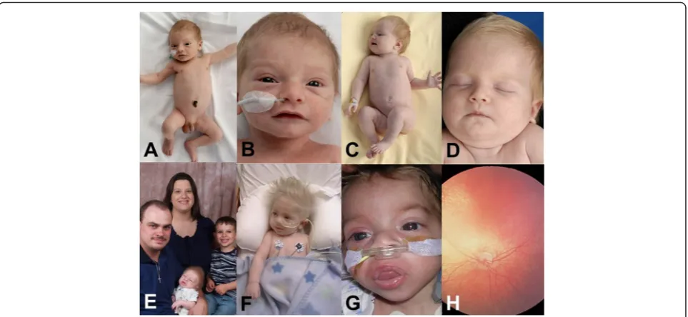
specific, are as consistently associated as the 5 main diagnostic features and highly supportive of the diag-nosis [14]. The common occurrence of structural con-genital abnormalities and acquired organ dysfunction (for example, congenital cardiac defects and cardiomyopathy later in life) is not infrequently observed in individual patients. Typical findings in Vici syndrome are outlined in detail below and summarized in Table 1. The characteris-tic features of Vici syndrome are illustrated in Fig. 1.
CNS
Development in Vici syndrome is profoundly delayed: Affected children may acquire a social smile, some de-gree of head control, and the ability to roll over, however there have been no reports of children sitting independ-ently, or acquiring speech. Where rolling has been attained, this skill may subsequently be lost. Almost two third of patients have seizures that are often difficult to control. Although head circumference is usually normal at birth, rapidly progressive microcephaly evolving within the first year of life suggests a neurodegenerative component superimposed on the principal neurodeve-lopmental defect.
In addition to agenesis of the corpus callosum, one of the five principal diagnostic features of Vici syndrome, other consistent radiological abnormalities include
pontine hypoplasia, reduced opercularisation of the Sylvian fissures, delayed myelination and general reduc-tion in white matter bulk [14]. Cortical malformareduc-tions and cerebellar abnormalities have been observed but are much less common. In few patients, distinct circum-scribed signal abnormalities (decrease in T2 with or with-out associated increase in T1 signal) have been noted within the thalami, similar to what has been described in patients with lysosomal storage disorders [15], also corre-sponding to some clinical overlap with these conditions.
Muscle
An associated skeletal muscle myopathy, already sug-gested by the presence of often profound hypotonia and variable hyperCKaemia in early case reports, was documented in detail by McClelland and colleagues in 2010 [7] and subsequently confirmed in other reports [2, 12]. Clinically, individuals with Vici syndrome are often profoundly hypotonic and weak, probably reflecting a combination of the progressive nature of the myopathy and/or ongoing neurodegeneration. Histopathologically, the myopathy associated with Vici syndrome is character-ized by marked variability in fibre size, increase in internal and centralized nuclei, type 1 fibre hypotrophy with nor-mally sized type 2 fibres (occasionally fulfilling the criteria for fibre type disproportion), increased glycogen storage and variable vacuoles on light microscopy [2, 7, 14]. Add-itional changes on electron microscopy may include abnormalities of mitochondrial structure and arrangement [4, 14] and, less frequently, sarcomeric disorganization. On the histopathological level there is considerable over-lap with the congenital myopathies, in particular Congeni-tal Fibre Type Disproportion (CFTD) and Centronuclear Myopathy (CNM), primary vacuolar myopathies, glycogen storage disorders and mitochondrial myopathies.
Nerves
Peripheral nerve involvement with almost complete ab-sence of myelinated axons has been reported in only one case to date [14]; however, an associated neuropathy may have been overlooked in other patients because of the overwhelming nature of other multisystem features. The majority of children have absent deep tendon re-flexes but those may be brisk in around a third.
Skin
Marked oculocutaneous hypopigmentation [16] is one of the cardinal features of Vici syndrome and has been noted in almost all cases reported to date. Affected indi-viduals are, however, not typically complete albinos and hypopigmentation is always relative to the familial and ethnic background (Fig. 1). Children with Vici syndrome have generally pale skin with light (often very blonde in those of Caucasian origin) hair, rather than discrete Table 1Clinical features of Vici syndrome
Feature Frequency
Principal diagnostic features
Absent corpus callosum ++++ Profound developmental delay ++++
Failure to thrive ++++
Hypopigmentation ++++
Immune problems ++++ Progressive microcephaly +++
Cardiomyopathy +++
Cataracts +++
Other features Presentation in neonatal period +++
Myopathy +++
Seizures ++
Absent reflexes (probable neuropathy)
++
Thymic aplasia +
Sensorineural deafness +
Optic atrophy +
Renal tubular acidosis +
Cleft lip/palate +
Coarse facial features +
Hepatomegaly +
hypopigmented patches. An intermittent, extensive maculopapular rash almost resembling Stevens-Johnson syndrome has been reported in few children [14].
Eyes
Bilateral cataracts are one of the “classical” diagnostic features of Vici syndrome, however, in a recent series of 50 patients those were only documented in three-quarters of affected individuals [14], probably reflecting evolution over time. Ocular features of Vici syndrome have been reviewed in detail by Filloux and colleagues [16] and include optic nerve hypoplasia, visual impair-ment, nystagmus and fundus hypopigmentation. Al-though individuals with Vici syndrome are usually only relatively hypopigmented, ocular features, in particular evidence of optic pathways misrouting on visually evoked potential (VEP) testing, and of a poorly defined and lesser depressed fovea on optical coherence tomog-raphy, are similar to those in individuals with typical al-binism [16].
Hearing
Sensorineural hearing loss was recognized in an isolated case in 2010 [7] and has been subsequently reported in other cases [6, 10] of Vici syndrome with or without confirmed EPG5 mutations. Sensorineural hearing loss is a feature that may be easily overlooked in Vici syn-drome due to profound developmental delay and over-whelming multisystem involvement, and should be actively investigated for.
Heart
Cardiac involvement is present in around 90 % of pa-tients with Vici syndrome and in around 80 % of cases a cardiomyopathy, one of the 5 main diagnostic features, has been documented. Minor congenital heart defects comprising persistent foramen ovale and atrial septal de-fects have been reported in around 10 % of patients. The associated cardiomyopathy usually develops early in life, although onset much later in childhood has been ob-served. Intermittent deterioration of cardiac function during intercurrent illness has also been noted (Patient 12.1 in [4]). Both hypertrophic and dilated forms of car-diomyopathy have been reported, always with left ventricular emphasis and occasionally in the same pa-tient subsequently evolving over time. In two unrelated patients where post mortem examination was performed [8, 11], changes in the heart also showed left ventricular emphasis, with variable degrees of interstitial fibrosis and cardiomyocytes containing vacuoles and membrane-bound cytoplasmic inclusions, possibly glycogen. In keeping with the underlying autophagy defect, cardio-myocytes showed increased staining for autophagy markers LC3 and p62 on immunohistochemistry [8].
Immune system
mucocutaneous candidiasis, sepsis and, less frequently, urinary tract infections, gastroenteritis, bacterial conjunc-tivitis, and perineal abscesses. Due to the severely reduced life expectancy, immune function has been assessed for-mally only in a few patients [6]. Abnormal findings re-ported to date include lymphopenia with variable T cell subset defects, neutropenia, leucopenia, hypogammaglo-bulinaemia, lack of response to recall antigens and a defect of memory B cells with lack of specific antibody response to certain immunizations such as those withtetanus and pneumococcal vaccine. Overall, these findings suggest prominent impairment of the humoral immune response with a milder defect of the T cell compartment, although further prospective studies will be required to delineate the immunological phenotype further. Immunological fea-tures of Vici syndrome, recommended immunological in-vestigations and potential treatment approaches have been outlined in detail by Finocchi and colleagues [6].
Thymus
Complete thymus aplasia or hypoplasia has been re-ported in around one fifth of patients [4, 14]. T-cell dys-function is part of the combined immunodeficiency observed in Vici syndrome although usually less promin-ent than B-cell dysfunction [6].
Lungs
Pulmonary hypoplasia has been reported in one patient with Vici syndrome [2]. Pulmonary involvement is common throughout life, due to recurrent respiratory infections sec-ondary to the associated combined immunodeficiency.
Thyroid
Thyroid agenesis and thyroid dysfunction have both been reported in rare patients with Vici syndrome [4, 14].
Liver
Hepatomegaly with or without associated liver dysfunc-tion has been reported in around 10 % of patients with Vici syndrome [4, 14] and is probably a reflection of in-creased glycogen storage, also reported on post mortem in few cases.
Kidneys
Renal involvement comprising hydronephrosis, renal dysfunction and/or signs of renal tubular acidosis with associated electrolyte imbalances, in particular marked hypokalaemia, have been reported in around 15 % of cases [4, 9, 14].
Blood
Some patients with Vici syndrome have been noted to develop profound anaemia [4, 14]; it is currently uncer-tain if this is a secondary feature (for example related to
recurrent severe infections) or, alternatively, reflects add-itional primary involvement of red cell lines.
Other features
Mildly dysmorphic, coarse facial features with full lips and macroglossia resembling those seen in (lysosomal) storage disorders have been noted in some patients with Vici syndrome [4, 14] (Fig. 1). Cleft lip and palate were a feature in Dionisi-Vici’s original siblings [1] but have subsequently been seen only in few families. Other minor dysmorphic features such as 2nd and 3rd toe syn-dactyly were a feature in two families reported [4, 14]. A long philtrum has been described in one family [17]. Marked failure to thrive evolving over time has been re-cently recognized as an almost universal feature [14]. One recent case report also suggests severe sleep abnor-malities that may have to be considered in Vici syn-drome [18].
Aetiology
Vici syndrome is due to recessive mutations inEPG5on chromosome 18q12.3, organized in 44 exons and encod-ing ectopic P granules protein 5 (EPG5), a protein of 2579 amino acids.EPG5(originally known asKIAA1632) was initially identified amongst a group of genes found to be mutated in breast cancer tissue [19] before its im-plication in Vici syndrome in 2013 [4].
To date, around 40EPG5 mutations have been identi-fied in families with Vici syndrome, distributed through-out the entire EPG5 coding sequence without clear genotype-phenotype correlations [13, 14]. Most EPG5
mutations associated with Vici syndrome are truncating with only few missense mutations on record. The large majority of EPG5 mutations are private to individual families, with only 3 recurrent mutations identified to date, p. Met2242CysfsX5 in an Italian and a Maltese family, p. Arg417X identified in the homozygous state in a patient from the Middle East and in the heterozygous state in a Caucasian child from the United States, and p. Gln336Arg identified in the homozygous (n= 3) and in the heterozygous (n= 1) state in four unrelated patients with definite or possible Ashkenazi ancestry [14]. Failure to identify an (or identification of one but not the allelic)
EPG5 mutation in a small number of cases with highly suggestive diagnostic features indicate the possibility of large copy number variations not detectable on Sanger se-quencing, or an altogether different genetic background.
The EPG5 protein has a key role as a regulator of au-tophagy in multicellular organisms, initially character-ized in C. elegans [20] and subsequently confirmed in
against infections and adaptation to changing metabolic demands (for review [[21–23]]). The autophagy pathway involves several tightly regulated steps, evolving from the initial formation of isolation membranes (or phagophores) to autophagosomes, whose fusion with lysosomes results in the final structures of degradation, autolysosomes (Fig. 2). The ultimate aim of the autophagy pathway is the effective delivery of an intracellular structure targeted for removal to the lysosome, and its ultimate intralysosomal degradation. Studies in EPG5-mutated fibroblasts from humans with Vici syndrome suggest that EPG5 deficiency results in failure of autophagosome-lysosome fusion [4] and, ultimately, impaired cargo delivery to the lysosome. It is currently uncertain if impaired autophagy is the only consequence of EPG5 deficiency, or only the most import-ant expression of a more generalized vesicular trafficking defect in Vici syndrome. Moreover, it remains unresolved if all manifestations of EPG5 deficiency are a direct conse-quence of the primary autophagy defect, or of the second-ary effects of defective autophagy such as reduced mitochondrial quality control and/or accumulation of de-fective proteins.
Autophagy is physiologically enhanced in neurons and muscle, probably explaining the prominent CNS and neuromuscular involvement in patients with Vici syn-drome and other conditions with primary autophagy de-fects. The phenotype of epg5-/-KO mice recapitulates the autophagy defect and the skeletal muscle myopathy seen in humans with Vici syndrome [24], and in addition exhibits clinical and pathological neurodegenerative
features, in particular progressive motor deficit, muscle atrophy and damage of cortical 5 layer and spinal motor neurones, resembling human amyotrophic lateral scler-osis (ALS). A recently generated conditional drosophila knockout also shows a marked autophagy defect and evi-dence of progressive neurodegeneration in retinal photo-neurons [14]. Taken together, these findings indicate Vici syndrome as a paradigm of a disorder linking neurode-velopment and neurodegeneration in the same pathway. Following the genetic resolution of Vici syndrome in 2013, a number of disorders associated with defects in primary autophagy regulators have now been identified– for example, Static Encephalopathy in childhood with NeuroDegeneration in Adulthood (SENDA) due to X-linked recessive mutations in WDR45, and early-onset syndromic ataxia due to recessive mutations in SNX14, suggesting congenital disorders of autophagy as a novel group of neurometabolic disorders with recognizable features, mechanistically linked in the same pathway (reviewed in, [25]).
Apart from the heart, the role of normally functioning autophagy in other organ systems involved in Vici syn-drome has been much less explored but poses interest-ing questions for future research, regardinterest-ing the normal biology of organ development but also organ-specific disease.
Diagnosis
The diagnosis of Vici syndrome is based on the presence of suggestive clinical features and confirmation of
recessive EPG5 mutations on diagnostic genetic testing. Based on binary logistic regression analysis, the presence of the eight key features as outlined above (absent cor-pus callosum, cataracts, hypopigmentation, cardiomyop-athy, immune dysfunction, profound developmental delay, progressive microcephaly, failure to thrive) has a specificity of 97 %, and a sensitivity of 89 % for a positive
EPG5 genetic test [14].EPG5testing is now offered as a diagnostic service [4]. Although the vast majority of
EPG5 mutations is unequivocally pathogenic, rarely
EPG5 variants of uncertain significance may require functional autophagy studies in fibroblast cultures that are currently only available on a research basis. In addition, introduction of complementary diagnostic gen-etic strategies (including high resolution CHG arrays, targeted MLPA testing, RNA studies) to investigate the possibility of copy number variations within the large
EPG5 gene are indicated in patients with suggestive diagnostic features where only one or no clearly patho-genic EPG5 variants have been identified on Sanger sequencing.
Other useful diagnostic investigations to document the extent of multisystem involvement (summarized in Table 2) include an MRI of the brain (in particular to document the callosal agenesis, one of the key diagnostic features), EEG, ophthalmology assesment including slit lamp examination and VEPs, chest x-ray, cardiac assess-ment including cardiac ultrasound, an abdominal ultra-sound to document the extent of organ involvement,
laboratory investigations assessing immune, thyroid, liver and renal function (see also paragraph on management). A muscle biopsy is not strictly needed to establish the diagnosis, however, in cases where this was performed before Vici syndrome was suspected, a certain combin-ation of consistent histopathological features as outlined above may be supportive ofEPG5involvement.
Differential diagnosis
Although in the presence of all principal features the clinical diagnosis of Vici syndrome should be straightfor-ward and prompt EPG5 testing, it is important to bear in mind that some of these features (in particular cata-racts, cardiomyopathy and immunodeficiency) may only evolve over time and are not necessarily present from birth. The differential diagnosis of Vici syndrome in-cludes a number of syndromes with overlapping clinical features, neurological and metabolic disorders with simi-lar CNS abnormalities (in particusimi-lar callosal agenesis) and primary neuromuscular disorders with a similar muscle biopsy appearance.
Amongst the syndromic conditions that may mimic Vici syndrome (Table 3), Marinesco-Sjoegren syndrome (MSS) and related disorders share cataracts and a skel-etal muscle myopathy with or without sensorineural deafness; however, failure to thrive and acquired micro-cephaly are uncommon and the degree of deve-lopmental delay is also usually less severe [26]. Hypopigmentation and immune defects are the typical
Table 2Recommended investigations for the diagnosis and surveillance of patients with Vici syndrome
Investigation Presentation/Diagnosis [expected key findings] Surveillance
EPG5 testing Baseline investigation [homozygous/ compound heterozygous mutation] Not required
MRI brain Baseline investigation [Congenital absence of corpus callosum, along with
other described features]a Not routinely required
Ophthalmology assessment
Baseline investigation [Cataracts, ocular albinism]b Required surveillance for cataracts
Cardiac ultrasound Baseline investigation [Structural defects and/or cardiomyopathy]a Required surveillance for progressive cardiomyopathy
Chest x-ray Baseline investigation [Thymus aplasia/hypoplasia] If clinically indicated
Immune function tests
Baseline investigationc Required surveillance for progressive
immunedeficiency
Renal function tests Baseline investigation If clinically indicated
Thyroid function tests Baseline investigation If clinically indicated
Liver function tests Baseline investigation If clinically indicated
Amino acids assessment
Baseline investigation If clinically indicated
Feeding study Often clinically indicated [most children require percutaneous feeding] If clinically indicated
EEG If clinically indicated If clinically indicated
Sleep study If clinically indicated If clinically indicated
Muscle biopsy No longer indicated if genetic diagnosis has been establisheda No longer indicated if genetic diagnosis has been established
features of Chédiak-Higashi (CHS) syndrome and re-lated primary immunodeficiency syndromes. Amongst the latter group, Griscelli syndrome (GS) most closely resembles Vici syndrome, and is further subdivided in 3 clinically and genetically distinct groups (for review, [27]), of which only GS type 2 due to recessive muta-tions in RAB27A features prominent immunological involvement and hemophagocytic lymphohistiocytosis (HLH), whereas GS type 1 due to recessive MYO5A, the allelic Elejalde syndrome (ES) and GS type 3 due to re-cessive MLPHmutations only feature pigmentary abnor-malities with or without primary neurological features, respectively, but not typically immunodeficiency. Interest-ingly, at least in a subset of patients, MSS, CHS, GS and ES are also neurodevelopmental disoders that, in common with Vici syndrome, may develop clinical features of early-onset neurodegeneration [28–32].
On the neuroradiological level, the differential diagno-sis of callosal agenediagno-sis is wide and in relation to Vici syndrome has been summarized by McClelland et al. [7]. Thalamic changes in some patients with Vici syndrome may resemble those seen in patients with primary (lyso-somal) storage disorders [15], a group of conditions also featuring some clinical overlap.
On the histopathological level, muscle biopsy findings in Vici syndrome may mimic a number of primary neuro-muscular disorders, in particular vacuolar myopathies [33] and the centronuclear myopathies [34], conditions that, interestingly, have been linked with primary and second-ary defects of the autophagy pathway [35]. The defects
implicated in Danon disease [36] and X-linked myopathy with excessive autophagy (MEAX) [37], in particular im-paired autolysosomal fusion and defective intralysosomal digestion, concern the same part of the autophagy path-way also affected in Vici syndrome. Considering common features of increased glycogen storage and abnormal mito-chondria, Vici syndrome (or indeed other disorders with primary autophagy defects) also ought to be considered in patients with suspected but genetically unresolved glyco-gen or mitochondrial disorder.
Management
There is currently no cure for Vici syndrome and man-agement is essentially supportive, aimed at alleviating the effects of extensive multisystem involvement.
As some of the associated features may only evolve over time, in addition to their usefulness at the point of diagno-sis, investigations that ought to be repeated at an interval include EEG, ophthalmology assesment including slit lamp examination, CXR, cardiac assessment including car-diac ultrasound, and laboratory investigations assessing immune, thyroid, liver and renal function (see also para-graph on diagnosis). Investigations recommended in pa-tients with suspected or established Vici syndrome are summarized in Table 2.
Management of the associatedimmunodeficiencyposes a particular challenge and may require regular
intraven-ous immunoglobulin infusions and antimicrobial
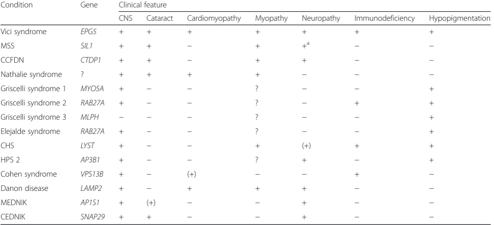
prophylaxis. It is also important to bear in mind that pa-tients with Vici syndrome may fail to respond to certain Table 3Syndromes showing phenotypical overlap with Vici syndrome (selection)
Condition Gene Clinical feature
CNS Cataract Cardiomyopathy Myopathy Neuropathy Immunodeficiency Hypopigmentation
Vici syndrome EPG5 + + + + + + +
MSS SIL1 + + − + +a − −
CCFDN CTDP1 + + − + + − −
Nathalie syndrome ? + + + + − − −
Griscelli syndrome 1 MYO5A + − − ? − − +
Griscelli syndrome 2 RAB27A + − − ? − + +
Griscelli syndrome 3 MLPH − − − ? − − +
Elejalde syndrome RAB27A + − − ? − − +
CHS LYST + − − + (+) + +
HPS 2 AP3B1 + − − ? + − +
Cohen syndrome VPS13B + − (+) − − + −
Danon disease LAMP2 + − + + + − −
MEDNIK AP1S1 + (+) − − + − −
CEDNIK SNAP29 + + − − + − −
immunizations such as those with tetanus or pneumococ-cal vaccines. An detailed overview of recommended im-munological investigations and possible management approaches is provided by Finocchi et al. [6].
More than half of patients with Vici syndrome have
seizuresthat ought to be managed with appropriate anti-convulsant therapy. Considering the profound autophagy abnormalities observed in patients with Vici syndrome, responses to anticonvulsants (or, indeed, other drugs) with potentially autophagy-modulating properties such as carbamazepine should perhaps be monitored closely following initiation of treatment.
If cataractsare present surgical removal may improve visual outcome but the indication for cataract surgery will have to be decided on an individual basis, based on overall severity and expected prognosis.
If a cardiomyopathy is identified on regular cardiac as-sessments, this may benefit from proactive medical man-agement; a deterioration of cardiac function during intercurrent illness has to be expected. Bothcentral and obstructive apnoea may require polysomnographic moni-toring, and non-invasive ventilatory support as indicated.
Hypothyroidismmay require thyroid hormone replace-ment. Renal dysfunction and electrolyte imbalances, in particular profound hypokalaemia, will have to be antici-pated and managed actively. Profound anaemiamay re-quire blood transfusion in some patients.
Counselling
Vici syndrome is inherited in an autosomal-recessive fashion. Genetic counselling should be offered to all families in whom a diagnosis of Vici syndrome has been established. Mutational analysis of theEPG5gene is now available on a diagnostic basis [4], andEPG5testing, the gold standard of antenatal diagnosis, can be offered to families where causative EPG5 mutations have been identified. It is important to bear in mind that foetal ultrasound applied for the detection of callosal agenesis may yield false positive and false negative results, there-fore when genetic testing is not readily available foetal MRI ought to be the preferred form of imaging.
Prognosis
Vici syndrome is a relentlessly progressive condition and survival beyond the first decade has not been reported. A large series recently demonstrated that death occurred at a median age of 42 months (range 1 to 102 months). Patients with homozygous mutations died sooner than patients with heterozygous mutations (median age nine months compared to 48 months) [14]. The degree of cardiac involvement and/or the extent of the associated immunodeficiency are the most important prognostic indicators.
Unresolved questions
Vici syndrome is the most extensive human multisystem disorder attributed to a primary autophagy defect reported to date. Although rare, the condition illustrates the impact of defective autophagy not only on neurodevelopment and neurodegeneration but also on a wide range of other organ systems where the role of normally functioning autophagy is currently only partially understood or not even considered yet. There are a number of unresolved questions of direct relevance to families affected by Vici syndrome but also for the wider field of autophagy research:
It is currently uncertain if Vici syndrome is genetically homogeneous, with the failure to identify two allelic muta-tions in some patients due toEPG5 copy number varia-tions not detectable on Sanger sequencing, or if there is genuine genetic heterogeneity with novel genetic back-grounds yet to be discovered in individuals with suggestive features but noEPG5mutations identified. Little is known about the physiological cellular interactions of the EPG5 protein, and it remains unclear if impaired autophagy is the only consequence of EPG5 deficiency, or just the most dramatic expression of a more generalized vesicu-lar trafficking defect in patients with Vici syndrome. The autophagy pathway is amenable to pharmacological manipulation, and delineating the precise defect in Vici syndrome will be important for the development of ra-tional therapies in future. The marked phenotypical over-lap between Vici and clinically related syndromes such as MSS or CHS is currently unexplained but suggests poten-tial interaction of the defective proteins in related cellular pathways, resulting in similar phenotypes.
Identification of new genotypes, further characterization of the precise biological role of EPG5 and the relation be-tween Vici and similar syndromes will further elucidate the role of defective autophagy in inherited multisystem disorders, and hopefully result in the development of ef-fective therapies for Vici syndrome and related conditions in future.
Consent
Written informed consent was obtained from the patient (s) for publication of this manuscript and accompanying images. A copy of the written consent is available for review by the Editor-in-Chief of this journal.
Competing interests
The authors declare that they have no competing interests.
Authors’contributions
Acknowledgements
LS was supported by a King’s Bioscience Institute PhD Fellowship. MG holds the BHF Chair of Molecular Cardiology; LS and MG are supported by the Leducq Foundation. HJ acknowledges grant support from the Myotubular Trust, Great Britain (Grant reference number 12KCL01).
Author details 1
Department of Paediatric Neurology, Neuromuscular Service, Evelina’s Children Hospital, Guy’s & St. Thomas’Hospital NHS Foundation Trust, London, UK.2Division of Metabolism and Laboratory of Molecular Medicine, Bambino Gesu Children’s Hospital IRCCS, Rome, Italy.3Randall Division of Cell and Molecular Biophysics, Muscle Signalling Section, King’s College, London, UK.4Department of Clinical and Basic Neuroscience, IoPPN, King’s College, London, UK.
Received: 20 August 2015 Accepted: 8 February 2016
References
1. Vici CD, Sabetta G, Gambarara M, Vigevano F, Bertini E, Boldrini R, Parisi SG, Quinti I, Aiuti F, Fiorilli M. Agenesis of the corpus callosum, combined immunodeficiency, bilateral cataract, and hypopigmentation in two brothers. Am J Med Genet. 1988;29(1):1–8.
2. Al-Owain M, Al-Hashem A, Al-Muhaizea M, Humaidan H, Al-Hindi H, Al-Homoud I, Al-Mogarri I: Vici syndrome associated with unilateral lung hypoplasia and myopathy. Am J Med Genet A. 2010;152A(7):1849–53. 3. Chiyonobu T, Yoshihara T, Fukushima Y, Yamamoto Y, Tsunamoto K,
Nishimura Y, Ishida H, Toda T, Kasubuchi Y. Sister and brother with Vici syndrome: agenesis of the corpus callosum, albinism, and recurrent infections. Am J Med Genet. 2002;109(1):61–6.
4. Cullup T, Kho AL, Dionisi-Vici C, Brandmeier B, Smith F, Urry Z, Simpson MA, Yau S, Bertini E, McClelland V et al. Recessive mutations in EPG5 cause Vici syndrome, a multisystem disorder with defective autophagy. Nat Genet. 2013;45(1):83–7.
5. del Campo M, Hall BD, Aeby A, Nassogne MC, Verloes A, Roche C, Gonzalez C, Sanchez H, Garcia-Alix A, Cabanas F et al. Albinism and agenesis of the corpus callosum with profound developmental delay: Vici syndrome, evidence for autosomal recessive inheritance. Am J Med Genet. 1999;85(5): 479–85.
6. Finocchi A, Angelino G, Cantarutti N, Corbari M, Bevivino E, Cascioli S, Randisi F, Bertini E, Dionisi-Vici C. Immunodeficiency in Vici syndrome: a heterogeneous phenotype.
Am J Med Genet A. 2012;158A(2):434–9.
7. McClelland V, Cullup T, Bodi I, Ruddy D, Buj-Bello A, Biancalana V, Boehm J, Bitoun M, Miller O, Jan W et al. Vici syndrome associated with sensorineural hearing loss and evidence of neuromuscular involvement on muscle biopsy. Am J Med Genet A. 2010;152A(3):741–7.
8. Miyata R, Hayashi M, Itoh E. Pathological changes in cardiac muscle and cerebellar cortex in Vici syndrome. Am J Med Genet A. 2014;164A(12):3203–5. 9. Miyata R, Hayashi M, Sato H, Sugawara Y, Yui T, Araki S, Hasegawa T, Doi S,
Kohyama J. Sibling cases of Vici syndrome: sleep abnormalities and complications of renal tubular acidosis. Am J Med Genet A. 2007;143(2): 189–94.
10. Ozkale M, Erol I, Gumus A, Ozkale Y, Alehan F. Vici syndrome associated with sensorineural hearing loss and laryngomalacia. Pediatr Neurol. 2012; 47(5):375–8.
11. Rogers CR, Aufmuth B, Monesson S: Vici Syndrome: A Rare Autosomal Recessive Syndrome with Brain Anomalies, Cardiomyopathy, and Severe Intellectual Disability.Case Reports in Genetics 2011, Volume 2011.
12. Said E, Soler D, Sewry C. Vici syndrome–a rapidly progressive
neurodegenerative disorder with hypopigmentation, immunodeficiency and myopathic changes on muscle biopsy. Am J Med Genet A. 2012; 158A(2):440–4.
13. Ehmke N, Parvaneh N, Krawitz P, Ashrafi MR, Karimi P, Mehdizadeh M, Kruger U, Hecht J, Mundlos S, Robinson PN. First description of a patient with Vici syndrome due to a mutation affecting the penultimate exon of EPG5 and review of the literature. Am J Med Genet A. 2014;164A(12): 3170–5.
14. Byrne S: EPG-related Vici syndrome: a paradigm of neurodevelopmental disorders with defective autophagy.Brain, in press.
15. Autti T, Joensuu R, Aberg L. Decreased T2 signal in the thalami may be a sign of lysosomal storage disease. Neuroradiology. 2007;49(7):571–8. 16. Filloux FM, Hoffman RO, Viskochil DH, Jungbluth H, Creel DJ.
Ophthalmologic features of Vici syndrome. J Pediatr Ophthalmol Strabismus. 2014;51(4):214–20.
17. Tasdemir S, Sahin I, Cayir A, Yuce I, Ceylaner S, Tatar A. Vici syndrome in siblings born to consanguineous parents. Am J Med Genet A. 2016;170(1):220–5. 18. El-Kersh K, Jungbluth H, Gringras P, Senthilvel E. Severe Central Sleep Apnea
in Vici Syndrome. Pediatrics. 2015;136(5):e1390–4.
19. Halama N, Grauling-Halama SA, Beder A, Jager D. Comparative integromics on the breast cancer-associated gene KIAA1632: clues to a cancer antigen domain. Int J Oncol. 2007;31(1):205–10.
20. Tian Y, Li Z, Hu W, Ren H, Tian E, Zhao Y, Lu Q, Huang X, Yang P, Li X et al. C. elegans screen identifies autophagy genes specific to multicellular organisms. Cell. 2010;141(6):1042–55.
21. Jiang P, Mizushima N. Autophagy and human diseases. Cell Res. 2014;24(1):69–79. 22. Klionsky DJ, Abdalla FC, Abeliovich H, Abraham RT, Acevedo-Arozena A,
Adeli K, et al. Guidelines for the use and interpretation of assays for monitoring autophagy. Autophagy. 2012;8(4):445–544.
23. Mizushima N, Komatsu M. Autophagy: renovation of cells and tissues. Cell. 2011;147(4):728–41.
24. Zhao H, Zhao YG, Wang X, Xu L, Miao L, Feng D, Chen Q, Kovacs AL, Fan D, Zhang H.. Mice deficient in Epg5 exhibit selective neuronal vulnerability to degeneration. J Cell Biol. 2013;200(6):731–41.
25. Ebrahimi-Fakhari D, Saffari A, Wahlster L, Lu J, Byrne S, Hoffmann GF, Jungbluth H, Sahin M: Congenital disorders of autophagy: An emerging class of inborn errors of neuro-metabolism
26. Krieger M, Roos A, Stendel C, Claeys KG, Sonmez FM, Baudis M, Bauer P, Bornemann A, de Goede C, Dufke A et al. SIL1 mutations and clinical spectrum in patients with Marinesco-Sjogren syndrome. Brain: a journal of neurology. 2013;136(Pt 12):3634–44.
27. Dotta L, Parolini S, Prandini A, Tabellini G, Antolini M, Kingsmore SF, Badolato R. Clinical, laboratory and molecular signs of immunodeficiency in patients with partial oculo-cutaneous albinism. Orphanet journal of rare diseases. 2013;8:168.
28. Silveira-Moriyama L, Moriyama TS, Gabbi TV, Ranvaud R, Barbosa ER. Chediak-Higashi syndrome with parkinsonism. Movement disorders: official journal of the Movement Disorder Society. 2004;19(4):472–5.
29. Byrne S, Dlamini N, Lumsden D, Pitt M, Zaharieva I, Muntoni F, King A, Robert L, Jungbluth H: SIL1-related Marinesco-Sjoegren syndrome (MSS) with associated motor neuronopathy and bradykinetic movement disorder. Neuromuscular disorders: NMD. 2015;25(7):585–8.
30. Duran-McKinster C, Rodriguez-Jurado R, Ridaura C, de la Luz O-CM, Tamayo L, Ruiz-Maldonando R. Elejalde syndrome–a melanolysosomal
neurocutaneous syndrome: clinical and morphological findings in 7 patients. Arch Dermatol. 1999;135(2):182–6.
31. Pastural E, Barrat FJ, Dufourcq-Lagelouse R, Certain S, Sanal O, Jabado N, Seger R, Griscelli C, Fischer A, de Saint Basile G. Griscelli disease maps to chromosome 15q21 and is associated with mutations in the myosin-Va gene. Nat Genet. 1997;16(3):289–92.
32. Pastural E, Ersoy F, Yalman N, Wulffraat N, Grillo E, Ozkinay F, Tezcan I, Gedikoglu G, Philippe N, Fischer A et al. Two genes are responsible for Griscelli syndrome at the same 15q21 locus. Genomics. 2000;63(3):299–306. 33. Malicdan MC, Nishino I. Autophagy in lysosomal myopathies. Brain Pathol.
2012;22(1):82–8.
34. Jungbluth H, Wallgren-Pettersson C, Laporte J. Centronuclear (myotubular) myopathy. Orphanet J Rare Dis. 2008;3:26.
35. Jungbluth H, Gautel M. Pathogenic mechanisms in centronuclear myopathies. Front Aging Neurosci. 2014;6:339.
36. Nishino I, Fu J, Tanji K, Yamada T, Shimojo S, Koori T, Mora M, Riggs JE, Oh SJ, Koga Y et al. Primary LAMP-2 deficiency causes X-linked vacuolar cardiomyopathy and myopathy (Danon disease). Nature. 2000;406(6798): 906–10.