RESEARCH ARTICLE
Priming stem cells with protein kinase C
activator enhances early stem cell-chondrocyte
interaction by increasing adhesion molecules
Dong‑Sik Chae
1,2, Chang Youn Lee
3, Jiyun Lee
4, Hyang‑Hee Seo
4, Chong‑Hyuk Choi
5, Seahyoung Lee
6*and Ki‑Chul Hwang
6*Abstract
Background: Osteoarthritis (OA) can be defined as degradation of articular cartilage of the joint, and is the most common degenerative disease. To regenerate the damaged cartilage, different experimental approaches including stem cell therapy have been tried. One of the major limitations of stem cell therapy is the poor post‑transplantation survival of the stem cells. Anoikis, where insufficient matrix support and adhesion to extracellular matrix causes apoptotic cell death, is one of the main causes of the low post‑transplantation survival rate of stem cells. Therefore, enhancing the initial interaction of the transplanted stem cells with chondrocytes could improve the therapeutic efficacy of stem cell therapy for OA. Previously, protein kinase C activator phorbol 12‑myristate 13‑acetate (PMA)‑ induced increase of mesenchymal stem cell adhesion via activation of focal adhesion kinase (FAK) has been reported. In the present study, we examine the effect PMA on the adipose‑derived stem cells (ADSCs) adhesion and spreading to culture substrates, and further on the initial interaction between ADSC and chondrocytes.
Results: PMA treatment increased the initial adhesion of ADSC to culture substrate and cellular spreading with increased expression of adhesion molecules, such as FAK, vinculin, talin, and paxillin, at both RNA and protein level. Priming of ADSC with PMA increased the number of ADSCs attached to confluent layer of cultured chondrocytes compared to that of untreated ADSCs at early time point (4 h after seeding).
Conclusion: Taken together, the results of this study suggest that priming ADSCs with PMA can increase the initial interaction with chondrocytes, and this proof of concept can be used to develop a non‑invasive therapeutic approach for treating OA. It may also accelerate the regeneration process so that it can relieve the accompanied pain faster in OA patients. Further in vivo studies examining the therapeutic effect of PMA pretreatment of ADSCs for articular cartilage damage are required.
Keywords: ADSC, PMA, Cellular adhesion, Chondrocyte, Osteoarthritis
© The Author(s) 2018. This article is distributed under the terms of the Creative Commons Attribution 4.0 International License (http://creativecommons.org/licenses/by/4.0/), which permits unrestricted use, distribution, and reproduction in any medium, provided you give appropriate credit to the original author(s) and the source, provide a link to the Creative Commons license, and indicate if changes were made. The Creative Commons Public Domain Dedication waiver (http://creativecommons.org/ publicdomain/zero/1.0/) applies to the data made available in this article, unless otherwise stated.
Background
Osteoarthritis (OA) is the most common degenerative disease and can be defined as degradation of articular cartilage of the joint [1]. Treatment for OA generally uses non-steroidal anti-inflammatory drugs (NSAIDs) and hyaluronic acid injections, but these options are mainly to relieve pain rather than regenerate the damaged
Open Access
*Correspondence: [email protected]; [email protected]
6 Institute for Bio‑Medical Convergence, College of Medicine, Catholic
Kwandong University, Gangneung‑si, Gangwon‑do, South Korea Full list of author information is available at the end of the article
damaged tissues, which decreases the therapeutic effi-cacy of the transplanted stem cells [7, 8].
The low survival rate of the transplanted stem cells in vivo has been linked to anoikis where insufficient matrix support and adhesion to extracellular matrix (ECM) causes apoptotic cell death [9]. To improve cell adhesion, number of approaches had been tried includ-ing the enhancement of anti-apoptotic signals [10–13], modulation of cell adhesion molecules [14–16], hypoxic preconditioning [17–19], and pretreatment of various growth factors, cytokines, and chemicals [20–23]. A previous study demonstrated that the protein kinase C (PKC) activator phorbol 12-myristate 13-acetate (PMA) increased cell adhesion of rat MSCs via activation of focal adhesion kinase (FAK) and the PMA-pretreated rat MSCs improved cardiac function following myocardial infarction [24].
The activation of PKC plays important roles in cytoskel-etal rearrangement, cell proliferation, survival, cell death [25], and the activation of FAK [26], which results in the modulation of cell adhesion and motility by recruiting other focal adhesion proteins including paxillin [27, 28]. Therefore, priming stem cells with PMA may enhance the initial interaction between transplanted stem cells and host tissue cells, further increasing the therapeutic efficacy. In the present study, we examined the effect of PMA pretreatment on ADSC adhesion and spreading and further investigated the effect of priming ADSCs with PMA on the initial interaction between ADSCs and chondrocyte.
Methods
Culture of human adipose‑derived stem cells (ADSCs)
Human adipose-derived stem cells (hADSCs) were pur-chased from Invitrogen (Waltham, MA, USA). hADSCs were cultured according to the manufacturer’s instruc-tions. High glucose-Dulbecco’s modified Eagle’s medium (DMEM; Gibco, Waltham, MA, USA) containing 10% fetal bovine serum (FBS; Gibco) and 1% antibiotics (Gibco) were used to culture and maintain the cells. The media were changed every 3 days, and the cells were pas-saged using 0.25% trypsin (Gibco) when they reached 80–90% confluency. The cells from passages 6–10 were used for experiments.
Culture of human chondrocytes
Human chondrocytes were purchased from PromoCell (Heidelberg, Germany). The cells were cultured according to the manufacturer’s instructions. Chondrocyte growth medium (c-27101, PromoCell) was used to culture and maintain the cells. The media were changed every 3 days,
and the cells were passaged using 0.25% trypsin (Gibco) when they reached 80–90% confluency. The cells from passages up to 6 were used for experiments.
PMA treatment
We examined the effect of PMA pretreatment on ADSC adhesion and spreading and further investigated the effect of priming ADSCs with PMA on the initial interac-tion between ADSCs and chondrocyte. To activate PKC in ADSCs, we used a well-known PKC activator PMA (Sigma-Aldrich, St. Louis, MO). PMA was dissolved in dimethyl sulfoxide (DMSO) at a concentration of 100 μM (stock solution). For cell treatment, 100 μM stock solu-tion was diluted in DMEM to the desired concentrasolu-tion.
Cell viability assay
To measure cell viability, 5 × 10³ hADSCs cells were
plated in a 96-well plate. The cells were treated with vary-ing concentrations of PMA (10, 20, 50, and 100 nM) PMA and incubated for 24 h. Highly water-soluble tetrazolium salt WST-8 (CCK-8, Dogen, Seoul, Korea) was added to each well to a final concentration of 0.5 mg/mL. WST-8 is reduced by dehydrogenases in cells to give an orange colored formazan, and the amount of the formazan dye generated by dehydrogenases in cells is directly propor-tional to the number of living cells. The WST-8-treated cells were incubated for additional 2 h. The amount of formazan formed was measured using a microplate reader at 450 nm (Thermo Fisher Scientific Waltham, MA, USA).
Evaluation of cell adhesion
To evaluate ADSC adhesion, after 4 h of pretreatment increasing concentrations of PMA (or DMSO for vehicle group) as a suspension in culture medium, the cells were centrifuged and washed with PBS. The cells were seeded in a 6 well plate (5 × 10⁴ cells/well) without any further
PMA treatment. Four 4 h after the seeding, the attached cells were carefully washed with PBS three times, and then four separate fields were photographed with a phase contrast microscope for counting.
Evaluation of cell spreading
For spreading assays, ADSCs were pretreated with PMA (or DMSO for vehicle group) as a suspension in culture medium for 4 h in a cell culture incubator. The PMA pretreated ADSCs were seeded in a 4 well chamber slide at a density of 1 × 10³ cells/well and cultured for 6 h. To
stained with Coomassie blue [0.1% solution of Coomassie brilliant blue R250 in 40% methanol: acetic acid: water, 46.5:7:46.5 (v/v/v)], and rinsed with distilled water [29]. Finally, the four separate fields were photographed using a phase contrast microscope.
F‑actin staining
In order to visualize ADSC’s cytoskeleton, the 4 h PMA (or DMSO for vehicle group) pretreated ADSCs were seeded in a 4 well chamber slide at a density of 1 × 10³ cells/well and cultured for 6 h. The cells were
washed with PBS and fixed in 10% formalin solution for 10 min at room temperature. After PBS wash, the cells were permeabilized in 0.1% Triton X-100 for 5 min and then PBS washed. After 30 min of blocking in 1% bovine serum albumin in PBS, and F-actin was stained with Texas Red-X phalloidin (Invitrogen, Waltham, MA, USA) overnight at 4 °C. Immunofluorescence was detected by confocal microscopy (LSM710; Carl Zeiss Microscopy GmbH, Jena, Germany).
Wound healing assay (migration assay)
ADSCs were plated at a density of 8 × 10⁴ cells/well in
six-well plates. After the cells had reached 70% conflu-ence, wounds were produced by scratching with 200 μL pipette tips. The medium was replaced with or with-out PMA (100 nM), and the cells were incubated for up to 24 h (1 day). Images were captured using an Axio-vert 40 °C inAxio-verted microscope (Carl Zeiss, Germany) equipped with a Powershot A640 digital camera (Canon, Japan) at 4 h and 24 h.
Seeding ADSCs on top of cultured chondrocytes
Chondrocytes were cultured in 6 well plates until they became confluent. Meantime, cultured ADSCs were trypsinized and the cells were suspended in 3 aliquots; (1) untreated ADSC, (2) PMA + ADSCs, and (3) PMA pretreated ADSCs (2 × 10³ cells/mL each). Only the PMA
pretreated ADSC group was incubated with 100 nM of PMA for 4 h. Meanwhile, the untreated ADSCs and PMA + ADSCs were kept in an incubator without PMA for the 4 h. After 4 h, Hoechst dye (2 μg/mL) was added to all groups and the cell suspensions were kept in an incubator for 30 min. The cell suspensions were centri-fuged and the cell pellets were washed with PBS 3 times. For the PMA + ADSC group, 100 nM of PMA was added to the cell suspension just prior to the seeding. The Hoe-chst-stained ADSCs (2 × 10³ cells per well) were seeded
on top of confluent chondrocytes and cultured for addi-tional 4 h. Unattached cells were washed 3 times with fresh medium, and Hoechst-stained ADSCs were imaged under fluorescent microscopy.
Reverse transcription polymerase chain reaction (RT‑PCR)
Briefly, the cells were seeded to a well of 6-well plate in DMEM containing 100 nM PMA and incubated in a humidified atmosphere of 95% air and 5% CO2 (Forma
Scientific, USA) at 37 °C. The cells were collected at 4 h after the initial seeding for reverse transcriptase PCR. Total RNA was extracted using TRIzol reagent (Ambion, Waltham, MA, USA) according to the manufacturer’s instructions. Complementary DNA (cDNA) was synthe-sized from RNA by AMV reverse transcriptase in pro-vided in a RT system kit (Promega, Fitchburg, WI, USA). The primer sequences used were as follows: human PTK2 (FAK, NCBI reference sequence: NM_001199649.1), for-ward: 5′-TTA TTG GCC ACT GTG GAT GA-3′, reverse: 5′-TAC TCT TGC TGG AGG CTG GT-3′; human TLN1 (Talin1, NCBI reference sequence: NM_006289.3), for-ward: 5′-TCT CCC AAA ATG CCA AGA AC-3′, reverse: 5′-CTC CAC TAG CCC TTG CTG TC-3′; human VCL (vinculin, NCBI reference sequence: NM_003373.3), forward: 5′-GCC AAG CAG TGC ACA GAT AA-3′, reverse: 5′-AGG TTC TGG GCA TTG TGA AC-3′, human PXN (paxillin, NCBI reference sequence: NM_001080855.2), forward: 5′-GGA GTC TAC CAC CTC CCA CA-3′, reverse: 5′-CCA CTG GTC TAA GGG GTC AA-3′, human GAPDH (NCBI reference sequence: NM_001256799.2), forward: 5′-CAT GGG TGT GAA CCA TGA GAA-3′, reverse: 5′-GGT CAT GAG TCC TTC CAC GAT-3′.
Western blot
For western blot, the cells were collected at 12 h after the initial seeding with or without PMA in the culture medium. Cells were washed once in PBS and lysed for 20 min in a lysis buffer (cell signaling) containing 20 mM Tris (pH 7.5), 150 mM NaCl, 1 mM Na2EDTA, 1 mM
EGTA, 1% Triton, 2.5 mM sodium pyrophosphate, 1 mM β-glycerophosphate, 1 mM Na3VO4, 1 mg/mL
(1:5000 dilution), goat anti-mouse-HRP (1:4000 dilution), and goat anti-rabbit-HRP (1:2000 dilution, Enzo Life Sci-ences, Farmingdale, NY). After thorough washing, a band was detected using enhanced chemiluminogenic (ECL) reagent (GE Healthcare Life Sciences). The intensity of the band was quantified using ImageJ 1.40g software (NIH).
Statistical analysis
Quantitative data were expressed as the mean ± S.E.M. For statistical analysis, Student’s t-test was used for 2 group comparison and one-way ANOVA with Bonfer-roni correction was performed using OriginPro 8 SR4 software (ver. 8.0951, OriginLab Corporation, USA) if there were more than 3 groups. A p value of < 0.05 was considered statistically significant.
Results
Effect of PMA on the viability of ADSCs
PMA cytotoxicity on ADSCs was assayed by treating with increasing concentrations of PMA (10, 20, 50, and 100 nM) over 24 h and determining cell viability using CCK-8 kit. As can be observed in Fig. 1, vehicle (0.1% DMSO) and PMA treatments did not induce statistically significant reductions of cell viability in the concentration range tested (Fig. 1).
Effect of PMA on the adhesion of ADSC to culture substrate
To examine the effect of PMA on ADSC adhesion to cul-ture substrate, cells were treated with varying concen-trations of PMA in suspension for 4 h, and seeded in a 6 well plate (5 × 10⁴ cells/well). The cells were allowed to
attach to the culture plate for 4 h and the images of cells were taken for counting (Fig. 2a). According to the data, PMA treatment significantly increased the number of attached ADSCs (32.64 ± 2.10% of initially seeded cells) compared to both untreated (22.18 ± 3.59%) and vehicle (25.38 ± 2.48%) treated cells. However, there was no sta-tistically significant dose-dependent effect among groups treated with different concentrations of PMA (Fig. 2b). Since the 100 nM group showed no significant cytotoxic-ity and had the smallest intra-sample variation, 100 nM of PMA was used for further experiments.
Effect of PMA pretreatment on the spreading of ADSCs
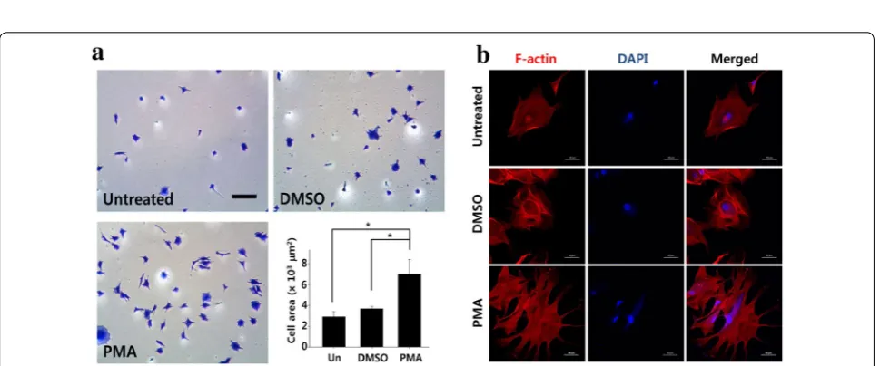
To further examine the effect of PMA on ADSC adhe-sion, the PMA pretreated ADSCs (100 nM, 4 h) were seeded and allowed to attach and spread for 6 h, fixed, and stained with Coomassie blue for clear visualiza-tion. Compared to both untreated controls and vehicle treated group, PMA (100 nM) treated group showed increased number of cells attached, and more spread
(blunt round vs. sharp pointy extensions) morphol-ogy (Fig. 3a). Furthermore, the cell area (Coomassie blue stained area) significantly increased with PMA treatment compared to that of both untreated control and vehicle treated group (2.89 ± 1.23 vs. 3.67 ± 0.50
vs. 6.90 ± 3.54 × 103 μm2, untreated, vehicle treated,
and PMA treated, respectively). To better visualize the cytoskeleton of ADSCs, F-actin staining was per-formed, and the results showed that PMA pretreated increased filopodia formation (Fig. 3b).
PMA has no significant effect on cellular migration of ADSCs
To examine whether PMA had any effect on ADSC migration, in vitro wound healing assay was conducted. When the cells reached approximately 70% confluence, wounds were created using a 200 μL pipette tip and the cells were allowed to migrate for additional 24 h with or without PMA (100 nM) in the culture media. As shown in the Fig. 4, there was no prominent effect of PMA on the migration of ADSCs observed, regardless of the time after the wound formation (Fig. 4).
PMA pretreatment enhances the initial interaction between ADSCs and chondrocytes
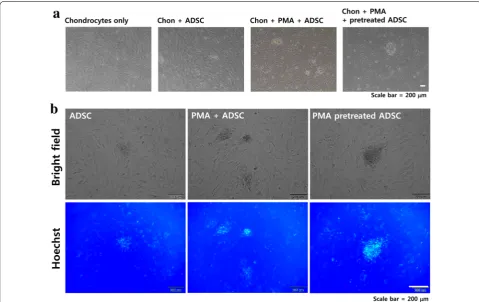
To examine whether PMA treatment could enhance the initial cell–cell interaction between ADSCs and chon-drocytes, Hoechst-labeled ADSCs with different PMA treatments (4 h of incubation with PMA prior to seeding and PMA treatment upon seeding) were seeded on top of
Fig. 1 The effect of varying concentrations of PMA on the viability of ADSCs. To test whether PMA itself has any cytotoxic effect on ADSCs, the cells were cultured in a 96 well plate (5 × 10³ cells/well) and
confluent layer of chondrocyte. According to the results, ADSCs pretreated with PMA for 4 h had a tendency to clump together compared to the untreated ADSCs as shown in Fig. 5a. Although ADSCs treated with PMA upon seeding also showed formation of clumps, it was smaller than ones formed in the PMA pretreated ADSCs. Further examination using fluorescent microscopy indi-cated that the number of ADSCs attached was increased
by both PMA treatments (4 h of incubation with PMA prior to seeding and PMA treatment upon seeding) but the location of Hoechst-positive ADSCs was not evenly distributed because they formed clumps (Fig. 5b). Regarding the effect of PMA on chondrocytes, there are only a handful of previous studies directly or indirectly examined the effect of PMA (or PKC activation) on chondrocytes. A relatively recent review indicated that a
Fig. 2 PMA pretreatment increases initial attachment of ADSCs to culture substrate. a Representative images of ADSCs attached to culture substrate with or without 4 h of PMA pretreatment. Scale bar = 200 μm. b Number of ADSCs attached to culture substrate was counted (per field). The quantitative data were expressed as the mean ± S.E.M of at least 3 independent experiments. *p < 0.05
Fig. 3 PMA pretreatment enhances cellular spreading of ADSCs. ADSCs pretreated with PMA (100 nM) for 4 h, and then the cells were seeded in a 4 well chamber slide (103 cells/well) and allowed to attach and spread for 6 h. a Attached cells were fixed and Coomassie blue stained for
chronic PMA treatment negatively affects chondrogen-esis by downregulating PKC expression [30]. However, since our experimental design involves 4 h of PMA treat-ment, no significant morphological changes or cell death was observed.
PMA increases the expression of cell adhesion molecules in ADSCs
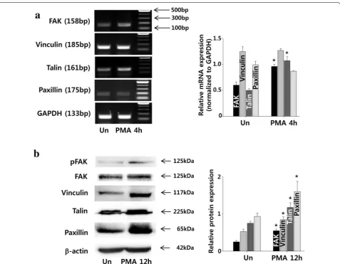
Examination of adhesion-related gene expression using RT-PCR indicated that the expressions of FAK and talin significantly increased with 4 h of PMA treatment, while the expressions of vinculin and paxillin remained unaf-fected (Fig. 6a). On the other hand, protein expressions of vinculin and paxillin increased at 12 h (Fig. 6b).
PMA increases the expression of anoikis resistant molecules in ADSCs
Examination of anoikis resistant extracellular signal-regulated kinase (ERK) and Akt activation using West-ern blot indicated that the expressions of phosphorylated ERK and Akt significantly increased by 4 h of PMA
Fig. 4 Effect of PMA on the migration of ADSCs. After ADSCs reached approximately 70% confluence, wounds were created using a yellow pipette tip. The cells were cultured with or without PMA (100 nM) for up to 24 h and the images were taken at 4 and 24 h after the wounds were created. Scale bar = 200 μm. White dotted line shows the migrating edge
treatment, while the expressions of phosphorylated p38 remained unaffected (Fig. 7).
Discussion
One of the major problems of stem cell-based therapy is the low post-transplantation survival rate due to insuf-ficient tissue integration called anoikis [31]. Therefore, preventing anoikis or enhancing initial cell adhesion may increase the therapeutic efficacy of stem cells for treat-ing number of diseases includtreat-ing OA [8]. In this study, the effect of PMA on the ADSC adhesion to culture substrate and to a layer of chondrocyte was examined. Previous reports have demonstrated that PMA exerted cytotoxic effect on certain types of cells such as pancre-atic cancer cells [32]. Therefore, we first verified whether there was any cytotoxic effect of PMA on ADSCs. As shown in the Fig. 1, PMA up to 100 nM did not cause any
significant cytotoxic effect, and we moved on to further experiments.
PMA is a non-physiological activator of PKC [33]. Among different isozymes of PKC, which are com-prised of conventional (PKCα, PKCβI, PKCβII and PKCγ), novel (PKCδ, PKCε, PKCη and PKCθ), and atyp-ical (PKCζ and PKCι) isozymes, PMA activates con-ventional and novel PKCs by mimicking diacylglycerol (DAG) [34]. Regarding integrin-mediated cell adhesion, the importance of PKC activation and subsequent FAK phosphorylation and focal adhesion formation is well known [35]. Our data demonstrated that activation of PKC using increased ADSC adhesion and subsequent spreading in vitro (Figs. 2 and 3). However, we did not examine which type of isoenzymes were actually acti-vated by PMA treatment so that it remains one of the limitations of this study, especially in elucidating the
underlying mechanisms of the PMA-mediated ADSC adhesion enhancement. For future studies, using PKC inhibitors specific for each isoenzyme will be helpful to identifying the exact mechanism.
On the other hand, although PMA induced the phos-phorylation of FAK required for cellular migration [36], it failed to enhance the migration of ADSCs (Fig. 4). This observation is somewhat contradictory to previous studies reported that PMA promoted cellular migration [37, 38]. Although we do not have data to back up at this point, one of the feasible explanations is that the use of 10% FBS supplemented DMEM for the experi-ment masked any possible effect of PMA on ADSC migration. In other words, if 0.1% FBS supplemented or serum free DMEM was used instead, the effect to PMA might have been detected. Another possibility is that we picked a wrong time point (4 h) to observe the early effect of PMA on ADSC migration. Since all the groups showed near-complete wound closing at 24 h, if there was any positive effect of PMA on cellular migration, such effect should have been observable between 4 and 24 h in our in vitro system. Therefore, for further stud-ies, it is required to include 0.1% serum supplemented group and increase the number of observations for bet-ter understanding of the effect of PMA on ADSCs.
The important integrin-mediated signaling molecules such as FAK and integrin linked kinase (ILK) have been associated with cell survival, and these signaling mol-ecules are also linked to PKC activity [39]. According to the data, PMA increased the transcription of key cel-lular adhesion molecules including FAK and talin after 4 h of treatment. Since both FAK and talin are critical components of integrin-containing adhesion complex
that mediate cell–matrix interaction during cell spread-ing [40], PMA-induced increase of FAK and talin maybe the major factors mediated PMA-induced adhesion and spreading of ADSCs observed in the present study.
Furthermore, the protein level, the same concentration of PMA increased the protein expression of other inte-grin binding adhesion molecules, such as paxillin and vinculin [41]. Within the extracellular matrix, integrin associates with paxillin and vinculin, and it crosslinks actomyosin stress fibers which attached to focal contacts [42]. Subsequently, integrin clustering leads to associa-tion of the protein tyrosine kinase Src and the adaptor protein p130C to facilitate cellular adhesion [43]. There-fore, we speculate that PMA mediated activation of PKC, in turn, increased the expression of important compo-nents of focal adhesion complex to facilitate the adhesion of ADSCs.
Additionally, PMA treatment activated both ERK and Akt that are known to facilitate resistance to anoikis [44] in the present study. ERK is one of the major signaling of the mitogen-activated protein kinase (MAPK) signaling, and MAPK-dependent protection against apoptotic cell death has been well reported. For example, Bim is one of the factors that regulates anoikis [45] and the activa-tion of MAPK suppresses Bim both at posttranslaactiva-tional [46] and transcriptional levels [47]. Furthermore, activa-tion of MAPK upregulates the pro-survival members of the Bcl-2 family, such as Bcl-2, Bcl-XL and Mcl-1 [48]. Akt activation also promotes cell survival by prevent-ing the release of cytochrome c from mitochondria [49] and inhibits the caspase cascade by phosphorylating the procaspase-9 [50]. Additionally PMA treatment did not affect p38 signaling, which is known to induce Anoikis
[51]. Therefore, PMA-induced activation of survival sign-aling such as ERK and Akt without affecting p38 could be beneficial to the survival of transplanted ADSCs.
Current articular cartilage repair procedures often involve invasive processes such as microfracture sur-gery where damaged cartilage is drilled or punched to expose the underlying bone [52], and such procedure itself is discomforting and necessarily requires long and demanding rehabilitation [53]. As a non-invasive alter-native approach, we envisioned that utilizing the PMA treated ADSCs for treating OA without invasive surgery, by directly injecting adhesion enhanced stem cells into the joint capsule without any major surgical procedure. Since the injected cells will be suspended in synovial fluid which is directly in contact with chondral heads (possi-bly damaged collagen fibers with exposed chondrocytes in OA patients), examining whether PMA treatment can enhance the interaction between ADSCs and chondro-cytes should be the first step if the aforementioned non-invasive alternative approach were to be established.
Another important consideration to be taken is the nature of chemicals for enhancing the initial adhesion of ADSCs due to a possible carcinogenic effect. For exam-ple, PMA that used in the present study is known as a tumor promoter [54]. According to a “two-stage hypoth-esis” of carcinogenesis, there are two types of cancer-promoting agents; tumor initiators and tumor promoters. Tumor initiators are literally mutagens, whereas tumor promoters simply increase the number of cells carrying genetic defects caused by tumor initiators [55]. There-fore, the probability that PMA itself, as a tumor pro-moter, causes progression to malignancy seems to be very low [56]. Nevertheless, another study demonstrated that PMA indirectly induced sister chromatid exchanges by forming intermediate active oxygen species [57]. These contradicting reports suggest that extensive search of alternative chemicals that has no carcinogenic effect may be necessary to guarantee the clinical safety of the non-invasive approach we proposed. For the time being, as a proof of concept study, we examined whether PMA pre-treated ADSCs had better interaction with chondrocytes. Although the bound cells formed focal clumps and were relatively low in numbers, PMA treatment increased the number of bound ADSCs compared to untreated ADSCs (Fig. 5). The possible explanation for the modest number of attached cells to chondro-cyte is the low seeding density. Since the experimental protocol was something we tried first without previ-ous reference, the seeding density might have not been optimized. For example, for the experiments shown in Fig. 3, the cells were seeded in a chamber slide at a density of 1 × 10³/well, and that was reasonable to
take pictures at lower magnification. However, for the
experiments shown in Fig. 5, the cells were seeded at a density of 2 × 10³/well (of a 6 well plate) and the
images were taken at higher magnification. The reason we did not used excessive amount of cells for seeding was that we expected most of the PMA pretreated cells would attach which was proven to be wrong. Therefore, the low seeding density might be one of the probable reasons of why the cells attached in modest numbers. Another possible reason may be the incubation time. Again, for the experiments shown in Fig. 3, the cells were allowed to attach for 6 h after the seeding while the cells were allowed for 4 h in experiment shown in Fig. 5. Therefore, for future experiments, higher seed-ing density and longer incubation time should be tried.
Nevertheless, Fig. 5 indicated that PMA may enhance the binding of ADSCs to the damaged chondral head for regeneration. In fact, according to our unpublished in vivo data, PMA-primed ADSC injection to the colla-genase-treated articular cartilage of rats, which simu-late wear and tear osteoarthritis in vivo [58], prevented further collagenase-induced degradation of the articu-lar cartilage and maintained thicker cartilage layer compared to untreated OA controls. Nevertheless, we so far observed only the final improvement (data not shown) and not yet were able to find proper time point and experimental methods that can clearly demonstrate the PMA-induced early tissue integration of trans-planted ADSCs compared to untreated ADSCs in vivo. Since our in vivo approach involves injecting cells into synovial fluid of knee joint rather than directly inject into cartilage tissues. Therefore, the injected cells have to superficially attach to the cartilage surface and then gradually be integrated. Because of this, it is always possible that loosely attached, or superficially attached cells, may be lost during sample preparation. To over-come such limitation of our current in vivo approach we used, we are trying to establish an ex vivo method of culturing explanted animal cartilage tissue in Matrigel. Once established, such method can be used to verify the effect of PMA pretreated ADSCs in treating OA.
Conclusions
Abbreviations
ADSC: adipose‑derived stem cell; OA: osteoarthritis; PMA: phorbol 12‑myristate 13‑acetate; MSC: mesenchymal stem cell; FAK: focal adhesion kinase; ERK: extracellular signal‑regulated kinase; MAPK: mitogen‑activated protein kinase.
Authors’ contributions
DSC conceived the study, participated in experiments, and drafted the manuscript. CL, JL, and HS participated in cell preparation and cell culture. CHC participated in the study design and, drafted the manuscript. SL drafted the manuscript and provided technical support. KH participated in the study design, provided technical support, and edited the manuscript. All authors read and approved the final manuscript.
Author details
1 Department of Medicine, The Graduate School, Yonsei University, Seoul,
South Korea. 2 Department of Orthopedic Surgery, International St. Mary’s
Hospital, Catholic Kwandong University College of Medicine, Incheon, South Korea. 3 Department of Integrated Omics for Biomedical Sciences, Yonsei
University, Seoul, South Korea. 4 Brain Korea 21 PLUS Project for Medical
Science, Yonsei University, Seoul, South Korea. 5 Department of Orthopedic
Surgery, Yonsei University College of Medicine, 50, Yonsei‑ro, Seodaemun‑gu, Seoul 03722, Republic of Korea. 6 Institute for Bio‑Medical Convergence, Col‑
lege of Medicine, Catholic Kwandong University, Gangneung‑si, Gangwon‑do, South Korea.
Competing interests
The authors declare that they have no competing interests.
Availability of data and materials
The datasets supporting the conclusion of this article are included within the article.
Consent for publication Not applicable.
Ethics approval and consent to participate Not applicable.
Funding
This study was supported by Grants funded by the Korea Ministry of Science, ICT and Future Planning (NRF‑2015M3A9E6029519, and NFR‑ 2015M3A9E6029407), Grants from the Ministry of Health & Welfare, Republic of Korea (HI18C0661010018 and HI18C0661020018), and a research fund of Catholic Kwandong University International St. Mary’s Hospital (CKURF‑201406820001).
Publisher’s Note
Springer Nature remains neutral with regard to jurisdictional claims in pub‑ lished maps and institutional affiliations.
Received: 12 April 2017 Accepted: 16 October 2018
References
1. Loeser RF, Collins JA, Diekman BO. Ageing and the pathogenesis of osteo‑ arthritis. Nat Rev Rheumatol. 2016;12:412–20.
2. Wood AM, Brock TM, Heil K, Holmes R, Weusten A. A review on the management of hip and knee osteoarthritis. Int J Chronic Dis. 2013;2013:845015.
3. Pers YM, Ruiz M, Noel D, Jorgensen C. Mesenchymal stem cells for the management of inflammation in osteoarthritis: state of the art and perspectives. Osteoarthr Cartil. 2015;23:2027–35.
4. Wyles CC, Houdek MT, Behfar A, Sierra RJ. Mesenchymal stem cell therapy for osteoarthritis: current perspectives. Stem Cells Cloning. 2015;8:117–24. 5. Bauge C, Boumediene K. Use of adult stem cells for cartilage tissue
engineering: current status and future developments. Stem Cells Int. 2015;2015:438026.
6. Zuk PA, Zhu M, Ashjian P, De Ugarte DA, Huang JI, Mizuno H, et al. Human adipose tissue is a source of multipotent stem cells. Mol Biol Cell. 2002;13:4279–95.
7. Sart S, Ma T, Li Y. Preconditioning stem cells for in vivo delivery. Biores Open Access. 2014;3:137–49.
8. Lee S, Choi E, Cha MJ, Hwang KC. Cell adhesion and long‑term survival of transplanted mesenchymal stem cells: a prerequisite for cell therapy. Oxid Med Cell Longev. 2015;2015:632902.
9. Frisch SM, Screaton RA. Anoikis mechanisms. Curr Opin Cell Biol. 2001;13:555–62.
10. Wang Y, Abarbanell AM, Herrmann JL, Weil BR, Manukyan MC, Poynter JA, et al. TLR4 inhibits mesenchymal stem cell (MSC) STAT3 activation and thereby exerts deleterious effects on MSC‑mediated cardioprotection. PLoS ONE. 2010;5:e14206.
11. Mangi AA, Noiseux N, Kong D, He H, Rezvani M, Ingwall JS, et al. Mesen‑ chymal stem cells modified with Akt prevent remodeling and restore performance of infarcted hearts. Nat Med. 2003;9:1195–201.
12. Wang X, Zhao T, Huang W, Wang T, Qian J, Xu M, et al. Hsp20‑engineered mesenchymal stem cells are resistant to oxidative stress via enhanced activation of Akt and increased secretion of growth factors. Stem Cells. 2009;27:3021–31.
13. Liu N, Zhang Y, Fan L, Yuan M, Du H, Cheng R, et al. Effects of transplanta‑ tion with bone marrow‑derived mesenchymal stem cells modified by survivin on experimental stroke in rats. J Transl Med. 2011;9:105. 14. Copland IB, Lord‑Dufour S, Cuerquis J, Coutu DL, Annabi B, Wang E, et al.
Improved autograft survival of mesenchymal stromal cells by plasmino‑ gen activator inhibitor 1 inhibition. Stem Cells. 2009;27:467–77. 15. Song H, Chang W, Lim S, Seo HS, Shim CY, Park S, et al. Tissue transglu‑
taminase is essential for integrin‑mediated survival of bone marrow‑ derived mesenchymal stem cells. Stem Cells. 2007;25:1431–8.
16. Benoit DS, Tripodi MC, Blanchette JO, Langer SJ, Leinwand LA, Anseth KS. Integrin‑linked kinase production prevents anoikis in human mesenchy‑ mal stem cells. J Biomed Mater Res A. 2007;81:259–68.
17. Hahn JY, Cho HJ, Kang HJ, Kim TS, Kim MH, Chung JH, et al. Pre‑treatment of mesenchymal stem cells with a combination of growth factors enhances gap junction formation, cytoprotective effect on cardio‑ myocytes, and therapeutic efficacy for myocardial infarction. J Am Coll Cardiol. 2008;51:933–43.
18. Yu J, Yin S, Zhang W, Gao F, Liu Y, Chen Z, et al. Hypoxia preconditioned bone marrow mesenchymal stem cells promote liver regeneration in a rat massive hepatectomy model. Stem Cell Res Ther. 2013;4:83. 19. Hu X, Yu SP, Fraser JL, Lu Z, Ogle ME, Wang JA, et al. Transplantation of
hypoxia‑preconditioned mesenchymal stem cells improves infarcted heart function via enhanced survival of implanted cells and angiogenesis. J Thorac Cardiovasc Surg. 2008;135:799–808.
20. Lu G, Haider HK, Jiang S, Ashraf M. Sca‑1+ stem cell survival and engraft‑ ment in the infarcted heart: dual role for preconditioning‑induced con‑ nexin‑43. Circulation. 2009;119:2587–96.
21. Chang W, Song BW, Lim S, Song H, Shim CY, Cha MJ, et al. Mesenchymal stem cells pretreated with delivered Hph‑1‑Hsp70 protein are protected from hypoxia‑mediated cell death and rescue heart functions from myocardial injury. Stem Cells. 2009;27:2283–92.
22. Song L, Yang YJ, Dong QT, Qian HY, Gao RL, Qiao SB, et al. Atorvasta‑ tin enhance efficacy of mesenchymal stem cells treatment for swine myocardial infarction via activation of nitric oxide synthase. PLoS ONE. 2013;8:e65702.
23. Huang F, Li ML, Fang ZF, Hu XQ, Liu QM, Liu ZJ, et al. Overexpression of MicroRNA‑1 improves the efficacy of mesenchymal stem cell transplanta‑ tion after myocardial infarction. Cardiology. 2013;125:18–30.
24. Song BW, Chang W, Hong BK, Kim IK, Cha MJ, Lim S, et al. Protein kinase C activation stimulates mesenchymal stem cell adhesion through activa‑ tion of focal adhesion kinase. Cell Transplant. 2013;22:797–809. 25. Reyland ME. Protein kinase C isoforms: multi‑functional regulators of cell
•fast, convenient online submission
•
thorough peer review by experienced researchers in your field
• rapid publication on acceptance
• support for research data, including large and complex data types
•
gold Open Access which fosters wider collaboration and increased citations maximum visibility for your research: over 100M website views per year
•
At BMC, research is always in progress.
Learn more biomedcentral.com/submissions
Ready to submit your research? Choose BMC and benefit from: 26. Bruce‑Staskal PJ, Bouton AH. PKC‑dependent activation of FAK and src
induces tyrosine phosphorylation of Cas and formation of Cas‑Crk com‑ plexes. Exp Cell Res. 2001;264:296–306.
27. Abedi H, Zachary I. Vascular endothelial growth factor stimulates tyrosine phosphorylation and recruitment to new focal adhesions of focal adhesion kinase and paxillin in endothelial cells. J Biol Chem. 1997;272:15442–51.
28. Berrier AL, Yamada KM. Cell‑matrix adhesion. J Cell Physiol. 2007;213:565–73.
29. Paddock SW. Incident light microscopy of normal and transformed cultured fibroblasts stained with coomassie blue R250. J Microsc. 1982;128:203–5.
30. Matta C, Mobasheri A. Regulation of chondrogenesis by protein kinase C: emerging new roles in calcium signalling. Cell Signal. 2014;26:979–1000. 31. Newland B, Welzel PB, Newland H, Renneberg C, Kolar P, Tsurkan M, et al. Tackling cell transplantation anoikis: an injectable, shape memory cryo‑ gel microcarrier platform material for stem cell and neuronal cell growth. Small. 2015;11:5047–53.
32. Bond JA, Gescher AJ, Verschoyle RD, Lemoine NR, Errington R, Wiltshire M, et al. Cytotoxic action of phorbol esters on human pancreatic cancer cells. Int J Cancer. 2007;121:1445–54.
33. Zhu L, McDavid S, Currie KP. ”Slow” voltage‑dependent inactivation of CaV2.2 calcium channels is modulated by the PKC activator phorbol 12‑myristate 13‑acetate (PMA). PLoS ONE. 2015;10:e0134117. 34. Way KJ, Chou E, King GL. Identification of PKC‑isoform‑specific biologi‑
cal actions using pharmacological approaches. Trends Pharmacol Sci. 2000;21:181–7.
35. Woods A, Couchman JR. Protein kinase C involvement in focal adhesion formation. J Cell Sci. 1992;101:277–90.
36. Hwang YP, Yun HJ, Choi JH, Han EH, Kim HG, Song GY, et al. Suppression of EGF‑induced tumor cell migration and matrix metalloproteinase‑9 expression by capsaicin via the inhibition of EGFR‑mediated FAK/ Akt, PKC/Raf/ERK, p38 MAPK, and AP‑1 signaling. Mol Nutr Food Res. 2011;55:594–605.
37. Nomura N, Nomura M, Sugiyama K, Hamada J. Phorbol 12‑myristate 13‑acetate (PMA)‑induced migration of glioblastoma cells is mediated via p38MAPK/Hsp27 pathway. Biochem Pharmacol. 2007;74:690–701. 38. Sumagin R, Robin AZ, Nusrat A, Parkos CA. Activation of PKCbetaII by
PMA facilitates enhanced epithelial wound repair through increased cell spreading and migration. PLoS ONE. 2013;8:e55775.
39. Song SW, Chang W, Song BW, Song H, Lim S, Kim HJ, et al. Integrin‑linked kinase is required in hypoxic mesenchymal stem cells for strengthening cell adhesion to ischemic myocardium. Stem Cells. 2009;27:1358–65. 40. Baumann K. Cell adhesion: FAK or talin: who goes first? Nat Rev Mol Cell
Biol. 2012;13:138.
41. Song H, Cha MJ, Song BW, Kim IK, Chang W, Lim S, et al. Reactive oxygen species inhibit adhesion of mesenchymal stem cells implanted into ischemic myocardium via interference of focal adhesion complex. Stem Cells. 2010;28:555–63.
42. Hwangbo C, Kim J, Lee JJ, Lee JH. Activation of the integrin effector kinase focal adhesion kinase in cancer cells is regulated by crosstalk between protein kinase Calpha and the PDZ adapter protein mda‑9/Syn‑ tenin. Cancer Res. 2010;70:1645–55.
43. Vuori K, Ruoslahti E. Activation of protein kinase C precedes alpha 5 beta 1 integrin‑mediated cell spreading on fibronectin. J Biol Chem. 1993;268:21459–62.
44. Paoli P, Giannoni E, Chiarugi P. Anoikis molecular pathways and its role in cancer progression. Biochim Biophys Acta. 2013;1833:3481–98. 45. Woods NT, Yamaguchi H, Lee FY, Bhalla KN, Wang HG. Anoikis, initiated by
Mcl‑1 degradation and Bim induction, is deregulated during oncogen‑ esis. Cancer Res. 2007;67:10744–52.
46. Ley R, Ewings KE, Hadfield K, Cook SJ. Regulatory phosphorylation of Bim: sorting out the ERK from the JNK. Cell Death Differ. 2005;12:1008–14. 47. Marani M, Hancock D, Lopes R, Tenev T, Downward J, Lemoine NR. Role of
Bim in the survival pathway induced by Raf in epithelial cells. Oncogene. 2004;23:2431–41.
48. Schlaepfer DD, Hauck CR, Sieg DJ. Signaling through focal adhesion kinase. Prog Biophys Mol Biol. 1999;71:435–78.
49. Datta SR, Katsov A, Hu L, Petros A, Fesik SW, Yaffe MB, et al. 14‑3‑3 proteins and survival kinases cooperate to inactivate BAD by BH3 domain phos‑ phorylation. Mol Cell. 2000;6:41–51.
50. Cardone MH, Roy N, Stennicke HR, Salvesen GS, Franke TF, Stanbridge E, et al. Regulation of cell death protease caspase‑9 by phosphorylation. Science. 1998;282:1318–21.
51. Wen HC, Avivar‑Valderas A, Sosa MS, Girnius N, Farias EF, Davis RJ, et al. p38alpha signaling induces anoikis and lumen formation during mam‑ mary morphogenesis. Sci Signal. 2011;4:ra34.
52. Gracitelli GC, Moraes VY, Franciozi CE, Luzo MV, Belloti JC. Surgical inter‑ ventions (microfracture, drilling, mosaicplasty, and allograft transplanta‑ tion) for treating isolated cartilage defects of the knee in adults. Cochrane Database Syst Rev. 2016;9:CD010675.
53. Wilk KE, Macrina LC, Reinold MM. Rehabilitation following microfracture of the knee. Cartilage. 2010;1:96–107.
54. Furstenberger G, Berry DL, Sorg B, Marks F. Skin tumor promotion by phorbol esters is a two‑stage process. Proc Natl Acad Sci USA. 1981;78:7722–6.
55. Irigaray P, Belpomme D. Basic properties and molecular mechanisms of exogenous chemical carcinogens. Carcinogenesis. 2010;31:135–48. 56. Thompson LH, Baker RM, Carrano AV, Brookman KW. Failure of the
phorbol ester 12‑O‑tetradecanoylphorbol‑13‑acetate to enhance sister chromatid exchange, mitotic segregation, or expression of mutations in Chinese hamster cells. Cancer Res. 1980;40:3245–51.
57. Emerit I, Cerutti PA. Tumour promoter phorbol‑12‑myristate‑13‑ac‑ etate induces chromosomal damage via indirect action. Nature. 1981;293:144–6.