Open Access
Research
Cathepsin D expression level affects alpha-synuclein processing,
aggregation, and toxicity
in vivo
Valerie Cullen
1,9, Maria Lindfors
2, Juliana Ng
7, Anders Paetau
4,
Erika Swinton
7, Piotr Kolodziej
7, Heather Boston
7, Paul Saftig
6,
John Woulfe
8, Mel B Feany
5, Liisa Myllykangas
3,4,
Michael G Schlossmacher*
1,7and Jaana Tyynelä*
2Address: 1Center for Neurologic Diseases, Department of Neurology, Brigham & Women's Hospital, Harvard Medical School, Boston, Massachusetts, USA, 2Institute of Biomedicine/Biochemistry, Helsinki University, Helsinki, Finland, 3Folkhälsan Institute of Genetics,
Biomedicum Helsinki, Helsinki, Finland, 4Department of Pathology, Haartman Institute, Helsinki University, Helsinki, Finland, 5Department of Pathology, Brigham & Women's Hospital, Harvard Medical School, Boston, Massachusetts, USA, 6Biochemical Institute, University of Kiel, Kiel, Germany, 7Division of Neuroscience, Ottawa Health Research Institute, Ottawa, Canada, 8Department of Pathology, University of Ottawa, Ottawa, Canada and 9Current affiliation : Link Medicine Corp., Cambridge, Massachusetts, USA
Email: Valerie Cullen - [email protected]; Maria Lindfors - [email protected]; Juliana Ng - [email protected]; Anders Paetau - [email protected]; Erika Swinton - [email protected]; Piotr Kolodziej - [email protected]; Heather Boston - [email protected]; Paul Saftig - [email protected]; John Woulfe - [email protected]; Mel B Feany - [email protected]; Liisa Myllykangas - [email protected];
Michael G Schlossmacher* - [email protected]; Jaana Tyynelä* - [email protected] * Corresponding authors
Abstract
Background: Elevated SNCA gene expression and intracellular accumulation of the encoded α-synuclein (aSyn)
protein are associated with the development of Parkinson disease (PD). To date, few enzymes have been examined for their ability to degrade aSyn. Here, we explore the effects of CTSD gene expression, which encodes the lysosomal protease cathepsin D (CathD), on aSyn processing.
Results: Over-expression of human CTSD cDNA in dopaminergic MES23.5 cell cultures induced the marked proteolysis of exogenously expressed aSyn proteins in a dose-dependent manner. Unexpectedly, brain extractions, Western blotting and ELISA quantification revealed evidence for reduced levels of soluble endogenous aSyn in ctsd knock-out mice. However, these CathD-deficient mice also contained elevated levels of insoluble, oligomeric aSyn species, as detected by formic acid extraction. In accordance, immunohistochemical studies of ctsd-mutant brain from mice, sheep and humans revealed selective synucleinopathy-like changes that varied slightly among the three species. These changes included intracellular aSyn accumulation and formation of ubiquitin-positive inclusions. Furthermore, using an established Drosophila model of human synucleinopathy, we observed markedly enhanced retinal toxicity in ctsd-null flies.
Conclusion: We conclude from these complementary investigations that: one, CathD can effectively degrade excess aSyn in dopaminergic cells; two, ctsd gene mutations result in a lysosomal storage disorder that includes microscopic and biochemical evidence of aSyn misprocessing; and three, CathD deficiency facilitates aSyn toxicity. We therefore postulate that CathD promotes 'synucleinase' activity, and that enhancing its function may lower aSyn concentrations in vivo.
Published: 9 February 2009
Molecular Brain 2009, 2:5 doi:10.1186/1756-6606-2-5
Received: 21 December 2008 Accepted: 9 February 2009
This article is available from: http://www.molecularbrain.com/content/2/1/5
© 2009 Cullen et al; licensee BioMed Central Ltd.
Background
α-Synuclein (aSyn) is a cytosolic and presynaptic protein strongly implicated in the pathogenesis of neurodegener-ative disorders. Point mutations in the corresponding
gene, SNCA, as well as over-expression of the wild-type
variant due to locus multiplication, cause autosomal-dominant forms of Parkinson disease (PD) [1-3]. Further-more, accumulation of aggregated, insoluble aSyn is a hallmark of many other neurodegenerative diseases, including sporadic PD, dementia with Lewy bodies (DLB), multiple system atrophy (MSA), the Lewy body
variant of Alzheimer's disease, and PANK2-linked
neuro-degeneration. Collectively, these disorders are referred to as synucleinopathies [4-7]. These observations from human studies and related insights from multiple
verte-brate and inverteverte-brate animal models of SNCA over
-expression (reviewed in: [8]) demonstrate that both wild-type and mutant forms of aSyn can induce neurodegener-ation [9-13].
Given that aSyn inclusions are a pre-requisite feature of synucleinopathies, the processing of aSyn has been exam-ined extensively in both ex vivo and in vivo models. These investigations have focused either on post-translational modifications of aSyn [14] or on mechanisms of degrada-tion. Initially, a key role had been postulated for the ubiq-uitin proteasome pathway (UPP) in the degradation of
aSyn, because mutations in two UPP-related genes, Parkin
and UchL-1 have been shown to influence PD risk [15-18] and because molecular, cellular and animal studies linked these genes to UPP-dependent processing of aSyn [19-21]. However, growing evidence has indicated that the lyso-some, as well as the protealyso-some, can mediate degradation of aSyn [22,23]. In general, proteins are sequestered
within lysosomes by one of three known methods, i.e.,
macroautophagy, microautophagy, and chaperone-medi-ated autophagy (CMA) (reviewed in [24]). Of these, it appears that aSyn can be a substrate for both macroau-tophagy and CMA [25-28].
Regardless of the exact autophagic pathway by which aSyn enters the lysosome, it is assumed that it undergoes rapid degradation by a proteolytic enzyme (or enzyme complex, referred to as 'synucleinase/s'). Cathepsins are lysosomal proteases whose enzymatic activity is conferred by critical residues, e.g., serine, cysteine or aspartic acid. Cathepsin D (CathD) is a major lysosomal aspartyl protease composed of two disulfide-linked polypeptide chains, both pro-duced from a single protein precursor [29]. Interestingly, CathD deficiency and its enzymatic inactivation in either humans, sheep, dogs or mice results in an early-onset, progressive and ultimately fatal neurodegeneration, which has been classified as one of several 'neuronal ceroid lipofuscinoses' (NCL) [30-34]. In vitro experiments indicated that the treatment of recombinant aSyn with
CathD resulted in partial aSyn proteolysis [35]. Recently, Sevlever et al. confirmed a proteolytic effect of CathD in in vitro digestion studies and extended their work to lyso-somal fractions using human neuroblastoma cells
over-expressing SNCA cDNA [36].
Here, we first examined the ability of CathD to regulate both wild-type and mutant aSyn in a dopaminergic cell culture system, and then examined the brains of several CathD-deficient mammals with NCL for evidence of mis-processing of endogenous aSyn. Finally, we tested the effects of ctsd expression on neuronal aSyn toxicity in a
well-described Drosophila model of synucleinopathy. Our
data indicate that ectopically expressed CathD enhances degradation of wild-type and mutant aSyn proteins, and that absence or deficiency of CathD promotes aSyn aggre-gation and toxicity in vivo.
Methods
Plasmid constructs and establishment of MES-hSNCA cell model
The human SNCA gene contained in the pcDNA3.1 vector
was a kind gift of Prof. P.T. Lansbury. The rat SNCA gene was cloned out of a rat brain cDNA library using the prim-ers GATATCGCCACCATGGATGTGTTCATGAAAGGACTT (forward) and CAAGACTATGAGCCTGAAGCCTCT-TCTAGA (reverse), and inserted into the pcDNA3.1 vector (Invitrogen) using the EcoRV and Xba1 restriction sites.
pCMV-XL5 vector with and without the human CTSD
gene was purchased from Origene's TrueClone collection. All constructs were isolated from DH5alpha E. coli using Qiagen's Maxiprep kit. To create the Ala30Pro, Glu46Lys, Ala53Thr, Ser129Ala, Ser129Asp, and Asp98Ala/
Gln99Ala mutants of human SNCA, site directed
muta-genesis was performed using Stratagene's QuikChange kit. All constructs were sequence verified prior to use.
The dopaminergic rodent mesencephalic cell culture sys-tem (MES 23.5 cells) was a gift of S Appel, Baylor College of Medicine. Cells seeded at a density of 6 million per 10 cm dish onto poly-D-Lysine coated plates (BD Falcon), were transiently transfected using Lipofectamine 2000 (Invitrogen Corp). Cells were routinely transfected with
0.5 μg (per 10 cm dish) of SNCA plasmid (either human
wt, rat wt, or human mutant) and with 0–5 μg (per 10 cm
dish) of wt CTSD plasmid. The total amount of DNA was
normalized to 5.5 μg (per 10 cm dish) using empty
routinely assayed; cellular metabolism was measured by the conversion of MTT [(3-(4,5-Dimethylthiazol-2-yl)-2,5-diphenyltetrazolium bromide) to formazan, as described [38]. For these standard toxicity assays, a posi-tive control leading to 100% cell lysis (0.1% Triton-X 100 treatment) was run in parallel.
Antibodies, Western blotting and ELISA
The rabbit polyclonal 7071AP, hSA-2 and mSA-1 antibod-ies (Abs) were raised and affinity-purified at Open Biosys-tems, Inc. (http://www.openbiosystems.com) against recombinant, full-length aSyn, which had been HPLC-and mass spectrometry-characterized HPLC-and subjected to amino acid composition and protein concentration anal-yses [39]. Cell lysates were analysed by SDS-PAGE as described [40]. The monoclonal aSyn antibody, Syn-1, was purchased from BD Clontech, the monoclonal anti-Ser129Phos antibody was from Dako, and the goat poly-clonal anti-CathD antibody was from Santa Cruz.
Lysates were analysed for aSyn content by a 384-well quantitative sandwich ELISA (enzyme-linked immune-adsorbant assay), as described in detail elsewhere [39,40], using hSA-2 Ab for coating and biotinylated Syn-1 Ab as the readout Ab. Color development was carried out by using Fast-p-Nitrophenyl Phosphate (Sigma) and moni-tored kinetically at OD 405 nm every 5 min for up to 60 min. For serial dilutions of cell lysates on the ELISA plate, blocking buffer containing 0.5% lysate from vector-trans-fected cells was used as diluent; the same was used to cre-ate the corresponding standard curve of recombinant human aSyn (Additional file 1, Fig. S1A). Saturation kinetics were examined for identification of time point(s) where standards and sample dilutions were in the log phase. When analyzing cell lysates by ELISA
[hSA2/Syn1-B], we recorded concentrations in MES-hSNCA cells that
showed the expected parallelism after serial dilution (e.g., the 1:2000 dilution gave a value approximately twice that of the 1:4000 dilution; Additional file 1, Fig. S1B), that
were SNCA cDNA dose-dependent (Additional file 1, Fig.
S1C), and that permitted the precise calculation of the total aSyn protein concentration (pg per total protein in μg) expressed in living cells.
Immunohistochemistry and histochemistry
Ctsd -/- mice produced by Saftig and colleagues [41] were maintained on a mixed C57BL6J strain background in the animal facility of the Helsinki University, Biomedicum,
where food and water were available ad libitum and the
light/dark cycle was 12/12 hours. The study protocol was approved by the Ethical Committee of Helsinki Univer-sity. Mice were decapitated at the age of 24 days and brains were collected. For immunohistochemistry, one hemisphere was immersion fixed in 4 per cent neutral buffered paraformaldehyde, dehydrated and embedded
in paraffin, while the other hemisphere was frozen for biochemistry.
Archival paraffin-embedded brain specimens of sheep with congenital ovine NCL (due to a homozygous
mis-sense mutation in the CTSD gene) and of three infants
with congenital NCL (due to a homozygous duplication
event in CTSD and an unknown gene defect leading to
absence of CathD in tissue) were analysed by immunohis-tochemistry, as described [30,31]. Four μm-thin paraffin sections were dewaxed in xylene and descending alcohol series. Endogenous peroxidase activity was blocked by incubation in methanol containing 1.6% hydrogen perox-ide, 30 min at RT. Sections were then rinsed 3 × with PBS and blocked with 15% normal serum, 30 min at RT, before O/N incubation at 4°C with primary antibody. Sections were washed 3 × 5 min with PBS and incubated with biotinylated secondary antibody and avidin coupled peroxidase for 30 min RT (mouse Vectastain Elite ABC kit, Vector laboratories, Peterborough, UK), and stained with aminoethyl carbazole as substrate. Sections were counter-stained with hematoxylin. Immunohistochemistry of ovine brain sections was carried out as described in detail elsewhere [37,42].
The SNCA-expressing and the CathD-deficient Drosophila
lines have been described previously [9,43]. These lines were crossed with each other to obtain flies expressing
human aSyn wild-type (WT) in the absence of CathD. Ctsd
genotypes were verified using a PCR-amplification of the fly ctsd locus on chromosome 2. Flies expressing either
human aSyn WT in wt background and ctsd-null flies not
expressing aSyn were used as controls. Flies were fixed in 4 per cent buffered paraformaldehyde, dehydrated and embedded in paraffin. Four mm-thin sections were stained with haematoxylin-eosin.
Mouse brain homogenization and fractionation
For initial examination of the central nervous system, fro-zen brains (without cerebellum and brain stem) from
24-day old wt and ctsd-/- mice were homogenized in 40 mM
Tris-Cl, pH 7.4 containing protease inhibitor cocktail (Complete, Amersham) and centrifuged at 1,500 g for 10 min at 4°C. These supernatants were used for Western analyses and called Tris-extracts. For serial extraction experiments, frozen brains (including the brain stem and
cerebellum) from 24-day old wt and ctsd-/- mice were
in 600 μl of STEN LB containing 1% formic acid (FA) and left overnight on a shaker at 4°C.
Results
Ectopic Cathepsin D effectively degrades wild-type and mutant α-synuclein in dopaminergic cells
To investigate whether CathD has any effect on aSyn turn-over, we utilized a rodent cell line of mesencephalic ori-gin, MES23.5, which constitutively synthesizes dopamine
[38,44,45]. At baseline, MES23.5 cells show very low lev-els of endogenous ctsd and snca gene expression (Fig. 1A). Therefore, we co-expressed untagged wild-type (wt)
human SNCA cDNA with increasing amounts of
untagged, wt human CTSD in these cells. After 24 hours of co-expression, we observed by Western blotting that as the levels of the mature, 32 kDa CathD protein increased, the signal intensity of the 16 kDa, full-length (FL) aSyn mon-omer decreased (Fig. 1A). Of note, this decrease in aSyn
Over-expression of Cathepsin D lowers α-synuclein in dopaminergic cells
Figure 1
Over-expression of Cathepsin D lowers α-synuclein in dopaminergic cells. (A) Western blot analyses under reduc-ing conditions of MES23.5 cells [38] expressreduc-ing untagged, wild-type human SNCA (hSNCA WT) or vector (vec) plasmid DNA following transfection with increasing amounts of CTSD cDNA. Lysates collected 24 hours after transfection show an inverse decrease in the level of full-length (FL) α-synuclein (aSyn), as shown with polyclonal antibodies (Ab), 7071AP [46], hSA-2 [39,40], and monoclonal Ab Syn-1 (not shown); anti-β-actin was used as loading control. Note, no low molecular fragments of aSyn are detected under these conditions. (B) ELISA [hSA2/Syn1-B] and statistical analyses of MES-aSyn cells transfected with increasing amounts of CTSD cDNA, demonstrating the quantitation of the aSyn-lowering effect by ectopic Cathepsin D (**, P < 0.01; ***, P < 0.001). Note, the addition of Pepstatin A to the cell lysis buffer did not change the observed reduction of the intracellular aSyn concentration (up to < 80 per cent). (C) ELISA [hSA2/Syn1-B] and statistical analyses of MES23.5 cells dem-onstrating the quantitation of ectopic Cathepsin D expression (5 μg per dish) on mutant aSyn proteins and wild-type rat aSyn (see text for details). Data in (B) and (C) are expressed as mean [± standard deviation] of the intracellular concentration of aSyn (in pg/per μg total protein) in MES23.5 cell lysates from each dish and graphically displayed in per cent numbers of vector control without ectopic CTSD expression. Graphs reflect six independent experiments.
! " # $
%&'
() $ *+, -./ 0 12 3 4 5 16 7 8 9 : ; < = > ? @ 8 9 : ; >= ? @ 8 9 : ; ?= A @
B CD
E F G H I J K L
M N OOP
Q R S Ͳ T U V W X Y YZ [ * $ -\ \ #\ ( ] ^^ ^ ^ ^ ^ ^ ^ ^ *' - (_# #_ Ͳ Ͳ ` ` ` `
a! b '
'
c dd!
ef 1 7 g 5 4 h h = g i2 f j 0 @ k l 16 1 g h 0 76 1 4 mf j @ kn o 6 p f 6 q r i s Ͳ 1 7 gf t p 4 5 1 4 u 56 f 1 76 h
%&'
v
'
monomer was not accompanied by an increase in lower molecular weight fragments of aSyn, as examined by longer exposure of Western blots with four different anti-bodies that had previously shown reactivity with both FL aSyn and C-terminally truncated aSyn species in vivo (Syn-1, 7071AP; hSA-2; mSA-1; Additional file 2, Fig. S2) [39,46]. Similarly, none of these four antibodies detected any CathD-induced cellular accumulation of insoluble or higher molecular weight aSyn species. Furthermore, we confirmed by LDH and MTT assays that the observed effect of CathD on lowering of aSyn was not caused by a decrease in cellular integrity or metabolic activity. Impor-tantly, co-expression of aSyn with plasmids encoding three other untagged, wild-type, human lysosomal enzymes (i.e., cathepsin E, cathepsin L and sulphotrans-ferase), which were expressed under the same promoter as
CTSD and analyzed as above, did not lower the intracellu-lar aSyn concentration (data not shown).
To quantify precisely the efficiency of CathD-induced reduction of aSyn in our cell culture model, we employed a sandwich-type enzyme-linked immunoabsorbent assay (ELISA [hSA2/Syn1-B pair]), that is both specific and sen-sitive for the quantification of cellular aSyn concentra-tions (Additional file 1, Fig. S1; [39,40]). As shown in Fig. 1B, we consistently recorded a significant, dose-depend-ent decrease in soluble aSyn with ectopic CathD
expres-sion, which peaked at 5 μg CTSD cDNA per 10 cm dish.
Importantly, the effect of CathD on aSyn occurred in liv-ing cells, because the addition of the specific CathD inhib-itor, pepstatin A, to our lysis buffer did not alter the outcome (Fig. 1B).
Heritable PD has been linked to distinct point mutations
within the SNCA gene; furthermore, sustained
phosphor-ylation of aSyn at residue Ser129 has been found in brain specimens of virtually all human synucleinopathies [14,47]. Therefore, we next examined the effect of human CathD on these aSyn variants by expressing degradation efficiency as a percentage of the signal obtained from each relevant mutant in the absence of CathD. As shown in Fig. 1C, CathD over-expression efficiently promoted the deg-radation of the three known PD-linked aSyn mutants, i.e., Ala30Pro, Glu46Lys and Ala53Thr. Furthermore, CathD over-expression also efficiently degraded two other aSyn variants, which we engineered to abrogate and mimic
phosphorylation at Ser129, i.e., Ser129Ala and
Ser129Asp, respectively. Similarly, exogenous CathD was able to lower the levels of full-length rat aSyn, using the same paradigm (Fig. 1C).
According to Cuervo and colleagues, residues Asp98 and Gln99 represent a putative motif by which aSyn is recog-nized by the hsc70 chaperone complex preceding chaper-one-mediated autophagy and degradation [28]. To
investigate the importance of this motif for CathD in low-ering aSyn ex vivo, MES23.5 cells were transfected with an aSyn variant where these two critical residues were changed to Alanine (DQ/AA-aSyn); CathD expression reduced DQ/AA aSyn as efficiently as wt aSyn in these MES23.5 cells when analyzed 24 hours after transfection (Fig. 1C).
Soluble α-synuclein levels are reduced in Cathepsin D-deficient mouse brain
To determine the role of CathD on endogenous aSyn metabolism in vivo, we utilized a well characterized ctsd
knock-out (ctsd-/-) mouse model. These mice carry two
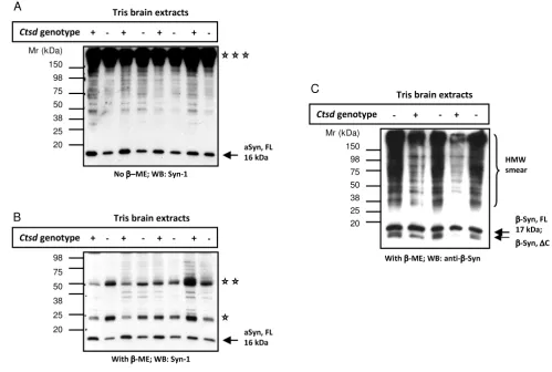
null alleles of murine ctsd and develop a fatal NCL leading to premature death at day 26 ± 1 [33,41]. We first exam-ined aSyn levels in the brain of ctsd-/- mice and their wild-type (wt) littermates by SDS/PAGE and Western blotting under both non-reducing (Fig. 2A) and reducing condi-tions (Fig. 2B). We found that the 16 kDa aSyn monomer was slightly but consistently less abundant in Tris-extracts of ctsd-/- brain (without cerebellum and brain stem) when compared with littermate controls (n = 4 per group for non-reducing conditions; n = 16 per group for reducing conditions). In these extracts, we found no evidence of truncated aSyn species by Western blotting (Fig. 2A, B; and data not shown). In parallel, the amount of mono-meric, full-length β-synuclein protein (17 kDa) appeared unchanged in ctsd-/- versus wt control mouse brains (Fig. 2C), although higher molecular weight species (and pos-sibly truncated variants) appeared increased (Fig. 2C; note, the latter findings could not be corroborated with another Ab due to lack of availability). In addition to the somewhat unexpected reduction of the 16 kDa aSyn pro-tein in ctsd-null brains, we observed that the mouse mon-oclonal aSyn Ab, Syn-1, also visualized bands that migrated at molecular weights of 25 kDa, 50 kDa (reduc-ing conditions; Fig. 2B) and > 150 kDa positions (non-reducing, Fig. 2A). These bands were interpreted as endog-enous immunoglobulin chains, as they were also detected by anti-mouse secondary Ab alone (not shown).
com-pared with their WT (n = 4) littermates; the reduction measured 18.6 and 20.0 per cent in NP-40 and SDS frac-tions, respectively (Fig. 3C). In addition, using densitom-etry we quantified the low molecular weight species of aSyn (migrating at 12.5 and 10 kDa positions) that were detected in NP-40 extracts by mSA-1 (Fig. 3A; Additional file 2, Fig. S2), but we found no apparent difference between the two genotypes (not shown).
Insoluble α-synuclein oligomers are elevated in Cathepsin D-deficient mouse brain
To complete the serial extraction protocol, we next searched for the presence of insoluble aSyn species in the formic acid extracts of SDS pellets from the ctsd-/- mouse brains shown in Fig. 3. There, we specifically searched for HMW aggregates of aSyn (referred to as oligomers) employing our most sensitive Abs (Fig. 4A). Independent Western blotting with hSA-2 and mSA-1 (Fig. 4A) and subsequent densitometry of the ratio, aSyn to actin,
revealed more insoluble, HMW oligomers in ctsd-/- than
in WT mouse brain (Fig. 4B). Of note, these oligomers
Mice lacking ctsd gene expression show reduced levels of endogenous α-synuclein in Tris-soluble brain extracts
Figure 2
Mice lacking ctsd gene expression show reduced levels of endogenous α-synuclein in Tris-soluble brain extracts. Western blot analyses of Tris brain extracts from 24-day old, wild-type (+) and ctsd-null mice (-) carried out under non-reduc-ing (A) and reducing (B) conditions. Probing with monoclonal aSyn Ab, Syn-1 (and rabbit anti-mouse IgG secondary Ab) dem-onstrates a subtle reduction in the full-length, monomeric aSyn protein (aSyn, FL) at the 16 kDa position in ctsd-null mice. Representative examples of 24 mouse brains are shown (n = 4 per genetic group under non-reducing conditions; n = 16 per genotype under reducing conditions). The same blots reveal a broad, high molecular weight (HMW) band above 150 kDa (tri-ple asterisks), and reactive bands at 50 kDa (double asterisks) and 25 (asterisk) kDa positions under non-reducing (A) and reducing (B) conditions, respectively. (C) Immunoblotting with anti-β-synuclein Ab under reducing conditions reveals no
differ-ence at the 17 kDa monomeric (β-Syn, FL) protein position, but indicates elevation of lower molecular weight fragment(s) at ~13 kDa (β-Syn, ΔC) and a HMW-immunoreactive smear in cathD-deficient mice. (Note, no additional anti-β-synuclein Ab is available for corroboration).
C
A
Ͳ
Ͳ
Ͳ Mr (kDa)
150 98 75 50 38 25 20
98
75
50
38
25
20
Mr (kDa) 150 98 75 50 38 25 20
B
! "
"
Ͳ " Ͳ
Ͳ "
Ͳ
#
$
%&
' ( )
#
! "
"
Ͳ " Ͳ
Ͳ "
Ͳ
#
$
%&
' ( )
#
!
Ͳ Ͳ "
Ͳ
"
Ͳ $
%&
* ( )
#
Ͳ $
+
Ͳ
#
Ͳ Ͳ
,
migrated predominantly at the interface of the stacking gel and running gel; their difference between the two gen-otypes was estimated to be ~15 per cent, as detected by both Abs (Fig. 4A and 4B; and data not shown). Intrigu-ingly, these formic acid extracted aSyn oligomers evi-dently did not co-localize with the HMW ubiquitin positive smears, as the latter were predominantly found in SDS extracts (Fig 4C).
Cathepsin D deficiency promotes intracellular α-synuclein accumulation in mouse brain
Our biochemical data showed a reduction of detergent soluble aSyn and an increase of formic-acid soluble aSyn
species in ctsd-/- mouse brains. In human PD, DLB and
MSA brain, the formic acid-soluble or urea-soluble frac-tions contain HMW aSyn aggregates. Therefore, we next explored whether any aSyn aggregates were also detecta-ble in brains of 24-day old ctsd-/- mice by
immunohisto-CathD-deficient mice show reduced α-synuclein levels in detergent soluble brain extracts
Figure 3
CathD-deficient mice show reduced α-synuclein levels in detergent soluble brain extracts. Western blot analyses of fractionated whole brain homogenates from 24-day old, wild-type (+) and ctsd-/- mice (-) carried out under reducing condi-tions showing NP-40 (A) and SDS extracts (B). Immunoblots were probed with anti-aSyn Abs (Syn-1; mSA-1), anti-β-actin and
anti-CathD Abs. Primary Ab detection was carried out with sheep anti-mouse and donkey anti-rabbit Ab, respectively. Under these immunoblotting conditions, variable amounts of the 16 kDa aSyn monomer (aSyn, FL) and traces of 12.5 kDa and 10 kDa (aSyn, ΔC) aSyn-reactive proteins are detectable; no reproducible > 150, 50 and 25 kDa aSyn-positive bands are seen.(C)
ELISA [hSA2/Syn1-B] quantification of NP-40 and SDS extracts from ctsd wild-type (WT) and mutant ctsd-/- (KO) mouse brains and of snca WT and snca-/- (KO) lysates serving as positive and negative controls. Ctsd-genotyped brain extracts were loaded in triplicates at three different dilutions each (1:500 to 1:2,000) onto a 384-well plate and read at 405 nm, as described [39,40]. Note, the absence of OD405 nm signals in snca KO mouse tissue. Bar graphs represent the mean group values (± SD) of mice that were individually analyzed (numbers as in A-C). The concentrations of total aSyn (in ng/μl) present in NP-40 extracts measured: ctsd WT, 1.34 ± 0.25; ctsd KO, 1.09 ± 0.26; and in the SDS fractions: ctsd WT, 0.60 ± 0.25; and ctsd KO, 0.48 ± 0.28.
Ͳ Ͳ Ͳ Ͳ Ͳ
Ͳ
0 0.5 1 1.5 2 2.5 3 3.5 4
1:128K 1:256K 1:512K 1:1024K 1:1K 1:1K ctsd WT ctsd KO ctsd WT ctsd KO
Recombinant synuclein standards snca WT snca KO NP-40 SDS
A
b
s
o
r
b
an
c
e
@ 405n
m
!" #
$% &'
( )
* + ,
!"
Ͳ&
!" #
$% !" #
- * + ,
. /
Ͳ&
01
)
#
. 02 34
56
( )
7 8
9 : 9
; < = >? @=A
B CDE F ; G H =I J ;
Ͳ K Ͳ Ͳ Ͳ Ͳ
Ͳ K
8
7 8
!" #
$% &'
( )
* + ,
!"
Ͳ&
7 8 Ͳ
L0M" #
5N
( )
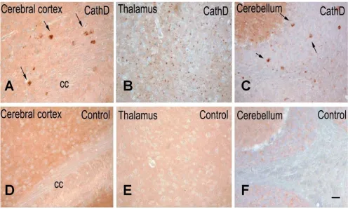
chemistry (Fig. 5). To avoid any cross-reactivity of the secondary anti-mouse Ab with endogenous immunoglob-ulins, we employed our rabbit polyclonal, affinity-puri-fied anti-aSyn Abs (mSA-1 and hSA-2), whose binding specificity for mammalian aSyn was confirmed using snca -null tissue (Additional file 2, Fig. S2) [39,40,48,49]. As shown in Fig. 5, we recorded prominent aSyn staining in the neuropil of the cerebral cortex, thalamus and cerebel-lum of control mice, as expected from its known presyn-aptic localization (Fig. 5D to 5F). In contrast, brain sections of ctsd-/- mice, processed in parallel, showed a less prominently stained neuropil, particularly in the tha-lamus (Fig. 5A, B). Intriguingly, in these CathD-deficient
mice, we also detected aSyn-positive aggregates within select neurons of the deep cortical laminae, superior col-liculi, the subiculum, as well as in the thalamus, deep cer-ebellar nuclei, and even within white matter tracts (Fig. 5A–C). Intra-neuronal cytoplasmic accumulations appeared large, while those within the thalamus were rel-atively small, thereby suggesting their localization within neurites. We concluded from these collective mouse data that: one, the distribution and processing of endogenous
aSyn is altered in ctsd-/- mice; and two, murine CathD
deficiency promotes an early-onset NCL phenotype that features the accumulation of a small pool of aSyn within an insoluble brain compartment.
CathD-deficient brains show increased HMW α-synuclein-positive oligomers in formic acid extracts
Figure 4
CathD-deficient brains show increased HMW α-synuclein-positive oligomers in formic acid extracts. (A) West-ern blot analyses carried out under reducing conditions of formic acid extracts obtained from previously generated SDS pellets of wild-type (+) and ctsd-/- (-) mouse brains (see Fig. 3 above). Immunoblots were probed with high sensitivity, polyclonal aSyn Abs, hSA-2 and mSA-1, and anti-β-actin Ab, as indicated. Under these conditions, HMW aSyn-positive smears and trace amounts of monomeric, 16 kDa aSyn (aSyn, FL) are detectable in these formic acid extracts.(B) Quantification of aSyn-reactive species by densitometry examining the ratio of HMW smears (blue bars) and 16 kDa monomer (empty bars) versus the signal of β-actin-positive bands. Note, an ~15 per cent increase in HMW oligomers of aSyn is detected in formic acid extracts of ctsd -/- brain by independent analysis of both mSA-1 (B) and hSA-2 immunoblots. (C) NP-40 and SDS extracts probed with anti-ubiquitin consistently reveal HMW immunoreactive smears in SDS brain extracts of ctsd-/- mice. Representative examples of twelve mouse brains are shown (n = 4, ctsd+/+; n = 8, ctsd-/-).
B
C
!
" # $
Ͳ
% & ' %
(
'
) % ) & * % * & * & & + ,- . /
0
1 2 3
Ͳ Ͳ 3 3
3
Ͳ Ͳ
4 5 6 7
8 9 8
!
" # :
Ͳ
;< =
;
: ' %
% &
( '
) %
) & * %
Ͳ
+ ,-. / 0
1 2
Ͳ 3 Ͳ Ͳ Ͳ
Ͳ 3
!
" #
>
$
Ͳ?
% &
Ͳ@:
AB
( '
' %
% &
Ͳ@:
AB
Mutant Cathepsin D expression alters α-synuclein immunohistochemistry in sheep brain
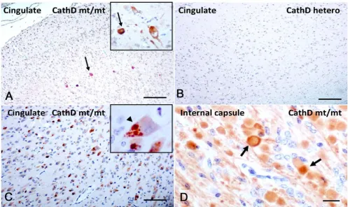
To validate these results in a second mammal, we carried out immunohistochemical studies in a sheep colony affected by congenital NCL (Fig. 6) [31]. This phenotype is caused by a homozygous missense mutation in the
ovine CTSD gene (Gln934Ala), which renders the enzyme
proteolytically inactive despite a 5- to 10-fold over-abun-dance of the mutant protein in the forebrain [50]. In sur-veys of sagittal hemibrain sections with our panel of mono- and polyclonal Abs to mammalian aSyn, overall staining of the neuropil appeared relatively normal in affected sheep (Fig. 6A; n = 3 for NCL sheep; n = 1 for het-erozygous sheep; n = 2 for wt control sheep). However, staining with our polyclonal Abs for ovine aSyn demon-strated widespread areas of elongated axonal swelling in deep white matter tracts in affected sheep (Fig. 6D) and revealed isolated aSyn aggregates in the thalamus, similar
to the mice (not shown). As expected, no axonal abnor-malities or aggregates were seen in heterozygous (Fig. 6B) and wt animals (not shown). Intriguingly, when we probed the sections with a well characterized monoclonal Ab to phosphoSer129-aSyn, we observed a small number of neurons in the cingulate cortex that showed intense
intracellular labeling in homozygous CTSD mutant sheep
(Fig. 6A), but not in heterozygous (Fig. 6B) and wt sheep. Of note, the axonopathy detected by the polyclonal Abs to aSyn was not matched by anti-phosphoSer129-aSyn reac-tivity. The changes in aSyn reactivity described above appeared specific, as staining of CathD-deficient sheep brain sections with Abs to Alzheimer's-linked phosphor-ylated tau, and TDP-43 did not reveal any obvious abnor-malities (not shown). In contrast, development of sections with a monoclonal anti-ubiquitin Ab revealed extensive inclusion formation that was present in NCL sheep brain throughout the cortex (Fig. 6C), including the
Mice lacking ctsd gene expression exhibit intracellular α-synuclein accumulation in several regions of the brain
Figure 5
cingulate cortex, but was absent in heterozygote and wt animals.
Cathepsin D deficiency promotes α-synuclein accumulation in human newborns
Next, we analyzed serial sections of paraffin-embedded,
post mortem human brains from three infants diagnosed with congenital NCL (Fig. 7). One of the three patients carried a homozygous duplication (c.764 dupA) in exon 6
of the CTSD gene, thereby creating a premature stop
codon and loss of CathD expression; another patient was his older sibling, who died of the same disease phenotype and thus, most probably, carried the same mutation, while the third, unrelated patient with congenital NCL had no detectable CathD protein present in her tissues although she remains genetically unidentified [30]. The brains of these three children revealed extreme atrophy with massive neuronal loss throughout the cortex,
accom-panied by generalized glial activation [30,51]. Using the same Ab panel employed above, we observed markedly reduced aSyn staining in the cortical neuropil of affected infants (Fig. 7B) compared to control brain (Fig. 7A),
sim-ilar to the changes seen in ctsd-/- mutant mouse brain
above. In addition, we identified strongly aSyn-reactive neurons scattered throughout the neocortex (Fig. 7B). These neurons carried cytoplasmic aSyn accumulations that appeared granular and were visible throughout the soma (Fig. 7B). Prominent 'synucleinopathy' was also observed in deep grey matter structures, thalamus (Fig. 7C, D) and basal ganglia of infants with congenital NCL. There, the aggregates varied greatly in size, with some appearing to localize to proximal axons (Fig. 7C), and some to the cell soma (Fig. 7D). Anti-hSA2-positive deposits could also be seen within white matter tracts, thus confirming their axonal localization. Within the cer-ebellum, aSyn deposits were observed in the granular cell
Sheep expressing a homozygous mutant of Cathepsin D exhibit accumulation of phosphorylated α-synuclein in cingulate cortex
Figure 6
layer and deep white matter. Of note, these inclusions were negative for Ser129-phosphorylation-specific aSyn reactivity, and for ubiquitin (not shown). As expected, no such aSyn abnormalities were seen in the control brain of a neurologically normal infant processed in parallel (Fig. 7A).
Neurotoxicity of human α-synuclein in Drosophila is enhanced in ctsd-mutant flies
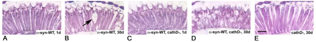
Over-expression of aSyn has been shown to be neurotoxic in a variety of cellular and animal models. In order to study the effect of endogenous CathD on aSyn-induced neurotoxicity, we generated flies expressing human aSyn in a ctsd-null background. In agreement with our previous results [9], the retina was normal in one-day-old trans-genic flies over-expressing wt human aSyn (Fig. 8A), but showed mild, vacuole-carrying changes by day 30 (Fig. 8B). Flies expressing human aSyn in the absence of CathD
also had normal retinas at day one (Fig. 8C), but devel-oped pronounced retinal degeneration within 30 days; these progressive changes included the formation of numerous vacuoles, thinning of the retina and loss of its architecture (Fig. 8D). Importantly, fly CathD deficiency by itself (without aSyn expression) did not cause this neu-rodegeneration phenotype of the retina at day 30 (Fig. 8E; [43]). These results indicated that neurotoxicity of aSyn is enhanced in the absence of endogenous CathD expres-sion.
Discussion
This report summarizes complementary investigations into the role of a key lysosomal enzyme, CathD, in the processing of aSyn. The in vivo results from three mamma-lian and one insect species described here provide addi-tional insights into the pathogenetic effects of CathD
Human neuronal ceroid lipofuscinosis due to Cathepsin D deficiency leads to intracellular accumulation of α-synuclein
Figure 7
deficiency, and identify in CathD a candidate for the much sought after 'synucleinase' activity in vivo.
Our initial set of experiments focused on a dopaminergic neuroblastoma cell culture model over-expressing aSyn
together with CathD (Fig. 1). CTSD expression reduced
aSyn concentrations in a manner that was dependent on the level of newly synthesized CathD (Fig. 1A and 1B). This effect could not be explained by a loss of cell viability, as the integrity of our culture model was unchanged from untransfected and vector-transfected cells in two standard viability assays. Similarly, the effect of CTSD cDNA could
not be explained by a competition effect with the SNCA
plasmid for the protein synthesis machinery, because co-expression of aSyn with Cathepsin L (CathL) or Cathepsin E (CathE) under the same conditions did not cause a reduction of aSyn concentrations (data not shown).
The simplest explanation for the effects of CTSD expres-sion on aSyn is that CathD-mediated degradation of aSyn within the lysosome was enhanced (see alternative expla-nations below). For this explanation to be valid, aSyn must have entered the lysosome by either macroau-tophagy or CMA, or both, given that CathD is thought to be active only at low pH. Several investigators have shown that the Asp98 and Gln99 residues of aSyn facilitate recog-nition by the Hsc70 chaperone and subsequent binding of the aSyn/Hsc70 complex to the Lamp2a receptor during CMA [26,28,52]. According to Cuervo et al., Ala30Pro and Ala53Thr mutants of aSyn are able to bind to isolated osomal membranes but unable to translocate into the lys-osomal lumen for degradation [28]. The fact that in our cell culture system, the D98A/Q99A double mutant of aSyn, as well as the Ala30Pro, Glu46Lys and Ala53Thr mutants of aSyn were degradable by exogenous CathD (Fig. 1C) suggests that, at the collection time point of 24 hours, the cells did not entirely rely on CMA as a means of transporting aSyn into lysosomes, and that, rather, macro-autophagy could be equally, or even more important than
CMA [26,52]. Likewise, changing the phosphorylation state of human aSyn at Ser129 did not alter the proteolytic synucleinase activity exhibited by CathD in our MES23.5 cell model. Of note, only select neurons in sheep cingulate cortex deficient for lysosomal CathD activity (but not in mouse or human brain) revealed evidence for abnormal anti-Ser129-aSyn reactivity (Fig. 7 and data not shown). Although the observed species difference could be caused by a variety of factors, our cellular data argue that Ser129 phosphorylation of aSyn is not a universal pre-requisite for CathD-induced aSyn degradation.
Western blotting analyses of our MES23.5 cell lysates demonstrated that the CathD-induced reduction in aSyn occurred without the concomitant appearance of either low-molecular weight (LMW) or HMW species, as exam-ined 24 hours after transfection. Of note, we had previ-ously confirmed by Western blotting that our Abs are capable of recognizing truncated aSyn species, (i.e., 10 to 12.5 kDa; see Additional file 2, Fig. S2) [46]. In particular, the appearance of LMW species of aSyn would have indi-cated incomplete proteolysis of aSyn, potentially at the
C-terminus. Such LMW aSyn-ΔC species are prone to
aggre-gate formation and have an enhancing effect on aSyn-mediated toxicity [46,53-55]. Previous studies by Dufty et
al. [56] showed that calpain-mediated digestion of aSyn
could induce the formation of aSyn-ΔC species and
for-mation of HMW species. Recently, Sevlever et al.
con-firmed a proteolytic effect of both enzymes, calpain and CathD, in in vitro biochemical studies and extended their
work to purified lysosomes from SNCA-expressing
neu-roblastoma cells [36].
In our SNCA and CTSD over-expression model, we
observed a maximum effect of CathD-induced reduction of excess aSyn levels that plateaued at ≤ 80 per cent of vec-tor-transfected cells (Fig. 1B). We concluded from these findings that a distinct pool of aSyn species (e.g., synaptic vesicle-associated) is not amenable to CathD-mediated
Cathepsin D deficiency enhances α-synuclein toxicity in the retina of fly brain
Figure 8
aSyn degradation. To explore whether over-expression of wt CTSD also reduces the concentration of endogenous aSyn, we carried out additional Western blotting and ELISA experiments in rodent MES23.5 and human SY5Y neuroblastoma cells. In the background of normal lyso-somal function and normal aSyn levels, we saw no con-sistent, further reduction of endogenous aSyn levels by ectopic CathD expression (not shown).
To examine the relevance of endogenous CathD
expres-sion on endogenous aSyn processing in vivo, we next
examined brain tissue from homozygous ctsd-/- mice [41]. We detected a consistent decrease in soluble aSyn proteins
in homozygous CathD deficient mouse brains versus those
of wt animals using Western blotting and ELISA (Figs. 2 and 3). This result was verified by quantitative 2D gel elec-trophoresis, which showed a significant reduction of the
16 kDa aSyn spot in ctsd-/- mice (data not shown). Our
quantitative ELISA showed a comparable decrease in the amount of soluble aSyn in NP-40 extracts (18.6 per cent) and in SDS-extracts (20.0 per cent) of ctsd-/- mice com-pared with controls (Fig. 3). This observation was in agreement with our immunohistochemistry results from the same mice that exhibited some reduction in aSyn staining in the neuropil of ctsd-/- mice, particularly within the thalamus (Fig. 5). Previously, we have confirmed that CathD deficient mice show a decrease in pre-synaptic markers in the ventral posterior nucleus of the thalamus, and a > 30 per cent loss of neurons in its corresponding target somatosensory cortex [34]. Because aSyn is a pre-dominantly pre-synaptic protein, we surmised that the reduction of soluble aSyn concentration recorded in our
ctsd-/- mice resulted – in part – from pre-synaptic abnor-malities.
However, in parallel to the reduction of soluble aSyn throughout the neuropil, we detected aSyn accumulation-carrying neurons within many brain regions of 24-day old
ctsd-/- mice. This finding was substantiated by our obser-vation of increased levels of insoluble species of oligo-meric aSyn in the formic acid extracts of ctsd-/- mice (15%; Fig. 4). (Of note, our current ELISA protocol is incompat-ible with the quantification of aSyn in the presence of for-mic acid). We concluded from these collective findings that the presence of murine CathD is important for the prevention of aSyn misprocessing and that its absence facilitates the formation of aSyn aggregation in vivo. In accordance, we speculate that the neuronal accumulation of murine aSyn in ctsd-/- mice may represent early precur-sor lesions to classical Lewy inclusions.
We next examined the role of mutant CathD protein expression on sheep aSyn levels (Fig. 6). Homozygous
missense mutation in the ovine CTSD gene (Gln934Ala)
essentially abolishes the enzyme's proteolytic activity
(residual level, < 5 per cent), while heterozygotes retain 40 per cent activity and are phenotypically normal [50]. The affected newborn lambs are weak, tremble and are unable to stand. When sacrificed shortly after birth, their brains are smaller and display mild cortical thinning, demyelina-tion, neuronal loss, reactive astrocytosis and macrophage infiltration, while visceral organs are spared. Classical NCL-type changes and widespread ubiquitin-positive inclusions within neurons were readily detected in the homozygous animals (Fig. 6). In contrast to the more extensive neuropil disruption and synucleinopathy seen in ctsd-/- mouse brain, we detected more select neuronal changes in homozygous CathD-mutant sheep, with abnormal anti-aSyn staining in the cingulate cortex and in the axons of deep white matter tracts. Intriguingly, synu-cleinopathy of the cingulate cortex is a hallmark of human dementia with Lewy bodies and advanced PD cases [6]. Unfortunately, no frozen material was available to deter-mine whether insoluble, HMW oligomeric species of ovine aSyn can be found biochemically in the cingulate cortex.
Humans with congenital NCL are affected at birth and
survive only for days. Two mutations in the human CTSD
gene have been described to underlie a subset of congeni-tal NCL cases [30,57]. Both mutations inactivate the CathD enzyme, in that the insertion mutation, c.764dupA, results in a premature stop-codon and rapid degradation of the protein, while the c.299C > T mutation leads to the production of an inactive but stable protein in
vivo. The brains of these infants show extreme cortical
atrophy, loss of neurons and myelin, as well as general-ized gliosis [30,51,57]. In brain specimens of two affected
siblings from a NCL pedigree linked to CTSD and of one
unrelated patient with congenital NCL, anti-aSyn staining of the neuropil was strongly reduced throughout the brain (Fig. 7). Accumulation of aSyn was particularly pro-nounced in the thalamus and basal ganglia, where the neuronal soma appeared to be loaded with aSyn aggre-gates, thereby morphologically resembling Lewy bodies of the forebrain; however, in contrast to classical Lewy inclu-sions these leinclu-sions were negative for ubiquitin and phos-pho-Ser129-specific reactivity in the affected newborns (not shown). Throughout the brains from these three infants, smaller aSyn-positive grains appeared to localize to neurites (Fig. 7B and 7C). Therefore, although the degree of brain atrophy and neuronal loss was much more severe in human infants with CathD deficiency than in
ctsd-/- mice, the aSyn-reactive histopathologies were very similar between the two species, possibly because of the complete loss of CathD protein expression in both.
different from that of lipofuscin deposition in NCL dis-eases. These autofluorescent accumulations are generally widespread within cortical neurons in humans and sheep affected by CathD deficiency and in the thalamus of ctsd -/- mice, while the observed aSyn accumulations occurred predominantly in the thalamus and basal ganglia in humans, in the cingulate cortex of affected sheep, and in the cerebral cortex of ctsd-/- mice.
Finally, our experiments using the established Drosophila
model of PD provided evidence for a modulatory effect of
endogenous CathD expression on aSyn toxicity in vivo
(Fig. 8). Deletion of the homologous CathD-encoding gene promoted toxicity of ectopically expressed human aSyn in the fly retina; these findings suggest that differ-ences in the expression level of CathD (and by inference, of other synucleinase-conferring proteases) may lower the threshold for synucleinopathy in other brain regions and species, too.
Importantly, our data do not demonstrate a direct interac-tion between CathD as an enzyme and aSyn as its
sub-strate. Although such a relationship was suggested by in
vitro experiments in one study [35], purification of lyso-somes from co-transfected MES23.5 cells and detailed imaging analyses will be required to further test this hypothesis. Intriguingly, aSyn transgenic mice show a decrease in aSyn inclusions when immunized with recom-binant aSyn, and this decrease was associated with a con-comitant increase in CathD levels as well as co-localization of CathD and aSyn [58]. Stefanis et al. [59] also noted co-localization of a pool of aSyn with CathD in their PC12 cell line-based experiments.
Indirect effects on aSyn accumulation by CathD defi-ciency are also possible. For example, CTSD gene expres-sion could activate another cathepsin or non-cathepsin protease, and this second enzyme may process aSyn for degradation. In this regard, it is interesting to note that CathD possesses activity that is similar to pepsin, and that pepsin can proteolytically activate Cathepsin F (CathF; [60]); intriguingly, in our cell culture system, over-expres-sion of CathF was able to reduce human aSyn levels, too, with an efficiency similar to that of CathD, whereas over-expression of CathE and CathL was ineffective (V.C., M.G.S., unpublished observations). It is noteworthy that mice deficient in CathF develop a late-onset neurological phenotype with gait disturbance, hind leg weakness and tremors, which is associated with brain and spinal cord pathology [61]; it will be interesting to discern whether these mice also display biochemical and/or immunohis-tochemical evidence of synucleinopathy.
Another possible mechanism for interaction between the
CTSD and SNCA genes in vivo is a change in lysosomal
constituency downstream of CathD protein expression. Perturbations in the activities of several lysosomal sphin-golipid-metabolising enzymes have been documented in
CathD-mutant sheep [31] and dogs [32], while ctsd
-/-mice accumulate phospho- and glyco-sphingolipids, including gangliosides [62]. Since aSyn is a lipid- and gan-glioside-binding protein [63-65], and since lipids can reg-ulate the oligomerization of aSyn [45,66], it is plausible that dysregulated lipid metabolism is a mechanism by which CathD deficiency leads to aSyn misprocessing. CathD is also known to exhibit activities that are unre-lated to its enzymatic function [67-69]. It is therefore pos-sible that non-proteolytic effects of CathD are essential in aSyn processing, akin to its effects on the ras-MAPK path-way [70]. Our data on aSyn processing in the cell soma and neurites of mutant sheep brain, where CathD is over-expressed but enzymatically disabled, would rather argue against this theory. However, one cannot exclude with cer-tainty that the homozygous mutation in CathD did not also compromise its non-enzymatic functions, although computational modeling did not predict the induction of a conformational change in the sheep mutant (J.T., data not shown).
Finally, our collective findings reported herein are in remarkable agreement with many (but not all) of the find-ings in the related report by Qiao and coworkers [71], who independently discovered CathD as a critical enzyme
in the metabolism of neuronal aSyn using both in vitro
and in vivo experiments. Of note, Qiao et al. examined off-spring of the same CathD-deficient mouse model that we employed [41], and observed a reduction of truncated aSyn species in ctsd-null mice, which we did not detect in our protocol. In addition to possible technical and Ab-related differences between the two reports, the slightly younger age of our animals at the time of euthanasia (post-natal day 24 versus 25) may also have contributed to this discrepancy.
Conclusion
These findings add NCL disorders to the growing list of human lysosomal storage diseases that feature mis-processing of neuronal aSyn [72-74]. Our evidence for altered processing and accumulation of insoluble species of endogenous aSyn in three different mammals with CathD deficiency indicates that its enzyme activity plays an important role in aSyn metabolism. This concept is supported by the pathogenesis replicated in our
inverte-brate Drosophila model, where the absence of CathD
markedly enhanced aSyn toxicity in vivo. Although we
The current treatment approaches for PD and related dis-orders are symptomatic at best, are not yet driven by molecular clues, and are invariably of limited duration. To date, progress in cause-directed treatment of synuclein-opathies has been prevented mostly by our limited knowl-edge of the normal regulators that control aSyn steady-state in vivo, from SNCA transcription to aSyn degrada-tion. Further exploration of the mechanism by which CathD promotes synucleinase activity holds the promise for novel drug targets to ultimately treat PD (and related disorders) at one if its root causes.
Competing interests
VC and MGS are listed as co-inventors of an application to the US Patent Office in 2007 related to the development of novel therapeutics for Parkinson disease. Otherwise, the authors have no competing interests.
Authors' contributions
VC and ML designed and conducted experiments; MGS and JT oversaw the project and together with VC wrote the manuscript; JN, AP, ES, PK and HB performed individual experiments; PS, JW, MBF and LM provided ideas, animal tissue and analysis. All authors reviewed the manuscript.
Additional material
Acknowledgements
We are grateful to the families of the CathD-deficient infants for tissue donation and Dr P. Stömme (Ullevål University Hospital, Norway) for co-ordinating patient contacts, and for the technical assistance of T. Inkinen and S. Kanerva; we thank RL. Nussbaum for tissue homogenates and PT. Lansbury for his gifts of recombinant aSyn and a SNCA plasmid.This study was supported by the Academy of Finland (to JT, #214343; to LM, #105861), the European Union (to JT & ML, EU-Lshm-CT2003-503051), the NIH/NINDS (to MGS, P50 NS38375), the Parkinson Research Consor-tium of Ottawa and Canada's Research Chair Program (to MGS). We thank Dr. J. Zhang for communication of her unpublished results.
References
1. Polymeropoulos MH, Lavedan C, Leroy E, Ide SE, Dehejia A, Dutra A, Pike B, Root H, Rubenstein J, Boyer R, et al.: Mutation in the alpha-synuclein gene identified in families with Parkinson's disease.
Science 1997, 276(5321):2045-2047.
2. Kruger R, Kuhn W, Muller T, Woitalla D, Graeber M, Kosel S, Przuntek H, Epplen JT, Schols L, Riess O: Ala30Pro mutation in the gene encoding alpha-synuclein in Parkinson's disease.
Nat Genet 1998, 18(2):106-108.
3. Singleton AB, Farrer M, Johnson J, Singleton A, Hague S, Kachergus J, Hulihan M, Peuralinna T, Dutra A, Nussbaum R, et al.: alpha-Synu-clein locus triplication causes Parkinson's disease. Science
2003, 302(5646):841.
4. Trojanowski JQ, Lee VM: Aggregation of neurofilament and alpha-synuclein proteins in Lewy bodies: implications for the pathogenesis of Parkinson disease and Lewy body dementia.
Arch Neurol 1998, 55(2):151-152.
5. Goedert M: Parkinson's disease and other alpha-synucleinopa-thies. Clin Chem Lab Med 2001, 39(4):308-312.
6. Spillantini MG, Schmidt ML, Lee VM, Trojanowski JQ, Jakes R, Goed-ert M: Alpha-synuclein in Lewy bodies. Nature 1997,
388(6645):839-840.
7. Klein C, Schlossmacher MG: Parkinson disease, 10 years after its genetic revolution: multiple clues to a complex disorder.
Neurology 2007, 69(22):2093-2104.
8. Chesselet MF: In vivo alpha-synuclein overexpression in rodents: a useful model of Parkinson's disease? Exp Neurol
2008, 209(1):22-27.
9. Feany MB, Bender WW: A Drosophila model of Parkinson's dis-ease. Nature 2000, 404(6776):394-398.
10. Masliah E, Rockenstein E, Veinbergs I, Mallory M, Hashimoto M, Takeda A, Sagara Y, Sisk A, Mucke L: Dopaminergic loss and inclusion body formation in alpha-synuclein mice:
implica-Additional file 1
Supplementary Figure 1 – Expression and quantification of α -synu-clein in MES23.5 cells. Recombinant, human α-synuclein (aSyn) (A) and lysates of MES23.5 cells expressing αS (B-C) were analyzed by ELISA [hSA2/Biotinylated Syn-1]. (A) A typical standard curve of serial dilu-tions of recombinant αS (mean of triplicate wells). (B) Following gener-ation of MES-aSyn cells with three different concentrgener-ations of human SNCA cDNA (μg/10 cm dish; total DNA transfected per dish, 5.5 μg), lysates were analyzed in 4 different dilutions, as indicated. Results show the mean absorbance signals of triplicates from a representative experi-ment. (C) Linear relationship between amount of SNCA-encoding cDNA transfected into MES23.5 cells and αS protein expression, as monitored by ELISA. Each lysate was analyzed in 4 different dilutions in triplicate. Linear regression of cDNA-to-protein was performed; results represent the mean aSyn values (μg) expressed from a representative experiment. Click here for file
[http://www.biomedcentral.com/content/supplementary/1756-6606-2-5-S1.ppt]
Additional file 2
Supplementary Figure 2 – Characterization of affinity-purified anti-bodies to α-synuclein using genotyped mouse brain. Whole brain extracts of genotyped mice were generated by lysis buffer that contained NP-40 and protease inhibitors [37]; increasing amounts of the NP-40 extract (μg/lane) were loaded onto SDS/PAGE gels under reducing con-ditions. Immunoblots were probed with polyclonal, affinity-purified anti-aSyn, hSA-2 (top panel) and mSA-1 (third panel). Loading controls showing IgG heavy chains are shown for both blots. Lysates were prepared from from snca knock-out mice without a transgene (no transgene; left half) and mice that carry a human, wild-type SNCA transgene (SNCA transgene; right half; brains kindly provided by Dr. Bob Nussbaum, UCSF). Note the specific detection of full-length aSyn (16 kDa), and of 12.5 kDa and ~10 kDa truncated species of aSyn in SNCA-expressing mice.
Click here for file
tions for neurodegenerative disorders. Science 2000,
287(5456):1265-1269.
11. Lee MK, Stirling W, Xu Y, Xu X, Qui D, Mandir AS, Dawson TM, Copeland NG, Jenkins NA, Price DL: Human alpha-synuclein-harboring familial Parkinson's disease-linked Ala-53 --> Thr mutation causes neurodegenerative disease with alpha-synuclein aggregation in transgenic mice. Proc Natl Acad Sci USA
2002, 99(13):8968-8973.
12. Giasson BI, Duda JE, Quinn SM, Zhang B, Trojanowski JQ, Lee VM:
Neuronal alpha-synucleinopathy with severe movement dis-order in mice expressing A53T human alpha-synuclein. Neu-ron 2002, 34(4):521-533.
13. Kahle PJ, Neumann M, Ozmen L, Muller V, Odoy S, Okamoto N, Jacobsen H, Iwatsubo T, Trojanowski JQ, Takahashi H, et al.: Selec-tive insolubility of alpha-synuclein in human Lewy body dis-eases is recapitulated in a transgenic mouse model. Am J
Pathol 2001, 159(6):2215-2225.
14. Anderson JP, Walker DE, Goldstein JM, de Laat R, Banducci K, Cac-cavello RJ, Barbour R, Huang J, Kling K, Lee M, et al.: Phosphoryla-tion of Ser-129 is the dominant pathological modificaPhosphoryla-tion of alpha-synuclein in familial and sporadic Lewy body disease. J
Biol Chem 2006, 281(40):29739-29752.
15. Leroy E, Anastasopoulos D, Konitsiotis S, Lavedan C, Polymeropou-los MH: Deletions in the Parkin gene and genetic heterogene-ity in a Greek family with early onset Parkinson's disease.
Hum Genet 1998, 103(4):424-427.
16. Kitada T, Asakawa S, Hattori N, Matsumine H, Yamamura Y, Minoshima S, Yokochi M, Mizuno Y, Shimizu N: Mutations in the parkin gene cause autosomal recessive juvenile
parkinson-ism. Nature 1998, 392(6676):605-608.
17. Carmine Belin A, Westerlund M, Bergman O, Nissbrandt H, Lind C, Sydow O, Galter D: S18Y in ubiquitin carboxy-terminal hydro-lase L1 (UCH-L1) associated with decreased risk of Parkin-son's disease in Sweden. Parkinsonism Relat Disord 2007,
13(5):295-298.
18. Maraganore DM, Lesnick TG, Elbaz A, Chartier-Harlin MC, Gasser T, Kruger R, Hattori N, Mellick GD, Quattrone A, Satoh J, et al.:
UCHL1 is a Parkinson's disease susceptibility gene. Ann Neu-rol 2004, 55(4):512-521.
19. Shimura H, Schlossmacher MG, Hattori N, Frosch MP, Trocken-bacher A, Schneider R, Mizuno Y, Kosik KS, Selkoe DJ: Ubiquitina-tion of a new form of alpha-synuclein by parkin from human brain: implications for Parkinson's disease. Science 2001,
293(5528):263-269.
20. Liu Y, Fallon L, Lashuel HA, Liu Z, Lansbury PT Jr: The UCH-L1 gene encodes two opposing enzymatic activities that affect alpha-synuclein degradation and Parkinson's disease suscep-tibility. Cell 2002, 111(2):209-218.
21. Lo Bianco C, Deglon N, Pralong W, Aebischer P: Lentiviral nigral delivery of GDNF does not prevent neurodegeneration in a genetic rat model of Parkinson's disease. Neurobiol Dis 2004,
17(2):283-289.
22. Webb JL, Ravikumar B, Atkins J, Skepper JN, Rubinsztein DC: Alpha-Synuclein is degraded by both autophagy and the
proteas-ome. J Biol Chem 2003, 278(27):25009-25013.
23. Shin Y, Klucken J, Patterson C, Hyman BT, McLean PJ: The co-chap-erone carboxyl terminus of Hsp70-interacting protein (CHIP) mediates alpha-synuclein degradation decisions between proteasomal and lysosomal pathways. J Biol Chem
2005, 280(25):23727-23734.
24. Ventruti A, Cuervo AM: Autophagy and neurodegeneration.
Curr Neurol Neurosci Rep 2007, 7(5):443-451.
25. Sarkar S, Davies JE, Huang Z, Tunnacliffe A, Rubinsztein DC: Treha-lose, a novel mTOR-independent autophagy enhancer, accelerates the clearance of mutant huntingtin and alpha-synuclein. J Biol Chem 2007, 282(8):5641-5652.
26. Vogiatzi T, Xilouri M, Vekrellis K, Stefanis L: Wild type alpha-synu-clein is degraded by chaperone-mediated autophagy and macroautophagy in neuronal cells. J Biol Chem 2008,
283(35):23542-23556.
27. Lee HJ, Khoshaghideh F, Patel S, Lee SJ: Clearance of alpha-synu-clein oligomeric intermediates via the lysosomal degrada-tion pathway. J Neurosci 2004, 24(8):1888-1896.
28. Cuervo AM, Stefanis L, Fredenburg R, Lansbury PT, Sulzer D:
Impaired degradation of mutant alpha-synuclein by chaper-one-mediated autophagy. Science 2004, 305(5688):1292-1295.
29. Hasilik A, Neufeld EF: Biosynthesis of lysosomal enzymes in fibroblasts. Synthesis as precursors of higher molecular weight. J Biol Chem 1980, 255(10):4937-4945.
30. Siintola E, Partanen S, Stromme P, Haapanen A, Haltia M, Maehlen J, Lehesjoki AE, Tyynela J: Cathepsin D deficiency underlies con-genital human neuronal ceroid-lipofuscinosis. Brain 2006,
129(Pt 6):1438-1445.
31. Tyynela J, Sohar I, Sleat DE, Gin RM, Donnelly RJ, Baumann M, Haltia M, Lobel P: A mutation in the ovine cathepsin D gene causes a congenital lysosomal storage disease with profound neuro-degeneration. EMBO J 2000, 19(12):2786-2792.
32. Awano T, Katz ML, O'Brien DP, Taylor JF, Evans J, Khan S, Sohar I, Lobel P, Johnson GS: A mutation in the cathepsin D gene (CTSD) in American Bulldogs with neuronal ceroid lipofus-cinosis. Mol Genet Metab 2006, 87(4):341-348.
33. Koike M, Nakanishi H, Saftig P, Ezaki J, Isahara K, Ohsawa Y, Schulz-Schaeffer W, Watanabe T, Waguri S, Kametaka S, et al.: Cathepsin D deficiency induces lysosomal storage with ceroid lipofuscin in mouse CNS neurons. J Neurosci 2000, 20(18):6898-6906. 34. Partanen S, Haapanen A, Kielar C, Pontikis C, Alexander N, Inkinen
T, Saftig P, Gillingwater TH, Cooper JD, Tyynela J: Synaptic changes in the thalamocortical system of cathepsin D-defi-cient mice: a model of human congenital neuronal ceroid-lipofuscinosis. J Neuropathol Exp Neurol 2008, 67(1):16-29. 35. Hossain S, Alim A, Takeda K, Kaji H, Shinoda T, Ueda K: Limited
proteolysis of NACP/alpha-synuclein. J Alzheimers Dis 2001,
3(6):577-584.
36. Sevlever D, Jiang P, Yen SH: Cathepsin D is the main lysosomal enzyme involved in the degradation of alpha-synuclein and generation of its carboxy-terminally truncated species.
Bio-chemistry 2008, 47(36):9678-9687.
37. Schlossmacher MG, Shimura H: Parkinson's disease: assays for the ubiquitin ligase activity of neural Parkin. Methods Mol Biol
2005, 301:351-369.
38. LaVoie MJ, Ostaszewski BL, Weihofen A, Schlossmacher MG, Selkoe DJ: Dopamine covalently modifies and functionally inacti-vates parkin. Nat Med 2005, 11(11):1214-1221.
39. Mollenhauer B, Cullen V, Kahn I, Krastins B, Outeiro TF, Pepivani I, Ng J, Schulz-Schaeffer W, Kretzschmar HA, McLean PJ, et al.: Direct quantification of CSF alpha-synuclein by ELISA and first cross-sectional study in patients with neurodegeneration.
Exp Neurol 2008, 213(2):315-325.
40. Scherzer CR, Grass JA, Liao Z, Pepivani I, Zheng B, Eklund AC, Ney PA, Ng J, McGoldrick M, Mollenhauer B, et al.: GATA transcription factors directly regulate the Parkinson's disease-linked gene alpha-synuclein. Proc Natl Acad Sci USA 2008,
105(31):10907-10912.
41. Saftig P, Hetman M, Schmahl W, Weber K, Heine L, Mossmann H, Koster A, Hess B, Evers M, von Figura K, et al.: Mice deficient for the lysosomal proteinase cathepsin D exhibit progressive atrophy of the intestinal mucosa and profound destruction of lymphoid cells. EMBO J 1995, 14(15):3599-3608.
42. Schlossmacher MG, Frosch MP, Gai WP, Medina M, Sharma N, Forno L, Ochiishi T, Shimura H, Sharon R, Hattori N, et al.: Parkin local-izes to the Lewy bodies of Parkinson disease and dementia with Lewy bodies. Am J Pathol 2002, 160(5):1655-1667. 43. Myllykangas L, Tyynela J, Page-McCaw A, Rubin GM, Haltia MJ, Feany
MB: Cathepsin D-deficient Drosophila recapitulate the key features of neuronal ceroid lipofuscinoses. Neurobiol Dis 2005,
19(1–2):194-199.
44. Crawford GD Jr, Le WD, Smith RG, Xie WJ, Stefani E, Appel SH: A novel N18TG2 × mesencephalon cell hybrid expresses prop-erties that suggest a dopaminergic cell line of substantia nigra origin. J Neurosci 1992, 12(9):3392-3398.
45. Sharon R, Goldberg MS, Bar-Josef I, Betensky RA, Shen J, Selkoe DJ:
alpha-Synuclein occurs in lipid-rich high molecular weight complexes, binds fatty acids, and shows homology to the fatty acid-binding proteins. Proc Natl Acad Sci USA 2001,
98(16):9110-9115.
46. Periquet M, Fulga T, Myllykangas L, Schlossmacher MG, Feany MB:
Aggregated alpha-synuclein mediates dopaminergic neuro-toxicity in vivo. J Neurosci 2007, 27(12):3338-3346.
47. Fujiwara H, Hasegawa M, Dohmae N, Kawashima A, Masliah E, Gold-berg MS, Shen J, Takio K, Iwatsubo T: alpha-Synuclein is phospho-rylated in synucleinopathy lesions. Nat Cell Biol 2002,
![Figure 1αing conditions of MES23.5 cells [38] expressing untagged, wild-type human Over-expression of Cathepsin D lowers -synuclein in dopaminergic cellsOver-expression of Cathepsin D lowers α-synuclein in dopaminergic cells](https://thumb-us.123doks.com/thumbv2/123dok_us/273983.1520269/4.612.59.555.208.559/conditions-expressing-expression-cathepsin-dopaminergic-expression-cathepsin-dopaminergic.webp)