Open Access
Research article
The prevalence of dental erosion in Nigerian patients with
gastro-oesophageal reflux disease
Adeleke O Oginni*
1, Elugwaraonu A Agbakwuru
2and Dennis A Ndububa
3Address: 1Department of Restorative Dentistry, College of Health Sciences, Obafemi Awolowo University, Ile-Ife, Nigeria, 2Department of Surgery,
College of Health Sciences, Obafemi Awolowo University, Ile-Ife, Nigeria and 3Department of Medicine, College of Health Sciences, Obafemi
Awolowo University, Ile-Ife, Nigeria
Email: Adeleke O Oginni* - adelekeoginni@yahoo.co.uk; Elugwaraonu A Agbakwuru - eagbakwu@oauife.edu.ng; Dennis A Ndububa - dndububa@oauife.edu.ng
* Corresponding author
Abstract
Background: In various people of the Western world, gastro-oesophageal reflux (GOR) has been reported to be a common problem. Various studies have also assessed the relationship between GOR and dental erosion. The authors are not aware of such studies in Nigerians. It is therefore the aims of the present study to estimate the prevalence of GOR; to estimate the prevalence of dental erosion in patients with GORD; to document the oral findings in patients diagnosed with GORD and to compare these findings with previous studies elsewhere.
Methods: A total of 225 subjects comprising of 100 volunteers and 125 patients diagnosed with GORD were involved in this study. History of gastric juice regurgitation and heartburn were recorded. Oral examination to quantify loss of tooth structure was done using the tooth wear index (TWI) designed by Smith and Knight (1984).
Results: Twenty patients with GORD presented with dental erosion in the maxillary anterior teeth with TWI scores ranging from 1–3. The prevalence of erosion was found to be statistically significant between GORD patients (16%) and control (5%) (p < 0.05), but not significant between endoscopic diagnostic groups (p > 0.05).
Conclusion: The present study supports the consideration of dental erosion as the extra-oesophageal manifestation of GORD. However the association between GORD and burning mouth sensation needs more investigation.
Background
Gastro-oesophageal reflux (GOR) is the passage of gastric contents into the oesophagus. Once past the upper oesophageal sphincter, the gastric juice may pass into the oral cavity. The continual exposure of the teeth and other oral structures to gastric refluxate may result in dental ero-sion and other soft tissue symptoms [1]. Any acid with a pH below the critical pH of dental enamel dissolution
(5.5) can dissolve the hydroxyapatite crystals in enamel. However the critical pH below which enamel dissolves is not constant but is rather inversely proportional to the concentrations of calcium and phosphate in the saliva and plaque fluid [2]. Gastric refluxate has a pH of less than 2.0 and thus has the potential to cause dental erosion [3]. Acid regurgitation is a common symptom of upper gastro-intestinal tract disorders and dysfunctions such as Published: 01 March 2005
BMC Oral Health 2005, 5:1 doi:10.1186/1472-6831-5-1
Received: 20 August 2004 Accepted: 01 March 2005
This article is available from: http://www.biomedcentral.com/1472-6831/5/1
© 2005 Oginni et al; licensee BioMed Central Ltd.
peptic ulcer (duodenal and gastric ulcers) and reflux oesophagitis [4]. Other symptoms include heartburn, non-cardiac epigastric and retrosternal pain [5]. Promi-nent among factors precipitating GOR and its complica-tions, gastro-oesophageal reflux disease (GORD) are fatty diets and alcohol.
Current understanding of GOR and GORD is that acid reflux into the oesophagus may be caused by three possi-ble mechanisms [6]:
(1) Transient spontaneous or inappropriate relaxations of the sphincter;
(2) Transient increase in intra-abdominal or intragastric pressure;
(3) Functional abnormality of the lower oesophageal sphincter (LOS).
Bargen and Austin in 1937 [7] first reported the link between dental erosion and gastro-intestinal distur-bances. Since then there has been several other studies confirming the relationship between this loss of tooth structure and GORD in the United Kingdom (UK) [8], United States of America (U.S.A) [9] and Canada [10]. A recent survey among young people in the U.K also revealed an association between dental erosion and symp-toms of GOR [11]. Although soft tissue sympsymp-toms (non-specific burning sensation in the mouth) have been men-tioned in the literature, pathognomonic soft tissue lesions have not been documented [1]. But some researchers have reported a lack of relationship between periodontal lesions and GOR, since the prevalence of periodontal lesions is similar in patients with GORD and in healthy volunteers [12]. In various people of the Western world, GOR has been reported to be a common problem, often related to meals and occurring in about 60% of the popu-lation at some point in their lives [13]. Whereas the prev-alence of GORD is estimated to range from 6% – 10% [14,15], Meurman et al [16] examined 117 patients with GORD, of whom 28 (24%) had dental erosion. Also, Schroeder et al [17] identified dental erosion in 11 (55%) of 20 patients with GORD. The authors are not aware of such figures in Nigerians. A search of the literature also revealed a dearth of information on this condition and its complications among Black Africans. It is therefore the aim of the present study to estimate the prevalence of GOR among patients attending the medical out-patient department of the Obafemi Awolowo University Teaching Hospital Complex Ile-Ife, Nigeria; to estimate the preva-lence of dental erosion in patients with GORD; to docu-ment the oral findings in patients diagnosed with GORD and to compare these findings with previous studies elsewhere.
Methods
A total of 225 subjects were involved in the study over a period of 6 months, January – June 2002. The subjects were made up of 100 volunteers attending the medical outpatient department (MOPD) and 125 patients diag-nosed with gastro-oesophageal reflux disease among whom the prevalence of GOR and GORD was determined respectively. Their age ranges from 18 – 72 years, with a mean age ± S.D. of 38 ± 10.87 years. Patients presenting primarily with symptoms of asthma, bronchitis and other respiratory disorders were excluded, since they are also at risk of dental erosion [18].
To assess the prevalence of GOR and GORD, consenting patients attending the medical outpatient department of the Obafemi Awolowo University Teaching Hospital's complex Ile-Ife, Nigeria, were questioned with reference to their experience regarding symptoms of GOR such as regurgitation of gastric juice, epigastric and non-cardiac pain (heartburn). The onset, frequency of occurrence and duration of each of the symptoms were ascertained and recorded. Patients presenting with a history of heartburn 2 or more times per week were diagnosed as having GORD10. Oral examination was carried out to quantify
any loss of tooth structure using the tooth wear index (TWI) designed by Smith and Knight (1984) [19].
To document the oral findings associated with GORD, patients referred to the Gastro-intestinal (GIT) endoscopic unit for investigation of gastro-oesophageal tract disease were also evaluated for dental erosion and other soft tis-sue symptoms such as non-specific burning sensation in the mouth and sensitivity in the tongue. The dental eval-uation included history to determine potential eatiologi-cal factors responsible for dental erosion. Patients were also examined clinically to quantify loss of tooth structure using the tooth wear index. The oral examination was per-formed by one of the authors (AO), blind as to the endo-scopic diagnosis of subjects. Patients with positive endoscopic findings plus the occurrence of heartburn two or more times per-week were assessed to meet the criteria for GORD. When a clear-cut endoscopic evidence of oesophageal inflammation was seen (marked redness, fibrinous membrane, and or ulcerations), a diagnosis of oesophagitis was made. Gastroduodenal ulcer was diag-nosed through endoscopic evidence of an ulcer with a necrotic base or a clear-cut scar. This was grouped into those with gastric ulcer (ulcer in the pylorus, antrum, cor-pus or fundus) and those with duodenal ulcer (ulcer in the bulbus). Only one diagnosis was given for each patient.
Results
Thirty-five of the 100 subjects attending the medical out-patient department had acid regurgitation and acidic taste sensation in the mouth. This occurs most of the time fol-lowing a full stomach meal and is associated with belch-ing. Table 1 shows the prevalence of GOR and GORD. Eleven reported a history of heartburn 2 or more times per week, while 16 and 10 reported weekly and monthly his-tory of heartburn respectively. Comparison of subjects presenting with heartburn 2 or more times per week (M = 5, F = 6) and those with less than 2 times per week (M = 12, F = 14) shows that more female presented with heart-burn. However, there was no statistical difference among the genders with regard to the symptoms recorded. None of these subjects presented with any burning sensation in the mouth or sensitivity in the tongue. In five of those that reported 2 or more weekly history of heartburn, there were minimal losses of tooth structure (TWI scores of 1
and 2 in three and in two subjects respectively) limited to the maxillary central incisors (Table 2).
One hundred and twenty five patients were diagnosed with GORD. Twenty presented with dental erosion in the maxillary anterior teeth with TWI scores ranging from 1– 3. The prevalence of erosion was found to be statistically significant between GORD patients (16%) and controls (5%) (Mantel Haenszel Chi-square 5.50, p < 0.05), Table 2. The result of the gastrointestinal endoscopy reported 41(32.8%) as having reflux oesophagitis, 36 (28.8%) with duodenal ulcer and 48 (38.4%) with gastric ulcer. Eight patients with dental erosion came from the group who had reflux oesophagitis, 7 from the group with duodenal ulcer and 5 from the group with gastric ulcer. Comparison of patients with oesophagitis and those without shows that the mean age of patients with oesophagitis was lower than in those without. The prevalence of erosion was not
Table 1: Prevalence of GOR and symptoms of GORD
Symptoms Male (N = 49) Female (N = 51) Total
Regurgitation and acidic taste in the mouth
16 19 35
Heartburn ≥ 2 per week 5 6 11
Heartburn per week 7 9 16
Heartburn per month 5 5 10
Chi Square = 0.10; df = 3; p = 0.99.
Table 2: Prevalence of dental erosion
Dental erosion
Subjects/patients No (%) TWI scores
M.O.P. (N = 100) 5 (5) 1 – 2
GORD (N = 125) 20 (16) 1 – 3
Mantel Haenszel chi-square = 5.50, p = 0.02.
Table 3: Endoscopic diagnostic groups: basic data and number of patients withdental erosion
Diagnostic groups Number Mean age (yrs) Gender Dental erosion
detected
No (%) ± S.D Male Female
Reflux oesophagitis 41 (32.8) 36.9 ± 9.7 24 17 8
Duodenal ulcer 36 (28.8) 39.2 ± 11.4 16 20 7
Gastric ulcer 48 (38.4) 38.1 ± 11.0 17 31 5
statistically significant between the endoscopic diagnostic groups (chi-square = 1.33, df = 2, p = 0.51), Table 3.
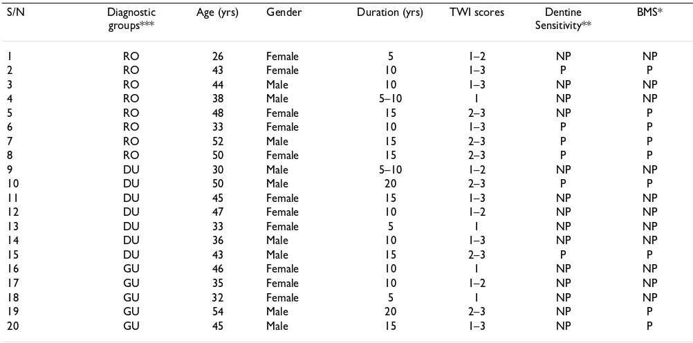
The details of the orodental findings in the twenty patients who had dental erosion are shown in Table 4. There seems to be a slight association between the dura-tion of gastrointestinal symptoms and the severity of ero-sion. TWI scores of 3 were seen only in those patients whose abdominal symptoms had lasted 10 years or more. The palatal surfaces of the maxillary anterior teeth were usually involved but the central incisors were the most severely affected. Six patients (4 from reflux oesophagitis and 2 from duodenal ulcer) had dental erosion related symptoms in their teeth: sensitivity to cold and heat. The teeth that presented with dentine sensitivity had TWI scores of 3.
Burning mouth sensation and peppery sensation in the tongue where reported by 9 patients who had had gas-trointestinal symptoms for 10–20 years.
Discussion
Various methods have been employed in the investigation of GOR including endoscopy with biopsy and contrast radiography. Although twenty-four hour oesophageal pH monitoring is considered the gold standard investigation of GOR [20], due to non-availability of the ambulatory
pH recorder in our center, GOR was diagnosed by endos-copy where visual identification of mucosal inflammation and oesophagitis was used to identify the existence of GOR. Several other authors have used this method [4,21].
The prevalence of GOR and GORD in the present study was 35% and 11% respectively. The prevalence of GOR was much lower than the 60% reported in the U.K. and other Western societies [13], whereas that of GORD was a little above the reported range of 6%–10% [14,15]. A lower percentage (10%) of the subject studied reported a monthly history of heartburn as compared to 59% of the population reported by Lock et al [22]. The present study examined 125 patients with GORD of which 20(16%) had dental erosion. This is also lower than the 24% and 55% reported by Meurman et al [16], and Schroeder et al [17] respectively. Although the authors cannot propose any reason for the reported lower prevalence of GOR and GORD, the low prevalence of dental erosion may be related to difference in diet. More so that the thrust of the European studies support the view that erosion (resulting from acidic and carbonated foods and beverages) is more important than attrition in the aetiology of tooth wear [23], whereas, attrition have been reported to be more important than erosion in the aetiology of tooth wear among Nigerians [24,25]. Also in the study of gastro-oesophageal reflux in children and its relationship to
Table 4: Orodental findings in the twenty patients with GORD who had erosions
S/N Diagnostic groups***
Age (yrs) Gender Duration (yrs) TWI scores Dentine Sensitivity**
BMS*
1 RO 26 Female 5 1–2 NP NP
2 RO 43 Female 10 1–3 P P
3 RO 44 Male 10 1–3 NP NP
4 RO 38 Male 5–10 1 NP NP
5 RO 48 Female 15 2–3 NP P
6 RO 33 Female 10 1–3 P P
7 RO 52 Male 15 2–3 P P
8 RO 50 Female 15 2–3 P P
9 DU 30 Male 5–10 1–2 NP NP
10 DU 50 Male 20 2–3 P P
11 DU 45 Female 15 1–3 NP NP
12 DU 47 Female 10 1–2 NP NP
13 DU 33 Female 5 1 NP NP
14 DU 36 Male 10 1–3 NP NP
15 DU 43 Male 15 2–3 P P
16 GU 46 Female 10 1 NP NP
17 GU 35 Female 10 1–2 NP NP
18 GU 32 Female 5 1 NP NP
19 GU 54 Male 20 2–3 NP P
20 GU 45 Male 15 1–3 NP P
*BMS = Burning mouth sensation and sensitivity in the tongue **NP = Not present, P = Present
erosion of primary and permanent teeth, diet was consid-ered to be a major contributory factor [26].
The consumption of acidic beverages among the study population was low, hence the silence on its contribution to the overall disease level. Although this is controversial, the authors are of the opinion that intrinsic (gastric) acid results in palatally eroded sites (as shown in Figure 1) while extrinsic (dietary) acids lead to labial or vestibular erosions. The authors observed some degree of tooth wear (tooth surface loss) on the molar teeth. It was not docu-mented because we believe they are more likely due to "attrition" resulting from rigorous mastication of the more fibrous Nigerian diet. However, acidic refluxate may have been a contributory factor, since tooth surface loss is a multi-factorial disease.
In accordance with the result of Gregory-Head et al [27], patients diagnosed with GORD in this study had higher TWI scores compared with control subjects. All the twenty patients that presented with dental erosions in the present
study had an underlying gastrointestinal pathosis (Gastric oesophagitis, Duodenal ulcer, and Gastric ulcer) with increased output of acid secretion into the stomach [28]. There was however no statistically significant difference in the prevalence of dental erosions in these diagnostic groupings, Table 3. This is in agreement with the study of Jarvinen et al [4]. The severity of dental erosions may depend on the frequency of regurgitation and duration of the gastro-oesophageal reflux. In the present study, patients with TWI scores of 3 had their symptoms for more than 10 years as shown in Table 4. This is supported by the study of Loffeld et al [21], which revealed a signifi-cant association between duration of complaints and presence of damage in the upper incisors, but in contrast to the findings of Jarvinen et al [4] who reported no direct association between the frequency of regurgitation symp-toms and the severity of the erosive lesions. Six patients presented with erosion related symptoms in their teeth, they had dentine sensitivity to cold and heat. All of these patients had dental erosions with TWI scores of 3 (Loss of enamel exposing dentine for more than one-third of the surfaces). This probably explains the sensitivity.
Nine patients reported burning mouth sensation and sen-sitivity in the tongue. They describe the sensation as pep-pery/burning feeling in the vestibule of the mouth and mostly on the dorsal surface of the tongue. These may have resulted from the prolonged effect of acidic gastric refluxate on the oral mucosa and on the papillae of the tongue.
Conclusion
The present study supports the consideration of dental erosion as the extra-oesophageal manifestation of GORD. However the association between GORD and burning mouth sensation needs more investigation.
Competing interests
The author(s) declare that they have no competing interests.
Authors' contributions
AOO conceived of the study, participated in its design, performed the dental examination including the applica-tion of the tooth wear index (TWI), and participated in the initial draft and final write-up of the manuscript. EAA and DAN performed the endoscopic examination of patients with gastro-oesophageal reflux disease, participated in the initial draft and final write-up of the manuscript.
References
1. Lazarchik DA, Filler SJ: Effects of gastroesophageal reflux on the oral cavity.Am J Med 1997, 103:107S-113S.
2. Dawes C: What is the critical pH and why does a tooth dis-solve in acid?J Can Dent Assoc 2003, 69:722-724.
Severe dental erosion affecting the palatal surfaces of the upper anterior teeth in a patient with reflux oesophagitis Figure 1
Publish with BioMed Central and every scientist can read your work free of charge
"BioMed Central will be the most significant development for disseminating the results of biomedical researc h in our lifetime."
Sir Paul Nurse, Cancer Research UK
Your research papers will be:
available free of charge to the entire biomedical community
peer reviewed and published immediately upon acceptance
cited in PubMed and archived on PubMed Central
yours — you keep the copyright
Submit your manuscript here: BioMedcentral
3. Lazarchik DA, Filler SJ: Dental erosion: predominant oral lesion in gastroesophageal reflux disease. Am J Gastroenterol 2000,
95:533-538.
4. Jarvinen V, Meurman JH, Hyvarinen H, Rytomaa I, Murtomaa H: Den-tal erosion and upper gastrointestinal disorders.Oral Surg Oral Med Oral Pathol 1988, 65:298-303.
5. Kjellen G, Tibbling L: Influence of body position, dry and water swallows, smoking and alcohol on oesophageal acid clearing. Scand J Gastroenterol 1978, 13:283-288.
6. Dodds WJ, Dent J, Hogan WJ: Mechanisms of gastroesophageal reflux in patients with reflux esophagitis.New Eng J Med 1982,
307:1547-1552.
7. Bargen JA, Austin LT: Decalcification of teeth as a result of obstipation with long continued vomiting: Report of a case.J AM Dent Assoc 1937, 24:1271-1276.
8. Bartlett DW, Evans DF, Smith BGN: The relationship between gastro-oesophageal reflux disease and dental erosion.J Oral Rehabil 1996, 23:289-297.
9. Ibarra G, Senna G, Cobb D, Denehy G: Restoration of enamel and dentin erosion due to gastroesophageal reflux disease: a case report.Pract proced aesthet Dent 2001, 13:297-304.
10. Barron RP, Carmichael RP, Marcon MA, Sandor GKB: Dental ero-sion in gastroesophageal reflux disease.J Can Dent Assoc 2003,
69:84-89.
11. Nunn JH, Gordon PH, Morris AJ, Pine CM, Walker A: Dental ero-sion – changing prevalence? A review of British National chil-dren surveys.Int J Paediatr Dent 2003, 13:98-105.
12. Munoz JV, Herreros B, Sanchiz V, Amoros C, Hernandez V, Pascual I, Mora F, Minguez M, Bagan JV, Benages A: Dental and periodontal lesions in patients with gastro-oesophageal reflux disease.Dig Liver Dis 2003, 35:461-467.
13. Jones R, Lydeard S: Prevalence of symptoms of dyspepsia in the community.B J 1989, 298:30-32.
14. Bloom BS, Glise H: What do we know about gastroesophageal reflux disease?Am J Gastroenterol 2001, 96:S1-56.
15. Talley NJ, Zinsmeister AR, Schleck CD, Melton LJ 3rd: The natural history of gastroesophageal reflux. Gastroenterology 1992,
102:A28.
16. Meurman JH, Toskala J, Nuutinen P, Klemetti E: Oral and dental manifestations in gastroesophageal reflux disease.Oral Surg Oral Med Oral Pathol 1994, 78:583-589.
17. Schroeder PL, Filler SJ, Ramirez B, Lazarchik DA, Vaezi MF, Richter JE:
Dental erosion and acid reflux disease.Ann Intern Med 1995,
122:809-815.
18. Sivasithamparam K, Young WG, Jirattanasopa V, Priest J, Khan F, Har-brow D, Daley TJ: Dental erosion in asthma: a case control study from southeast Queensland.Aust Dent J 2002, 47:298-303. 19. Smith BGN, Knight JK: An index for measuring the wear of
teeth.Br Dent J 1984, 156:435-438.
20. Dawes AEM, Sandhu BK: Diagnosis and treatment of gastro-oesophageal reflux.Arch Dis Child 1995, 73:82-86.
21. Loffeld RJ: Incisor teeth status in patients with reflux oesophagitis.Digestion 1996, 57:388-390.
22. Locke GR 3rd, Talley NJ, Fett SJ, Zinsmeister AR, Melton LJ 3rd:
Prevalence and clinical spectrum of gastroesophageal reflux: a population-based study in Olmsted County, Minnesota. Gastroenterology 1997, 112:1448-1456.
23. Bartlett D, Phillips K, Smith B: A difference in perspective – the North American and European Interpretation of tooth wear.Int J Prosthodont 1999, 12:401-408.
24. Akpata ES: Molar tooth attrition in a selected group of Nigerians.Community Dent Oral Epidemiol 1975, 3:132-135. 25. Oginni AO, Olusile AO: The prevalence, aetiology and clinical
appearance of tooth wear: the Nigerian experience.Int Dent J
2002, 52:268-272.
26. O' Sullivan EA, Curzon MEJ, Roberts GJ, Milla PJ, Stringer MD: Gas-troesophageal reflux in children and its relationship to ero-sion of primary and permanent teeth. Eur J Oral Sci 1998,
106:765-769.
27. Gregory-Head BL, Curtis DA, Kim L, Cello J: Evaluation of dental erosion in patients with gastroesophageal reflux disease.J Prosthet Dent 2000, 83:675-680.
28. Richardson CT: Pathogenetic factors in peptic ulcer disease. Am J Med 1985, 79:1-7.
Pre-publication history
The pre-publication history for this paper can be accessed here: