Detection of Staphylococcus Aureus and Streptococcus
Agalactiae: Subclinical Mastitis Causes in Dairy Cow
and Dairy Buffalo (Bubalus Bubalis)
Muslimin Lucia1, Sri Rahayu1, DzulHaerah1, and Wahyuni1
Veterinary Medicine, Faculty of Medicine, Hasanuddin University, Makassar, South Sulawesi Corresponding author: [email protected]
Abstract
This study aims to detect the presence of Staphylococcus aureus (S. aereus) and Streptococcus agalactiae (S. agalactiae) in subclinical mastitis infection in dairy cows and buffalo in Enrekang (region in South Sulawesi). Subclinical mastitis was pre-examined using California Mastitis Test (CMT) reagent and 33 samples were positively detected. The positive samples were then isolated with culture test on the Baird Parker Agar media (BPA), identified with catalyst, Gram staining, coagulase test, and isolated in Mannitol Salt Agar (MSA) media. To distinguish the S. aureus from others Staphylococcusspecies, the samplewas tested on blood agar media to observe the presence of hemolysis. For S. agalactiae identification, culture methods, Gram staining as well as biochemical tests with catalyst, Christie Atkins Munch-Peterson (CAMP) test, eskulin bile test, glucose, maltose, sucrose, motility test, indole test, urea and blood agar test were used. The result showed that 4 samples were positive of S. aureus characterized with grayish colonies observed on the BPA, positive result on gram staining (formed purple cocci bacteria chain), catalyst test, coagulase test and blood agar test. Glucose, maltose, lactose tests were also positive, while the catalyst test, eskulin bile test, indole, urease test showed negative results. The result also showed that S. agalactiae was deteccted inone milk sample while the others 28 samples were negatively detected for both bacteria.Key words: S. aureus, S. agalactiae, dairy cattle, dairy buffaloes
1. Introduction
As one of the dairy products, milk is an animal protein
sourse that is increasing demand in improving life quality.
Staphylococcus aureus is a gram-positive bacterium that colonizes a variety of animal species . S. aureus infections in animals are most commonly reported as a cause of mastitis in dairy-producing animals (including cattle and goats) and “bumblefoot” in chickens (24). Staphylococcus aureus is the key causative agent for mastitis and responsible for sub-clinical cases although it is also responsible for different forms of the disease.( 19.) Using bacteriological, biochemical and PCR-based identification schemes, 12 (25.53%) isolates were confirmed as S. aureus. All the isolates showed β-hemolysis on 5% sheep blood agar. S. aureus specific nuc 27gene (target size 279-bp) was amplified in the cases of all isolates (12). Animal
S aureus
always evolve from human strains, such that every human strain may be the ancestor of a novel animal-adapted strain. The zoonotic transfer of IMI- and milk-associated strains ofStaph. aureus
between cattle and humans seems to be very limited and different hosts are not considered as a source for mutual, spontaneous infections (6).According to ( 31) isolated S. agalactiae caused 83% subclinical mastitis in Bogor (region in West Java), 82% in Boyolali (region in Central Java), and 80% in Malang
(region in East Java). S. agalactiae is an important cause of mastitis, it was considered desirable to obtain additional
information on the factors that can influence its growth in milk (21). Streptococcus agalactiae continues to be a major cause of subclinical mastitis in dairy. Cattle and as cource of economic loss for the industry.Veterinarians are often asked to provide information on herd level control and eradication of Staph.agalactiae mastitis (14). Strep. Agalactiae have attracted most attention due to their economic significance (10) According to (1). Streptococcus agalactiae, one of Streptococci species that infecting both terrestrial and aquatic animals. The organisms have been isolated from numerous fish species in natural disease outbreaks and showed to be pathogenic to several fish species
Mastitis is a common disease and is a major problem in the world of dairy farming because it cause huge economic losses to the dairy farm in the world (4) The economic losses caused by mastitis, especially subclinical mastitis, includes declining milk production and quality, increasing maintenance costs and treatment, and early livestock culling. Expressed by ( 8), the decline in milk production due to mastitis reach about 15-20% of the total milk production, while according to ( 27), milk production declined to about 30%. Mortality from infections associated with
S. aureus
bacteraemia can range from as low as 2.5% to as high as 40% (28)Staphylococcus aureus (S. aureus) mastitis is extremely difficult to control by treatment alone. To date, successful control is gained only through prevention of new infections and culling of infected animals (32) S. aureus
infection can occur through milkers’ hands, washcloths, teat cup liners, and flie
2. Materials and Methods
Sample was collected during the afternoon milking. Before the samples were taken, they first performed Mastitis Test using a reagent california mastitis test (CMT). A total of 2 ml of milk is placed on the paddle and added with 2 ml reagent CMT, and then horizontally and gently shaken for 10-15 seconds. The test result is considered negative if the mixture remain homogeneous, positive 1 if sedimentation is visible, positive 2 if mixture thickens rapidly and gel moves to the middle, and positive 3 if a lot of gel formed which causes the surface turns convex .
2.1. Streptococcus agalactiae Test
Milk samples which is positive with CMT were grown in a Brain Heart Infusion Broth media (BHIB) and then incubated for 18-24 hours at 37° C, and later with Nutrient Agar (NA) media. Colonies suspected contain
Streptococcus sp. characterized by round, small, smooth, convex, transparent, and 0.5-1 mm in diameter. Bacterial culture on the suspected Streptococcus sp., were then identified through several biochemical tests as follows: Gram staining: Streptococcus sp., were characterized by
purple, round (cocci) and long chain
Catalase test: S. agalactiae is negative if no bubbling formation.
Christie, Atkins, Munch-Peterson test (CAMP Test): CAMP test was conducted on Media Blood Agar with
S. aureus as a marker. Staphylococcus aureus is streaked in the middle of the media and then streak the sample suspected with S. Agalactiae across a plate perpendicular to the S. Aureus streak. Cultures were then incubated for 24 to 48 hours at 37 C. Establishment of an arrow-like zone at the junction of 2 organisms indicated the presence of S. agalactiae
colonies.
Sugar Test: positive results for glucose, sucrose, and maltose tests indicate the presence of S. agalactia. Bile Bacteria Test: S. agalactiae not change to black
color, indicating a negative result.
Motility test, indol test, urease test: negative results indicate the absent of S. agalactiae
Hemolysis test of S. agalactiae were performed on a Blood Agar Plate.
2.2. S. Aureus Test
Staphylococci are gram positive, non-motile and non sporeforming bacteria. Pathogenic staphylococci are identified by their ability to produce coagulase thus clot the blood (11)
Detection and differentiation of S. aureus were conducted on Baird Parker Agar (BPA) media. S. aureus
colonies on BPA characterized by round, slimy/smooth, convex with diameter 2 mm-3mm, grayish to black, with a clear halo develops around the edge of the colony. Further, the presence of S. aureus was confirmed by the following tests:
Fermentation test on Mannitol Salt Agar media (MSA): acid condition were produced as the color changes from pink to medium yellow which indicates the presence of
S. aureus
Coagulation Test: the case of plasmatic clotting showing the presences of S. aureus
Hemolysis Test (Test Pathogenicity): the pathogenic S. aureus is characterized by the ability to lyse red blood cells, with the presence of a transparent zone around the colonies on Blood Agar media.( 16)
The ability of Staphylococcus aureus to adhere to the extracellular matrix and plasma proteins deposited on biomaterials is a significant factor in the pathogenesis of orthopaedic-device related infections. S. aureus possesses adhesion proteins on its surface, but it is not known how they interact with each other to form stable interactions with the substrate (11), also has emerged as a significant public health problem both in human and veterinary medicine (13). Methicillin-resistant Staphylococcus aureus (MRSA) is a critically important human pathogen that is also an emerging concern in veterinary medicine and animal agriculture. It is present in a wide range of animal species, including dogs, cats, rabbits, horses, cattle, pigs, poultry, and exotic species, both as a cause of infection and in healthy carriers (29)
The prevalence of S. aureus can most likely be attributed to the wide distribution of the organism inside mammary glands and on the skin of teats and udders.
S. aureus adapts very well in the udder and establishes chronic and subclinical infections. From there it is shed into the milk, which serves as a source of infection for healthy cows during the milking process. (3)
3. Result and Discussion
Subclinical mastitis examination was performed using CMT reagent on each of the udder quarters of cows and buffaloes.The test results of subclinical mastitis shown in Table 1.
Table 1. Number of research samples and the results of subclinical mastitis with CMT test in Enrekang
No Name No. of samples (per cow) No. of quarters that tested CTM (quarters) No. of quarters CTM positive (quarter) No. of patients with subclinical mastitis (tail) 1. L 17 68 19 (27.94%) 14 (82.35%) 2. P 11 44 14 (31.81%) 9 (81.81%) Number 28 112 33 (29.46%) 23 (82.14%)
A total of 33 quarters (29.46%) out of 112 tested udder quarters were positive subclinical mastitis using CMT test, while number of subclinical mastitis cows were 23 individuals (82.14%) out of 28 tested dairy cows. Based on the results in Table 1, the incidence of subclinical mastitis in Enrekang is quite high at 82.14%. These results are in accordance with some previous research which reported that the incidence of subclinical mastitis is quite high. The incidence of subclinical mastitis in dairy cows
in Indonesia is as high as 95-98%. While ( 31), mentioned the incidence of subclinical mastitis in Indonesia is 85%.
3.1
Isolation
and
Identification
of
Stapylococcus agalactiae
The 33 subclinical mastitis milk samples were tested positive using the CMT reagent. Total of 19 milk samples were collected from the Lebang village and 14 from the Pinang village. Isolation and identification of bacteria were performed by inoculating milk sample into medium brain heart infusion broth (BHIB). On the other hand, identification of bacteria was carried out followed by Gram staining, resulted in 11 colonies of Gram-positive was revealed, proven by a group of bacteria that looks purple with a necklace of cocci-shaped colonies.
Results of Gram staining sample confirming the grown bacterial colonies was Streptoccous based on the cell morphology that showed purple color or Gram positive, and a chain of cocci shaped.The next identification stage was performed by catalase test on 11 samples which obtained negative catalase results, characterized by the formation of air bubbles on the colony which indicates these samples are belongs to a Streptococcus sp. group. The identification of the samples were then performed by CAMP test to pre-identify whether they were Streptococcus Group B or S. agalactiae, which obtained 98-100% positive results. This positive CAMP test results showed perfect hemolysis zones forming an arrow-like or a crescent shape in the area close to S. aureus colonies (vertical line). From 11 samples tested, only one isolate showed positive result which was originated from sample S.07.
Strain of S. agalactiae increases the hemolytic activity on Staphylococcal ß- toxins formed as an arrow-like shape in CAMP reaction. According to (23) Streptococcus Group B has CAMP factor, that is the extracellular protein that produces a synergistic hemolysis on sheep blood agar with Staphylococcal ß-lysine (sphingomyelinase C) shared by S. aureus. A complete hemolysis phenomenon of CAMP test will establish an arrow-head zone. ( 17), stated that the sphingomyelinase initiates sphingomyelin into ceramide which makes erythrocytes easily lysed by CAMP factor activity. Mammalian erythrocytes affect the CAMP factors performance in different ways, depending on the sphingomielin content in the cell membrane. Sphingomielin content in sheep blood is 51% while in the blood of rabbits and humans are 26 and 19%. The greater the sphingomielin content, the clearer the positive reaction formed the CAMP test.
Fig 1. CAMP test results compared to literature. A= 1) Staph.aureus. 2) isolates showed positive results CAMP test 3) isolates demonstrated
negative results CAMP test B) 1) Staph.aureus. 2) Staph.agalactiae CAMP positive.3) Streptococcus pyogenesnegative
CAMP test results performed on isolates originating from two samples: samples with the code S.07 (2) and S.09 (3). Only one colony formed a vertical line identified as S. aureus and 1 colony forming vertical crescent zone towards S. aureus is the colonies suspected of S. agalactiae. The identification phase was then followed with several biochemical tests, among others, eskulin bile test, sugars (glucose, maltose, sucrose) test, motility test, and indole urea test. Based on (5), the results of S. agalactiae biochemical test shows negative eskulin bile test, positive glucose test, positive maltose test, positive sucrose test, negative motility test, negative indole test, and negative urease test. Results from the 11 isolates matched to Bergeys manual showed that isolates from the samples with the code S.07 is S. agalactiae. The biochemical results are shown in Table 2.
Table 2. Results of biochemistry test Streptococcus agalactiae
C ode S am pl e Forms G ra m C ata la se B il e E skul in Sugars Test M ot il it y Indol Ure as e C A M P G lu M al S uk S 04 Streptococcus + - - + + + - - - - S 05 Streptococcus + - + - + + - - - - S 07 Streptococcus + - - + + + - - - + S 09 Streptococcus + - + - + + - - - - S 11 Streptococcus + - - + + + - - - - S 14 Streptococcus + - - + + + - - - - S 16 Streptococcus + - + + + + - - - - S 21 Streptococcus + - + + + + - - - - S 23 Streptococcus + - - + + + - - - - S 29 Streptococcus + - + - + + - - - - S30 Streptococcus + - + - + + - - - -
After the S. agalactiae identification test, colonies were cultivated in blood agar (BA) to examine the ability of bacteria in performing hemolysis on erythrocyte proven by the occurring of transparent zone around the colony. According to (25) on blood agar media S. agalactiae is round, transparent, convex and form hemolysis area that is slightly larger than its own colonies (0.5-1 mm in diameter). The morphology of the colony suspected S. agalactiae is shown on sample S.07. Based on the results of laboratory tests on 33 samples of dairy cows showed
only one sample (3.03%) was positive S. agalactiae, while the other 32 samples were negative.
Two pathogenic bacterias that often cause subclinical mastitis are S. agalactiae (92%) and S. aureus (67%). They are grouped into non environment pathogens (obligate parasites) and environment pathogen. Bacteria S. agalactiae, in this classification, is an obligate parasites (33). According to (7) S. agalactiae is highly infectious and easily transmitted from cows to other lactating cows. The main reservoir of the bacterial infection is glandules of mammae. Bacteria S. agalactiae can survive in its host in a high temperature, depending on its ability to resist phagocytosis. Isolates from S. agalactiae produces a polysaccharide capsule which function as an important virulence factor. The capsules block phagocytosis and act as a complement in the absence of antibody (20). Meanwhile, according to (30) S. agalactiae has a capsule composed by sialic acid and other carbohydrate compounds that form the structure of oligosaccharides. This capsule, as one of the S. agalactiae virulence factors,
prevents phagocytosis, controls bacteria survive ability and prevents the bacteria killing process.
3.2. Isolation and Identification of S. aureus
Mastitis positive milk is further tested to determine whether there is any further S. aureus bacterium. Milk samples were isolated then followed by identification test including the observation of the colony characteristics, catalase test, Gram staining, coagulase test, mannitol fermentation test using media mannitol salt agar (MSA), and pathogenicity test in media blood agar (BA). Isolation is conducted in media Baird Parker agar (BPA), which is a specific medium for staphylococcus, due to the content of sodium pyruvate that stimulates staphylococcus growth. The characteristic of S. aureus colonies are round, slippery, grassy/sticky, gray to black color, and convex. Results of bacteria in media Baird Parker agar (BPA) .
Identification test is then performed starting from catalase test to distinguish between Streptococcus sp. and
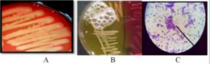
Staphylococcus sp. Staphylococcus genus is characterized by positive catalase result. Catalase enzyme functions as a catalyst which diffused hydrogen peroxide (H2O2) into H2 and O2, thus, when the bacteria colonies are mixed up with H2O2, the gas bubbles produced. Figure7 (A) shows positive catalase test result which is characterized by gas bubbles. Staphylococcus genus is Gram-positive bacteria, which morphologically cocci and clustered together like grapes (Figure 2C).
Fig 2. Streptococcus agalactiae hemolysis in Blood Agar (A), Katalase positive test (B) and the morphology of Staphylococcus sp purple and
clustered (C)
The identification results of catalase test and Gram staining from 33 positive mastitis milk samples, there are 20 samples (60.6%) containing the bacteria
Staphylococcus sp. and from the 2 negative samples mastitis, there is one sample containing Staphylococcus bacteria.
Coagulase test is an important identification test. The production of coagulase enzyme becomes pathogenic factor of S. aureus that differentiates them from other Staphylococcus. Coagulase enzyme produced by S. aureus
is able to clot blood plasma since it resembles prothrombin, which can converts fibrinogen to fibrin. Figure 3 shows no clots in the blood plasma indicating negative coagulation.
Fig 3. Positif coagulase (A) S. aureus culture Yellow colour on MSA (B) and hemolysis of S. aureus culture on BA
Identification test is then followed by mannitol fermentation tests using MSA as culture medium. This is a
major test procedure which is commonly used after coagulation test in identifying S. aureus. The high concentration of sodium chloride (NaCl) in MSA media makes this media selective to S. aureus, since other bacteria not survive in this condition. Figure 3B shows culture results on MSA media, S. aureus colonies has golden color and change the color from pink to golden yellow. It caused by the S. aureus ability in producing acid which modify the medium.
Other bacteria from Staphylococcus genus which grows on this medium is S. epidermidis, yet, this bacteria cannot fermenting mannitol and therefore the colony looks white and the media remains pink. The test results showed that four (25%) out of 16 samples are positive Staphylococcus
sp. proven by positive coagulase and capable in fermenting mannitol on the MSA media. Whereas, the mastitis negative samples does not grow on mannitol salt agar (MSA) media and negative coagulase.
Following the identification of S. aureus, the colonies were then streak onto blood agar (BA) aimed to observe the ability of bacteria to hemolysis blood erythrocyte, thus the hemolysis zone around colonies will be visible
(
Figure 3C)Subclinical mastitis is caused by various types of bacteria, for example S. aureus. Results of observations during milking process showed that there is still feces/dirt around the cows or the cage. Milking is performed using simple tools such as a plastic bucket milk container but some breeders were used old plastic bucket which were used to paint the walls, and the milk collective containers were not closed during milking. Additionally, the farmers’ habit of not washing the nipples (dipping) before and after milking may increase the risk of mastitis.
Examination of subclinical mastitis using cmt reagent offer a high level of sensitivity and specificity. this is also supported by (26), which compares the diagnostic for the detection of subclinical mastitis, and showed the result of 100% sensitivity for somatic cell count and 96% for CMT reactions. Sensitivity is the ability of a reagent to show positive results in cows suffering from subclinical mastitis, while specificity is the ability of a reagent to indicate a negative result in cows suffering from subclinical mastitis. The CMT reagent contains detergent or surfactant which is a composition wherein the surfactant can be used to detect the increasing degree of somatic cells in mastitis milk. According to ( 34) different types of surfactants have different effects on milk which contains Aril CMT alkyl sulfonate (3%), sodium hydroxide (1.5%) and Bromocresol purple. Aryl alkyl sulfonate (3%) had a great sensitivity to the milk pH, while bromocresol purple is a color indicator to highlight the reaction during observation. In the milk affected by mastitis, there will be an increasing number of leukocytes therefore the pH will be more alkaline. If an active substance such as alkyl sulfonate Aril is added (3%), it will react with milk somatic cells, including leukocytes, resulting in the increasing of milk concentration to becomes more viscous (thick) and forming a gel. Laboratory test on 33 positive mastitis samples revealed that 60.6% has bacteria Staphylococcus sp. characterized with catalase positive. In the catalase test, most of the bacteria producing enzyme catalase that can decompose H2O2 into H2O and O2. Hydrogen peroxide is toxic to cells because this material is inactivating enzymes in the cells. Hydrogen peroxide is formed during aerobic
metabolism, so that microorganisms grow in aerobic environment needs to decompose the toxic material. Catalase is one of the enzymes used micro-organisms to decompose hydrogen peroxide. This means that H2O2 is not decomposed by catalase-negative bacteria, thus it does not produce oxygen. Catalase-negative bacteria do not contain the catalase enzyme which decompose H2O2, including the genus of Streptococcu22s. (18) suggests that mastitis caused by Staphylococcus can reach 70% within a farm. The inhibitory titer was progressively increased when 5 to 30
μ
g of ammonium thiocyanate per ml of milk was added to pasteurized milk and the diluent ((9). (15), found the bacteria S. aureus in 145 (48.33%) of their 300 mastitis milk samples. Based on previous research conducted by ( 2), it can be concluded that the incidence of mastitis caused by S. aureus on a farm is quite high. S. aureus is a bacterium contagious which can be isolated from human or animal skin surface. Its role as mastitis causes in cows is related with the environmental sanitation and milking hygiene. Observation of environmental conditions showed that some cages do not have proper sanitation, where sewer and fesses container are too close to the water or feed storage areas. In such conditions, the possibility of S. aureus strains infection derived not from humans and animals are very high. In accordance with the ( 22) besides being able to be isolated from skin, S. aureuscan also be isolated from the equipment in a cage, cage environment and milking tools. Generally, strains of S.
aureus found in milk were not derived from cows or human contamination. Based on the results of 28 samples of milk from the dairy buffaloes spread in Enrekang, no milk samples found positive for S. aureus and S. agalactiae.
4. Conclusion
From 33 buffaloes (Bubalus bubalis) milk samples which were positive subclinical mastitis in Enrekang, 1 milk samples (3.03%) was positive for S. agalactiae and 4 samples (12.12%) were positive for S. aureus, whereas 28 milk samples from the dairy were negative for both S. agalactiae and S. aureus bacteria.
References
[1] Abuseliana ,A.F., Hassan Hj Mohd Daud , Saleha Abdul Aziz , Siti Khairani Bejo and Milud Alsaid.2011. Pathogenicity of Streptococcus agalactiae Isolated from a Fish Farm in Selangor to Juvenile Red Tilapia (Oreochromis sp.). Journal of Animal and Veterinary Advances , 2011 Volume: 10 Issue: 7 Page No.: 914-919.
[2] Agus, M. 1991. Mastitis study in dairy cattle in Baturraden. Hemerazoa. 74:21-24.
[3] Anueyiagu, K. N.and Isiyaku A. W.2016. Isolation, identification of Staphylococcus aureusfrom bovine milk and its antibiotics susceptibility. International Journal of Livestock Production. Vol. 6(6), pp74-77, June 2015DOI: 10.5897/IJLP. 2015.0248 Article Number:3EE6E8B53585 ISSN 2141-2448
[4] Bannerman, D.D., and R.J. Wall. 2005. A Novel Strategy for the Prevention of Staphylococcus aureus-Induced Mastitis in Dairy Cows. Information Systems for Biotechnology News Report. Virginia Tech University. USA.
[5] Bergey DH, RE Buchanan, NE Gibbons, and American Society for Microbiology. 1975. Bergey’s Manual of Determinative Bacteriology. Eight Edition. Baltimore : Williams and Wilkins Co.
[6] Boss,A. A. Cosandey, M. Luini, K. Artursson, M. Bardiau, F. Breitenwieser, E. Hehenberger, Th. Lam, M. Mansfeld, A. Michel, G. Mösslacher, J. Naskova, S. Nelson, O. Podpečan, A. Raemy,
E. Ryan, O. Salat, P. Zangerl, A. Steiner, H.U. Graber.2016. Bovine Staphylococcus aureus: Subtyping, evolution, and zoonotic transfer .Journal of Dairy Science.. January 2016 Volume 99, Issue 1, Pages 515–528
[7] Blowey, R. 1995. Mastitis Control in Dairy Herds an Illustrated and Practical Guide. Farming Press, USA
[8] Bray, D.R., and J.K. Shearer. 2003. Milking Machines and Mastitis Control Handbook. Florida
[9] Brown, Richard W.1967. Streptococci and enterococci as animal pathogens. Journal of Applied MicrobiologyVolume 83, Issue S1
[10] Chanter,N. 2003. Factors Affecting Growth of Streptococcus agalactiae in Milk. Journal of Dairy Science.October 1967Volume 50, Issue 10, Pages 1572–1584
[11] Harris ,L.G., S.J. Foster, and R.G. Richards.2002. An Introduction to
Staphylococcus aureus and Techniques for identifying and Quantifyings.S. aureus adhesins in relation to adhesion to biomaterials. European Cells and Materials Vol. 4. 2002 (pages 39-60). ISSN 1473-2262
[12] Jahan,M., Marzia Rahman, Md. Shafiullah Parvej, Shah Md. Ziqrul Haq Chowdhury, Md. Enamul Haque, Md. Abdul Khaleque Talukder, Sultan Ahmed.2015. Isolation and characterization of Staphylococcus aureus from raw cow milk in Bangladesh. Journal Advance Veterinary Animal Research . 2015; 2(1): 49-55
[13] Joshi L.R.and Shiva Prasad Devkota.2014. Methicillin Resistant Staphylococcus Aureus (MRSA) In Cattle. Epidemiology and Zoonotic implications International Journal Applied Science Biotechnology, Vol 2(1): 29-33 DOI: 10.3126/ijasbt.v2i1.9861
[14] Keefe, G. P. (1997). Streptococcus agalactiae mastitis: a review. The Canadian Veterinary Journal.La Revue Veterinaire Canadienne, 38(7), 429-437.
[15] Kirkan, S., E.O. Goksoyand, and O. Kaya. 2003. Identification and antimicrobial susceptibility of S. aureus and coagulase negative Staphylococci from bovine mastitis in the aydin region of Turkey. Turky Journal. Veterinary and Animal Science. 29(2005):791-796.
[16] Lowy,F.D.1998.Staphylococcus aureus Infections..The New England Journal of Medicine 1998; 339:520-532
[17] Lang, S. and M. Palmer. 2003. Characterization of S. agalactiae CAMP factor as a pore-forming toxin. Journal. Biological Chemistry. 278:38167-73
[18] Oliver, S.P. 2000. Mastitis in Heifers: Prevalence, Strategy for Control during the Periparturient Period, and Economic Implications. Proceeding British Mastitis Conference. Institute for Animal Health/Milk Development Council. Tennesee,USA:1-13.
[19] PadhyA., Amit Ranjan Sahu, Shashank Shekhar, Saraswat Sahoo, Abhishek Sahoo, Nirupama Dalai..2016.Staphylococcus aureus: An Emergent Cause of Bovine Mastitis in India . International Journal of Livestock Research. Year: 2016, Volume: 6,
[20] Quinn,P.J., B.K. Markey, M.E.Carter, W.J. Donnelly, and F.C. Leonard. 2002. Veterinary Microbiology and Microbial Disease. Blackwell Publishing. USA.
[21] Richard,W, Brown.1967. Factors Affecting Growth of Streptococcus agalactiae in Milk. Journal.of Dairy Science. October 1967 .Volume 50, Issue 10, Pages 1572–1584
[22] Roberson, J.R., L.K., Fox, D.D., Hancock, J.M., Gay and T.E. Besser. 1994. Ecology of Staph.aureus Isolated from various sites on dairy farms. Journal. Dairy Science. 77(11):3354-3364.
[23] Ruoff, K.L. 1995. Streptococcus. In Manual of Clinical Microbiology. Murray, P.R., E.J. Baron, M.A. Pfaller, F.C. Tenover, and R.H. Yolken. 6th ed. ASM Press, Washington DC.Murray, P.R., E.J. Baron, M.A. Pfaller, F.C. Tenover, and R.H. Yolken. 6th ed. ASM Press, Washington DC.
[24] Smith TC (2015) Livestock-Associated Staphylococcus aureus: The United States Experience. PLoS Pathog 11(2): e1004564. doi:10.1371/journal.ppat.1004564
[25] Songer JG, Post KW. 2005. Veterinary Microbiology Bacterial and Fungal Agents of Animal Disease 1st Ed. Elsevier Saunders.
[26] Tanwar, R.K., S.K. Vyas, Fakhruddin, and A.P. Singh. 2001. Comparative efficacy of various diagnostic tests in diagnosis of SCM in Rathi cows. Advancement of Veterinary Research (IAAVR). Izatnagar, India. pp 161-163.
[27] Taylor, R.E. and T.G. Field. 2009. Scientific Farm Animal Production: An Introduction to Animal Science. 9th ed. Pearson
Prentice Hall. New Jersey.
[28] Turnige,J.D., Despina K, Wendy M, Sally, Catherine M B, Graeme R N,
[29] Geoffrey W C, Ronan J M, Benjamin H, Paul D R J, Kate D 2009. Staphylococcus aureus bacteraemia: a major cause of mortality in
Australia and New Zealand MedicalJournal Australia 2009; 191 (7): 368-373
[30] Weese J.S.2010. Methicillin-Resistant Staphylococcus aureus in Animals. ILAR Journal Oxford, Volume 51, Number 3 2010
[31] Wibawan, I.W.T. dan Ch. Laemmler. 1990. Properties of Grup B Streptococci with protein surface antigen X and R. Journal. Clinical Microbiol.28:2834-836.
[32] Wibawan IWT, Pasaribu FH, Huminto H, Estuningsih S. 1995. Ciri Biovar Streptococcus Agalactiae Sebagai Petunjuk Infeksi Silang Antara Sapi dan Manusia. Laporan Hibah Bersaing IV Tahap-1. Fakultas Kedokteran Hewan, Institut Pertanian Bogor
[33] Wibawan IWT, Pasaribu FH, Huminto H, Estuningsih S. 1995 (Biovar Streptococcus agalactiae characteristic as cross-infection indicator between cow and human. Competitive Grant Report IV, Phase I. Facutly of Veterinary Science, Institut Pertanian Bogor)
[34] Wolfe, C. S. Peterson. I. K. Mullarky, G. M. Jones2010. Staphylococcus aureus Mastitis: Cause, Detection, and Control. Journal of Dairy Science 65(2): 27174.
[35] Wyder, A.B., Boss, R., Naskova, J., Kaufmann, T., Steiner, A. and Graber, H.U., 2011. Streptococcus spp. and related bacteria: their identification and their pathogenic potential for chronic mastitis–a molecular approach. Research in veterinary science, 91(3), pp.349-357.
[36] Xia, S.S. 2006. The Rheology of Gel Formed During the California Mastitis Test. Thesis. The University of Waikato. New Zealand.
[37] Fogg, B.J, Persuasive technology: using computers to change what we think and do, Morgan Kaufmann Publishers, Boston, 2003, 30-35.
[38] Hirsh, H., Coen, M.H., Mozer, M.C., Hasha, R. and Flanagan, J.L,
“Room service, AI-style,” IEEE intelligent systems, 14 (2). 8-19. Jul.2002.
[39] T. Eckes, The Developmental Social Psychology of Gender, Lawrence Erlbaum, 2000. [E-book] Available: netLibrary e-book.