ETDB-Caltech: a blockchain-based distributed public database for electron
1tomography
2 3 4 5Davi R. Ortega1, Catherine M. Oikonomou1, H. Jane Ding1, Prudence Rees-Lee1, Alexandria^, 6 Grant J. Jensen1,2,* 7 8 9 10 11 12
1 Division of Biology and Biological Engineering, California Institute of Technology, Pasadena,
13
California, USA 14
2 Howard Hughes Medical Institute, Pasadena, California, USA
15 16 *Corresponding author 17 Email: [email protected] (GJJ) 18 19
^Membership of Alexandria is provided in the Acknowledgments. 20
Abstract
22
Three-dimensional electron microscopy techniques like electron tomography provide valuable 23
insights into cellular structures, and present significant challenges for data storage and 24
dissemination. Here we explored a novel method to publicly release more than 11,000 such 25
datasets, more than 30 TB in total, collected by our group. Our method, based on a peer-to-peer 26
file sharing network built around a blockchain ledger, offers a distributed solution to data 27
storage. In addition, we offer a user-friendly browser-based interface, https://etdb.caltech.edu, for 28
anyone interested to explore and download our data. We discuss the relative advantages and 29
disadvantages of this system and provide tools for other groups to mine our data and/or use the 30
same approach to share their own imaging datasets. 31
32
Introduction
33
Three-dimensional electron microscopy (3D EM) techniques produce large and information-rich 34
datasets about biological samples. In electron tomography (ET), samples are imaged as they are 35
tilted incrementally – typically 1-2 degrees between images. The resulting tilt-series of 2D 36
projection images can then be computationally combined into a 3D reconstruction, or tomogram, 37
of the sample with nanometer-scale resolution. ET has both biological [1] and materials science 38
applications [2]. ET is frequently performed on frozen samples (cryo-ET) such as intact, small 39
cells. Cryo-ET has revealed many details about cell ultrastructures that are inaccessible by other 40
techniques, either because they cannot be purified intact or because they are not preserved by 41
traditional EM sample preparations [3]. Another 3D EM technique, single particle analysis, also 42
yields 3D information about cellular complexes [4]. 43
Biological applications of 3D EM techniques are rapidly increasing, with an explosive rise in the 45
number of datasets published [5] and excitement about the field (e.g. [6-8]). In addition, 46
technological advances such as increased automation for higher-throughput data collection and 47
movie acquisition with direct detectors are increasing the information content of datasets [9, 10], 48
which makes management of these datasets a mounting challenge [11]. At the same time, public 49
accessibility is of critical importance [12]. 3D EM techniques, while burgeoning, are still 50
inaccessible to most cell biologists due to the expensive equipment (several million dollars to 51
purchase and maintain, in a customized space) and specialized expertise required. In addition, the 52
technology is still in a phase of active development, in both hardware and software. To facilitate 53
software development efforts, programmers need access to large and varied test datasets. 54
55
Public dissemination outlets for 3D EM datasets address two fundamentally different missions: 56
(1) to provide curated, validated data for peer review and education [13]; and (2) to provide large 57
quantities of possibly problematic data to facilitate biological discovery and software 58
development. The first mission is well served by resources such as the Electron Microscopy Data 59
Bank (EMDB) and the Cell Image Library. The EMDB, an invaluable community tool for 60
deposition of 3D EM data [14], is part of the EMDataBank [15], a global resource for 3D EM 61
managed by the worldwide Protein Data Bank (PDB) consortium [16]. Like its counterpart, the 62
PDB [17], it is the standard repository for published structures, such as single particle 63
reconstructions and subtomogram averages [18]. To encourage public access, the EMDB 64
developed web-based visualization tools to interact with data [19, 20]. The Cell Image LibraryTM 65
is an open-source catalog of curated images, animations and videos aimed at disseminating cell 66
biology to the broader public [21]. Entries include light and electron microscopy imaging, as 67
well as correlated datasets. The resource includes datasets previously available as the Cell 68
Centered Database (CCDB), an online repository of high-resolution, often 3D, light and electron 69
microscopy data, including many electron tomograms [22-24]. 70
71
The second mission is currently served in a more piecemeal fashion, largely by initiatives from 72
single labs and imaging centers to release a subset of their raw datasets for public access. 73
Unfortunately, these resources often suffer from a lack of permanence due to lapsed maintenance 74
of published websites. Recognizing the need for a centralized public repository of the raw EM 75
datasets from which EMDB structures are derived, in 2016 the European PDB announced a sister 76
site to the EMDB: the Electron Microscopy Public Image Archive, or EMPIAR [25]. EMPIAR 77
collects tilt-series related to reconstructions deposited in the EMDB. It therefore offers an ideal 78
resource for benchmarking software with verified, published datasets, but it is not designed for 79
large-scale releases of unpublished, problematic and/or complicated datasets: datasets must be 80
associated with an EMDB deposition; only tilt-series can be deposited (the resulting 81
reconstructions are available in the EMDB, but associated files such as correlated light 82
microscopy images or digital segmentations cannot be included); and much of the metadata is 83
entered manually [26], a daunting task for a large batch of data. 84
85
While releasing data of unverified quality may seem to be of dubious value, we would argue that 86
it is necessary for the progress of the field. As pointed out by the developers of the CCDB, ET 87
datasets that currently yield poor-quality reconstructions offer opportunities for developing better 88
reconstruction methods [24]. Also, biological insights often come from unexpected places; as a 89
single anecdotal example, years ago our lab collected electron tomograms of bacteria to study 90
chromosome segregation and observed novel tubes inside cells; we shared the images and a cell 91
biologist made a connection to a secretion system he was studying, allowing us together to figure 92
out its mechanism [27]. 93
94
Since 2003, our lab has collected more than 30,000 ET datasets. Each dataset consists of a tilt-95
series of 2D TEM projection images and the resulting 3D tomographic reconstruction, as well as 96
additional image, video, and segmentation files. Each dataset is 1-5 GB, and the full collection 97
adds up to ~110 TB of data. To store and curate this volume of data for internal use by our 98
group, we developed the Caltech Tomography Database, a central repository linked to a browser-99
based interface for lab members to browse, search, and download data [28]. To further 100
streamline data handling, we integrated the internal Caltech Tomography Database with an 101
automatic processing pipeline that uploads and processes datasets as they are acquired by the 102
microscope [28]. The majority of our ET datasets come from cryo-preserved cells. They 103
represent more than 100 unique species of bacteria, archaea, and eukaryotes and have led to 104
dozens of publications about diverse aspects of cell ultrastructure. The nature of whole-cell 105
imaging, though, means that these datasets are far from exhausted. While we collected them for a 106
specific study, they contain information about many other aspects of cell biology that may be 107
useful to other researchers. 108
109
While we have been sharing our data by publishing papers and depositing representative 110
tomograms in the EMDB, we have also received many requests–from software developers, 111
biologists, and EMPIAR–to share more of our data. We filled these individual requests, but 112
wanted to explore a broader solution to enable our lab and others to share large amounts of data 113
of unverified quality in a persistent and decentralized fashion. The approach we describe here 114
uses a distributed peer-to-peer file network tracked by an ownerless ledger (blockchain) system. 115
We describe how we used this method to release more than 11,000 electron tomography datasets 116
(excluding those that are still part of ongoing studies), representing 85 species and encompassing 117
more than 30 TB. We discuss the advantages and drawbacks of our approach, and how it can be 118
adopted by other groups that wish to share their own datasets. 119
120
Results & Discussion
121 122
Approach
123
In recent years, decentralized cryptographic ledgers, or blockchains, have been explored as a 124
method to securely record data (typically cryptocurrency transactions, for which they were first 125
conceived [29]). Rather than relying on a trusted central authority, blockchains employ a security 126
model that builds consensus from a system of distributed users, none of whom necessarily need 127
to trust one another. Originally developed to solve the problem of double-spending, blockchain 128
technology has since been adapted to other uses. For instance, the Republic of Georgia uses the 129
bitcoin blockchain to record land transfer titles, one of several countries using the cryptographic 130
ledger to improve the security of property rights [30]. In the United States, blockchains have 131
been proposed as a way for patients to control access to their digital medical records [31, 32]. 132
Blockchains are used by Nasdaq in the U.S. and stock exchanges in other countries to record 133
private securities transactions [33]. 134
In 2013, an anonymous developer announced a fork from a cryptocurrency called Litecoin to 136
create a new cryptocurrency, FlorinCoin (FLO), whose ledger features a descriptive transaction 137
comment line similar to that found on a traditional check. The text entered in this transaction 138
comment is stored in the FLO blockchain along with the details of the transaction. Each 139
comment can contain up to 528 characters [34]. In 2014, a company called Alexandria proposed 140
to use this feature as a public record of information and developed an open source protocol 141
termed the Open Index Protocol (OIP) [35]. They first used this protocol to record public social 142
media status in the FLO blockchain and later, using a peer-to-peer distributed file-sharing 143
network, they expanded the specifications of the protocol to register the metadata of videos and 144
music in the FLO blockchain while storing the files in the peer-to-peer file-sharing network 145
BitTorrent, allowing artists to prove ownership of these digital assets. From September 2017 to 146
May 2018 FlorinCoin passed through a series of upgrades. It was renamed FLO, its code was 147
updated to version 0.15 of Bitcoin (still retaining the sCrypt algorithm for proof-of-work), and 148
the comment field was expanded to 1,040 characters. The current OIP specification (0.42) is 149
optimized for the new FLO comment field size, encompasses a variety of data types, and uses a 150
peer-to-peer file system called the InterPlanetary File System (IPFS) [36] to store files. File 151
metadata is thereby cryptographically secured, and completely searchable, allowing anyone to 152
discover and download the files from the IPFS. 153
154
We were curious to see if this blockchain-based data distribution model would be effective to 155
openly and securely share our scientific imaging data. In the scheme, each dataset would be 156
distributed to IPFS and its metadata recorded in the FLO blockchain. Any interested party, 157
typically through a user-friendly front-end in their web browser, could query the blockchain for 158
datasets of interest and retrieve them from IPFS. We called the resulting distributed database the 159
public Electron Tomography Database - Caltech (ETDB-Caltech), and its information flow is 160
schematized in Figure 1. 161
162
Figure 1. Information flow in the ETDB-Caltech file-sharing network. Datasets hosted from 163
a local server are distributed to IPFS, a network of seeding nodes that includes the local server. 164
The associated metadata and locations of the files are recorded in the FLO blockchain using the 165
OIP specification. Users can query this ledger to locate and retrieve desired files from the IPFS. 166
167
We worked with Alexandria to develop a digital record type tailored to the metadata of our 168
datasets that could be encoded easily in the FLO transaction comment. The result, Research-169
Tomogram, contains fields corresponding to the information we store about each dataset in our 170
internal database. This information includes details about the user who collected the data, 171
descriptions of the sample and its preparation, and data acquisition and processing parameters. 172
Where appropriate, this information follows standard conventions for the 3D EM field [37]. We 173
wrote a simple GoLang script to automatically read this information from the record in the 174
internal lab database and translate it into an OIP Research-Tomogram record. If other groups 175
want to adopt this approach, they can use a subset of these fields and/or add their own as 176
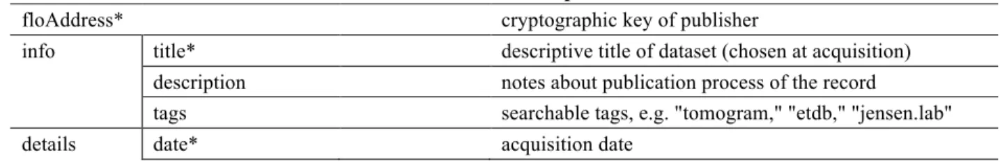
necessary to match their local recordkeeping. Table 1 lists the currently available fields in the 177
Research-Tomogram record. 178
179
Table 1. Fields in the Research-Tomogram record. 180
181
Description
floAddress* cryptographic key of publisher
info title* descriptive title of dataset (chosen at acquisition) description notes about publication process of the record tags searchable tags, e.g. "tomogram," "etdb," "jensen.lab"
NCBItaxID NCBI taxonomy identifier artNotes notes about the dataset
scopeName acquisition microscope, e.g. "Caltech Polara" speciesName* species of cell imaged
strain information about the specimen strain
tiltSingleDual single-axis or dual-axis tilt acquisition scheme defocus imaging defocus (µm)
dosage imaging electron dosage (e/Å2)
tiltConstant 1: if constant angular increment; 0: if other method tiltMin minimum of acquisition tilt range (degrees) tiltMax maximum of acquisition tilt range (degrees) tiltStep tilt increment (degrees)
swAquisition software used for acquisition swReconstruction software used for reconstruction magnification acquisition magnification (X)
emdb EMDB code if record is also available on EMDB microscopist scientist who acquired tilt-series
institution e.g. "Caltech" lab e.g. "Jensen Lab"
sid internal database identifier (laboratory specific) storage network* e.g. "IPFS"
files** fname* file name
dname name to be displayed in interface fsize file size (bytes)
type e.g. "Tomogram" or "Image" subtype e.g. "Tiltseries" or "Reconstruction"
ctype content type, e.g. "image/jpeg" or "video/mp4" location* hash of file locations for retrieval
payment payment information (N/A for this blockchain use) timestamp* time of publication to blockchain
type* "Research"
subtype "Tomogram"
182
* mandatory field
183
**stores the indicated information for each file associated with the dataset
184 185
As in other peer-to-peer networks, files can be chunked and hosted from multiple nodes in the 186
network. Users who download a file and participate in IPFS can choose to host it in this fashion 187
for other users. This feature makes the distribution model scalable; if many users are 188
downloading a file, multiple seeds speed up those downloads, avoiding a bottleneck from a 189
single server. In our case, we expect relatively light file traffic, so at the current time, files are 190
downloaded solely from our server, as in a traditional distribution model. In the rare event that a 191
dataset is published in error, OIP offers the option of deactivating a published record. This action 192
will not erase the metadata published in the blockchain, but the record will no longer be available 193
to anyone using the OIP API to search the blockchain. In that case, if a user were interested in an 194
unavailable tomogram, they would have to search the raw data in the blockchain, and hope that 195
the files were still in the IPFS network. 196
197
There are two ways that users can download our datasets. The first is through a direct query of 198
the blockchain and IPFS. We built a command-line application that facilitates this approach; see 199
Materials & Methods for details. To increase public accessibility, we added a second route: a
200
browser-based front-end. This graphical interface, which can be found at https://etdb.caltech.edu, 201
provides an intuitive, interactive experience for anyone to browse ETDB-Caltech datasets, view 202
images and videos they contain, and download part or all of each dataset. A sample dataset 203
display page is shown in Figure 2. 204
205
Figure 2. Sample entry page in the browser-based ETDB-Caltech interface. A sample 206
electron cryotomography dataset from a Vibrio cholerae cell is shown. An embedded video of 207
the reconstruction appears at left and plays automatically. The metadata is shown at right. Files 208
associated with the dataset are listed at the bottom of the page, where they can be downloaded 209
individually. 210
211
The ETDB-Caltech front-end offered us a chance to highlight scientific challenges for target user 212
groups – cell biologists and software developers. We hope cell biologists will find novel features 213
in the imaged cells, and identify those that remain mysterious. Electron tomograms contain a 214
wealth of information, not all of which is currently interpretable; recently, for instance, we 215
published a paper describing some of the cellular features we have observed in our electron 216
tomograms but could not identify [38]. We hope software developers will use the released 217
datasets to improve image-processing algorithms. In particular, we hope the availability of these 218
datasets contributes to the development of software that can: (1) more reliably find and track the 219
fiducial markers used for alignment in tomographic reconstruction; (2) automatically and 220
accurately segment the boundaries of cells; and (3) automatically segment large macromolecular 221
complexes in cells. In addition to their usefulness to experts in the field, the datasets in ETDB-222
Caltech may be of interest to students and the general public. To welcome these users, we 223
designed the front-end of ETDB-Caltech to be accessible and educational, with information 224
about the data and technology, as well as a Featured Tomograms page highlighting various 225
features of bacterial and archaeal cells that are visible in electron tomograms (Figure 3). 226
227
Figure 3. Featured Tomograms page of the ETDB-Caltech interface. Targeting students and 228
others unfamiliar with ET data, the page highlights cellular features of bacteria and archaea 229
visible by cryo-ET. Selecting a category takes the user to a page with a brief description of the 230
structure and a few datasets containing examples. 231
232
Outlook
233
Here we tested a new approach to publicly share a large amount of ET data. If our goal was 234
simply to continue honoring requests from the community to make our datasets public, it would 235
have been cheaper and easier to simply host the data from a local MySQL database, as we do for 236
our internal group users. However, we also wanted to make a broader resource that could 237
encompass data from many ET labs into a flexible repository that does not rely on a central 238
authority. If ETDB is ultimately successful in enabling large-scale community data sharing, we 239
believe it will complement (but never replace) the mission of curated repositories like EMDB 240
and EMPIAR by providing varied datasets with a wide range of quality and content for 241
biological and technological projects. 242
243
Compared to more centralized models of data storage, this dissemination model offers several 244
attractive points. The first is flexibility. Multiple file types can be combined in a single OIP 245
record, allowing, for example, light micrographs from correlative light and electron microscopy 246
experiments and annotated segmentations to be included in EM datasets; this has been cited as a 247
key feature lacking in some current repositories [12, 39]. Other file types from different imaging 248
modalities can be accommodated with similar ease. The OIP specification of the Research-249
Tomogram record type requires few mandatory fields (Table 1). These fields can be adapted to 250
the metadata collected by other groups, who may be using different internal databases (e.g. [40, 251
41]). The flip side of this flexibility is that, compared to repositories of validated datasets like 252
EMDB/EMPIAR [26], ETDB entries may be missing information like pixel size or contain 253
errors in metadata. This caveat should be kept in mind when using the data in further studies; 254
information critical to interpretation should be verified with the depositor. 255
256
Another appealing feature of distributed file sharing is the distribution of storage and cost. 3D 257
EM datasets are large, as reflected by EMPIAR, which has grown to accommodate >80 TB of 258
stored data in 5 years [42]. These datasets are associated with only 168 studies [43]. The 259
popularity of 3D EM methods, particularly cryo-ET [8], is growing rapidly: the number of 260
entries in the EMDB has more than doubled over the last three years [5, 44]. There are currently 261
more than 6,500 entries in the EMDB [44]; if each of these was associated with a similarly-sized 262
dataset in EMPIAR, more than 3 PB of centralized storage space would be required. In a 263
distributed distribution model, each contributing lab is responsible for storing their own data, 264
which they presumably already do. In our case, we could have implemented the system using our 265
existing server, which hosts our internal database, at no added cost. For extra security, we chose 266
to keep the server with the internal database behind a local firewall and mirror the relevant 267
datasets on an additional server outside the firewall hosting ETDB. This second server, which is 268
larger than necessary to accommodate additional applications and future growth, cost 269
~US$7,000. 270
271
In addition to the local server, files should be available from other nodes of the IPFS. This 272
ensures data persistence in the event of, for instance, a local disk failure. Of course, how well 273
this feature works depends on whether the system is widely adopted. In addition to users hosting 274
IPFS nodes, institutions can also easily archive ETDB data through the IPFS. The more nodes 275
are hosting a file in the IPFS, the higher the bandwidth for users to download it; this scalability is 276
a major feature of peer-to-peer networks. Currently, however, the IPFS is still experimental and, 277
like many new technologies, unstable. For that reason, we serve the files in our front-end directly 278
from the IPFS node running on our local server, not through the full IPFS peer-to-peer network. 279
However, IPFS is in rapid development and we expect soon to update the front-end to fetch and 280
serve the files from the IPFS. Our command line application for bulk download, ETDB-281
downloads, already retrieves the files from the IPFS network. 282
283
The maintenance of the ownerless ledger used to store the ETDB metadata, the FLO blockchain, 284
depends on a distributed network of miners and users. This feature facilitates adoption as anyone 285
can publish tomograms to the ETDB without having to seek permission from a central authority. 286
However, as in other cryptocurrencies, miners and users have an incentive to participate in the 287
FLO network depending on a combination of factors including the costs of hardware and 288
electricity, and the value of FLO in the cryptocurrency market. Although FLO has been in 289
circulation for over 5 years, a relatively long time by cryptocurrency standards, its eventual 290
success is difficult to predict. If FLO becomes an inviable option, it may be necessary to switch 291
to a different ledger system in the future (Ethereum, Namecoin, and Bitcoin Cash are all capable 292
of storing text). Note, however, that metadata already published remains accessible as long as at 293
least one copy of the FLO blockchain exists; we host one ourselves. 294
295
For us, the project took a few months to complete and the cost for the cryptocurrency 296
transactions we used to publish 11,293 datasets was US$17.89 (see Materials and Methods). 297
Most of the development effort was invested in the user interface as well as the scripts to 298
automatically upload datasets to the IPFS and the metadata to the FLO blockchain using OIP. If 299
other groups wish to adopt the same approach to make their data public, they would only need to 300
slightly modify these scripts (available on GitHub, see Materials & Methods) to match their 301
internal database descriptors. Our front-end code is similarly available on GitHub so that other 302
groups can easily adapt it to taste and use it to display: (1) their own data, (2) all ETDB datasets 303
in the IPFS, or (3) a custom subset (e.g. data from a single species or technique). In addition, 304
individuals interested in web applications for visualization and manipulation of tomograms can 305
use the ETDB as a distributed database of content without needing to host any tomograms 306
themselves. Outlets (e.g. science educators) can stream tomogram videos directly from the IPFS 307
network. 308
Ultimately, we believe the relationship between the ETDB and curated central repositories like 310
the EMDB is complementary. We will continue to support the invaluable mission of the EMDB 311
and EMPIAR in safeguarding scientific data by submitting representative curated datasets we use 312
in our publications. We hope that the ETDB can in turn help facilitate broader releases of large 313
batches of electron tomography data for community use. If successful, the ETDB could even be 314
integrated into centralized repositories by their hosting an IPFS node, enhancing accessibility of 315
the data. The flexible features of this blockchain-based, distributed scheme of data sharing may 316
also make it useful for other types of scientific data. 317
318
Materials & Methods
319
ETDB-Caltech Distribution
320
The ETDB-Caltech database is fed by a MySQL database (version 14.14 distribution 5.7.21) 321
hosted on an Ubuntu Server (Artful Aardvark kernel version 4.3.0-37). The MySQL database 322
contains the metadata of entries from the Caltech Tomography Database [28] that have been 323
designated for publication. Associated files are stored in a RAID6 ext4 file system. Each night, 324
the internal server hosting the internal Caltech Tomography Database executes a script to find 325
datasets newly edited or marked for publication and copy them to the external ETDB-Caltech 326
server, updating the MySQL database. 327
328
The ETDB-Caltech server runs a full node of the FLO blockchain, a node of the IPFS and a 329
MySQL database. Upon changes in the MySQL database, a custom-built GoLang script (go-330
etdb, available on Github: https://github.com/theJensenLab/go-etdb) makes the new files 331
publicly accessible through the InterPlanetary File System (IPFS, version 0.4.15-dev) [36]. The 332
IPFS daemon calculates a unique identifier to the dataset directory called a hash which is 333
cryptographically dependent on the contents of the directory and makes the directory available to 334
other nodes of the IPFS. This hash is combined with the metadata of each dataset and formatted 335
according to Open Index Protocol (OIP, version 0.42) specification to create a JSON record (see 336
Table 1). Each record generated this way is signed with a cryptographic key unique to the Jensen 337
lab (the private key associated with public address
338
FTSTq8xx8yWUKJA5E3bgXLzZqqG9V6dvnr) and published to the FLO blockchain by a 339
daemon (OPId) on the server, attaching the record to the "floData" field of one or more 340
transactions. The cost to publish the full set of 11,293 tomograms (at then-current rates of 341
exchange) was US$17.89. 342
343
To search for ETDB-Caltech data, any user can use the cryptographic key given above to query 344
the blockchain and retrieve matching ETDB records. This procedure is facilitated by an OIP 345
daemon that scans and indexes the FLO Blockchain and exposes an Application Programming 346
Interface (API) for public use. The API is accessible by a package (oip-js) deposited on the node 347
package manager (npm). We also developed a command-line application for Unix-related 348
environments (ETDB-downloads, manual available on Github:
349
https://github.com/theJensenLab/etdb-downloads/blob/master/userManual.md) designed to allow 350
users to download all or a subset of ETDB-Caltech datasets. Unlike the ETDB-Caltech website 351
(see below), this application launches a temporary IPFS node and fetches the files from the IPFS 352 network. 353 354 ETDB-Caltech Interface 355
The front-end was built using node.js (version 9.1), react (16.2.0), webpack (4.1.1), and Twitter 356
Bootstrap. It uses the oip-js package (https://github.com/oipwg/oip-js) to connect to an 357
OIPdaemon Representational State Transfer (REST) API, which scans the FLO blockchain for 358
valid OIP records and indexes them into an internal database. Currently, oip-js queries 359
OIPdaemon for a list of records with type "Research" and subtype "Tomogram" published by our 360
lab (the private key associated with public address:
361
FTSTq8xx8yWUKJA5E3bgXLzZqqG9V6dvnr). In the future, queries could also search for the 362
cryptographic keys of different groups. Alternatively, records could be retrieved by a full-node 363
search of the FLO blockchain (available on GitHub: https://github.com/floblockchain/flo) with 364
OIPdaemon. Files are served for download from this interface directly from the IPFS node on the 365
ETDB-Caltech server. 366
367
The interface was designed to be easily navigable by scientists and non-scientists, and is 368
optimized for viewing on all common web-enabled devices. We expect that in the future, some 369
users and other labs may wish to customize this web interface. They can either copy and modify 370
our template (available on GitHub: https://github.com/theJensenLab/etdb-react) or develop their 371
own. While the Caltech ETDB interface displays only entries from our lab, other users may wish 372
to build front-ends to display data from all labs sharing data using Open Index Protocol or to 373
display only a subset of interest, for instance only those datasets corresponding to a particular 374
species. In that case, instead of serving the files directly from the ETDB-Caltech IPFS node, 375
those websites would use the peer-to-peer feature of the IPFS to search for the files in multiple 376
nodes. 377
Acknowledgments
379
We thank members of the Jensen lab for helpful comments on the ETDB interface, as well as 380
past and present lab members (Morgan Beeby, Ariane Briegel, Yi-Wei Chang, Songye Chen, 381
Megan Dobro, Lu Gan, Gregory Henderson, Cristina Iancu, Andreas Kjær, Zhuo Li, Alasdair 382
McDowall, Gavin Murphy, Martin Pilhofer, Rasika Ramdasi, Jian Shi, Poorna Subramanian, 383
Matthew Swulius, William Tivol, Elitza Tocheva, Cora Woodward, Qing Yao, Zhiheng Yu, and 384
Elizabeth Wright who generously allowed data they collected to be made public. We also thank 385
other lab members whose data will be published in the future. The Alexandria team is composed 386
of Devon Read James, Amy James, Jeremiah Buddenhagen, Sky Young, Ryan Chacon and 387
Anthony Stewart. Thanks also to past Alexandria contributors Ryan Jordan, Ryan Taylor, and 388
Joseph Fiscella for their work on the Open Index Protocol specification. This work was made 389
possible through the support of the National Institutes of Health (grant R35 GM122588 to G.J.J.) 390
and the John Templeton Foundation as part of the Boundaries of Life Initiative (grant 51250 to 391 G.J.J.). 392 393
References
3941. He, W. and He, Y. (2014). Electron tomography for organelles, cells, and tissues. 395
Methods Mol Biol 1117, 445-83.
396
2. Ercius, P., Alaidi, O., Rames, M. J., and Ren, G. (2015). Electron Tomography: A Three-397
Dimensional Analytic Tool for Hard and Soft Materials Research. Adv Mater 27, 5638-398
63. 399
3. Oikonomou, C. M. and Jensen, G. J. (2017). Cellular Electron Cryotomography: Toward 400
Structural Biology In Situ. Annu Rev Biochem 86, 873-896. 401
4. Elmlund, D., Le, S. N., and Elmlund, H. (2017). High-resolution cryo-EM: the nuts and 402
bolts. Curr Opin Struct Biol 46, 1-6. 403
5. Patwardhan, A. (2017). Trends in the Electron Microscopy Data Bank (EMDB). Acta
404
Crystallogr D Struct Biol 73, 503-508.
405
6. Callaway, E. (2015). The revolution will not be crystallized: a new method sweeps 406
through structural biology. Nature 525, 172-4. 407
7. Prize, N. (2017). The 2017 Nobel Prize in Chemistry - Press Release (Nobelprize.org). 408
8. Marx, V. (2018). Calling cell biologists to try cryo-ET. Nat Methods 15, 575-578. 409
9. Frank, J. (2017). Advances in the field of single-particle cryo-electron microscopy over 410
the last decade. Nat Protoc 12, 209-212. 411
10. Baldwin, P. R., Tan, Y. Z., Eng, E. T., Rice, W. J., Noble, A. J., Negro, C. J., Cianfrocco, 412
M. A., Potter, C. S., and Carragher, B. (2017). Big data in cryoEM: automated collection, 413
processing and accessibility of EM data. Curr Opin Microbiol 43, 1-8. 414
11. Patwardhan, A., Carazo, J. M., Carragher, B., Henderson, R., Heymann, J. B., Hill, E., 415
Jensen, G. J., Lagerstedt, I., Lawson, C. L., Ludtke, S. J., Mastronarde, D., Moore, W. J., 416
Roseman, A., Rosenthal, P., Sorzano, C. O., Sanz-Garcia, E., Scheres, S. H., 417
Subramaniam, S., Westbrook, J., Winn, M., Swedlow, J. R., and Kleywegt, G. J. (2012). 418
Data management challenges in three-dimensional EM. Nat Struct Mol Biol 19, 1203-7. 419
12. Patwardhan, A., Ashton, A., Brandt, R., Butcher, S., Carzaniga, R., Chiu, W., Collinson, 420
L., Doux, P., Duke, E., Ellisman, M. H., Franken, E., Grunewald, K., Heriche, J. K., 421
Koster, A., Kuhlbrandt, W., Lagerstedt, I., Larabell, C., Lawson, C. L., Saibil, H. R., 422
Sanz-Garcia, E., Subramaniam, S., Verkade, P., Swedlow, J. R., and Kleywegt, G. J. 423
(2014). A 3D cellular context for the macromolecular world. Nat Struct Mol Biol 21, 841-424
5. 425
13. Berman, H. M., Burley, S. K., Chiu, W., Sali, A., Adzhubei, A., Bourne, P. E., Bryant, S. 426
H., Dunbrack, R. L., Jr., Fidelis, K., Frank, J., Godzik, A., Henrick, K., Joachimiak, A., 427
Heymann, B., Jones, D., Markley, J. L., Moult, J., Montelione, G. T., Orengo, C., 428
Rossmann, M. G., Rost, B., Saibil, H., Schwede, T., Standley, D. M., and Westbrook, J. 429
D. (2006). Outcome of a workshop on archiving structural models of biological 430
macromolecules. Structure 14, 1211-7. 431
14. Tagari, M., Newman, R., Chagoyen, M., Carazo, J. M., and Henrick, K. (2002). New 432
electron microscopy database and deposition system. Trends Biochem Sci 27, 589. 433
15. Lawson, C. L., Baker, M. L., Best, C., Bi, C., Dougherty, M., Feng, P., van Ginkel, G., 434
Devkota, B., Lagerstedt, I., Ludtke, S. J., Newman, R. H., Oldfield, T. J., Rees, I., Sahni, 435
G., Sala, R., Velankar, S., Warren, J., Westbrook, J. D., Henrick, K., Kleywegt, G. J., 436
Berman, H. M., and Chiu, W. (2011). EMDataBank.org: unified data resource for 437
CryoEM. Nucleic Acids Res 39, D456-64. 438
16. Berman, H., Henrick, K., and Nakamura, H. (2003). Announcing the worldwide Protein 439
Data Bank. Nat Struct Biol 10, 980. 440
17. Bernstein, F. C., Koetzle, T. F., Williams, G. J., Meyer, E. F., Jr., Brice, M. D., Rodgers, 441
J. R., Kennard, O., Shimanouchi, T., and Tasumi, M. (1977). The Protein Data Bank. A 442
computer-based archival file for macromolecular structures. Eur J Biochem 80, 319-24. 443
18. Editorial (2003). A database for 'em. Nat Struct Biol 10, 313. 444
19. Lagerstedt, I., Moore, W. J., Patwardhan, A., Sanz-Garcia, E., Best, C., Swedlow, J. R., 445
and Kleywegt, G. J. (2013). Web-based visualisation and analysis of 3D electron-446
microscopy data from EMDB and PDB. J Struct Biol 184, 173-81. 447
20. Salavert-Torres, J., Iudin, A., Lagerstedt, I., Sanz-Garcia, E., Kleywegt, G. J., and 448
Patwardhan, A. (2016). Web-based volume slicer for 3D electron-microscopy data from 449
EMDB. J Struct Biol 194, 164-70. 450
21. Orloff, D. N., Iwasa, J. H., Martone, M. E., Ellisman, M. H., and Kane, C. M. (2013). 451
The cell: an image library-CCDB: a curated repository of microscopy data. Nucleic Acids
452
Res 41, D1241-50. 453
22. Martone, M. E., Gupta, A., Wong, M., Qian, X., Sosinsky, G., Ludascher, B., and 454
Ellisman, M. H. (2002). A cell-centered database for electron tomographic data. J Struct
455
Biol 138, 145-55. 456
23. Martone, M. E., Zhang, S., Gupta, A., Qian, X., He, H., Price, D. L., Wong, M., Santini, 457
S., and Ellisman, M. H. (2003). The cell-centered database: a database for multiscale 458
structural and protein localization data from light and electron microscopy. 459
Neuroinformatics 1, 379-95.
460
24. Martone, M. E., Tran, J., Wong, W. W., Sargis, J., Fong, L., Larson, S., Lamont, S. P., 461
Gupta, A., and Ellisman, M. H. (2008). The cell centered database project: an update on 462
building community resources for managing and sharing 3D imaging data. J Struct Biol
463
161, 220-31. 464
25. Iudin, A., Korir, P. K., Salavert-Torres, J., Kleywegt, G. J., and Patwardhan, A. (2016). 465
EMPIAR: a public archive for raw electron microscopy image data. Nat Methods 13, 466
387-8. 467
26. Henrick, K., Newman, R., Tagari, M., and Chagoyen, M. (2003). EMDep: a web-based 468
system for the deposition and validation of high-resolution electron microscopy 469
macromolecular structural information. J Struct Biol 144, 228-37. 470
27. Basler, M., Pilhofer, M., Henderson, G. P., Jensen, G. J., and Mekalanos, J. J. (2012). 471
Type VI secretion requires a dynamic contractile phage tail-like structure. Nature 483, 472
182-6. 473
28. Ding, H. J., Oikonomou, C. M., and Jensen, G. J. (2015). The Caltech Tomography 474
Database and Automatic Processing Pipeline. J Struct Biol 192, 279-86. 475
29. Nakamoto, S. (2008). Bitcoin: A Peer-to-Peer Electronic Cash System (White Paper). 476
30. Shin, L. The first government to secure land titles on the bitcoin blockchain expands
477
project. 2017 February 7, 2017 [cited 2018 August 7]; Available from:
478
https://www.forbes.com/sites/laurashin/2017/02/07/the-first-government-to-secure-land-479
titles-on-the-bitcoin-blockchain-expands-project/. 480
31. Cunningham, J. and Ainsworth, J. (2017). Enabling Patient Control of Personal 481
Electronic Health Records Through Distributed Ledger Technology. Stud Health Technol
482
Inform 245, 45-48.
483
32. Patel, V. (2018). A framework for secure and decentralized sharing of medical imaging 484
data via blockchain consensus. Health Informatics J. 10.1177/1460458218769699, 485
1460458218769699. 486
33. Bajpai, P. How stock exchanges are experimenting with blockchain technology. 2017 487
June 12, 2017 [cited 2018 August 7]; Available from: 488
https://www.nasdaq.com/article/how-stock-exchanges-are-experimenting-with-489
blockchain-technology-cm801802. 490
34. FLO. FLO. [cited 2018 August 7]; Available from: https://www.flo.cash/. 491
35. Open Index Protocol Wiki. [cited 2018 August 7]; Available from: https://oip.wiki/.
492
36. Benet, J. (2014). IPFS - content addressed, versioned, P2P file system. arXiv
493
arXiv:1407.3561. 494
37. Heymann, J. B., Chagoyen, M., and Belnap, D. M. (2005). Common conventions for 495
interchange and archiving of three-dimensional electron microscopy information in 496
structural biology. J Struct Biol 151, 196-207. 497
38. Dobro, M. J., Oikonomou, C. M., Piper, A., Cohen, J., Guo, K., Jensen, T., Tadayon, J., 498
Donermeyer, J., Park, Y., Solis, B. A., Kjaer, A., Jewett, A. I., McDowall, A. W., Chen, 499
S., Chang, Y. W., Shi, J., Subramanian, P., Iancu, C. V., Li, Z., Briegel, A., Tocheva, E. 500
I., Pilhofer, M., and Jensen, G. J. (2017). Uncharacterized bacterial structures revealed by 501
electron cryotomography. J Bacteriol. 10.1128/JB.00100-17. 502
39. Gutmanas, A., Oldfield, T. J., Patwardhan, A., Sen, S., Velankar, S., and Kleywegt, G. J. 503
(2013). The role of structural bioinformatics resources in the era of integrative structural 504
biology. Acta Crystallogr D Biol Crystallogr 69, 710-21. 505
40. Fellmann, D., Pulokas, J., Milligan, R. A., Carragher, B., and Potter, C. S. (2002). A 506
relational database for cryoEM: experience at one year and 50 000 images. J Struct Biol
507
137, 273-82. 508
41. Rees, I., Langley, E., Chiu, W., and Ludtke, S. J. (2013). EMEN2: an object oriented 509
database and electronic lab notebook. Microsc Microanal 19, 1-10. 510
42. PDBe. EMPIAR yearly data storage. 2018 May 9, 2018]; Available from: 511
https://www.ebi.ac.uk/pdbe/emdb/statistics_empiar_yearly_size.html/. 512
43. PDBe. EMPIAR entry releases. 2018 [cited 2018 August 9]; Available from: 513
https://www.ebi.ac.uk/pdbe/emdb/statistics_empiar_entry_releases.html/. 514
44. PDBe. EMDB map releases. 2018 [cited 2018 August 9]; Available from: 515
https://www.ebi.ac.uk/pdbe/emdb/statistics_releases.html/. 516
Figures:
518
Figure 1 519
Figure 2 521
Figure 3 523