PHD THESIS DANISH MEDICAL JOURNAL
This review has been accepted as a thesis together with four previously published
papers and one manuscript by the University of Copenhagen 15th of April 2013 and defended on 21st of June 2013.
Tutor(s): Gunnar H. Gislason, Christian Torp-Pedersen, Morten Lock Hansen, Michael Vinther & Jim Hansen
Official opponents: Jesper Hastrup Svendsen, David Arnar & Henning Mølgaard
Correspondence: Department of Cardiology, Gentofte Hospital, Niels Andersens Vej 65, 2900 Hellerup, Denmark.
E-mail: [email protected]
Dan Med J 2013;60(9): B4702
THIS THESIS IS BASED ON THE FOLLOWING FIVE PAPERS
• Paper I: Martin Huth Ruwald, Morten Lock Hansen, Morten Lamberts, Søren Lund Kristensen, Mads Wis-senberg, Anne-Marie Schjerning Olsen, Stefan Bisgaard Christensen, Michael Vinther, Lars Køber, Christian Torp-Pedersen, Jim Hansen, Gunnar Gislason. Accuracy of the ICD-10 discharge diagnosis for syncope [1]. Eu-ropace. 2013 Apr;15(4):595-600.
• Paper II: Martin Huth Ruwald, Morten Lock Hansen,
Morten Lamberts, Michael Vinther, Christian Torp-Pedersen, Jim Hansen, Gunnar Gislason. Unexplained syncope after discharge and workup according to the ICD-10 discharge diagnosis for syncope. Manuscript.
• Paper III: Martin Huth Ruwald, Morten Lock Hansen, Morten Lamberts, Carolina Malta Hansen, Michael Vin-ther, Lars Køber, Christian Torp-Pedersen, Jim Hansen, Gunnar Gislason. The relation between age, sex, co-morbidity, and pharmacotherapy and the risk of syn-cope: a Danish nationwide study [2]. Europace. 2012 Oct; 14(10):1506-14.
• Paper IV: Martin Huth Ruwald, Morten Lock Hansen, Morten Lamberts, Carolina Malta Hansen, Michael Vin-ther, Lars Køber, Christian Torp-Pedersen, Jim Hansen, Gunnar Gislason. Prognosis among healthy individuals discharged with a primary diagnosis of syncope [3]. J Am Coll Cardiol. 2013 Jan;61(3):325-332.
• Paper V: Martin Huth Ruwald, Anne-Christine Huth Ru-wald, Christian Jons, Morten Lamberts, Morten Lock Hansen, Michael Vinther, Lars Køber, Christian Torp-Pedersen, Jim Hansen, Gunnar Gislason. Evaluation of the CHADS2 risk score on short and long-term all-cause and cardiovascular mortality after syncope [4]. Clinical Cardiology. 2013 May; 36(5):262-8.
INTRODUCTION
Syncope or “fainting” is defined as a total loss of consciousness due to transient global cerebral hypoperfusion characterized by a short duration, fast onset and a spontaneous complete recovery [5]. Today, this definition of syncope has been set by the Euro-pean Society of Cardiology in 2001 [6] and thereby excluding mimics of syncope such as epilepsy, intoxication, hypoglycemia, cerebral insults etc. This allows more stringent comparisons of future studies but the implementation of this definition in the discharge coding of patients is unknown. Syncope is a common medical condition associated with frequent hospitalizations and decreased quality-of-life [7-13]. It is a symptom that is generally difficult to evaluate and is associated with a high mortality rate in selected subgroups of patients [14-20]. The specific cause of syncope is naturally related to the outcome, which is a problem in epidemiological research as the cause of syncope is rarely known or reported. Epidemiological research of syncope is important to identify specific risks in patients in order to strengthen the use of risk stratification and prognostic tools and in the end to lower hospitalization, diagnostic costs and reduce mortality. Hospital discharge diagnoses are frequently used to identify subjects in epidemiological observational studies based on na-tional registries and at discharge each hospital contact is coded with one primary diagnosis and if appropriate one or more sec-ondary diagnoses according to the ICD-10 system. Syncope is in the International Classification of Diseases (ICD) coding system coded as R55.9 (‘syncope and collapse’). Prospective syncope observational programs are generally rare, primarily because of the high cost and therefore administrative registries have become a highly sought source of data for disease observation, assess-ment of health resource consumption and evaluation of out-comes. The potential benefits of administrative databases are their sizes and rich contents, particularly when combined with other administrative databases. The use of registry based data provide information on patients with syncope but lacks important clinical variables and evaluation of the etiology. Furthermore for the reliable use of registry data, validation is required for optimal data interpretation. We examined the use and validity of the registries for the diagnosis of syncope in Paper I and II.
Epidemiology
Syncope comprises approximately one percent of all attendances to European emergency departments (ED) and up to six percent of all hospital admissions [7, 8, 21, 22]. The reported incidence is known to be distributed in a bimodal fashion with peaks in the young and the elderly with incidence rates ranging from 2.6 to 19.5 per 1000 person-years [14, 23, 24].
Epidemiological studies on syncope
- A register based approach
To reliably report the prognosis of syncope, comparable defini-tions of etiological categories between studies has to be used, but the retrospective studies conducted are diverse in this field. The prevalence of the causes of syncope differs depending on the clinical setting in which the patient is evaluated. Furthermore as noted, within recent years, the definition of syncope has changed and older studies have previously included unconsciousness due to head trauma or cerebral insults.
The often cited Framingham offspring study [14] provided some data on prognosis from the general population– basing the etiol-ogy of syncope on patients’ recollection of symptoms, revealing a large proportion (21%) of syncope to be reflex, 10% to be or-thostatic hypotension, 10% were cardiac and 37% were unex-plained. Several studies conducted in EDs [7, 9, 25-28] found wide ranges in these etiological allocations, but generally reflex syn-cope was the most frequent cause, orthostatic hypotension was rare among younger patients and common among the elderly. Cardiac syncope was generally associated with higher ages and was more frequent in cardiology settings.
Cardiovascular disease and syncope
Previous studies have shown that patients with cardiac syncope have increased mortality compared to patients with non-cardiac syncope [14, 18, 29]. Cardiac arrhythmias are the most common cardiac cause of syncope. The arrhythmias, particularly in patients with inadequate vascular response mechanisms, reduce cardiac output causing hemodynamic instability and consequently de-crease the cerebral blood flow [30, 31]. If the arrhythmia and inadequate hemodynamics persists and no recovery is obtained syncope develops into cardiac arrest. Syncope caused by struc-tural cardiac disease is typically caused by a dynamic obstruction of left ventricular blood flow due to hypertrophy, valvular aortic stenosis among others. The interwoven relationship of structural cardiac disease, arrhythmias and inappropriate vasomotor re-sponse, however, poses a diagnostic challenge and the mecha-nism is often deemed multifactorial.
Non-cardiovascular disease and syncope
Non-cardiovascular syncope is broadly categorized into two major groups, reflex mediated syncope and orthostatic hypotension, which are pathophysiological distinct. The main risk of non-cardiac syncope seems related to physical harm that may occur especially if the patient has recurrent events [32, 33] and most of the deaths and poor outcomes of non-cardiovascular syncope seem associated with the severity of the underlying disease ra-ther than the syncope in itself [19, 34]. Differentiation between benign and malignant causes remains challenging and numerous of the previous studies are limited by small cohorts and by the setting of either tertiary syncope clinics or emergency depart-ments introducing selection bias. The pathophysiological classifi-cation is summarized in Figure 1.
Figure 1. The pathophysiological basis of syncope classification (after Moya et al.[5]).
Despite the high incidence of syncope, very few studies have evaluated the characteristics of morbidity and pharmacotherapy in patients with syncope and little is known about the relationship between syncope and underlying (pre-disposing) conditions. We examined this relationship in Paper III.
The primary purpose in the evaluation of the patient with syn-cope is to determine whether the patient is at increased risk and implement preventive measures. Previous studies have shown conflicting results regarding the risk of death [5, 14, 17-19, 23, 29, 35-40], and limited data reflect the risk of syncope in patients who are otherwise healthy. We tested the hypothesis in Paper IV, that individuals who are otherwise healthy have increased risk of death and cardiovascular adverse outcome after an episode of syncope compared to the background population.
The CHADS2 score is a validated clinical tool used for prediction of stroke in the presence of atrial fibrillation and used to guide initiation of oral anticoagulant therapy. The score evaluates the risk of stroke through the sum of certain individual risk factors for stroke (Congestive heart failure, Hypertension, Age ≥ 75 years, Diabetes and previous Stroke [doubled])[41, 42]. The CHADS2 score may, however, also be predictive of myocardial infarction and cardiovascular mortality [43-47]. Syncope patients, particu-larly with cardiovascular disease remain at high risk of arrhyth-mias and sudden death [48-50], with risk of death markedly higher than the general population [14]. The association between CHADS2 score and the prevalence of death in the post-syncope setting is unknown and consequently we tested the hypothesis in Paper V that the CHADS2 score predicts long and short term cardiovascular and all-cause mortality in syncope and we evalu-ated if the CHADS2 score may be applicable in risk stratification. The overall purpose of the present thesis was to use the Danish registries for detailed information on incidence, prevalence, comorbidities and prognosis of syncope in large scale and to establish the validity and etiology of the diagnosis in a represen-tative cohort.
OBJECTIVES
The present thesis aimed to investigate the following:
• Analyze the use, validity and accuracy of the ICD-10
di-agnosis of syncope R55.9 in the National Patient Regis-try for the use of this diagnosis in the epidemiology of syncope (Paper I).
Hypothesis: R55.9 is a valid discharge diagnosis with a high positive predictive value.
• Retrospective analysis on the diagnostics used and
eti-ology of a random selection of patients who had a dis-charge diagnosis of R55.9 (Paper II).
Hypothesis: The etiology of syncope is difficult to estab-lish even with the wide use of diagnostic tests.
• Analysis of the incidence, prevalence and cardiovascular
factors associated with the risk of syncope (Paper III). Hypothesis: Syncope is a common cause for hospital contact in Denmark and patients have higher morbidity and receive markedly more medications than a compa-rable background population without syncope.
• The prognosis in healthy individuals discharged after
syncope (Paper IV).
Hypothesis: Syncope is associated with an unfavorable prognosis even in healthy persons across age groups.
• Prognosis of patients after syncope and evaluation of
the CHADS2 score as a tool for short and long-term risk prediction (Paper V).
Hypothesis: The use of the CHADS2 score can predict the outcome of syncope patients, improving clinical risk stratification
METHODS
Local registries
Hospital patient management systems: ‘Opus Work-place 2005, CSC Scandihealth System’ (Opus) and ‘Grønt System’ (GS) have information on individual patient data such as name, personal registration number (CPR), admission dates and departments, discharge diagnoses and procedural codes as well as many other data, i.e. address, marital status and phone number. Data from Opus and Grønt System was available for three hospitals in Den-mark; Gentofte, Herlev and Glostrup Hospitals from 1st of January 2007 to 1st of December 2011.
Individual charts are accessible through Opus using the personal CPR number for each patient for review of the chart. Here the data quality relies on the descriptions made by the physicians in the charts on information such as medication, history and dis-eases. Only a few tests performed were available through Opus (x-rays, electrocardiograms, lab results) since raw data results from other tests are stored in separate databases. Results of these tests were, however, usually described in the written charts in Opus and thus accessible for data collection.
For Paper I and II data from Opus, concerning patients discharged with the specific diagnosis of R55.9 (750 patients) denoted Sam-ple-1, was collected by one single investigator (Martin Huth Ru-wald) on a pre-specified database with pre-specified variables. Data concerning all patients discharged with various discharge
codes from all medical admissions from 1st of January 2008 to 31st of January 2008 (5262 patients), denoted Sample-2, were collected by all authors of Paper I. The charts were divided amongst the authors who all used the latest definition of syncope according to the European Society of Cardiology [5] as a screening tool for syncope in the recorded history of the actual cause of hospitalization or referral. This sample was used for calculation of the sensitivity, negative predictive value and specificity. Inter-observer variation was done by blinding two reviewers and com-paring their results from the review of 200 charts.
All charts from discharged patients in Sample-1 were read inde-pendently, reviewing all notes from the physician history and physical examination, physician notes during rounds and at dis-charge, imaging reports, laboratory tests, electrocardiogram or echocardiography reports, and other investigations (e.g., tilt-testing, electrophysiological studies, orthostatic hypotension test or carotid sinus massage). The most likely cause of the syncope according to the discharging physician was noted both at the initial discharge and after workup. The workup of each individual patient was reviewed during December 2011 by examining the patient charts from the date of the initial syncope, which had the discharge diagnosis of R55.9 in the study period. The etiology was determined and noted if definite diagnosis was made within the study period, otherwise the syncope had to be classified as un-known.
Of the three hospitals, one is a major center of cardiology with a specialized syncope unit and catheter laboratory for pacemaker implantations, coronary angiography and cardiac ablations, while the other two are representative of large volume hospitals with large open-referral emergency departments and designated departments of internal medicine and neurology.
Nationwide registries
Denmark possesses nationwide and complete administrative registers providing a wide range of health care related variables. The registers are built on an individual level and access to anon-ymous data from these registers (for Paper III, IV and V) was reached via a secure encrypted access to Statistics Denmark. The CPR registry includes information on date of birth and gender of all individuals living in
Denmark.
The Danish National Patient Register includes information on dates and causes of all hospitalizations in Denmark since 1978 [51]. Every hospitalization was registered according to the Inter-national Classification of Diseases system
(www.who.int/classifications/icd/en); the 8th version between 1978 and 1993 and the 10th version from 1994 and onwards. The diagnoses of myocardial infarction, heart failure, stroke, bleeding and renal disease have previously been validated and used widely [52-58].
The validity of R55.9 has not been done previously and therefore this was performed in Paper I.
The Danish Register of Medicinal Product Statistics includes data on every dispensed prescription in Denmark since 1995 [59]. All drugs are registered on the Anatomical Therapeutic Classification (ATC) system with information available on date of dispensing and amount of tablets dispensed. Because of partial reimburse-ment of drug expenses, the accuracy of the register is very good [59]. The following ATC codes were used for identification of pharmacotherapy up to 180 days prior to syncope in the present
thesis and Paper III, IV and V: statins (C10A), beta-blockers (C07), angiotensin converting enzyme inhibitors (C09), loop diuretics (C03C), spironolactone (C03D), thiazides (C03A), calcium channel blockers (C08), digoxin (C01AA05), class I antiarrhythmic drugs (C01BC), class III antiarrhythmic drugs (C01BD and C07AA) class IV antiarrhythmic drugs (C08DA), morphine (N02AA), glucose lower-ing medication (A10), clopidogrel (B01AC04), acetylsalicylic acid (B01AA0), vitamin K antagonists (B01AA0), antiepileptic drugs (N03), antiparkinson drugs (N04), antidepressants (N06A), seda-tives and anxiolytics (N05B, N05C), antipsychotic agents (N05A), bronchial dilators (R04) and alpha-blockers (C02C).
Codes and combinations of codes from The Danish National Pa-tient Register and The Danish Register of Medicinal Product Sta-tistics were used in the description of comorbidity and pharmaco-therapy as follows in Paper III, IV and V.
Identification and information on comorbidities related to syn-cope up to 5 years prior to initial date for synsyn-cope admission or ED visit was based on hospital discharge diagnosis codes accord-ing to a modified Charlson’s Comorbidity Index [60, 61]. We ob-tained information through the Danish National Patient Register based on the primary diagnosis for the following ICD-10 codes: peripheral vascular disease (I70, I74), cerebral vascular disease (I60-69), ischemic heart disease (I20-25), previous myocardial infarction (I21-22), cardiac conduction disorders (I44-45), atrial fibrillation (I48), other cardiac arrhythmias (I47, I49), heart failure (I50,I42), chronic renal failure (N18,I12-13), acute renal failure (N17,N19,R34), peptic ulcer (K25-K28), diabetes with or without complications (E10-14), pulmonary edema (J81), shock ( R57, A41), chronic obstructive pulmonary disease (J42-44), dementia (G30), malignancies and metastatic cancer (C00-97).
Hypertension was based on at least two prescriptions in the classes of antihypertensive treatment as defined and validated earlier [42] by: α adrenergic blockers (C02A, C02B, C02C), non-loop diuretics (C02DA, C02L, C03A, C03B, C03D, C03E, C03X, C07C, C07D, C08G, C09BA, C09DA, C09XA52), vasodilators (C02DB, C02DD, C02DG, C04, C05), β blockers (C07), calcium channel blockers (C07F, C08, C09BB, C09DB), and renin-angiotensin system inhibitors (C09).
The National Causes of Deaths Register includes information about all deaths occurring in Denmark (date of occurrence and causes; primary or contributing, coded according to the ICD-10 system). The validity of the register for specific causes of deaths has not been investigated.
Study populations and population definitions
Paper I
We retrospectively identified a cohort of subjects who underwent admission or was seen in the ED for all medical reasons from January 1st 2008 to 31st of January 2008 on three hospitals in the Capital Region of Copenhagen (Herlev Hospital, Gentofte Hospital and Glostrup Hospital). A total of 4045 charts of admitted pa-tients and 1255 ED papa-tients were identified through the elec-tronic patient management system (Opus and Grønt System), of which 38 charts could not be accessed yielding a total of reviewed charts of 5262.
Patients only entered the analysis by a subjective analysis of the patient history by the individual reviewer. If syncope and total loss of consciousness was present in the history the case was affirmed by a ‘yes’, if not a ‘no’ was assigned the patient which then became the outcome measures. Also included in the analysis of Paper I was the cohort of patients described below.
Paper I and II
We included a cohort of patients who underwent a hospitaliza-tion and discharge for syncope according to the R55.9 diagnosis (primary discharge diagnosis only) in the period from January 1st 2007 to 31st of December 2010, identified through the electronic patient management system (Opus and GS). A total of 1,223 charts were identified, 23 (2%) charts were insufficient for docu-mentation or the chart could not be accessed by the reviewer. From this overall syncope population of 1,200, we randomly selected 50% from each hospital of the total discharged patients for individual chart review, while a random selection of 50 pa-tients per hospital for a total of 150 was used for emergency department validation. Of the 600 admitted patients, 570 (95%) satisfied the definition of total loss of consciousness and syncope [5] and entered the analysis and baseline model. Outcomes for Paper II were primarily etiology in terms of the most likely cause of the syncope according to the discharging physician as noted both after initial admission and after workup. Secondary meas-ures were types and number of tests performed during workup. Paper III
From the Danish National Patient Register we identified all Danish residents with a first-time hospitalization for syncope, R55.9, when classified as the primary discharge diagnosis between Janu-ary 1st 1997 and December 31st 2009. All hospital admissions, ED contacts and non-acute referrals i.e. outpatients were included. To assess differences in comorbidity, hospital admissions and pharmacotherapy among syncope patients and the general popu-lation, every syncope patient was matched on age and sex with 5 random controls from the Danish population. The controls were assigned the same date of syncope as the case they were matched upon. Patients were stratified according to comorbidity, pharmacotherapy and age. Patients with one of the following diseases were grouped as having cardiovascular disease: ischemic heart disease, cerebral vascular disease, previous myocardial infarction, cardiac arrhythmias, electrical conduction disorders, pulmonary edema, congestive heart failure, cardiogenic shock, peripheral vascular disease, diabetes and atrial fibrillation. Pa-tients not included in this group were categorized as having non-cardiovascular disease. Prescriptions claimed for the following agents were categorized as cardiovascular specific medication: anti-angina medication, angiotensin converting enzyme inhibitors, digoxin, class I antiarrhythmic drugs, class III antiarrhythmic drugs, class IV antiarrhythmic drugs, calcium channel blockers, beta-blockers, cholesterol lowering agents, vitamin K antagonists, clopidogrel, glucose lowering medication and diuretics. Prescrip-tions claimed for the residual medicaPrescrip-tions were categorized as non-cardiovascular medication.
Paper IV
From the Danish National Patient Register we identified patients with the primary discharge diagnosis code R55.9 either after an admission or an emergency department visit between January 1st 2001 and December 31st 2009.
To define the population of patients with no comorbidity we therefore excluded all patients previously hospitalized (up to five years prior to admission) for comorbidities listed in ‘Methods’ by the modified Charlson’s Comorbidity index and of selected pharmacotherapy of importance defined by records of claimed prescriptions up to 180 days prior to admission for any of the following drugs: beta-blockers (C07), angiotensin converting enzyme inhibitors (C09), loop diuretics (C03C), spironolactone (C03D), thiazides (C03A), calcium channel blockers (C08), digoxin
(C01A), class I antiarrhythmic drugs (C01BC), class III antiarrhyth-mic drugs (C01BD and (C07AA) class IV antiarrhythantiarrhyth-mic drugs (C08DA), glucose lowering medication (A10), clopidogrel (B01AC04), (B01AA0), vitamin K antagonists (B01AA0, antipsy-chotic agents (N05A) and bronchodilators (R04).
To compare this defined healthy population with the background population we used a matched control population where every syncope patient was matched on age and sex with 5 random controls from the Danish population as defined in Paper III. Primary outcomes were all-cause mortality within one year and during follow (long-term). Secondary outcomes were implanta-tion of an implantable cardioverter defibrillator or pacemaker, stroke, cardiovascular hospitalization and/or recurrent syncope. Paper V
All patients discharged from emergency departments with a primary discharge diagnosis code R55.9 were identified in the period between January 1st 2001 and December 31st 2009. The patients were risk stratified according to the CHADS2 score. Every syncope patient was matched on age and sex with 5 random controls as defined in Paper III.
Information on comorbidity was collected as noted in ‘Methods’. The CHADS2 score was calculated with one point given for each of the following parameters if present at inclusion according to information on comorbidity: Congestive heart failure (CHF), hy-pertension, age ≥ 75 years and diabetes. Two points were given for prior transient ischemic attack (TCI) or stroke. CHF was de-fined as a combination of a diagnosis with CHF and the use of loop diuretics as done previously [62]. Patients were defined as having hypertension if receiving at least two types of antihyper-tensive drugs as noted in ‘Methods’ and done previously [42]. We defined diabetes mellitus as a claimed prescription for a glucose lowering drug (ATC code A10) and/or admission for diabetes with or without complications (E10-14) as done in previous studies [63]. The primary outcome measures were one week, one year and long-term cardiovascular death and all-cause mortality.
Data protection
The studies were accepted by the Danish Data Protection Agency (ref. 2007-58-0015, int. ref: GEH-2010-001). Ethical approval is not required for register based studies in Denmark and all data are anonymous and encrypted.
Statistical analysis
The analyses in all papers were performed using SAS statistical software package, version 9.2 (SAS Institute Inc., Cary, NC, USA). Findings with a two-sided p-value below 0.05 were considered statistically significant.
Paper I
Patient demographics were analyzed for all patients identified as having syncope based on the administrative coding in the data-base. Student’s t-tests and chi-square tests were used to assess baseline differences between patients included in the chart re-view. The validity of the coding was described as the positive predictive value defined as the proportion of patients with an administrative coding of syncope that actually had the condition by a positive history of syncope in the chart. The sensitivity was calculated in Sample-1 by dividing the total amount of patients with syncope recorded as R55.9 with the total amount of syncope as found in the review.
Paper II
Patient demographics at baseline were the same as in Paper I. Age, distributed in a non-gaussian fashion, was expressed as median and interquartile ranges. T-test and non-parametric Mann-Whitney U test where used where appropriate on vari-ables. Comparison between proportions was by means of the Chi-square test. For patients included in the chart review, information collected on the abstraction form regarding the use of diagnostic test and treatment procedures was reported by frequencies and descriptively.
Paper III
Data are presented as numbers and percentages or means with standard deviation (SD). Data without normal distribution are presented as medians with interquartile range (IQR). Differences between categorical variables were analyzed with Chi-square test and differences between continual variables with the Wilcoxon ranked sum test or the Kruskal-Wallis test. The crude incidence rate of syncope was calculated by dividing the number of patients with syncope by the total number of 1000 person-years in the Danish population in the period 1997-2009.
Multivariable conditional logistic regression models were con-structed to analyze odds ratios for admission with syncope ac-cording to age, gender, comorbidity and pharmacotherapy. Three models were applied. In the first model we adjusted for sex and age. In the second we adjusted for comorbidities and in the third we adjusted for concomitant pharmacotherapy. Association between variables is given as odds ratios (OR). All models were tested for linearity of continuous variables and interactions. We chose the three different models to test the robustness of the individual covariates consecutively added to the models. In the first model we tested the effect of either cardiovascular disease or use of cardiovascular pharmacotherapy across different age groups. The second model analyzes the effect of more specific comorbid conditions on the risk of admission for syncope across age groups and the third model analyses the effect of specific cardiovascular and non-cardiovascular pharmacotherapy on the risk of syncope. The rationale for using this approach was to gain information on covariates of significance after testing in univari-ate models.
Paper IV
Baseline data are presented as numbers and percentages or means with standard deviation (SD). Data not normal distributed were presented as medians with interquartile range (IQR). Differ-ences between categorical variables were analyzed with Chi-square test and differences between continual variables with the Wilcoxon ranked sum test or the Kruskal-Wallis test.
Events and outcomes were individually evaluated and then ana-lyzed by Cox proportional hazard regression models and compet-ing risk model analysis and cumulative incidence calculations. Kaplan-Meier plots were used for graphic presentation of mortal-ity and cumulative incidence plots were used for graphic presen-tation of secondary outcomes, not involving death but adjusted for the competing risk of death. All models were tested for linear-ity of continuous variables and found to be valid.
Paper V
Patients and controls were divided into four groups based on CHADS2 score 0, CHADS2 score 1-2, CHADS2 score 3-4 and CHADS2 score ≥5.
Chi square test was used to determine p-values between CHADS2 groups for comparison in baseline characteristics. Outcomes were displayed by Kaplan-Meier plots and compared using log-rank
tests. Two Cox proportional regression analyses were used. One to determine hazard ratios and their 95 % confidence intervals for measured end points using CHADS2 = 0 as reference group in the patient population. The adjusted hazard ratios have been ad-justed for year of admission, sex and baseline comorbidities ex-cluding those involved in the CHADS2 risk score calculation. The second Cox model was used to compare between the individual CHADS2 groups in the patients with the CHADS2 groups in the controls. This model was adjusted for sex, age, year of admission and baseline comorbidities. In all Cox regression models, the model assumptions (proportional hazards, linearity of continuous covariates, and lack of interactions) were found to be valid.
MAIN RESULTS AND CONCLUSIONS
Paper I
Results
In the review of a total of 150 ED charts and 600 charts from discharged patients with syncope (R55.9) 140 and 570 cases were found fulfilling syncope criteria, respectively. The calculated positive predictive value was 93% in the ED and 95% in admitted patients. The sensitivity was calculated through the review of 5262 charts revealing 75 (1.4%) cases of syncope of which 47 (62.7%) were coded as R55.9. The negative predictive value and the specificity were 99% (Figure 1:1).
Figure 1:1. Relationship between terms in syncope.
Conclusion
ICD-10 coding for the identification of those with total loss of consciousness and syncope who visit the ED or who are admitted to hospital, is highly specific. ICD-10 coding R55.9 can be used with a positive predictive value of 95% and a sensitivity of 63%.
Paper II
Results
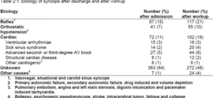
After assessment of 570 admitted patients the final diagnosis after workup was reflex syncope 21% and cardiac cause 18% Orthostatic hypotension accounted for 10%, other non-syncopal causes 4% and ultimately 48% remained of unknown etiology (Table 2:1). The mean number of tests per patient was 4.7 (SD: +/- 2.0).
Conclusion
The discharge diagnosis of R55.9 covers wide etiological manifes-tations, but mostly, it covers syncope due to unknown cause even after work-up. Many tests were used in the care path and more studies are needed to improve patient selection.
Paper III
Results
In the period from 1997 to 2009, a total of 127,508 patients were either seen in the ED, hospitalized or handled as outpatients due to syncope according to the Danish National Patient Register. Syncope accounted for 0.9% of the total admissions in the period and 0.6% of the total ED visits.
The age distribution of the patients showed two large peaks and one smaller peak (Figure 3:1). The first peak was represented primarily by females around 20 years of age, a second and quite smaller peak in older patients around 60 years of age and a third peak around 80 years of age. The third peak was left shifted in males compared to females; peaking 5-7 years earlier.
The incidence rates showed a bimodal distribution being higher in the youngest and highest in the elderly, with a distinct rise at 70 years to 40.2 per 1000 person-years increasing to 81.2 in the age group above 80 years (shown in Figure 3:2).
The risk of syncope in patients in presence of concomitant cardio-vascular disease and specific cardiocardio-vascular medication was sig-nificantly increased across all age groups and more pronounced in the young (Table 3:1).
Conclusion
Cardiovascular comorbidity and pharmacotherapy was signifi-cantly associated with the risk of syncope. The incidence rates were markedly higher than previously reported and the age dis-tribution of syncope differed according to gender.
Paper IV
Results
We identified 88,335 patients with syncope of which 37,017 patients had no known previous hospitalization for comorbidities and no concomitant use of selected pharmacotherapy. Compared
to an age and gender matched background population the long-term mortality was significantly increased in the syncope popula-tion across all age groups less than 75 years (Figure 4:1). Figure 4:1. Cumulative incidence curve in selected age groups comparing healthy individuals with syncope to the age and gen-der matched background population. Inserted in the graph are the hazard ratios (HR) from the multivariate Cox regression analy-ses of two selected age groups (25-44 years) and (45-74 years).
The results also showed that syncope as a first symptom signifi-cantly predicted several adverse cardiac outcomes such as an increased risk of stroke, pacemaker or defibrillator implantation and cardiovascular hospitalizations.
Conclusion
The first discharge after syncope in a population without prior comorbidity significantly predicted the risk of all-cause mortality, stroke, cardiovascular hospitalization, device implantation and recurrent syncope. The study suggested that syncope in seem-ingly healthy persons may be a first symptom of more severe underlying cardiovascular disease.
Paper V
Results
A total of 37,705 syncope patients from emergency departments and 188,525 controls were included in the study. Increasing CHADS2 score was significantly associated in a proportional man-ner with increased cardiovascular and all-cause mortality in ad-justed analyses. Overall there was a low absolute risk of death due to cardiovascular cause in patients with a CHADS2 score of 0, but on short as well as long-term syncope patients were at higher risk of death. Comparing the CHADS2 syncope population with the control population it was evident that syncope per se was associated with an increased risk of all-cause mortality and car-diovascular death thus yielding additional prognostic information than CHADS2 score in controls, while however a CHADS2 score of 5-6 did not provide prognostic value (Table 5:1).
Conclusion
The CHADS2 score significantly predicts short and long-term all-cause and cardiovascular mortality in patients discharged after syncope. Syncope was associated with increased risk of death but in patients with very high CHADS2 scores (5-6) syncope only predicted long-term all-cause mortality compared to the back-ground population. A CHADS2 score of 0 was associated with a very low absolute mortality but a higher relative risk than that of the background population suggesting that other significant risk factors are of importance in a syncope population.
DISCUSSION
In the present thesis, the epidemiology of syncope was investi-gated using local and national Danish registries. Through a hy-pothesis-based approach basic epidemiological factors of syncope were studied in a diverse field of pitfalls. The prognosis and out-come of syncope is tightly coupled to the cause, (which often remains unknown). Taken that into account, many things pro-foundly associated with the cause, such as electrocardiogram and precise history, were unavailable in the registries. Furthermore, it was not known whether the diagnosis ‘syncope’ consisted of other conditions, i.e. dizziness, head trauma, vertigo, hypoglyce-mia etc. in the patient registries. To fully describe and investigate the epidemiology of syncope on these conditions, validation had to be done.
In Paper I, validation of the discharge diagnosis R55.9 was per-formed in a large retrospective study of patient charts from emergency departments and hospital admissions. The study in Paper I was the first to systematically validate the discharge diag-nosis of syncope, thus strengthening the general use of the ICD-10 diagnosis of syncope in epidemiological research. Other valida-tion studies have examined various discharge diagnoses, and as a general rule the sensitivity and positive predictive values cannot be extrapolated to all diseases or discharge diagnoses, as these are being used differently in different settings [64-68]. Taken the findings of Paper I into consideration, the high positive predictive value suggests that the coding of syncope can be used to identify patients with syncope in administrative databases with a high level of accuracy. Accordingly a small percentage of the patients are non-syncopal total loss of consciousness or other conditions. Finally, approximately one third of all patients with syncope are represented by other ICD-10 diagnoses, some as specific cardiac diagnoses, others as unspecific observational codes.
In Paper II, the etiology of syncope and use of diagnostic tests during admission and workup were demonstrated in a relatively
large sample size. Major finding of this study was the relatively high number of unexplained cases (50%) despite workup and the use of many tests. The findings of etiology were in agreement with the results from other smaller retrospective and prospective studies [7, 25-28, 33, 69, 70], but importantly – these studies were not based on the ICD-10 code of syncope. Therefore the case definition of syncope was not completely comparable. Gen-erally Paper II found a higher percentage of unexplained cases of syncope than the other studies, which could be explained by the fact that the discharging physician after discharge or workup had to account for etiology, and if not claimed in the chart, the case was classified as unknown. Another explanation lies in the general use of R55.9 as discharge diagnosis. As previously stated one third of syncope cases is represented by other discharge diagnoses, most likely if explanation of the syncope is discovered during the admission period, i.e. ventricular tachycardia, myocardial infarc-tion or orthostatic hypotension etc. Thus there may be a higher concentration of unexplained etiology inherited in the routine use of R55.9. Paper II showed that the use of diagnostic tests in this setting was high and not according to guideline recommendations [5, 71, 72], thus comparable to other smaller studies [70, 73-76]. In perspective, the absence of a gold standard clinical test able to provide an easy, cheap and certain diagnosis and the extensive use of multiple diagnostic tests, termed the ‘shotgun approach’, contributes to increased costs and use of medical resources. The results from Paper III demonstrated that the risk of syncope is closely associated with the use of cardiovascular medications and concomitant cardiovascular disease, particularly in the young. This is consistent with common perception, but the association has not been well established in larger studies and data is sparse in unexplained syncope [77, 78]. The increased risk of syncope associated with cardiovascular disease or use of cardiovascular medications in the younger age groups, where reflex syncope is very prevalent, is a new finding. Paper III also established clear and significantly higher incidence rates of first syncope than previously reported [14, 16, 22, 23] even from the general popu-lation. The fraction of syncope as defined by R55.9 in the emer-gency departments and hospital departments are consistent with recent larger studies from the US and Europe [7, 17]. The high incidence rates were found despite the fact that we only counted the initial syncope and did not take recurrent admissions into account, and secondarily that R55.9, as noted previously, missed one third of all cases. The real fraction of syncope of all visits to emergency departments and admissions is thus believed to be even higher than reported in Paper III.
Syncope was more common in the elderly, females and generally a diagnosis associated with considerable comorbidity. Finally a novel age distribution of syncope was described for hospitalized patients and revealed a gender related parallel shift in the third peak, where the distribution of men suffering syncope peaked five years earlier than for women, representing an age difference in the general disease burden.
In Paper IV we evaluated the prognosis of healthy individuals after syncope. The main finding was that syncope per se was associated with higher risk of death when compared to a back-ground population with various degrees of comorbidities. Fur-thermore syncope was associated with increased risk of adverse cardiovascular outcomes and hospitalization. The absolute risk of death in this low risk population was indeed very low and not that much different from the control group. This is in agreement with smaller studies suggesting that syncope (generalized to all etiolo-gies) is rather benign and that excess mortality is driven by car-diovascular comorbidities [17, 19, 39]. However the
interpreta-tion of the increased risk in mortality in certain age groups de-serves extra attention. It may not be safe to rely on a history of no comorbidity when evaluating a patient with syncope. The relative risk of death as well as the other outcomes in these patients was increased and even these low risk patients with no apparent comorbidities at presentation need to be carefully evaluated. This underlines the difficulties associated with the assessment of syncope and why there is a need for proper risk stratification in these patients in order to balance the limited health care resources [79].
In Paper V we tested the hypothesis that the CHADS2 score is a useful tool for risk stratification of cardiovascular and all-cause mortality. The CHADS2 score predicted short and long-term mor-tality in this population which was expected as the CHADS2 score probably predicts mortality in many subsets of populations [43-47]. The evaluation of the score revealed that the CHADS2 score may provide additional prognostic information on risk stratifica-tion in syncope patients when compared to a control populastratifica-tion stratified by the same CHADS2 scores. Also importantly Paper V showed that a CHADS2 score of 0 represented a very low absolute risk, but on the other hand that a score of 0 was associated with a worse prognosis in patients with syncope. An important factor of these results was the low mean age of the population with a CHADS2 score of 0 and that the score does not take other impor-tant covariates into account. Taken into consideration the results from Paper IV, where syncope, as the only factor in otherwise healthy people, remained an independent risk factor of mortality and development of cardiovascular disease, it is not recom-mended to rely on a CHADS2 of 0 as a safe prognostic marker. Secondarily the CHADS2 score did not proportionally predict the mortality in the very high scores of 5-6, making it a tool too sim-ple to predict mortality in general. In the past 10 years several other risk-stratification schemes for syncope have emerged. The schemes include the OESIL score [80], the San Francisco rule [81], the STePS study [29], the Rose rule [82], and the Boston criteria [83, 84]. They resulted in different covariates of prognostic value and consequently the guidelines tried to incorporate all of them [5]. None have been widely implemented; they have not im-proved diagnostic discrimination and have failed validation [85-87]. Clinicians are thus in dire need for proper prospective stud-ies, pre-specified to assess risk stratification in the emergency department in various subsets of patient populations on short-term risk (< 1 week) and long-short-term risk.
In general most of the recommendations in the management of syncope patients are based on class IC and IIC levels of evidence, reflecting the difficulties of dealing with syncope, not only in daily practice but also in larger studies, randomized or observational. The papers presented in this thesis reflect this divergence but contributes significantly to establishment and continued use of registries in observational studies of syncope. Implementation and incorporation of other databases, such as electronically re-corded laboratory results (cardiac enzymes, biomarkers and hemoglobin levels), ECG’s and echocardiograms will contribute significantly to a future risk stratification algorithm based on substantial data in a large population.
The evaluation of syncope is a major and increasing annual health-care cost [79, 88] calling for two important factors. Firstly, a durable risk stratification and secondly, an optimization of diagnostic pathways and introduction of interdisciplinary collabo-ration in syncope clinics. The present thesis discusses these themes and the present and future use of registries for improve-ment of overall manageimprove-ment of syncope.
LIMITATIONS AND EVALUATION OF METHODS IN THE THESIS
Observational studies are able to detect small, but clinically sig-nificant differences between groups quickly and with limited resources compared to randomized controlled trials (RCT). Obser-vational studies reflect real-life settings to a higher degree than RCTs which makes the generalization more straightforward and often provide new information and questions that can later be evaluated in RCTs. There are several important limitations of the methods used in the present papers of this thesis. All studies were retrospective and based on local and national registries. Many clinically important and relevant covariates are missing in the national registries (i.e. creatinine levels, biomarkers such as NT-proBNP, weight, blood pressure, echocardiograms and elec-trocardiograms) and may confound the results. In Paper II, we have information on these covariates and these may be represen-tative proportionately, but not useful on the individual level for the large registries. All validation from the local registries was performed on pre-existing charts written or described by physi-cians with different quality. Many factors in a clinically useful description of a syncopal event could be lost depending on the quality of work and experience of the given physician. A specific limitation of the sensitivity of the diagnosis in the na-tional registries was acquired and thus taken into account in the conclusions of Paper III-V from the validation made in Paper I. Sensitivity of the diagnosis plays an important role, mainly be-cause the severity of syncope may be underestimated in prognos-tic studies as one third of patients with syncope receive other, and sometimes more severe diagnoses, i.e. heart failure, aortic stenosis, myocardial infarction etc. This is particularly important in Paper V, as the evaluation of the CHADS2 score did not seem to predict excess mortality in the CHADS2 score of 5-6 compared to the controls. In general this may be due to important selection bias towards non-cardiac conditions, essentially biasing the re-sults. Other major limitations in the registries are unvalidated causes of death in the registry along with less autopsies being performed. In the three Papers based on national registries we used arbitrary cut-off limits of when patients were on baseline pharmacotherapy. We used prescriptions redeemed within 180 days before admission, but the actual accuracy of this approach is completely unknown. The cut-off limits of five years of previous discharges set as the limit of baseline comorbidity is just as arbi-trary and we do not know the actual accuracy of this approach either. We mention that many of the discharge diagnoses are well validated, but these were all validated in different ways and none of them were validated with limits of comorbidity set at ‘5 years prior to admission’. A general limitation in epidemiological stud-ies is also the risk of unmeasured confounding. Even though we take many confounders into account in the adjusted models, unmeasured confounding may still bias the results.
Future research
As noted briefly, prospective larger trials of patients with syncope should be undertaken with the goal of optimal risk stratification. For high risk cardiac patients with syncope, but with unclear implantable defibrillator indications implantable loop recorders may answer critical questions in terms of short term risks. More studies are also needed to asses optimal care pathways and op-timal strategy for diagnostic work-up. This is important not only in the high risk patient but also, as we have demonstrated, in low risk patients.
Funding and acknowledgements
Martin Huth Ruwald was supported by the Danish Heart Associa-tion (grant 12-04-R90-A3806-22701), the Lundbeck FoundaAssocia-tion (grant R108-A104415), Helsefonden (grant 2012B018), Forskning og Udvikling Kardiologisk Afdeling P Gentofte Hospital (FUKAP), support from Section of Electrophysiology, Department of Cardi-ology, Gentofte Hospital, Knud Hoejgaards Fond, Arvid Nilssons Fond, Snedkermester Sophus Jacobsens og Hustru Astrid Jacob-sens Fond, Boehringer Ingelheim Educational Travel Grant, Astra-Zeneca Travel Grant, Holger Rabitz Fond, The Capital Region of Denmark Research Foundation, Copenhagen University, Faculty of Health Sciences and Else og Mogens Wedell-Wedellsborgs Fond. None of the funding sponsors were involved in the management, analysis or interpretation of the data or the manuscripts pre-sented in this thesis.
SUMMARY
The epidemiology and prognosis of ‘fainting’ or syncope has puzzled physicians over the years. Is fainting dangerous? This is a question often asked by the patient– and the answer is ‘it de-pends on a lot of things’. The diverse pathophysiology of syncope and the underlying comorbidites of the patients play an essential role. In epidemiology these factors have major impact on the outcome of the patients. Until recently, even the definition of syncope, differed from one study to another which has made literature reviews difficult. Traditionally the data on epidemiology of syncope has been taken from smaller studies from different clinical settings with wide differences in patient morbidity. Through the extensive Danish registries we examined the charac-teristics and prognosis of the patients hospitalized due to syncope in a nationwide study.
The aims of the present thesis were to investigate: 1) the use, validity and accuracy of the ICD-10 diagnosis of syncope R55.9 in the National Patient Registry for the use of this diagnosis in the epidemiology of syncope, 2) diagnostics used and etiology of a random selection of patients who had a discharge diagnosis of R55.9, 3) the incidence, prevalence and cardiovascular factors associated with the risk of syncope, 4) the prognosis in healthy individuals discharged after syncope, and 5) the prognosis of patients after syncope and evaluation of the CHADS2 score as a tool for short and long-term risk prediction.
The first studies of the present thesis demonstrated that the ICD-10 discharge diagnosis could reliably identify a cohort of patients admitted for syncope and that the discharge code carried a high number of unexplained cases despite use of numerous tests. The last studies showed that syncope is a common cause for hospital contact in Denmark and that the risk of syncope is tightly associ-ated with cardiovascular comorbidities and use of pharmacother-apy. Furthermore in patients with no comorbidities (or healthy individuals), syncope is a significant and independent prognostic factor of adverse cardiovascular outcome and death compared to the background population. Lastly evaluation of the CHADS2 score, as a tool for risk stratification, showed that it provided additional prognostic information on short and long-term cardio-vascular mortality in syncope patients compared to controls.
REFERENCES
1. Ruwald MH, Hansen ML, Lamberts M, Kristensen SL, Wissenberg M, Olsen AM, Christensen SB, Vinther M, Kober L, Torp-Pedersen C, Hansen J, Gislason GH. Accu-racy of the ICD-10 discharge diagnosis for syncope. Eu-ropace. 2013;15:595-600
2. Ruwald MH, Hansen ML, Lamberts M, Hansen CM, Ho-jgaard MV, Kober L, Torp-Pedersen C, Hansen J, Gis-lason GH. The relation between age, sex, comorbidity, and pharmacotherapy and the risk of syncope: a Danish nationwide study. Europace. 2012;14:1506-14
3. Ruwald MH, Hansen ML, Lamberts M, Hansen CM, Vin-ther M, Kober L, Torp-Pedersen C, Hansen J, Gislason GH. Prognosis among healthy individuals discharged with a primary diagnosis of syncope. J Am Coll Cardiol. 2013;61:325-2
4. Ruwald MH, Ruwald AC, Jons C, Lamberts M, Hansen ML, Vinther M, Kober L, Torp-Pedersen C, Hansen J, Gis-lason GH. Evaluation of the CHADS Risk Score on Short- and Long-Term All-Cause and Cardiovascular Mortality After Syncope. Clin Cardiol. 2013 May;36(5):262-8 5. Moya A, Sutton R, Ammirati F, Blanc JJ, Brignole M,
Dahm JB, Deharo JC, Gajek J, Gjesdal K, Krahn A, Massin M, Pepi M, Pezawas T, Ruiz Granell R, Sarasin F, Ungar A, van Dijk JG, Walma EP, Wieling W. Guidelines for the diagnosis and management of syncope (version 2009). Eur Heart J. 2009;30:2631-71
6. Brignole M, Alboni P, Benditt D, Bergfeldt L, Blanc JJ, Bloch Thomsen PE, Fitzpatrick A, Hohnloser S, Kapoor W, Kenny RA, Theodorakis G, Kulakowski P, Moya A, Ra-viele A, Sutton R, Wieling W, Janousek J, van Dijk G. Task force on syncope, European Society of Cardiology. Part 1. The initial evaluation of patients with syncope. Europace. 2001;3:253-60
7. Sarasin FP, Louis-Simonet M, Carballo D, Slama S, Ra-jeswaran A, Metzger JT, Lovis C, Unger PF, Junod AF. Prospective evaluation of patients with syncope: a pop-ulation-based study. Am J Med. 2001;111:177-84 8. Quinn J, McDermott D, Kramer N, Yeh C, Kohn MA, Stiell
I, Wells G. Death after emergency department visits for syncope: how common and can it be predicted? Ann Emerg Med. 2008;51:585-90
9. Day SC, Cook EF, Funkenstein H, Goldman L. Evaluation and outcome of emergency room patients with tran-sient loss of consciousness. Am J Med. 1982;73:15-23 10. Sun BC, Derose SF, Liang LJ, Gabayan GZ, Hoffman JR, Moore AA, Mower WR, Mangione CM. Predictors of 30-day serious events in older patients with syncope. Ann Emerg Med. 2009;54:769-78 e1-5
11. Rose MS, Koshman ML, Spreng S, Sheldon R. The rela-tionship between health-related quality of life and fre-quency of spells in patients with syncope. J Clin Epide-miol. 2000;53:1209-16
12. Linzer M, Pontinen M, Gold DT, Divine GW, Felder A, Brooks WB. Impairment of physical and psychosocial function in recurrent syncope. J Clin Epidemiol. 1991;44:1037-43
13. Linzer M, Gold DT, Pontinen M, Divine GW, Felder A, Brooks WB. Recurrent syncope as a chronic disease: preliminary validation of a disease-specific measure of functional impairment. J Gen Intern Med. 1994;9:181-6 14. Soteriades ES, Evans JC, Larson MG, Chen MH, Chen L,
Benjamin EJ, Levy D. Incidence and prognosis of syn-cope. N Engl J Med. 2002;347:878-85
15. Alshekhlee A, Shen WK, Mackall J, Chelimsky TC. Inci-dence and mortality rates of syncope in the United States. Am J Med. 2009;122:181-8
16. Lipsitz LA, Wei JY, Rowe JW. Syncope in an elderly, insti-tutionalised population: prevalence, incidence, and as-sociated risk. Q J Med. 1985;55:45-54
17. Getchell WS, Larsen GC, Morris CD, McAnulty JH. Epi-demiology of syncope in hospitalized patients. J Gen In-tern Med. 1999;14:677-87
18. Kapoor WN. Evaluation and outcome of patients with syncope. Medicine (Baltimore). 1990;69:160-75 19. Kapoor WN, Hanusa BH. Is syncope a risk factor for poor
outcomes? Comparison of patients with and without syncope. Am J Med. 1996;100:646-55
20. Kapoor W, Snustad D, Peterson J, Wieand HS, Cha R, Karpf M. Syncope in the elderly. Am J Med. 1986;80:419-28
21. Sun BC, Emond JA, Camargo CA, Jr. Characteristics and admission patterns of patients presenting with syncope to U.S. emergency departments, 1992-2000. Acad Emerg Med. 2004;11:1029-34
22. Colman N, Nahm K, Ganzeboom KS, Shen WK, Reitsma J, Linzer M, Wieling W, Kaufmann H. Epidemiology of re-flex syncope. Clin Auton Res. 2004;14 Suppl 1:9-17 23. Savage DD, Corwin L, McGee DL, Kannel WB, Wolf PA.
Epidemiologic features of isolated syncope: the Fram-ingham Study. Stroke. 1985;16:626-9
24. Ganzeboom KS, Mairuhu G, Reitsma JB, Linzer M, Wiel-ing W, van Dijk N. Lifetime cumulative incidence of syn-cope in the general population: a study of 549 Dutch subjects aged 35-60 years. J Cardiovasc Electrophysiol. 2006;17:1172-6
25. Ammirati F, Colivicchi F, Santini M. Diagnosing syncope in clinical practice. Implementation of a simplified diag-nostic algorithm in a multicentre prospective trial - the OESIL 2 study (Osservatorio Epidemiologico della Sin-cope nel Lazio). Eur Heart J. 2000;21:935-40
26. Blanc JJ, L'Her C, Touiza A, Garo B, L'Her E, Mansourati J. Prospective evaluation and outcome of patients admit-ted for syncope over a 1 year period. Eur Heart J. 2002;23:815-20
27. Disertori M, Brignole M, Menozzi C, Raviele A, Rizzon P, Santini M, Proclemer A, Tomasi C, Rossillo A, Taddei F, Scivales A, Migliorini R, De Santo T. Management of pa-tients with syncope referred urgently to general hospi-tals. Europace. 2003;5:283-91
28. Olde Nordkamp LR, van Dijk N, Ganzeboom KS, Reitsma JB, Luitse JS, Dekker LR, Shen WK, Wieling W. Syncope prevalence in the ED compared to general practice and population: a strong selection process. Am J Emerg Med. 2009;27:271-9
29. Costantino G, Perego F, Dipaola F, Borella M, Galli A, Cantoni G, Dell'Orto S, Dassi S, Filardo N, Duca PG, Mon-tano N, Furlan R. Short- and long-term prognosis of syn-cope, risk factors, and role of hospital admission: results from the STePS (Short-Term Prognosis of Syncope) study. J Am Coll Cardiol. 2008;51:276-83
30. Leitch JW, Klein GJ, Yee R, Leather RA, Kim YH. Syncope associated with supraventricular tachycardia. An ex-pression of tachycardia rate or vasomotor response? Circulation. 1992;85:1064-71
31. Brignole M, Gianfranchi L, Menozzi C, Raviele A, Oddone D, Lolli G, Bottoni N. Role of autonomic reflexes in syn-cope associated with paroxysmal atrial fibrillation. J Am Coll Cardiol. 1993;22:1123-9
32. Goldschlager N, Epstein AE, Grubb BP, Olshansky B, Pry-stowsky E, Roberts WC, Scheinman MM. Etiologic con-siderations in the patient with syncope and an appar-ently normal heart. Arch Intern Med. 2003;163:151-62 33. Chen-Scarabelli C, Scarabelli TM. Neurocardiogenic
syn-cope. BMJ. 2004;329:336-41
34. Naschitz JE, Rosner I. Orthostatic hypotension: frame-work of the syndrome. Postgrad Med J. 2007;83:568-74 35. Ungar A, Mussi C, Del Rosso A, Noro G, Abete P, Ghirelli
L, Cellai T, Landi A, Salvioli G, Rengo F, Marchionni N, Masotti G. Diagnosis and characteristics of syncope in older patients referred to geriatric departments. J Am Geriatr Soc. 2006;54:1531-6
36. Linzer M, Pontinen M, Gold DT, Divine GW, Felder A, Brooks WB. Impairment of physical and psychosocial function in recurrent syncope. J Clin Epidemiol. 1991;44:1037-43
37. van Dijk N, Sprangers MA, Colman N, Boer KR, Wieling W, Linzer M. Clinical factors associated with quality of life in patients with transient loss of consciousness. J Cardiovasc Electrophysiol. 2006;17:998-1003 38. Suzuki M, Hori S, Aikawa N. Application of the recent
American practice resources for risk stratification sys-tem for patients presenting to a Japanese emergency department because of syncope. Int Heart J. 2007;48:513-22
39. Middlekauff HR, Stevenson WG, Stevenson LW, Saxon LA. Syncope in advanced heart failure: high risk of sud-den death regardless of origin of syncope. J Am Coll Cardiol. 1993;21:110-6
40. Roussanov O, Estacio G, Capuno M, Hill J, Kovesdy C, Jarmukli N. Outcomes of unexplained syncope in the el-derly. Am J Geriatr Cardiol. 2007;16:249-54
41. Gage BF, Waterman AD, Shannon W, Boechler M, Rich MW, Radford MJ. Validation of clinical classification schemes for predicting stroke: results from the National Registry of Atrial Fibrillation. JAMA. 2001;285:2864-70 42. Olesen JB, Lip GY, Hansen ML, Hansen PR, Tolstrup JS,
Lindhardsen J, Selmer C, Ahlehoff O, Olsen AM, Gislason GH, Torp-Pedersen C. Validation of risk stratification schemes for predicting stroke and thromboembolism in patients with atrial fibrillation: nationwide cohort study. Bmj. 2011;342:d124
43. Crandall MA, Horne BD, Day JD, Anderson JL,
Muhlestein JB, Crandall BG, Weiss JP, Osborne JS, Lappe DL, Bunch TJ. Atrial fibrillation significantly increases to-tal morto-tality and stroke risk beyond that conveyed by the CHADS2 risk factors. Pacing Clin Electrophysiol. 2009;32:981-6
44. Kim YD, Cha MJ, Kim J, Lee DH, Lee HS, Nam CM, Nam HS, Heo JH. Ischaemic cardiovascular mortality in pa-tients with non-valvular atrial fibrillation according to CHADS(2) score. Thromb Haemost. 2011;105:712-20 45. Nakagawa K, Hirai T, Takashima S, Fukuda N, Ohara K,
Sasahara E, Taguchi Y, Dougu N, Nozawa T, Tanaka K, Inoue H. Chronic kidney disease and CHADS(2) score in-dependently predict cardiovascular events and mortal-ity in patients with nonvalvular atrial fibrillation. Am J Cardiol. 2011;107:912-6
46. Khumri TM, Idupulapati M, Rader VJ, Nayyar S, Stoner CN, Main ML. Clinical and echocardiographic markers of mortality risk in patients with atrial fibrillation. Am J Cardiol. 2007;99:1733-6
47. Ruiz-Nodar JM, Marin F, Manzano-Fernandez S, Valen-cia-Martin J, Hurtado JA, Roldan V, Pineda J, Pinar E, So-gorb F, Valdes M, Lip GY. An evaluation of the CHADS(2) stroke risk score in patients with atrial fibrillation who undergo percutaneous coronary revascularization. Chest. 2011;139:1402-9
48. Pouleur AC, Barkoudah E, Uno H, Skali H, Finn PV, Ze-lenkofske SL, Belenkov YN, Mareev V, Velazquez EJ, Rouleau JL, Maggioni AP, Kober L, Califf RM, McMurray JJ, Pfeffer MA, Solomon SD. Pathogenesis of sudden un-expected death in a clinical trial of patients with myo-cardial infarction and left ventricular dysfunction, heart failure, or both. Circulation. 2010;122:597-602 49. Huikuri HV, Castellanos A, Myerburg RJ. Sudden death
due to cardiac arrhythmias. N Engl J Med. 2001;345:1473-82
50. Yap YG, Duong T, Bland M, Malik M, Torp-Pedersen C, Kober L, Connolly SJ, Marchant B, Camm J. Temporal trends on the risk of arrhythmic vs. non-arrhythmic deaths in high-risk patients after myocardial infarction: a combined analysis from multicentre trials. Eur Heart J. 2005;26:1385-93
51. Andersen TF, Madsen M, Jorgensen J, Mellemkjoer L, Olsen JH. The Danish National Hospital Register. A valu-able source of data for modern health sciences. Dan Med Bull. 1999;46:263-8
52. Krarup LH, Boysen G, Janjua H, Prescott E, Truelsen T. Validity of stroke diagnoses in a National Register of Pa-tients. Neuroepidemiology. 2007;28:150-4
53. Madsen M, Davidsen M, Rasmussen S, Abildstrom SZ, Osler M. The validity of the diagnosis of acute myocar-dial infarction in routine statistics: a comparison of mor-tality and hospital discharge data with the Danish MON-ICA registry. J Clin Epidemiol. 2003;56:124-30
54. Kumler T, Gislason GH, Kirk V, Bay M, Nielsen OW, Ko-ber L, Torp-Pedersen C. Accuracy of a heart failure diag-nosis in administrative registers. Eur J Heart Fail. 2008;10:658-60
55. Hommel K, Rasmussen S, Madsen M, Kamper AL. The Danish Registry on Regular Dialysis and Transplantation: completeness and validity of incident patient registra-tion. Nephrol Dial Transplant. 2010;25:947-51 56. Olesen JB, Lip GY, Lindhardsen J, Lane DA, Ahlehoff O,
Hansen ML, Raunso J, Tolstrup JS, Hansen PR, Gislason GH, Torp-Pedersen C. Risks of thromboembolism and bleeding with thromboprophylaxis in patients with atri-al fibrillation: A net clinicatri-al benefit anatri-alysis using a 'reatri-al world' nationwide cohort study. Thromb Haemost. 2011;106:739-49
57. Olesen JB, Lip GY, Kamper AL, Hommel K, Kober L, Lane DA, Lindhardsen J, Gislason GH, Torp-Pedersen C. Stroke and bleeding in atrial fibrillation with chronic kidney disease. N Engl J Med. 2012;367:625-35 58. Sorensen R, Hansen ML, Abildstrom SZ, Hvelplund A,
Andersson C, Jorgensen C, Madsen JK, Hansen PR, Ko-ber L, Torp-Pedersen C, Gislason GH. Risk of bleeding in patients with acute myocardial infarction treated with different combinations of aspirin, clopidogrel, and vita-min K antagonists in Denmark: a retrospective analysis of nationwide registry data. Lancet. 2009;374:1967-74 59. Gaist D, Sorensen HT, Hallas J. The Danish prescription
registries. Dan Med Bull. 1997;44:445-8
60. Charlson ME, Pompei P, Ales KL, MacKenzie CR. A new method of classifying prognostic comorbidity in longitu-dinal studies: development and validation. J Chronic Dis. 1987;40:373-83
61. Nuttall M, van der Meulen J, Emberton M. Charlson scores based on ICD-10 administrative data were valid in assessing comorbidity in patients undergoing urologi-cal cancer surgery. J Clin Epidemiol. 2006;59:265-73 62. Andersson C, Norgaard ML, Hansen PR, Fosbol EL,
Schmiegelow M, Weeke P, Olesen JB, Raunso J, Jorgen-sen CH, Vaag A, Kober L, Torp-PederJorgen-sen C, Gislason GH. Heart failure severity, as determined by loop diuretic dosages, predicts the risk of developing diabetes after myocardial infarction: a nationwide cohort study. Eur J Heart Fail. 2010;12:1333-8
63. Schramm TK, Gislason GH, Kober L, Rasmussen S, Ras-mussen JN, Abildstrom SZ, Hansen ML, Folke F, Buch P, Madsen M, Vaag A, Torp-Pedersen C. Diabetes patients requiring glucose-lowering therapy and nondiabetics with a prior myocardial infarction carry the same car-diovascular risk: a population study of 3.3 million peo-ple. Circulation. 2008;117:1945-54
64. Kokotailo RA, Hill MD. Coding of stroke and stroke risk factors using international classification of diseases, re-visions 9 and 10. Stroke. 2005;36:1776-81
65. Jette N, Reid AY, Quan H, Hill MD, Wiebe S. How accu-rate is ICD coding for epilepsy? Epilepsia. 2010;51:62-9 66. Stickler DE, Royer JA, Hardin JW. Validity of hospital
dis-charge data for identifying cases of amyotrophic lateral sclerosis. Muscle Nerve. 2011;44:814-6
67. Pedersen M, Klarlund M, Jacobsen S, Svendsen AJ, Frisch M. Validity of rheumatoid arthritis diagnoses in the Danish National Patient Registry. Eur J Epidemiol. 2004;19:1097-103
68. Nielsen EH, Lindholm J, Laurberg P. Use of combined search criteria improved validity of rare disease (cranio-pharyngioma) diagnosis in a national registry. J Clin Epi-demiol. 2011;64:1118-26
69. Alboni P, Brignole M, Menozzi C, Raviele A, Del Rosso A, Dinelli M, Solano A, Bottoni N. Diagnostic value of his-tory in patients with syncope with or without heart dis-ease. J Am Coll Cardiol. 2001;37:1921-8
70. Baron-Esquivias G, Martinez-Alday J, Martin A, Moya A, Garcia-Civera R, Paz Lopez-Chicharro M, Martin-Mendez M, del Arco C, Laguna P. Epidemiological characteristics and diagnostic approach in patients admitted to the emergency room for transient loss of consciousness: Group for Syncope Study in the Emergency Room (GESINUR) study. Europace. 2010;12:869-76 71. Brignole M, Ungar A, Bartoletti A, Ponassi I, Lagi A,
Mussi C, Ribani MA, Tava G, Disertori M, Quartieri F, Al-boni P, Raviele A, Ammirati F, Scivales A, De Santo T. Standardized-care pathway vs. usual management of syncope patients presenting as emergencies at general hospitals. Europace. 2006;8:644-50
72. Sanders NA, Jetter TL, Brignole M, Hamdan MH. Stan-dardized Care Pathway Versus Conventional Approach in the Management of Patients Presenting with Faint at the University of Utah. Pacing Clin Electrophysiol. 2012 73. Grossman SA, Fischer C, Bar JL, Lipsitz LA, Mottley L,
Sands K, Thompson S, Zimetbaum P, Shapiro NI. The yield of head CT in syncope: a pilot study. Intern Emerg Med. 2007;2:46-9
74. Sun BC, Emond JA, Camargo CA, Jr. Inconsistent electro-cardiographic testing for syncope in United States emergency departments. Am J Cardiol. 2004;93:1306-8 75. Giglio P, Bednarczyk EM, Weiss K, Bakshi R. Syncope and
head CT scans in the emergency department. Emerg Radiol. 2005;12:44-6
76. Brignole M, Vardas P, Hoffman E, Huikuri H, Moya A, Ricci R, Sulke N, Wieling W, Auricchio A, Lip GY, Almen-dral J, Kirchhof P, Aliot E, Gasparini M, Braunschweig F, Botto GL. Indications for the use of diagnostic implant-able and external ECG loop recorders. Europace. 2009;11:671-87
77. Hess DS, Morady F, Scheinman MM. Electrophysiologic testing in the evaluation of patients with syncope of undetermined origin. Am J Cardiol. 1982;50:1309-15 78. Linzer M, Yang EH, Estes NA, 3rd, Wang P, Vorperian VR,
Kapoor WN. Diagnosing syncope. Part 2: Unexplained syncope. Clinical Efficacy Assessment Project of the American College of Physicians. Ann Intern Med. 1997;127:76-86
79. Sun BC, Emond JA, Camargo CA, Jr. Direct medical costs of syncope-related hospitalizations in the United States. Am J Cardiol. 2005;95:668-71
80. Colivicchi F, Ammirati F, Melina D, Guido V, Imperoli G, Santini M. Development and prospective validation of a risk stratification system for patients with syncope in the emergency department: the OESIL risk score. Eur Heart J. 2003;24:811-9
81. Quinn J, McDermott D, Stiell I, Kohn M, Wells G. Pro-spective validation of the San Francisco Syncope Rule to predict patients with serious outcomes. Ann Emerg Med. 2006;47:448-54
82. Reed MJ, Newby DE, Coull AJ, Prescott RJ, Jacques KG, Gray AJ. The ROSE (risk stratification of syncope in the emergency department) study. J Am Coll Cardiol. 2010;55:713-21
83. Reed MJ, Newby DE, Coull AJ, Jacques KG, Prescott RJ, Gray AJ. The Risk stratification Of Syncope in the Emer-gency department (ROSE) pilot study: a comparison of existing syncope guidelines. Emerg Med J. 2007;24:270-5
84. Reed MJ, Henderson SS, Newby DE, Gray AJ. One-year prognosis after syncope and the failure of the ROSE
de-cision instrument to predict one-year adverse events. Ann Emerg Med. 2011;58:250-6
85. Serrano LA, Hess EP, Bellolio MF, Murad MH, Montori VM, Erwin PJ, Decker WW. Accuracy and quality of clini-cal decision rules for syncope in the emergency de-partment: a systematic review and meta-analysis. Ann Emerg Med. 2010;56:362-73 e1
86. Quinn JV, Stiell IG, McDermott DA, Kohn MA, Wells GA. The San Francisco Syncope Rule vs physician judgment and decision making. Am J Emerg Med. 2005;23:782-6 87. Birnbaum A, Esses D, Bijur P, Wollowitz A, Gallagher EJ.
Failure to validate the San Francisco Syncope Rule in an independent emergency department population. Ann Emerg Med. 2008;52:151-9
88. Malasana G, Brignole M, Daccarett M, Sherwood R, Hamdan MH. The prevalence and cost of the faint and fall problem in the state of Utah. Pacing Clin Electrophy-siol. 2011;34:278-83
![Figure 1. The pathophysiological basis of syncope classification (after Moya et al.[5])](https://thumb-us.123doks.com/thumbv2/123dok_us/1151577.2654192/2.892.457.810.138.394/figure-pathophysiological-basis-syncope-classification-moya-et-al.webp)