AASLD PRACTICE GUIDELINE
The Diagnosis and Management of Non-Alcoholic Fatty
Liver Disease: Practice Guideline by the American
Association for the Study of Liver Diseases, American
College of Gastroenterology, and the American
Gastroenterological Association
Naga Chalasani, MD, FACG,1Zobair Younossi, MD, FACG,2 Joel E. Lavine, MD, PhD,3Anna Mae Diehl, MD,4 Elizabeth M. Brunt, MD,5Kenneth Cusi, MD,6Michael Charlton, MD,7and Arun J. Sanyal, MD8
Preamble
These recommendations are based on the following: (1) a formal review and analysis of the recently published world literature on the topic [Medline search up to June 2011]; (2) the American College of Physicians’ Manual for Assessing Health Practices and Designing Practice Guide-lines;1(3) guideline policies of the three societies approv-ing this document; and (4) the experience of the authors and independent reviewers with regards to NAFLD.
Intended for use by physicians and allied health profes-sionals, these recommendations suggest preferred approaches to the diagnostic, therapeutic and preventive aspects of care. They are intended to be flexible and ad-justable for individual patients. Specific recommendations are evidence-based wherever possible, and when such evi-dence is not available or inconsistent, recommendations are made based on the consensus opinion of the authors. To best characterize the evidence cited in support of the recommendations, the AASLD Practice Guidelines Com-mittee has adopted the classification used by the Grading of Recommendation Assessment, Development, and Evaluation (GRADE) workgroup with minor modifica-tions (Table 1).2The strength of recommendations in the GRADE system is classified as strong (1) or weak (2). The quality of evidence supporting strong or weak rec-ommendations is designated by one of three levels: high
(A), moderate(B) or low-quality(C).2This is a practice
guideline for clinicians rather than a review article and interested readers can refer to several comprehensive reviews published recently.3-8
Definitions
The definition of nonalcoholic fatty liver disease (NAFLD) requires that (a) there is evidence of hepatic steatosis, either by imaging or by histology and (b) there are no causes for secondary hepatic fat accumula-tion such as significant alcohol consumpaccumula-tion, use of steatogenic medication or hereditary disorders (Table 2). In the majority of patients, NAFLD is associated with metabolic risk factors such as obesity, diabetes mellitus, and dyslipidemia. NAFLD is histologically further cate-gorized into nonalcoholic fatty liver (NAFL) and nonal-coholic steatohepatitis (NASH) (Table 3). NAFL is defined as the presence of hepatic steatosis with no evi-dence of hepatocellular injury in the form of ballooning of the hepatocytes. NASH is defined as the presence of hepatic steatosis and inflammation with hepatocyte injury (ballooning) with or without fibrosis.
Incidence and Prevalence in the General Population
The incidence of NAFLD has been investigated in a limited number of studies. Two Japanese studies9,10
Submitted for Governing Board approval by AASLD, ACG, and AGA on February 22, 2012
Abbreviations: NAFLD, Nonalcoholic Fatty Liver Disease; NAFL,Nonalcoholic Fatty Liver; NASH, Nonalcoholic Steatohepatitis; T2DM, Type 2 Diabetes Mellitus; AST, Aspartate Aminotransferase; ALT, Alanine Aminotransferase; HOMA,Homeostatic Model Assessment; RCT; Randomized Controlled Trial; PIVENS: Pioglitazone versus Vitamin E versus Placebo for the Treatment of Non-diabetic patients with Nonalcoholic steatohepatitis; TONIC; Treatment of Nonalcoholic Fatty Liver Disease in Children; NAS, NAFLD Activity Score; CK18; Cytokeratin 18 Fragments; ELF, Enhanced Liver Fibrosis Panel; TZD; Thiazolidinediones; UDCA: Ursodeoxycholic Acid; ANA; Anti Nuclear Antibody; ASMA: Anti Smooth Muscle Antibody; US; Ultrasound; CT: Computerized Tomography; MR; Magnetic Resonance.
From the1Indiana University School of Medicine, Indianapolis, IN;2Center for Liver Disease and Department of Medicine, Inova Fairfax Hospital, Falls Church;
3
Columbia University, New York, NY;4Duke University, Durham, NC;5Washington University, St. Louis, MO;6University of Florida, Gainesville, FL;7Mayo Clinic, Rochester, MN;8Virginia Commonwealth University, Richmond, VA
Received April 3, 2012; accepted April 3, 2012.
reported an incidence rate of 31 and 86 cases of sus-pected NAFLD per 1,000 person-years respectively, whereas another study from England showed a much lower incidence rate of 29 cases per 100,000 person-years.11 More studies are needed to better understand the incidence of NAFLD across different age, ethnic, and geographic groups.
The reported prevalence of NAFLD varies widely depending on the population studied and the defini-tion used. The prevalence of histologically-defined NAFLD was 20% and 51% in two different studies comprised of potential living liver donors.12,13 The reported prevalence of NAFLD when defined by liver ultrasound ranged between 17% and 46% depending on the population studied.4 In a study consisting of nearly 400 middle aged individuals, the prevalence of NAFLD defined by ultrasonography was 46% and the prevalence of histologically confirmed NASH was 12.2%.14 In the Dallas Heart Study, when assessed by MR spectroscopy the prevalence of NAFLD in the general population was 31%.15 The prevalence of sus-pected NAFLD when estimated using aminotransfer-ases alone without imaging or histology ranged between 7% and 11%, but aminotransferases can be normal in individuals with NAFLD.4 In summary, estimates of the worldwide prevalence of NAFLD ranges from 6.3% to 33% with a median of 20% in
the general population, based on a variety of assess-ment methods.4 On the other hand, the estimated prevalence of NASH is lower, ranging from 3 to 5%.4 The prevalence of NASH cirrhosis in the general pop-ulation is not known.
Prevalence of NAFLD in High Risk Groups (Table 4)
Obesity is a common and well documented risk factor for NAFLD. Both excessive BMI and visceral obesity are recognized risk factors for NAFLD. In patients with severe obesity undergoing bariatric surgery, the preva-lence of NAFLD can exceed 90% and up to 5% of patients may have unsuspected cirrhosis.4,16-20 There is a very high prevalence of NAFLD in individuals with type 2 diabetes mellitus (T2DM).4An ultrasonographic study of patients with T2DM showed a 69% prevalence of NAFLD.21 In another study, 127 of 204 diabetic patients displayed fatty infiltration on ultrasound, and 87% of the patients with fatty infiltration who con-sented to biopsy had histologic confirmation of NAFLD.22High serum triglyceride levels and low serum HDL levels are very common in patients with NAFLD. The prevalence of NAFLD in individuals with dyslipide-mia attending lipid clinics was estimated to be 50%.23
Age, gender and ethnicity are also associated with a differential prevalence for NAFLD.4 A number of
Table 1. Grading of Recommendations, Assessment, Development and Evaluation (GRADE)
Criteria
Strength of Recommendation
Strong [1] Factors influencing the strength of the recommendation included the quality of the evidence, presumed patient-important outcomes, and cost
Weak [2] Variability in preferences and values, or more uncertainty. Recommendation is made with less certainty, higher cost or resource consumption
Quality of Evidence
High [A] Further research is unlikely to change confidence in the estimate of the clinical effect Moderate [B] Further research may change confidence in the estimate of the clinical effect Low [C] Further research is very likely to impact confidence on the estimate of clinical effect
Address reprint requests to: Naga Chalasani, MD, FACG, Professor of Medicine and Cellular & Integrative Physiology, Director, Division of Gastroenterology and Hepatology, Indiana University School of Medicine, RG 4100, 1050 Wishard Boulevard, Indianapolis, IN 46202. E-mail: [email protected]; fax: 317-278-1949.
CopyrightVC2012 by the American Association for the Study of Liver Diseases. Published online in Wiley Online Library (wileyonlinelibrary.com).
DOI 10.1002/hep.25762
Potential conflict of interest: Naga Chalasani, MD, FACG has received compensation for providing consulting related to NAFLD and NASH from Amylin, Gilead, Genentech, and Mochida and he has received research support from Amylin, Eli Lilly, Intercept, and Cumberland Pharmaceuticals in the last 3 years. Over the last 3 years, he has received compensation for providing consultation related to drug hepatotoxicity from J & J, Merck, GlaxoSmithKline, Karo Bio, Salix, Advanced Life Sciences, BMS, Teva Pharmaceuticals, Abbott, Biolex, Sanofi-Aventis, and Vertex. Zobair Younossi, MD has received consulting fees from Salix, Tibotec, and Vertex. Anna Mae Diehl, MD has received compensation for providing consulting related to NAFLD from Vertex, Norgine, and Celgene. Elizabeth Brunt, MD has received compensation from Amylin, Pfizer, and Geneva Foundation for NASH consulting. Kenneth Cusi, MD has received compensation from Merck, Daichi-Sankyo, and Roche for providing consulting. Michael Charlton, MD has received compensation from Gilead and Genentech for providing consulting related to NAFLD and NASH. Joel Lavine, MD, PhD has received compensation for providing consultations related to NAFLD from Quark Pharmaceuticals and Synageva BioPharma, and received research support from Raptor Pharmaceuticals, all in the last 3 years. Arun Sanyal, MD has served as an ad hoc advisor to Roche, Takeda, Merck, Astella, Sanofi, Exhalenz, and Immuron. He serves as the global PI for trials for Exhalenz and Immuron.
studies have shown that the prevalence of NAFLD increases with age.24-28 The likelihood of disease pro-gression to advanced fibrosis or mortality increases in older patients with NAFLD.29-31 Many recent studies have reported that male gender is a risk factor for fatty liver disease.4 For example, in a study of 26,527 sub-jects undergoing medical checkups, the prevalence of NAFLD was 31% in men and 16% in women.32 Compared to non-Hispanic whites, Hispanic individu-als have significantly higher and non-Hispanic blacks have significantly lower prevalence of NAFLD.15,33-35 The prevalence of NAFLD in American-Indian and Alaskan-Native populations appears lower, ranging from 0.6% to 2.2%, although the lack of histologic definition makes it likely that is an underestimate.36,37
There are data to suggest that hypothyroidism, hypopituitarism, hypogonadism, sleep apnea, and poly-cystic ovary syndrome independent of obesity are important risk factors for the presence of NAFLD (Table 4).3
Natural History
The evolution of hepatic histologic changes in patients with NAFL and NASH has been investigated by several studies, but these generally included smaller
number of patients and had relatively modest duration of follow-up.4,7 Nonetheless, it is generally agreed that patients with simple steatosis have very slow, if any, histological progression, while patients with NASH can exhibit histological progression to cirrhotic-stage disease.4,7
The long term outcomes of patients with NAFLD and NASH have been reported in several studies.31,38-45 Their detailed discussion is beyond the scope of this guideline, but their findings can be summarized as fol-lows; (a) patients with NAFLD have increased overall mortality compared to matched control populations, (b) the most common cause of death in patients with NAFLD, NAFL and NASH is cardiovascular disease, and (c) patients with NASH (but not NAFL) have an increased liver-related mortality rate.
Another piece of indirect evidence that supports the progressive nature of NASH is in the features of cryp-togenic cirrhosis which is closely related to NAFLD.46,47 Patients with cryptogenic cirrhosis have disproportionately high prevalence of metabolic risk factors (T2DM, obesity, metabolic syndrome) typical of patients with NAFLD, their liver biopsies frequently show one or more features of NASH, and studies have demonstrated the loss of histological features of NASH with the development of cirrhosis.4,7,46,47
Patients with NAFLD are at increased risk for HCC, but this risk is likely limited to those with advanced fibrosis and cirrhosis.48-53 Several studies investigated the natural history of NASH cirrhosis in comparison to patients with hepatitis C cirrhosis.54-57 One large prospective US-based study55 observed a lower rate of decompensation and mortality in patients with NASH cirrhosis as compared to patients with hepatitis C cirrhosis. However, a more recent interna-tional study56 of 247 NAFLD patients with advanced fibrosis and cirrhosis followed over a mean duration of 85.6 6 54.5 months showed an overall 10-year sur-vival of 81.5% that was not different from matched patients with hepatitis C cirrhosis. Importantly, both
Table 2. Common Causes of Secondary Hepatic Steatosis
Macrovesicular steatosis
- Excessive alcohol consumption - Hepatitis C (genotype 3) - Wilson’s disease - Lipodystrophy - Starvation - Parenteral nutrition - Abetalipoproteinemia
- Medications (e.g., amiodarone, methotrexate, tamoxifen, corticosteroids)
Microvesicular steatosis
- Reye’s syndrome
- Medications (valproate, anti-retroviral medicines) - Acute fatty liver of pregnancy
- HELLP syndrome
- Inborn errors of metabolism (e.g., LCAT deficiency, cholesterol ester storage disease, Wolman disease)
Table 3. Nonalcoholic Fatty Liver Disease and related definitions
Nonalcoholic Fatty Liver Disease (NAFLD) Encompasses the entire spectrum of fatty liver disease in individuals without significant alcohol consumption, ranging from fatty liver to steatohepatitis and cirrhosis.
Nonalcoholic Fatty Liver (NAFL) Presence of hepatic steatosis with no evidence of hepatocellular injury in the form of ballooning of the hepatocytes or no evidence of fibrosis. The risk of progression to cirrhosis and liver failure is minimal. Nonalcoholic steatohepatitis (NASH) Presence of hepatic steatosis and inflammation with hepatocyte injury (ballooning) with or without fibrosis.
This can progress to cirrhosis, liver failure and rarely liver cancer.
NASH Cirrhosis Presence of cirrhosis with current or previous histological evidence of steatosis or steatohepatitis Cryptogenic Cirrhosis Presence of cirrhosis with no obvious etiology. Patients with cryptogenic cirrhosis are heavily enriched with
metabolic risk factors such as obesity and metabolic syndrome.
NAFLD Activity Score (NAS) An unweighted composite of steatosis, inflammation, and ballooning scores. It is a useful tool to measure changes in liver histology in patients with NAFLD in clinical trials.
studies have shown that patients with NASH cirrhosis are at significantly lower risk for HCC than patients with hepatitis C cirrhosis.55,56
Alcohol Consumption & Definition of NAFLD
By definition, NAFLD indicates the lack of any evi-dence of ongoing or recent consumption of significant quantities of alcohol. However, the precise definition of significant alcohol consumption in patients with suspected NAFLD is uncertain. A recent consensus meeting58 concluded that, for NASH clinical trials candidate eligibility purposes, significant alcohol con-sumption be defined as >21 drinks per week in men and >14 drinks per week in women over a 2-year pe-riod prior to baseline liver histology. Furthermore, this group recommended that validated questionnaires should be used to quantify the amount of alcohol con-sumption in the context of clinical trials. The defini-tion of significant alcohol consumpdefini-tion in the pub-lished NAFLD literature has been inconsistent and ranged from >1 alcoholic drink ( 10 grams of alco-hol per one drink unit) per day to > 40 grams per day, and published studies have not always used gen-der-specific definitions.59 If self-reported alcohol con-sumption details are not consistent with clinical suspi-cion when evaluating a patient with suspected NAFLD, confirmation with a family member or a close friend should be considered.
Recommendation
1. Ongoing or recent alcohol consumption > 21 drinks on average per week in men and> 14 drinks on average per week in women is a reasonable defi-nition for significant alcohol consumption when evaluating patients with suspected NAFLD in clini-cal practice. (Strength – 2, Quality - C)
Evaluation of Incidentally Discovered Hepatic Steatosis
Some patients undergoing thoracic and abdominal imaging for reasons other than liver symptoms, signs or biochemistry may demonstrate unsuspected hepatic steatosis. While this phenomenon is not uncommon in clinical practice, studies have not systematically exam-ined the characteristics or natural history of NAFLD in this patient population.
Recommendations
2. When patients with unsuspected hepatic steato-sis detected on imaging have symptoms or signs attributable to liver disease or have abnormal liver biochemistries, they should be evaluated as though they have suspected NAFLD and worked-up accord-ingly. (Strength – 1, Evidence -A)
3. In patients with unsuspected hepatic steatosis detected on imaging who lack any liver-related symptoms or signs and have normal liver biochemis-tries, it is reasonable to assess for metabolic risk fac-tors (e.g., obesity, glucose intolerance, dyslipidemia) and alternate causes for hepatic steatosis such as sig-nificant alcohol consumption or medications. (Strength – 1, Evidence -A)
4. In patients with unsuspected hepatic steatosis detected on imaging who are asymptomatic and have normal liver biochemistries, a liver biopsy can-not be recommended. (Strength – 1, Evidence -B) Screening in Primary Care, Diabetes, and Obesity Clinics
It can be argued that there should be systematic screening for NAFLD, at least among higher-risk indi-viduals attending diabetes and obesity clinics. How-ever, at present there are significant gaps in our knowl-edge regarding the diagnosis, natural history, and treatment of NAFLD. As liver biochemistries can be within normal ranges in patients with NAFLD and NASH, they may not be sufficiently sensitive to serve as screening tests, whereas liver ultrasound is poten-tially more sensitive but it is expensive and cumber-some as a screening test.
Recommendation
5. Screening for NAFLD in adults attending primary care clinics or high-risk groups attending diabetes or obesity clinics is not advised at this time due to uncertainties surrounding diagnostic tests and treatment options, along with lack of knowledge related to the long-term benefits and cost-effectiveness of screening. (Strength – 1, Evi-dence -B)
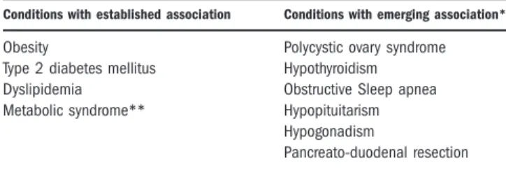
Table 4. Risk Factors Associated with NAFLD
Conditions with established association Conditions with emerging association*
Obesity Polycystic ovary syndrome Type 2 diabetes mellitus Hypothyroidism Dyslipidemia Obstructive Sleep apnea Metabolic syndrome** Hypopituitarism
Hypogonadism
Pancreato-duodenal resection *Few studies suggested that individuals with type1 diabetes have increased prevalence of hepatic steatosis based on liver imaging, but there is limited his-tological evidence.
**The Adult Treatment Panel III clinical definition of the metabolic syndrome requires the presence of three or more of the following features: (1) waist cir-cumference greater than 102 cm in men or greater than 88 cm in women; (2) triglyceride level 150 mg/dL or greater; (3) high-density lipoprotein (HDL) cho-lesterol level less than 40 mg/dL in men and less than 50 mg/dL in women; (4) systolic blood pressure 130 mm Hg or greater or diastolic pressure 85 mm Hg or greater; and (5) fasting plasma glucose level 110 mg/dL or greater.199
Screening of Family Members
Anecdotal experience and some published studies suggest familial clustering and heritability of NAFLD,60-63 but conclusive studies are lacking. In a retrospective cohort study, Willner et al. observed that 18% of patients with NASH have a similarly affected first degree relative.61 A small familial aggregation study observed that patients with NAFLD have a sig-nificantly higher number of first degree relatives with cirrhosis and a trend towards familial clustering of NAFLD or cryptogenic cirrhosis than matched healthy controls.62 In another familial aggregation study63 of overweight children with and without NAFLD, after adjusting for age, gender, race, and BMI, the heritabil-ity of MR-measured liver fat fraction was 0.386, and fatty liver was present in 18% of family members of children with NAFLD despite normal ALT and lack of obesity.
Recommendation
6. Systematic screening of family members for NAFLD is currently not recommended. (Strength – 1, Evidence - B)
Initial Evaluation
The diagnosis of NAFLD requires that (a) there is hepatic steatosis by imaging or histology, (b) there is no significant alcohol consumption, (c) there are no competing etiologies for hepatic steatosis, and (d) there are no co-existing causes for chronic liver disease.
Common alternative causes of hepatic steatosis are significant alcohol consumption, hepatitis C, medica-tions, parenteral nutrition, Wilson’s disease, and severe malnutrition (Table 2). When evaluating a patient with newly suspected NAFLD, it is important to exclude co-existing etiologies for chronic liver disease including hemochromatosis, autoimmune liver disease, chronic viral hepatitis, and Wilson’s disease.3 Mildly elevated serum ferritin is common in patients with NAFLD and it does not necessarily indicate increased iron stores.3,64 Elevated serum ferritin and transferrin saturation in patients with suspected NAFLD should lead to testing for genetic hemochromatosis. Mutations in the HFE gene occur with variable frequency in patients with NAFLD and their clinical significance is unclear.64 One should consider a liver biopsy to assess hepatic iron concentration and to exclude significant hepatic injury and fibrosis in a patient with suspected NAFLD with elevated serum ferritin and a homozy-gote or compound heterozyhomozy-gote C282Y mutation in the HFE gene.65 Elevated serum autoantibodies are common in patients with NAFLD and are generally considered to be an epiphenomenon.3 In a recently
published large study from the NASH Clinical Research Network, positive serum autoantibodies, defined as ANA > 1:160 or ASMA >1:40 were pres-ent in 21% of patipres-ents with well-phenotyped NAFLD and were not associated with more advanced histologic features.66
Recommendations
7. When evaluating a patient with suspected NAFLD, it is essential to exclude competing etiolo-gies for steatosis and co-existing common chronic liver disease. (Strength – 1, Evidence - A)
8. Persistently high serum ferritin and increased iron saturation, especially in the context of homozy-gote or heterozyhomozy-gote C282Y HFE mutations may warrant a liver biopsy. (Strength – 1, Evidence - B)
9. High serum titers of autoantibodies in associa-tion with other features suggestive of autoimmune liver disease (very high aminotransferases, high globu-lin) should prompt a more complete work-up for auto-immune liver disease. (Strength – 1, Evidence - B) Non-invasive assessment of steatohepatitis and advanced fibrosis in NAFLD
The natural history of NAFLD is fairly dichoto-mous – NAFL is generally benign whereas NASH can progress to cirrhosis, liver failure, and liver cancer. Existing dogma posits that liver biopsy is the most reliable approach for identifying the presence of steato-hepatitis and fibrosis in patients with NAFLD, but it is generally acknowledged that biopsy is limited by cost, sampling error, and procedure-related morbidity and mortality. Serum aminotransferase levels and imag-ing tests such as ultrasound, CT, and MR do not reli-ably assess steatohepatitis and fibrosis in patients with NAFLD. Therefore, there has been significant interest in developing clinical prediction rules and non-invasive biomarkers for identifying steatohepatitis in patients with NAFLD,7 but their detailed discussion is beyond the scope of this practice guideline.
The presence of metabolic syndrome is a strong pre-dictor for the presence of steatohepatitis in patients with NAFLD3,7,67-69 and may be used to best identify patients with persistently abnormal liver biochemistries who would benefit diagnostically and prognostically from a liver biopsy.
There has been intense interest in non-invasive methods to identify advanced fibrosis in patients with NAFLD7;these include the NAFLD Fibrosis Score70, Enhanced Liver Fibrosis (ELF) panel70 and transient elastography. The NAFLD Fibrosis Score is based on six readily available variables (age, BMI, hyperglyce-mia, platelet count, albumin, AST/ALT ratio) and it is
calculated using the published formula (http://naflds-core.com). In a meta-analysis of 13 studies consisting of 3,064 patients,7 NAFLD Fibrosis Score has an AUROC of 0.85 for predicting advanced fibrosis (i.e., bridging fibrosis or cirrhosis) and a score < 1.455 had 90% sensitivity and 60% specificity to exclude advanced fibrosis whereas a score > 0.676 had 67% sensitivity and 97% specificity to identify the presence of advanced fibrosis. The ELF panel consists of plasma levels of three matrix turnover proteins (hyaluronic acid, TIMP-1, and PIIINP) had an AUROC of 0.90 with 80% sensitivity and 90% specificity for detecting advanced fibrosis.71
Circulating levels of cytokeratin-18 (CK18) frag-ments have been investigated extensively as novel bio-markers for the presence of steatohepatitis in patients with NAFLD.7,72 Wieckowska et al., measured CK18 fragments in plasma that had been obtained from 44 consecutive patients with suspected NAFLD at the time of liver biopsy, and correlated the findings with hepatic immunohistochemistry data.70Plasma CK18 fragments were markedly increased in patients with NASH com-pared with patients with simple steatosis or normal biopsies (median 765.7 U/L versus 202.4 U/L or 215.5 U/L, respectively; P <0.001), and independently pre-dicted NASH (OR 1.95; 95% CI 1.18-3.22 for every 50 U/L increase). This observation was reproduced in sev-eral subsequent studies and a recent meta-analysis esti-mated that plasma CK18 levels have a sensitivity of 78%, specificity of 87%, and an area under the receiver operating curve (AUROC) of 0.82 (95% CI: 0.78-0.88) for steatohepatitis in patients with NAFLD.7 Although these are very encouraging results, currently this assay is not commercially available. Furthermore, as each study utilized a study-specific cut-off value, there is not an established cut-off value for identifying steatohepatitis.
Transient elastography, which measures liver stiffness non-invasively, has been successful in identifying advanced fibrosis in patients with hepatitis B and hepati-tis C. Although a recent meta-analysis showed high sen-sitivity and specificity for identifying fibrosis in NAFLD,7it has a high failure rate in individuals with a higher BMI. Furthermore, it is not commercially avail-able in the United States. Other imaging tools such as MR elastography, although commercially available in the United States, is rarely used in clinical practice.
A major limitation of these prediction models and biomarkers is that they have largely been investigated in cross-sectional studies and thus their utility in mon-itoring disease natural history, predicting outcomes or response to therapeutic intervention is unknown.
Recommendations
10. As the metabolic syndrome predicts the pres-ence of steatohepatitis in patients with NAFLD, its presence can be used to target patients for a liver bi-opsy. (Strength – 1, Evidence - B)
11. NAFLD Fibrosis Score is a clinically useful tool for identifying NAFLD patients with higher likelihood of having bridging fibrosis and/or cirrho-sis. (Strength – 1, Evidence - B)
12. Although serum/plasma CK18 is a promising biomarker for identifying steatohepatitis, it is pre-mature to recommend in routine clinical practice. (Strength – 1, Evidence - B)
When to obtain a liver biopsy in patients with NAFLD?
Liver biopsy remains the gold standard for charac-terizing liver histology in patients with NAFLD. How-ever, it is expensive and carries some morbidity and very rare mortality risk. Thus, it should be performed in those who would benefit the most from diagnostic, therapeutic guidance, and prognostic perspectives.
Recommendations
13. Liver biopsy should be considered in patients with NAFLD who are at increased risk to have stea-tohepatitis and advanced fibrosis. (Strength – 1, Evi-dence - B)
14. The presence of metabolic syndrome and the NAFLD Fibrosis Score may be used for identifying patients who are at risk for steatohepatitis and advanced fibrosis. (Strength – 1, Evidence - B)
15. Liver biopsy should be considered in patients with suspected NAFLD in whom competing etiologies for hepatic steatosis and co-existing chronic liver dis-eases cannot be excluded without a liver biopsy. (Strength – 1, Evidence - B)
MANAGEMENT OF PATIENTS WITH NAFLD
The management of patients with NAFLD consists of treating liver disease as well as the associated meta-bolic co-morbidities such as obesity, hyperlipidemia, insulin resistance and T2DM. As patients with NAFLD without steatohepatitis have excellent progno-sis from a liver standpoint, treatments aimed at improving liver disease should be limited to those with NASH.Lifestyle intervention
Many studies indicate that lifestyle modification may reduce aminotransferases and improve hepatic ste-atosis when measured either by ultrasound73-80 or MR
imaging and spectroscopy.81-94 In a meta-analysis of 15 early case series and clinical studies spanning between 1967 through 2000, most studies reported reductions in aminotransferases and hepatic steatosis by ultrasound across a broad spectrum of diets of dif-ferent caloric restriction intensities and macronutrient composition (low vs. high carbohydrate, low vs. high fat, saturated vs. unsaturated fat diets).95 However, these early studies were inconclusive as a result of being small, largely uncontrolled and few using histol-ogy as the primary endpoint. More recent uncon-trolled studies also showed an improvement in amino-transferases and hepatic steatosis on histology with lifestyle modification.96-98
Orlistat (an enteric lipase inhibitor) in conjunction with lifestyle modification was investigated in two randomized controlled trials. In the study by Ziegler-Sagi et al.,99 orlistat reportedly improved ALT and ste-atosis by US, but its effect on liver histology could not be evaluated because the majority of patients did not undergo a follow-up liver biopsy. However, in the study by Harrison et al.,100 orlistat did not improve body weight or liver histology.
The best evidence for weight loss as a means to improve liver histology in NASH comes from a trial that randomized 31 obese persons with NASH to in-tensive lifestyle changes (diet, behaviour modification and 200 minutes a week of moderate physical activity for 48 weeks) versus structured basic education alone.101The intensive arm had 9.3% weight loss (ver-sus 0.2% in the dietary counseling alone arm) and led to an improvement in steatosis, necrosis and inflam-mation, but not fibrosis. Importantly, participants with
7% weight loss had significant improvement in stea-tosis, lobular inflammation, ballooning, and NAFLD Activity Score (NAS).101 There was a similar pattern in the study by Harrison et al.,100 where participants who lost > 5% body weight improved steatosis, whereas individuals with 9% weight loss had signifi-cant improvement in steatosis, lobular inflammation, ballooning, and NAS.
A number of recent studies used MR spectroscopy to assess changes in hepatic fat in response to lifestyle modification. The results from these studies using a variety of interventions, either by diet alone81,83,84,89,92,93 or in combination with different exercise prescriptions,82,85-88,92,94 have consistently reported a significant reduction in liver fat by an aver-age of40% (ranging from 20% to 81%). The degree of hepatic fat reduction was proportional to the inten-sity of the lifestyle intervention and generally required a body weight loss between5 to 10%.82,88,92
The effect of exercise without dietary modification on hepatic steatosis was investigated in four studies using MR spectroscopy.102-105 Exercise programs con-sisted of 2-3 sessions a week of 30-60 minutes over a period of 6 to 12 weeks. In all but one study101 liver fat content diminished without a significant change in body weight.
Recommendations
16. Weight loss generally reduces hepatic steatosis, achieved either by hypocaloric diet alone or in con-junction with increased physical activity. (Strength – 1, Evidence - A)
17. Loss of at least 3-5% of body weight appears necessary to improve steatosis, but a greater weight loss (up to 10%) may be needed to improve necroin-flammation. (Strength – 1, Evidence - B)
18. Exercise alone in adults with NAFLD may reduce hepatic steatosis but its ability to improve other aspects of liver histology remains unknown. (Strength – 1, Evidence - B)
INSULIN SENSITIZING AGENTS
Metformin
Several studies investigated the effect of metformin on aminotransferases and liver histology in patients with NASH. Early small, open-label studies demon-strated a reduction in insulin resistance and amino-transferases106-108 but no significant improvement in liver histology.107,108 An open-label trial consisting of 110 patients with NASH received either metformin 2 grams/day (55 patients), vitamin E 800 IU/day (28 patients) or dietary-induced weight loss (27 patients) for 12 months.109 Aminotransferases improved more with metformin than with vitamin E or diet alone. However, there was only a modest improvement in he-patic steatosis and inflammation in the subset of 17 patients undergoing paired liver biopsies with metfor-min treatment. In a 48-week open-label study in 26 patients, metformin improved NASH activity in only 30% of patients, although interpretation of the study was confounded by a significant weight loss in the res-ponders (19% lost more than 10 kilograms).110 Hau-keland et al.112 reported a similar lack of efficacy in a larger (n¼48) randomized control trial (RCT) of met-formin vs. placebo with a similar dietary and exercise intervention in both groups. Other studies also failed to show major benefit for metformin on hepatic insu-lin sensitivity, aminotransferases111-116 or liver histol-ogy.111,113,116 A recent meta-analysis4 concluded that 6-12 months of metformin plus lifestyle intervention did not improve aminotransferases or liver histology,
compared with lifestyle intervention alone, independ-ently of metformin dose or the presence of diabetes.
Recommendation
19. Metformin has no significant effect on liver histology and is not recommended as a specific treat-ment for liver disease in adults with NASH. (Strength – 1, Evidence - A)
Thiazolidinediones
Several studies investigated the effect of pioglitazone and rosiglitazone on aminotransferases and liver histol-ogy in adults with NASH. In an early uncontrolled open-label study117 in 22 subjects with biopsy-proven NASH, rosiglitazone improved aminotransferases and hepatic steatosis, ballooning and inflammation scores, but not fibrosis. But in a subsequent RCT, Ratziu et al.118observed that rosiglitazone improved aminotrans-ferases and hepatic steatosis, but not necroinflamma-tion or fibrosis and its two-year open-label extension phase also showed similar results.119 Belfort et al.120 conducted a RCT of pioglitazone (45 mg/day) in patients with NASH who had impaired glucose toler-ance or T2DM. Although there was a significant weight gain (2.5 60.5 kg) with pioglitazone, it signif-icantly improved aminotransferases, steatosis, balloon-ing, and inflammation. The NAS improved with pio-glitazone in 73% compared to 24% of placebo-treated patients (P<0.001) and there was a trend towards improvement in fibrosis among patients randomized to pioglitazone (P¼0.08). Aithal et al.121 performed a RCT of lifestyle intervention with either pioglitazone 30 mg/day or placebo for 12 months in a total of 74 patients with NASH. While steatosis did not improve significantly compared to placebo, hepatocellular injury and fibrosis improved significantly.1210 The PIV-ENS122study is a large multicenter RCT that random-ized 247 non-diabetic patients with NASH to pioglita-zone (30 mg/day), vitamin E (800 IU/day), or placebo for 24 months. The primary endpoint was an improvement in NAS 2 points with at least 1 point improvement in hepatocellular ballooning and 1-point improvement in either the lobular inflammation or ste-atosis score, and no increase in the fibrosis score.122 It was achieved in 19% in the placebo group compared to 34% in the pioglitazone group (P¼0.04vs. placebo) and 43% in the vitamin E group (P¼0.001 vs. pla-cebo).122 Because this study consisted of two primary comparisons (pioglitazone vs. placebo and vitamin E vs. placebo), a P-value of 0.025 was considered to be significant a priori. Therefore, although there were his-tological benefits associated with pioglitazone, this study concluded that pioglitazone did not meet the
primary end point. However, resolution of NASH, a key secondary end point, was achieved in significantly higher number of patients receiving pioglitazone than receiving placebo (47% vs. 21%, P¼0.001).122 Of note, pioglitazone was associated with a 4.7 kg weight gain compared to placebo (P<0.001). Vitamin E and pioglitazone were well tolerated and there were no dif-ferences in other adverse events.
A recent meta-analysis4 that included 5 RCTs showed that pioglitazone significantly improved steato-sis (OR 4.05, 95% CI 2.58-6.35) and inflammation (OR 3.53, 95% CI 2.21-5.64), but not fibrosis (OR 1.40, 95% CI 0.87-2.24).
There has been considerable debate about the long-term safety of TZDs regarding cardiovascular disease, congestive heart failure (CHF), bladder cancer, and bone loss. In a recent meta-analysis123 of 19 trials enrolling a total of 16,390 patients with T2DM, pio-glitazone treatment was associated with a significant reduction (18%) in the primary outcome of death, myocardial infarction, or stroke (P¼0.005). However, there was also a higher rate of CHF with pioglitazone (2.3% vs. 1.8% in the control group, P¼0.002), so caution must be exercised when considering its use in patients with impaired myocardial function. Due to increased risk of coronary events, rosiglitazone is no longer marketed in Europe and its use is highly re-stricted in the United States.
Recommendation
20. Pioglitazone can be used to treat steatohepati-tis in patients with biopsy-proven NASH. However, it should be noted that majority of the patients who participated in clinical trials that investigated pio-glitazone for NASH were non-diabetic and that long term safety and efficacy of pioglitazone in patients with NASH is not established. (Strength – 1, Evi-dence - B)
Vitamin E
Oxidative stress is considered to be a key mecha-nism of hepatocellular injury and disease progression in subjects with NASH. Vitamin E is an anti-oxidant and has been investigated to treat NASH.124-128 Com-parison between these trials is difficult due to varying criteria for entry into the study, different doses of vita-min E and unclear formulations of vitavita-min E used which could affect its bioavailability, the additional use of other anti-oxidants or other drugs and limited histo-logic data to assess outcomes. Also, most studies were relatively under-powered and did not meet or publish CONSORT criteria for clinical trials. Despite these limitations, it can be summarized that (1) the use of
vitamin E is associated with a decrease in aminotrans-ferases in subjects with NASH, (2) studies where histo-logic endpoints were evaluated indicate that vitamin E causes improvement in steatosis, inflammation, and ballooning and resolution of steatohepatitis in adults with NASH, and (3) vitamin E has no effect on he-patic fibrosis. Although two meta-analyses8,129 failed to observe significant histological benefits with vitamin E in patients with NASH, these analyses were con-ducted before PIVENS122 and TONIC130 trials were published. In the largest clinical trial (PIVENS)122 reported to date, the pure form of rrr a-tocopherol was orally administered at a dose of 800 IU/day for 96 weeks. The primary endpoint as stated previously was achieved in a significantly greater number of par-ticipants receiving vitamin E compared to placebo (42% vs. 19%, P< 0.001, number needed to treat¼ 4.4).
One concern with vitamin E is the controversial issue of whether it increases all-cause mortality. Some meta-analyses have reported an increase in all-cause mortality with high dose vitamin E,131,132 but others failed to confirm such an association.133-135 A recently published RCT showed that vitamin E administered at a dose of 400 IU/day increased the risk of prostate cancer in relatively healthy men (absolute increase of 1.6 per 1000 person years of vitamin E use).136
Recommendation
21. Vitamin E (a-tocopherol) administered at daily dose of 800 IU/day improves liver histology in non-diabetic adults with biopsy-proven NASH and therefore it should be considered as a first-line pharmacotherapy for this patient population. (Strength -1, Quality - B)
22. Until further data supporting its effectiveness become available, vitamin E is not recommended to treat NASH in diabetic patients, NAFLD without liver biopsy, NASH cirrhosis, or cryptogenic cirrhosis (Strength - 1, Quality - C)
Ursodeoxycholic acid (UDCA), Omega-3 fatty acids, and Miscellaneous Agents
Several studies126,137-140 investigated UDCA (con-ventional and high doses) to improve aminotransfer-ases and steatosis in patients with NAFLD and liver histology in patients with NASH. All but one study139 have been proof-of-concept studies with small numbers of participants and/or surrogate endpoints. Notably, a single large multicenter RCT convincingly showed that UDCA offers no histological benefit over placebo in patients with NASH.139Omega-3 fatty acids, currently approved in the United States to treat
hypertriglyceri-demia, have been investigated to treat NAFLD both in animal models and in humans.141 A recent review by Masterton et al.,142 of published literature related to omega-3 fatty acids in NAFLD, found experimental evi-dence to support their use but the interpretation of human studies was limited by small sample size and methodological flaws. A large multicenter study of one omega-3 fatty acid (eicosapentanoic acid) to treat NASH is ongoing in the United States.. More than a dozen other miscellaneous agents have been investigated in small, proof-of-concept studies and their detailed evaluation is beyond the scope of this guideline.
Recommendations
23. UDCA is not recommended for the treatment of NAFLD or NASH. (Strength – 1, Quality – B)
24. It is premature to recommend omega-3 fatty acids for the specific treatment of NAFLD or NASH but they may be considered as the first line agents to treat hypertriglyceridemia in patients with NAFLD. (Strength – 1, Quality – B)
Bariatric Surgery
As the majority of patients undergoing bariatric sur-gery have associated fatty liver disease, there has been an interest in foregut bariatric surgery as a potential treatment option for NASH. There are no RCTs that evaluated any type of foregut bariatric surgical proce-dure to specifically treat NAFLD or NASH. However, there are several retrospective and prospective cohort studies that compared liver histology in the severely obese individuals before and after bariatric surgery. Unfortunately, in the majority of these studies, post-bypass liver biopsies were performed at varying inter-vals and only in selected patients undergoing surgical procedures such as hernia repair or adhesiolysis. One exception is the study by Mathurin et al.,143 that pro-spectively correlated clinical and metabolic data with liver histology before and 1 and 5 years after bariatric surgery in 381 adult patients with severe obesity. Gas-tric band, bilio-intestinal bypass, and gasGas-tric bypass were done in 56%, 23%, and 21%, respectively. Com-pared to baseline, there was a significant improvement in the prevalence and severity of steatosis and balloon-ing at 1 and 5 years followballoon-ing bariatric surgery. In patients with probable or definite NASH at baseline (n¼99), there was a significant improvement in steato-sis, ballooning, and NAS and resolution of probable or definite NASH at 1 and 5 years following bariatric surgery. Most histological benefits were evident at 1 year with no differences in liver histology between 1 and 5 years following bariatric surgery. Intriguingly, a minor but statistically significant increase in mean
fibrosis score was noted at 5 years after the bariatric surgery (from 0.276 0.55 at baseline to 0.36 6 0.59, P¼0.001). Despite this increase, at 5 years 96% of patients exhibited fibrosis score F1 and 0.5% had F3, indicating there is no clinically significant worsen-ing in fibrosis that can be attributed directly to the procedure. In the important subgroup of patients with probable or definite NASH at baseline, there was no worsening of fibrosis at 1 and 5 years, compared to baseline liver biopsy. As no patient in the study had F3 or F4 at baseline, the effect of bariatric surgery in those with advanced fibrosis and cirrhosis could not be evaluated.
Two meta-analyses144,145evaluated the effect of bari-atric surgery on the liver histology in patients with NAFLD. The meta-analysis by Mummadi et al.,144 showed that steatosis, steatohepatitis, and fibrosis appear to improve or completely resolve after bariatric surgery. However, a recently published Cochrane review145 concluded that lack of randomized clinical trials or quasi-randomized clinical studies prevents de-finitive assessment of benefits and harms of bariatric surgery as a therapeutic approach for patients with NASH.
Recommendations
25. Foregut bariatric surgery is not contraindi-cated in otherwise eligible obese individuals with NAFLD or NASH (but without established cirrho-sis). (Strength – 1, Quality – A)
26. The type, safety and efficacy of foregut bariat-ric surgery in otherwise eligible obese individuals with established cirrhosis due to NAFLD are not established. (Strength – 1, Quality – B)
27. It is premature to consider foregut bariatric surgery as an established option to specifically treat NASH (1B)
Alcohol use in patients with NAFLD and NASH
Heavy alcohol consumption is a risk factor for chronic liver disease and should be avoided by patients with NAFLD and NASH. The National Institute on Alcohol Abuse and Alcoholism (NIAAA) defines heavy or at-risk drinking as more than 4 drinks on any day or more than 14 drinks per week in men or more than 3 drinks on any day or 7 drinks per week in women.146 Several recent cross-sectional studies147-153 suggest a beneficial effect of light alcohol consumption (on average less than one drink per day) on the pres-ence (defined either biochemically or by imaging) and severity of NAFLD. There are no studies reporting the effect of ongoing alcohol consumption on disease se-verity or natural history of NAFLD or NASH. The
effects of light drinking on the cardiovascular system and cancer risks, if any, have not been investigated in individuals with NAFLD.
Recommendations
28. Patients with NAFLD should not consume heavy amounts of alcohol (Strength -1, Quality – B)
29. No recommendation can be made with regards to non-heavy consumption of alcohol by individuals with NAFLD. (Strength – 1, Quality – B)
Statin use in patients with NAFLD and NASH
Patients with NAFLD and NASH are at increased risk for cardiovascular disease and several studies have established cardiovascular disease as their most com-mon cause of death.6 Patients with NAFLD should be risk stratified for cardiovascular disease, and their car-diovascular risk factors should be managed accord-ingly.154 The treatment of dyslipidemia should be con-sidered in the overall frame work of cardiovascular risk reduction in patients with NAFLD.154
Statins are an important class of agents to treat dys-lipidemia, and yet there is continued reluctance to use statins in patients with suspected or established chronic liver disease, including NAFLD and NASH. Although elevated aminotransferases are not uncommon in patients receiving statins, serious liver injury from sta-tins is rarely seen in clinical practice. Over the last dec-ade, one RCT and several retrospective and prospective studies155-159 have established that (a) statins are safe in patients with liver disease and (b) there is no evi-dence that patients with chronic liver disease including at NAFLD and NASH are at higher risk for serious liver injury from statins than those without liver disease.
Several studies have suggested that statins may improve liver biochemistries and histology in patients with NASH.159-167 These studies consisted of small numbers of patients and have not been rigorously designed. A recent post-hoc analysis of the cardiovascu-lar outcomes study, GREACE,165 observed that statins significantly improve liver biochemistries and cardio-vascular outcomes in patients with elevated liver enzymes likely due to NAFLD. There are no RCTs with histological endpoints which investigated statins to treat NASH.
Recommendations
30. Given the lack of evidence to show that patients with NAFLD and NASH are at increased risk for serious drug-induced liver injury from sta-tins, statins can be used to treat dyslipidemia in patients with NAFLD and NASH. (Strength – 1, Quality – B)
31. Until RCTs with histological endpoints prove their efficacy, statins should not be used to specifi-cally treat NASH. (Strength – 1, Quality – B) NAFLD in patients with other chronic liver diseases
Because of the high prevalence of risk factors for NAFLD and NASH, it is not uncommon for patients with other chronic liver diseases to exhibit co-existing his-tological features of NAFLD.168 Coexistent hepatic stea-tosis is common in chronic hepatitis C (HCV) infection and is strongly associated with more advanced liver dis-ease.169-171 Another large study showed high prevalence of steatosis (40.5%) and steatohepatitis (15%) in patients with primary biliary cirrhosis (PBC),172although at least some of the steatosis and steatohepatitis in that study was suspected to be due to alcohol consumption. In clinical practice, it is not uncommon for obese and/or diabetic patients with autoimmune liver disease to exhibit steatosis and steatohepatitis in their liver biopsies.
Previous studies have shown that obesity, insulin re-sistance, and hepatic steatosis are associated with a lower response to pegylated interferon and ribavirin for the treatment of HCV.173-175 Obesity does not have a similar negative impact on the response to newer protease-inhibitor based anti-viral regimens,176-180 but the impact of insulin resistance and hepatic steatosis has not yet been investigated sufficiently. It is not known if the treatment of steatosis and steatohepatitis alters the natural history of other chronic liver diseases such as HCV and PBC. Furthermore, it is not known if agents such as vitamin E and pioglitazone are effective to treat steatosis and steatohepatitis when present in patients with other chronic liver diseases.
Recommendations
32. When steatosis and steatohepatitis are evident in patients with other types of chronic liver disease, it is important to assess for metabolic risk factors and alternate etiologies for hepatic steatosis. (Strength – 1, Quality – B)
33. In patients with other types of chronic liver diseases who have co-existing NAFLD and NASH, there are no data to support the use of vitamin E or pioglitazone to improve the liver disease. (Strength – 1, Quality – B)
Miscellaneous Recommendations Pertinent to Clinical Practice
34. Patients with NASH cirrhosis should be screened for gastroesophageal varices according to the AASLD/ACG practice guidelines.181(Strength – 1, Quality – B)
35. Patients with NASH cirrhosis should be consid-ered for HCC screening according to the AASLD/ACG practice guidelines.182(Strength – 1, Quality – B)
36. Current evidence does not support routinely repeating a liver biopsy in patients with NAFL or NASH. (Strength – 2, Quality – C)
Aspects of NAFLD Specific to Children and Adolescents
Recognition of NAFLD in children is essential to understanding the origin of disease in those likely to be most genetically or environmentally susceptible. Adults with onset of NAFLD in childhood may be most at risk for early or severe complications. Definition of NAFLD in childhood is the same as in adults. Children are reported with NAFLD as early as 2 years and with NASH-related cirrhosis as early as age 8.183,184
Prevalence and Risk Factors
Estimation of population prevalence in children presents difficulties for the same reasons detailed above in adults. Estimates vary based upon the type of test or imaging, the cut-points for detection, and the age, sex, race and ethnicity of the geographic region sampled. A school-based study of obese children in Minnesota, California, Texas and Louisiana, using abnormal serum ALT as a surrogate marker (>40U/L), found that 23% of 17-18 year olds had elevated unex-plained ALT.183 An autopsy study using the ‘‘gold standard’’ of liver histology examined 742 children between the ages of 2-19 y who died from unnatural causes. The estimated NAFLD prevalence was 9.6% when adjusted for age, gender, race and ethnicity.184 Multivariate analyses showed that obesity, older age (in adolescence), male gender, and Hispanic ethnicity are independent predictors of fatty liver prevalence.
Natural History of NAFLD in Children
A single retrospective single center report has been published on the natural history of NAFLD in 66 chil-dren.185Only 5 had serial biopsies, obtained for unspeci-fied reasons over varying intervals, averaging 41 months between biopsies. Of these 5 children, 4 had progression of fibrosis. Four of the 5 underwent liver transplantation and 2 died of cirrhosis. Clearly, more robust prospective data are needed on larger number of children to better understand the natural history of NAFLD in children.
Screening for NAFLD in Children
NAFLD is under-diagnosed in children due to lack of recognition, screening or appreciation of associated complications by health care providers. One study
showed that less than a third of obese children were screened for NAFLD at clinic visits.186 Children may not be recognized as obese at visits and age-appropriate norms for body mass index may go unacknowledged. Abdominal adiposity may mask detection of hepato-megaly by palpation during physician examination. As in adults, children with features of metabolic syndrome such as obesity, hypertension, insulin resistance and dyslipidemia63 are at higher risk for NAFLD and par-ticular histopathological features of NAFLD correlate with components of metabolic syndrome.187 Thus, identification of children at risk for NAFLD could occur in general health provider settings as well as in specialty clinics for nutrition, gastroenterology, hepato-logy, endocrinology and bariatric surgery. Children may also exhibit NAFLD incidentally discovered while undergoing imaging, but there are no studies evaluat-ing how to proceed with children identified in this fashion. Recently, the summary report of an expert committee suggested biannual screening for liver dis-ease with serum ALT and AST starting at age 10 years in obese children and those with BMI of 85th to 94th percentile with other risk factors.188
Penetrance of NAFLD has been demonstrated in family members of children with NAFLD.63 The like-lihood of first, second and third degree relatives exhib-iting abnormally high fat fractions (by MRI estima-tion) relative to body mass index is much more highly correlated in those related to a child with NAFLD than to those who are related to an age, gender and BMI-matched child without NAFLD.
Diagnosis in children
Given the relatively early onset, caregivers must give additional consideration to the possibility of mono-genic disorders that present as fatty liver disease in very young children. Considerations include inborn errors of fatty acid or carnitine metabolism, peroxiso-mal disorders, lysosoperoxiso-mal storage disorders, Wilson’s disease, and cystic fibrosis.189 However, as in adults, positive serum autoantibodies are present in a signifi-cant population of children with biopsy-proven NAFLD and on some occasion liver biopsy is required to discriminate between autoimmune hepatitis and NAFLD.63 Obviously, the confounding factor of alco-holism is much less common in children and standard questionnaires for quantifying alcohol intake are usu-ally unnecessary.
Recommendations
37. Children with fatty liver who are very young or not overweight should be tested for monogenic causes of chronic liver disease such as fatty acid
oxi-dation defects, lysosomal storage diseases and peroxi-somal disorders, in addition to those causes consid-ered for adults. (Strength – 2, Quality – C)
38. Low serum titers of autoantibodies are often present in children with NAFLD, but higher titers, particularly in association with higher serum amino-transferases and high globulin should prompt a liver biopsy to evaluate for possible autoimmune hepati-tis. (Strength – 2, Quality – B)
39. Due to a paucity of evidence, a formal recom-mendation cannot be made with regards to screening for NAFLD in overweight and obese children despite a recent expert committee recommendation for bian-nual screening for liver disease with liver enzyme measurements in this population. (Strength –1, Quality – B).
When to obtain a liver biopsy for suspected pediatric NAFLD?
The decision to perform a liver biopsy in a child to confirm the diagnosis of NAFLD must be weighed against the risks associated with biopsy and the likeli-hood that the result will impact management. In chil-dren with an uncertain diagnosis, biopsy may rule out potential drug hepatotoxicity or lack of clarity due to presence of serum autoantibodies. When there is an interest in grading or staging NAFLD, instead of sub-mitting all children with NAFLD to a liver biopsy it would be optimal to identify those children who are more likely to have NASH. The paucity of natural his-tory data confounds the decision to biopsy since altera-tion of long-term outcomes with treatment based on severity of histology at baseline is unknown.
As in adults, development of noninvasive bio-markers or imaging to identify those at risk for more rapid progression or severe disease onset is desirable. Particularly, accurate markers of cellular injury and fi-brosis are needed. Two studies suggested that ELF score can be used to accurately predict fibrosis in chil-dren with NAFLD, but both studies consisted of rela-tively small numbers of children and fewer with advanced fibrosis.190,191 There is reported benefit in predicting fibrosis stage in pediatric patients, with a AUROC of 0.92, although only 9 of the 76 subjects studied had fibrosis stage 3 or more.190 Validation of the serum CK18 levels to evaluate NASH needs to be undertaken in children with NAFLD.
Recommendations
40. Liver biopsy in children with suspected NAFLD should be performed in those where the di-agnosis is unclear, where there is possibility of multi-ple diagnoses, or before starting therapy with
potentially hepatotoxic medications. (Strength – 1, Quality – B)
41. A liver biopsy to establish a diagnosis of NASH should be obtained prior to starting children on pharmacologic therapy for NASH. (Strength – 2, Quality – C)
NAFLD Histology in Children
Histopathology of children with NAFLD can differ from that found in adults.192 As in adults, children can present with pronounced features of hepatocellular injury, lobular inflammation, and peri-sinusoidal fibro-sis, but there is a unique pattern of unclear significance also recognized in children. This pattern is typified by marked macrovesicular hepatocellular steatosis, portal inflammation and portal fibrosis in the absence of ballooning.192,194
Recommendation:
42. Pathologists interpreting pediatric NAFLD biopsies should recognize the unique pattern fre-quently found in children to not misidentify pediat-ric NAFLD. (Strength – 1, Quality – B)
Treatment in Children
Recommendations for pediatric treatment options are limited by a small number of randomized clinical trials and insufficient information on natural history to assess risk-benefit. The overall goal is to improve a child’s quality of life and reduce longer term cardiovas-cular and liver morbidity and mortality. Given that early-onset likely indicates higher likelihood of later complications, attempts should be made to identify children who will benefit from intervention.
Lifestyle modification
Since most pediatric NAFLD patients are obese, addressing their obesity is the first step. An open label study 195 in 84 Italian children with biopsy-proven NAFLD showed that >20% body weight reduction over 12 months resulted in improvement in serum ALT and steatosis by ultrasonography in most children with NAFLD. Reportedly, 94% of the 70 enrolled subjects were able to achieve this weight loss goal using caloric restriction and exercise advice. Since liver biop-sies were not performed at the end of the study, the effect of lifestyle intervention on liver histology could not be determined. In another study, Nobili et al.196 randomized 53 children with biopsy-proven NAFLD to lifestyle modification plus antioxidant therapy or lifestyle modification and placebo. Antioxidant therapy did not improve liver histology, but children in both groups showed significant improvement in steatosis,
inflammation, ballooning, and the NAS. Although there are no randomized controlled trials of intensive lifestyle modification compared to standard-of-care advice, these two studies indicate that lifestyle modifi-cation is beneficial in children with NAFLD.
No information exists on recommending any partic-ular type of diet or exercise. Further studies are needed to assess the efficacy of specific diets. Recommenda-tions for overweight pediatric NAFLD patients should include consultation with a registered dietitian to assess quality of diet and measurement of caloric intake, adoption of American Heart Association dietary strat-egies, and regular aerobic exercise progressing in diffi-culty as fitness allows.197,198 Enlisting other willing family members to adopt diet and exercise goals may aid compliance.
Pharmacotherapy
As in adults, clinical trials for pediatric NAFLD generally targeted insulin resistance or oxidative stress. Open-label proof-of-concept studies have utilized changes in serum ALT or liver brightness on ultra-sound as endpoints.189 Agents evaluated thus far include metformin, vitamin E, ursodeoxycholic acid and delayed-release cysteamine.189 Recently, a large multicenter RCT using change in histology as a sec-ondary endpoint was published.130 This study, called TONIC, compared the efficacy of vitamin E or met-formin to placebo in 8-17 year olds with NAFLD.130 Although the primary outcome of sustained reduction of ALT was not different among the 3 groups, there were statistically significant improvements in NAS and resolution of NASH (P<0.006) with vitamin E treat-ment compared to placebo over 96 weeks.130 In this study, metformin administered at 500 mg twice daily dose had no effect on liver biochemistries or liver histology.
Recommendations
43. Intensive lifestyle modification improves ami-notransferases and liver histology in children with NAFLD and thus should be the first line of treat-ment. (Strength – 2, Quality – B)
44. Metformin at 500 mg twice daily offers no benefit to children with NAFLD and thus should not be prescribed. The effect of metformin administered at a higher dose is not known. (Strength – 1, Qual-ity – B)
45. Vitamin E 800 IU/day (RRR a–tocopherol) offers histological benefits to children with biopsy-proven NASH or borderline NASH but confirmatory studies are needed before its use can be
recommended in clinical practice (Strength – 1,Quality – B)
Acknowledgment: This practice guideline was devel-oped in collaboration with the AASLD Practice Guide-lines Committee, and was approved by the ACG Practice Parameters Committee, and the AGA Institute Clinical Practice and Quality Management Commit-tee. Raphael B. Merriman, MD, MRCPI and Benja-min L. Shneider, MD served as primary reviewers for the AASLD Practice Guidelines Committee. Dr. Mer-riman declared no relevant conflicts of interest. Dr. Shneider serves as a scientific consultant with Bristol-Myers Squibb and the advisory board for Ikaria. Exter-nal review was provided by Jean P. Molleston, MD and Stephen A. Harrison, MD. Dr. Molleston received research support from Schering-Plough and Roche. Dr. Harrison serves as a consultant to Amylin Pharma-ceuticals and has received research support from Rotta-pharm and Mochida.
References
1. Eddy DM. A manual for assessing health practices and designing practice guidelines. Philadelphia. American College of Physicians. 1996;1-126. 2. Guyatt GH, Oxman AD, Vist GE, Kunz R, Falck-Ytter Y,
Alonso-Coello P, Schunemann HJ. GRADE: an emerging consensus on rating quality of evidence and strength of recommendations. BMJ 2008;336: 924-926.
3. Vuppalanchi R, Chalasani N. Nonalcoholic fatty liver disease and non-alcoholic steatohepatitis: selected practical issues in their manage-ment. Hepatology 2009;49:306-317.
4. Vernon G, Baranova A, Younossi ZM. Systematic review: the epidemi-ology and natural history of alcoholic fatty liver disease and non-alcoholic steatohepatitis in adults. Aliment Pharmacol Ther 2011;34: 274-285.
5. Neuschwander-Tetri BA. Hepatic lipotoxicity and the pathogenesis of nonalcoholic steatohepatitis: the central role of nontriglyceride fatty acid metabolites. HEPATOLOGY2010;52:774-788.
6. Targher G, Day CP, Bonora E. Risk of cardiovascular disease in patients with nonalcoholic fatty liver disease. N Engl J Med 2010; 363:1341-1350.
7. G, Gambino R, Cassader M, Pagano G. Meta-analysis: Natural his-tory of non-alcoholic fatty liver disease (NAFLD) and diagnostic accu-racy of non-invasive tests for liver disease severity. Annals of Medicine 2011;43(8):617-49.
8. Musso G, Gambino R, Cassader M, Pagano G. A meta-analysis of randomized trials for the treatment of nonalcoholic fatty liver disease. HEPATOLOGY2010;52:79-104.
9. Suzuki A, Angulo P, Lymp J, St Sauver J, Muto A, Okada T, Lindor K.. Chronological development of elevated aminotransferases in a nonalcoholic population. HEPATOLOGY. 2005;41(1):64-71.
10. Hamaguchi M, Kojima T, Takeda N, et al. The metabolic syndrome as a predictor of non-alcoholic fatty liver disease. Ann Intern Med 2005;143(10):722-8.
11. Whalley S, Puvanachandra P, Desai A, Kennedy H. HEPATOLOGY out-patient service provision in secondary care: a study of liver disease incidence and resource costs. Clin Med. 2007;7(2):119-24.
12. Lee JY, Kim KM, Lee SG, Yu E, Lim YS, Lee HC, Chung YH, Lee YS, Suh DJ. Prevalence and risk factors of non-alcoholic fatty liver disease in potential living liver donors in Korea: a review of 589 con-secutive liver biopsies in a single center. J Hepatol. 2007;47(2): 239-44.
13. Marcos A, Fischer RA, Ham JM, Olzinski AT, Shiffman ML, Sanyal AJ, Luketic VA, Sterling RK, Olbrisch ME, Posner MP. Transplanta-tion 2000;69:2410-2415.
14. Williams CD, Stenger J, Asike MI, Torres DM, Shaw J, Contreras M, Landt CL, Harrison SA. Prevalence of nonalcoholic fatty liver disease and nonalcoholic steatohepatitis among a largely middle-aged popula-tion utilizing ultrasound and liver biopsy: a prospective study. Gastro-enterology 2011;140:124-131.
15. Browning JD, Szczepaniak LS, Dobbins R, Nuremberg P, Horton JD, Cohen JC, Grundy SM, Hobbs HH. Prevalence of hepatic steatosis in an urban population in the United States: impact of ethnicity. H EPA-TOLOGY. 2004;40:1387-95.
16. Boza C, Riquelme A, Iba~nez L, Duarte I, Norero E, Viviani P, Soza A,Fernandez JI, Raddatz A, Guzman S, Arrese M. Predictors of nonal-coholic steatohepatitis (NASH) in obese patients undergoing gastric bypass. Obes Surg. 2005;15:1148-53.
17. Haentjens P, Massaad D, Reynaert H, Peeters E, Van Meerhaeghe A, Vinken S, Poppe K, Velkeniers B. Identifying non-alcoholic fatty liver disease among asymptomatic overweight and obese individuals by clini-cal and biochemiclini-cal characteristics. Acta Clin Belg. 2009;64:483-93. 18. Machado M, Marques-Vidal P, Cortez-Pinto H. Hepatic histology in
obese patients undergoing bariatric surgery. J Hepatol. 2006;45:600-6. 19. Colicchio P, Tarantino G, del Genio F, Sorrentino P, Saldalamacchia G,Finelli C, Conca P, Contaldo F, Pasanisi F. Non-alcoholic fatty liver disease in young adult severely obese non-diabetic patients in South Italy. Ann Nutr Metab. 2005;49:289-95.
20. Beymer C, Kowdley KV, Larson A, Edmonson P, Dellinger EP, Flum DR. Prevalence and predictors of asymptomatic liver disease in patients undergoing gastric bypass surgery. Arch Surg. 2003;138: 1240-4.
21. Leite NC, Salles GF, Araujo AL, Villela-Nogueira CA, Cardoso CR. Prevalence and associated factors of non-alcoholic fatty liver disease in patients with type-2 diabetes mellitus. Liver Int. 2009;29:113-9. 22. Prashanth M, Ganesh HK, Vima MV, John M, Bandgar T, Joshi SR,
Shah SR, Rathi PM, Joshi AS, Thakkar H, Menon PS, Shah NS. Prevalence of nonalcoholic fatty liver disease in patients with type 2 diabetes mellitus. J Assoc Physicians India. 2009;57:205-10. 23. Assy N, Kaita K, Mymin D, Levy C, Rosser B, Minuk G. Fatty
infil-tration of liver in hyperlipidemic patients. Dig Dis Sci 2000;45: 1929-1934.
24. Li H, Wang YJ, Tan K, Zeng L, Liu L, Liu FJ, Zhou TY, Chen EQ, Tang H. Prevalence and risk factors of fatty liver disease in Chengdu, Southwest China. Hepatobiliary Pancreat Dis Int. 2009;8(4):377-82. 25. Amarapurkar D, Kamani P, Patel N, Gupte P, Kumar P, Agal S, Baijal
R, Lala S, Chaudhary D, Deshpande A. Prevalence of non-alcoholic fatty liver disease: population based study. Ann Hepatol. 2007;6(3):161-26. Park SH, Jeon WK, Kim SH, Kim HJ, Park DI, Cho YK, Sung IK, Sohn CI, Keum DK, Kim BI. Prevalence and risk factors of non-alco-holic fatty liver disease among Korean adults. J Gastroenterol Hepatol. 2006;21(1 Pt 1):138-43.
27. Frith J, Day CP, Henderson E, Burt AD, Newton JL. Non-alcoholic fatty liver disease in older people. Gerontology. 2009;55(6):607-13. 28. Chen CH, Huang MH, Yang JC, Nien CK, Yang CC, Yeh YH, Yueh
SK. Prevalence and etiology of elevated serum alanine aminotransfer-ase level in an adult population in Taiwan. J Gastroenterol Hepatol. 2007;22:1482-9.
29. Ong JP, Pitts A, Younossi ZM. Increased overall mortality and liver-related mortality in non-alcoholic fatty liver disease. J Hepatol. 2008; 49:608-12.
30. Hashimoto E, Yatsuji S, Kaneda H, Yoshioka Y, Taniai M, Tokushige K, Shiratori K. The characteristics and natural history of Japanese patients with nonalcoholic fatty liver disease. Hepatol Res. 2005; 33(2):72-6.
31. Adams LA, Lymp JF, St Sauver J, Sanderson SO, Lindor KD, Feld-stein A, Angulo P. The natural history of nonalcoholic fatty liver dis-ease: a population-based cohort study. Gastroenterology. 2005 l;129: 113-21.