HPV DNA Testing:
Technical and Programmatic
Issues for Cervical Cancer
Prevention in Low-Resource
Settings
Curt Malloy, M.S.
Jacqueline Sherris, Ph.D.
Cristina Herdman
Preparation of this paper was supported by the Bill & Melinda Gates Foundation through the Alliance for Cervical Cancer Prevention
Table of Contents
I. Introduction 1
II. HPV and Cervical Cancer 3
III. Techniques for Detecting HPV 5
Signal-amplified techniques Target-amplified techniques Non-amplified techniques
Potential for faster, cheaper methods
IV. Programmatic Considerations 16
Clinical challenges
Sociocultural considerations
V. Conclusion 21
List of Acronyms 22
Online Information Related to HPV Diagnostics 23
I. Introduction
Cervical cancer kills approximately 230,000 women annually, with the vast majority of deaths occurring in developing countries. Worldwide, cervical
carcinoma is the fifth most common cancer-related cause of death among women; in the developing world, it is the leading cause of cancer death in women.1 The global distribution of cervical cancer varies, with Africa, Asia, and Latin America bearing a substantial burden of this disease.1,2
Cervical cancer prevention programs in both developed and developing nations generally have relied on cytological testing using the Papanicolaou (Pap) smear test. Pap smears require that a health care provider obtain a sample of cells from the uterine cervix of each woman screened. Trained technicians then examine the specimen for cellular changes (dysplasia) known to precede the development of cervical cancer. Such screening programs can be expensive, prone to error, and logistically difficult to implement—particularly in developing countries.
Furthermore, cultural barriers and client perceptions may limit women’s participation in cytology-based screening programs.
Research worldwide has clearly shown that virtually all cervical cancer is caused by human papillomavirus (HPV) infection. HPV is a sexually transmitted infection (STI) that is very common among young men and women in many parts of the world. In most women, the infection becomes undetectable relatively quickly. Women persistently infected with certain carcinogenic types are at increased risk of developing severe dysplasia* and cervical cancer.
The direct detection of HPV in cervical specimens may offer an alternative or complement to population-based cytological screening. Recent studies have demonstrated that HPV test results are more sensitive (although they are less specific) than Pap smears in detecting high-grade dysplasia in older women.3,4 In most scenarios women with positive HPV tests still have Pap tests or a diagnostic procedure to provide cytological or histological confirmation of their disease. Several technologies exist for the molecular detection of HPV infection. Most of these technologies, while sensitive and specific, are too costly and cumbersome to incorporate into large-scale screening programs. Recently, molecular diagnostic
*Classified as high-grade squamous intraepithelial lesions (HSIL) according to the Bethesda Classification system, or cervical intraepithelial neoplasia (CIN) II–III or carcinoma in situ (CIS) according to the CIN classification system.
kits have been tested in developed and developing countries through several large research projects incorporating HPV screening into cervical dysplasia detection programs.3–5 Currently, two commercially available molecular diagnostic kits have been approved by the United States Food and Drug Administration (US FDA) and field-tested in the developing world. Both kits, the Hybrid Capture Tube test and the Hybrid Capture II test (HC II), are produced by the Digene Corporation (Gaithersburg, Maryland).
Molecular-based HPV diagnostics remain largely untested in broad screening programs, however. Research and pilot projects are under way in several
countries to clarify the diagnostic, clinical, and programmatic implications of HPV screening for cervical cancer prevention.
This document provides a summary of HPV as it relates to cervical cancer and examines the molecular technologies currently available for detecting HPV. Particular emphasis is placed on assessing the suitability of these diagnostic tests for use in cervical cancer prevention programs in developing countries.
II. HPV and Cervical Cancer
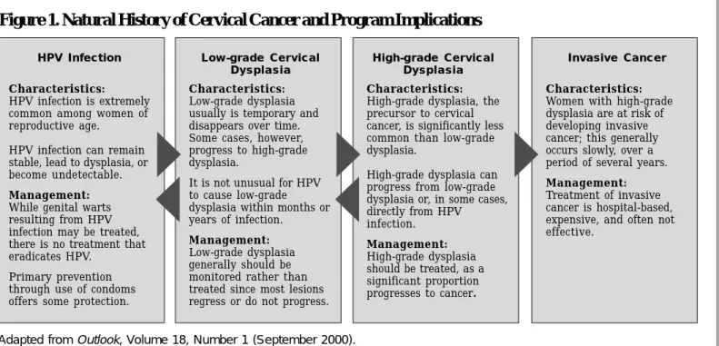
HPV, one of the most common STIs,6 has been established as the main cause of cervical cancer. While HPV infection is considered a necessary precursor of both cervical cancer and associated precancerous lesions,7,8 it is not a sufficient cause, as the majority of women with the infection will not progress to cancer (see Figure 1). It is estimated that less than five percent of women infected with HPV who receive no health intervention ultimately develop cervical cancer.9
Infections with HPV are common in both men and women. Some estimates indicate that more than 50 percent of sexually active adults in the United States have experienced an infection with one or more HPV viral types.10 One study, using prevalence data among Finnish women, estimated a woman’s lifetime risk of HPV infection at 75 percent.11
Although no effective treatment is available for HPV, the infection is transient in the majority of cases. One study suggests that in up to 70 percent of those initially diagnosed, the infection is undetectable within two years.12 A significant
proportion of women with HPV infection develops low-grade cervical lesions. Most of these low-grade lesions regress spontaneously; one study suggests that
approximately 15 percent progress to high-grade cervical lesions within two years. High-grade cervical lesions have a strong malignant potential; one study found that about one-third of high-grade lesions progress to cancer within ten years.13
.6
HPV Infection
Characteristics:
HPV infection is extremely common among women of reproductive age.
HPV infection can remain stable, lead to dysplasia, or become undetectable.
Management:
While genital warts resulting from HPV infection may be treated, there is no treatment that eradicates HPV.
Primary prevention through use of condoms offers some protection.
Low-grade Cervical Dysplasia
Characteristics:
Low-grade dysplasia usually is temporary and disappears over time. Some cases, however, progress to high-grade dysplasia.
It is not unusual for HPV to cause low-grade dysplasia within months or years of infection.
Management:
Low-grade dysplasia generally should be monitored rather than treated since most lesions regress or do not progress.
Characteristics:
High-grade dysplasia, the precursor to cervical cancer, is significantly less common than low-grade dysplasia.
High-grade dysplasia can progress from low-grade dysplasia or, in some cases, directly from HPV infection. Management: High-grade dysplasia should be treated, as a significant proportion progresses to cancer. Invasive Cancer Characteristics:
Women with high-grade dysplasia are at risk of developing invasive cancer; this generally occurs slowly, over a period of several years.
Management:
Treatment of invasive cancer is hospital-based, expensive, and often not effective.
Figure 1. Natural History of Cervical Cancer and Program Implications
High-grade Cervical Dysplasia
There are over 50 viral types of HPV that infect the genital tract. Only a small portion appears to cause most cervical neoplasias and cancers. Of the 15 to 20 types associated with cervical cancer, a worldwide study determined that four types—16, 18, 31, and 45—accounted for 80 percent of cervical cancers.6 HPV 16 was detected in half of cervical cancers. Other types identified as high-risk are 33, 35, 39, 51, 52, 56, 58, 59, and 68. Virtually all genital warts are caused by types 6 and 11.
A woman’s HPV status and information on the viral type(s) involved in infection have important clinical significance. At the same time, other factors, including age, persistence of detectable infection, and parity likely influence clinical outcome.
HPV infection is most common in younger women. Although prevalence varies among regions, it reaches a peak of at least 20 percent among women between the ages of 20 and 24 years of age, with a subsequent decline to approximately three percent among women over 30 years of age.14,15 HPV infection among younger women generally appears to be self-limiting; in most, the infection becomes undetectable within a year or two.
Despite a decline in HPV prevalence among women over the age of 25 years, the risk for cervical cancer increases until women reach their fifties, probably due to risks associated with persistent HPV infection. Women over 30 years of age who are infected with high-risk HPV may be up to 116 times more likely to develop severe dysplasia than similar, uninfected women.14
Other determinants of the progression of HPV infection to cervical cancer relate to a woman’s immune status. Women who are co-infected with the human immunodeficiency virus (HIV), or those with an immune system compromised as a result of malnutrition, pregnancy, or immunosuppressive chemotherapy, appear to be at increased risk of progression.16–18
III. Techniques for Detecting HPV
HPV cannot be cultured reliably in a laboratory setting; therefore, HPV diagnostics rely on molecular technologies that detect HPV DNA in cervical/vaginal samples.
Molecular techniques can be broadly divided into those technologies that are not amplified, such as nucleic acid probe tests, and those that utilize amplification, such as polymerase chain reaction (PCR). Amplification techniques can be further divided into three separate categories: (1) target amplification, in which the assay amplifies the target nucleic acids (for example, PCR); (2) signal amplification, in which the signal generated from each probe is increased by a compound-probe or branched-probe technology; and (3) probe amplification, in which the probe molecule itself is amplified (for example, ligase chain reaction). To date, target and signal amplification techniques, in addition to non-amplified techniques, have been applied to the detection of HPV.
Because there are many HPV types with differing oncogenic potential, diagnostic tests must not only detect HPV DNA, they also must determine the type(s) present in each specimen. Several diagnostic technologies also are able to
estimate a specimen’s viral load, which approximates the average number of viral genomes in the cervical cells sampled. It has not been determined whether such semi-quantitative data yield clinically relevant information. Some studies have found no association between viral load and disease progression;19–22 research is ongoing to further define this issue.
Signal-amplified techniques for detecting HPV include hybrid capture and
branched DNA approaches. The most widely used technique is the hybrid capture technology as described below.
Hybrid Capture Technology. Hybrid capture technology (HC), developed by the Digene Corporation, detects nucleic acid targets directly, using signal
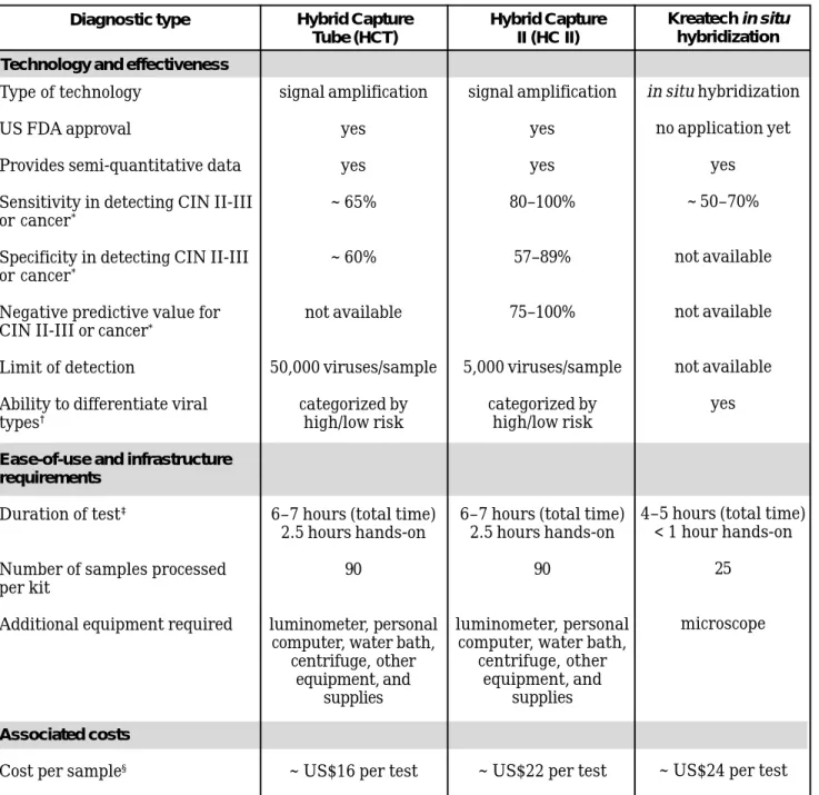
amplification to provide sensitivity comparable to target amplification methods. Digene has developed two products for the detection of HPV: the first-generation Hybrid Capture Tube (HCT) test and the more recent Hybrid Capture II (HCII) assay (see Table 1). Both assays detect “high-risk” HPV types. The HCT test detects the following high-risk types (as initially defined by Digene and supported by epidemiological studies): 16, 18, 31, 33, 35, 45, 51, 52, and 56. HCT was granted US FDA approval in May 1995. In March 1999, the US FDA approved Digene’s second-generation HPV detection kit (HC II). Four additional viral types were
Signal-amplified techniques
added to the high-risk category in the HC II test: 39, 58, 59, and 68. The level of detection of the second-generation HC II is rated at 5,000 viral copies per sample, or one picogram of HPV DNA per sample (in contrast to HCT, which detects 10 picograms).
To perform the HC assay (Figure 2), cervical or vaginal clinical specimens— collected through self-sampling or obtained by a health care provider during a pelvic examination—are combined with an extraction buffer to release and
denature the target HPV DNA. The released target DNA then combines with specific RNA probes to create RNA-DNA hybrids, which are captured onto a solid phase by an antibody specific for the hybrids. These captured RNA-DNA hybrids are then tagged with antibody reagents linked to alkaline phosphatase. A chemiluminescent substrate then produces light that is measured on a luminometer in relative light units (RLUs). The amount of light generated is proportional to the amount of target DNA in the original specimen.23 Processing the HCII test requires the following
equipment:
! a refrigerator to store unused test kits at 2°C (if stored for longer than 2 weeks)*;
! a centrifuge, required for sample preparation; ! a vortexor (a device that agitates the sample and is
required for sample preparation);
! a waterbath to incubate the samples at 65°C; ! a shaker, required during the hybrid capture
process;
! a luminometer integrated with a personal computer (sold by the Digene Corporation); and
! miscellaneous laboratory supplies, including microtubes, gloves, parafilm®, pipettors, and microtips.
* Note: Once samples are taken, they can be stored at room temperature for two weeks, at 4°C for one additional week, and at -20°C for up to three months.
DNA and Hybridization
Deoxyribonucleic acid (DNA) is composed of two complementary strands of
nucleotides, which are attached and unattached fairly easily—generally by heating or by adding specific chemicals. The term denaturation refers to the process of separating the two
complementary strands. Hybridization occurs when a new strand of DNA links with a complementary strand. The new strand can be a genetic probe designed to link with a specific, targeted sequence. Most diagnostic techniques employ these probes as a means of detecting the presence of a strand of DNA that matches with a given pathogen—in this case certain types of HPV.
In many settings, these requirements are burdensome enough to limit the use of Digene’s technology to regional hospitals and laboratories.
The Digene Corporation has announced it is seeking US FDA approval for a four-in-one-sample assay, which will concurrently test for HPV, Neisseria gonorrhoeae, Chlamydia trachomatis, and herpes simplex virus using the procedure described above.24 The cost implications of this test are unknown at this time.
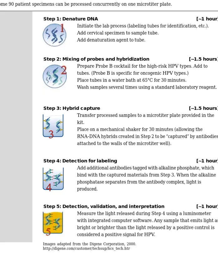
Figure 2: Laboratory process for Digene’s HC II
Digene’s HC II test involves a five-part process (described below). The hybrid capture assay takes an estimated 6 to 7 hours, approximately 2.5 hours of which require the direct attention of a technician. Some 90 patient specimens can be processed concurrently on one microtiter plate.
Step 1: Denature DNA [~1 hour]
! Initiate the lab process (labeling tubes for identification, etc.). ! Add cervical specimen to sample tube.
! Add denaturation agent to tube.
Step 2: Mixing of probes and hybridization [~1.5 hours]
! Prepare Probe B cocktail for the high-risk HPV types. Add to tubes. (Probe B is specific for oncogenic HPV types.)
! Place tubes in a water bath at 65°C for 30 minutes.
! Wash samples several times using a standard laboratory reagent.
Step 3: Hybrid capture [~1.5 hours]
! Transfer processed samples to a microtiter plate provided in the kit.
! Place on a mechanical shaker for 30 minutes (allowing the RNA-DNA hybrids created in Step 2 to be “captured” by antibodies attached to the walls of the microtiter well).
Step 4: Detection for labeling [~1 hour]
! Add additional antibodies tagged with alkaline phosphate, which bind with the captured materials from Step 3. When the alkaline phosphatase separates from the antibody complex, light is produced.
Step 5: Detection, validation, and interpretation [~1 hour]
! Measure the light released during Step 4 using a luminometer with integrated computer software. Any sample that emits light as bright or brighter than the light released by a positive control is considered a positive signal for HPV.
Images adapted from the Digene Corporation, 2000. http://digene.com/customer/techsup/hcs_tech.htr
Research using Digene’s hybrid capture assays. Numerous articles have reported on experience with Digene’s two HPV tests. Several have shown good agreement when comparing the findings of HC II/HCT tests with the PCR tests (see page 10).25,26 In the past few years, a number of studies also have produced promising results regarding the sensitivity and specificity of HC II/HCT tests for identifying dysplasia and cervical cancer.
In general, studies have found that Digene’s hybrid capture tests for HPV DNA have the potential for use in screening for cervical dysplasia, in conjunction with other screening or diagnostic tests (most frequently Pap smears and/or
colposcopy) or alone. Of particular interest for low-resource settings, study results suggest that the tests may have potential as a primary screen among older
women (age 30 to 35 or older). In this age group, the sensitivity range of the HC II test in detecting high-grade dysplasia has been recorded at about 80 to 90 percent (higher than for Pap smears), and specificity has ranged from 57 to 89 percent.27 It is important to note, however, that most studies have not controlled for
verification bias. Sensitivities therefore may be inflated.
Two recent studies carried out in developing country settings are of particular interest. One, conducted by Schiffman et al.,3 evaluated use of the HC II test to identify women likely to have high-grade dysplasia and cancer among more than 9,000 sexually active women age 18 and older in Guanacaste Province, Costa Rica. The study found that HPV testing detected 88.4 percent of high-grade cervical lesions and cancers, with a specificity of 89 percent. When results were calculated by age tertile (18 to 30, 31 to 40, and 41 and older), specificity was highest
(94 percent) for older women. Overall, HPV DNA testing using the HC II test was more sensitive than conventional Pap testing (88.4 versus 77.7 percent) for detection of high-grade lesions and cancers, but less specific (89 versus
94 percent). The authors concluded that, while more data are needed on various factors that can impact the results of HPV testing (such as analytic cutoffs and geographic variation in HPV types), the method clearly has “come of age” technically, and should be increasingly useful in cervical screening efforts. The other study, conducted by Wright et al.,4 evaluated use of the HC II test (using self-collected and clinician-obtained samples) to identify women likely to have HSIL and cancer among more than 1,400 previously unscreened black South African women aged 35 to 65. The sensitivity of HPV DNA testing of self-collected vaginal samples was 66.1 percent for detection of high-grade lesions and cancer; the false-positive rate was 17.1 percent. The sensitivity of HPV DNA testing of
clinician-collected samples was 83.9 percent; the false-positive rate was
15.5 percent. In comparison, the sensitivity of conventional Pap smear (with low-grade LSIL and higher cytologic abnormalities classified as positive) was
60.7 percent, with a false-positive rate of 3.2 percent. In summary, HPV DNA testing using self-collected vaginal samples was less specific but as sensitive in this study as conventional Pap testing for detecting high-grade cervical disease in women age 35 or older. The authors concluded that HPV DNA testing using self-collected vaginal samples holds promise for simplifying cervical cancer screening in many settings, although low specificity remains a problem. They discussed the need to assess two-stage screening protocols (for example HPV testing followed by a Pap smear or visual inspection) to improve specificity.
DNA target amplification is a laboratory-based procedure that duplicates DNA fragments from a target sequence of a gene, thus providing concentrated samples of a specific genetic sequence. Several types of DNA target amplification
technologies exist; however, PCR is the most commonly employed in HPV detection. PCR is a standard laboratory procedure, which can be adapted for the detection and typing of HPV.
PCR frequently is used as a diagnostic tool in epidemiologic investigations of HPV, but the associated costs and technology requirements often are inappropriate for large screening programs. PCR technology requires a cyclic, three-step reaction. A master mix of
chemicals, including all the necessary catalysts and enzymes required for the process, is placed into a reaction vessel, and the biochemical process is then automated in a thermocycler. With each thermal cycle, the amount of target DNA theoretically doubles. For example, within approximately one hour, 20 cycles can amplify the target a million-fold.
Target-amplified techniques
The Polymerase Chain Reaction (PCR)
PCR is a chemical reaction that results in the synthesis of a large number of target DNA strands, in this case HPV DNA. The reaction cycles through the following three-step process:
Step 1: Step 1:Step 1:
Step 1:Step 1: Once cervical cells are obtained and prepared, the sample is heated to approximately 95°C. This heating denatures the DNA, resulting in two single strands.
Step 2: Step 2:Step 2:
Step 2:Step 2: The temperature of the reaction is decreased to 55°C and HPV-specific primers bind to target DNA.
Step 3: Step 3:Step 3:
Step 3:Step 3: The reaction is then heated to 72°C and an enzyme present in the mixture catalyzes the extension of the probes and promotes the creation of two complementary strands of the targeted HPV DNA sequence.
The inherent strength of the PCR-based methodology lies in its capacity to detect very small amounts of HPV DNA. At the same time, strict laboratory procedures and controls are critical in reducing contamination-related false-positive findings.28 Non-amplified technologies rely on a variety of laboratory-based molecular
diagnostic methodologies, and include Southern blot hybridization, dot blot hybridization, and in situ hybridization, among others. Several factors limit the incorporation of non-amplified techniques into large-scale screening programs, particularly in developing countries: in addition to their relatively low sensitivity, they generally are time-consuming, require trained technicians, and demand an array of laboratory reagents and equipment.
Southern blot and dot blot hybridizations. Southern blot hybridization is an important research tool and has been the technique generally used to classify newly identified viral types.29 However, the method is restricted by a time-consuming and labor-intensive process,30 as well as a reliance on radiolabeled probes (isotopes). Commercial kits are not marketed for this method; rather, the process is entirely laboratory-based, using existing reagents and well-established methodologies. This intensive identification method therefore requires a
sophisticated laboratory, with access to appropriate reagents and personnel skilled in advanced laboratory techniques.
In Southern blot hybridization, the HPV genome is extracted from a specimen and the DNA chain is broken using enzymes. The product is integrated into a gel, which is subjected to an electric current—a process referred to as gel
electrophoresis. The electrophoretic process separates the DNA based on the size of each fragment. The DNA fragments separated by this method are transferred to a nitrocellulose membrane and hybridized with cloned HPV genomic probes. These probes are then labeled, often using radioisotopes. The detection of the labeled DNA hybrids indicates HPV is present in a given sample.
Dot blot hybridization employs a simpler laboratory method than Southern blot but is rarely used due to its low sensitivity.29,31 This method is similar to Southern blot hybridization, except that it does not include electrophoresis. Dot blot
hybridization techniques formerly were used in two commercial HPV detection kits, Virapap and Viratype. These kits, previously available through the Digene Corporation, are no longer marketed.32
Non-amplified techniques
In situ hybridization. In situ hybridization applies hybridization techniques to
the intact DNA of infected cells. The process is performed on a microscope slide, and can be applied to archived cervical smears. Numerous studies have used in situ hybridization methods in HPV DNA detection and typing, at times in tandem with PCR.33–35
After preprocessing the sample to remove cellular components other than the targeted DNA, the specimen is heated to denature the DNA. Probes are
introduced and bind to the HPV DNA, if present. Antibodies are then introduced that attach to these probes. Enzymes are added, which stain the sample if HPV DNA is present. Identification of positive or negative findings is accomplished visually, using a microscope.
Kreatech Biotechnology B.V. (Amsterdam, The Netherlands) has developed a detection kit for use in the detection of HPV DNA using the in situ hybridization techniques (see Table 1). This product currently is available as a research tool.36 The Kreatech kit contains all slides, coverslips, and reagents necessary for performing the diagnostic procedure, and takes four to five hours to process. HPV probes are marketed for use together or separately; viral types included are 1, 2, 6, 11, 16, 18, 31, and 33. Only one peer-reviewed publication to date has
specifically examined the use of Kreatech’s in situ hybridization kits for the detection of HPV;37 no direct comparison was undertaken between this method and other technologies.
*Sensitivity, specificity, and negative predictive values were adapted from Cuzick et al. Only one study was cited as having a sensitivity/specificity for HCT38; six studies reported on use of HC II.27 The negative predictive value of the HC II (ranging from
75 to 100 percent) indicates that the majority of those testing negative using HC II are classified correctly.
†Viral types identified:
HCT: 16, 18, 31, 33, 35, 45, 51, 52, 56
HC II: 16, 18, 31, 33, 35, 39, 45, 51, 52, 56, 58, 59, 68 (highlighted italics indicate additional types new to HC II) Kreatech in situ hybridization: 1, 2, 6, 11, 16, 18, 31, 33
‡Testing times were obtained from the manufacturers.
Hybrid Capture Tube (HCT) Hybrid Capture II (HC II) Kreatech in situ hybridization Diagnostic type
Technology and effectiveness
Table 1. Overview of HPV diagnostic technologies available in kit form, as of November 2000
signal amplification yes yes ~ 65% ~ 60% not available 50,000 viruses/sample categorized by high/low risk
6–7 hours (total time) 2.5 hours hands-on
90
luminometer, personal computer, water bath,
centrifuge, other equipment, and
supplies
~ US$16 per test
signal amplification yes yes 80–100% 57–89% 75–100% 5,000 viruses/sample categorized by high/low risk
6–7 hours (total time) 2.5 hours hands-on
90
luminometer, personal computer, water bath,
centrifuge, other equipment, and
supplies
~ US$22 per test
in situ hybridization no application yet yes ~ 50–70% not available not available not available yes
4–5 hours (total time) < 1 hour hands-on
25
microscope
~ US$24 per test Type of technology
US FDA approval
Provides semi-quantitative data Sensitivity in detecting CIN II-III or cancer*
Specificity in detecting CIN II-III or cancer*
Negative predictive value for CIN II-III or cancer*
Limit of detection
Ability to differentiate viral types†
Ease-of-use and infrastructure requirements
Duration of test‡
Number of samples processed per kit
Additional equipment required
Associated costs Cost per sample§
To date, only Digene Corporation’s two hybrid capture assays have received US FDA approval and have undergone testing in a range of environments.3–5 While such studies demonstrate the potential for the success of HPV screening programs, the technical, financial, and logistic requirements of the tests are beyond the capacity of many developing-country health programs.
There are two major restrictions that may impede the use of current HPV testing technologies in screening programs: (1) the methods and instrumentation
required to process cervical specimens, and (2) the technical equipment requirements for interpreting test results.
Regarding the first restriction, it is possible that instrumentation and processing of samples may be simplified by developments in isothermal amplification of the target HPV DNA. As implied by its name, isothermal amplification does not require the constant change in temperature generally needed to separate, hybridize, and amplify target DNA. Instead, enzymes catalyze the formation of “daughter” strands identical to the targeted section of DNA. These enzymes are effective in all three phases of amplification, which can proceed at room
temperature. This technology is still in development.39
The second restriction ultimately may be addressed through adaptations of
current approvals, and/or development of simple, rapid, endpoint read-out systems using a lateral flow (immunochromatographic) technology.
Potential for faster, cheaper methods
Micro-arrays (“DNA chips”)
Recent developments in combining molecular probes with silicon-based chips ultimately may lead to quick, relatively inexpensive diagnostics. This technology requires the use of silicon chips created through well-established techniques similar to those applied in computer microchip fabrication. The surface of the chip often is covered with a fine layer of gold, and molecular probes are attached to the chip’s surface. Such an arrangement is referred to as a micro-array. Each of the molecular probes differs slightly in the target DNA they are designed to hybridize.
A product to detect and type HPV DNA using micro-arrays may incorporate a diagnostic process similar to the following:
! A sample of cervical cells is prepared for micro-array analysis and added to the surface of the chip.
! Primers on the micro-array bind to the target sequences of the HPV DNA.
! An instrument measures binding of DNA targets on the micro-array. ! If binding is detected, the sample would be considered positive for HPV. While micro-array technology holds promise for the detection of a broad range of infectious diseases (as well as the early detection of some cancers), such technologies are in the research phase and thus currently unavailable. The creation of specialized DNA primers is still evolving, as is an efficient fabrication process for creating functional silicon-based micro-arrays. There is no public information indicating that products using this technology are in development.
IV. Programmatic Considerations
Cervical cancer is now primarily a disease of marginalized women, particularly women in developing countries.2 These countries often lack access to resources necessary to implement successful cervical cancer prevention programs. In countries with limited funds for disease prevention, cancer screening programs compete with other pressing health needs.40 Currently, the costs related to HPV testing place the technology out of the reach of developing countries. While comprehensive cost analysis has yet to be undertaken, new technical and
programmatic approaches to use of HPV tests might someday reduce the costs of such programs and improve access to cervical cancer prevention services.
Women in developing countries who are at highest risk of developing cervical cancer often have the most restricted access to information and services they need to protect themselves. A broad array of clinical, social, and cultural issues influence where, how, or even whether cervical cancer screening services are provided. Programs must carefully evaluate provision of services that have the greatest impact on the largest number of women. Clearly, a thorough
understanding of the complexities of HPV testing must be carefully considered before HPV tests are incorporated into widespread programs.
All cervical cancer screening approaches face common challenges to successful implementation. Cytology, visual inspection with acetic acid (VIA), HPV testing, and other screening approaches face barriers such as logistic and infrastructure inadequacies, cost concerns, poor follow-up, and sociocultural constraints. Health care planners who are considering implementing any type of cervical cancer screening must develop clinical protocols that are responsive to the natural history of cervical disease, the diagnostic characteristics of the screening technology, disease prevalence in the target population, and women’s’ and providers’ needs and concerns. For example, some of the topics protocols need to address include:
• age of the target population to be screened;
• screening coverage and frequency;
• use of single- or dual-screening methodology (for example, HPV testing in conjunction with cytology or VIA); and
• conditions for which outpatient or inpatient treatment is recommended. To be effective, any cervical cancer screening program must be offered within an array of education and treatment services (including palliative care) that will
Clinical challenges
reach the majority of targeted women. Screening should be initiated only when adequate diagnostic and treatment services are readily accessible to women who need them.
Effective HPV testing programs must develop clinical protocols based upon a clear understanding of the natural history of HPV. Findings from recent research indicate that HPV infection in older women is strongly associated with risk of HSIL and cancer, that more persistent infections are likely to be higher risk HPV types, and that a woman with a compromised immune system is more likely to have persistent HPV and to experience a more rapid onset and course of illness. HPV infection usually is transient among young women. Potential approaches for use of HPV testing are outlined in the box on the following page.
Any program considering HPV testing must recognize that the currently available commercial test is associated with special technical barriers. Its reliance upon technology and infrastructure support could have multiple repercussions and will influence decisions regarding whether to implement HPV testing at the local or district level. Clinics operating at the local level generally can obtain good cervical specimens for processing (see box, page 20), but they likely will lack the
infrastructure and capacity needed to run the test on these specimens. Likewise, program planners will need to weigh the advantages of integrating HPV testing into an existing array of health services as opposed to introducing HPV testing as an independent health intervention.
All cervical cancer screening programs share challenges in their efforts to educate women about disease prevention and to persuade women to accept screening. In order to implement screening strategies that are acceptable, accessible, and effective, program planners must understand and respond to the cultural and social factors that influence women’s health-seeking behavior.40,41 Programmatic understanding of current levels of knowledge about and perceptions of cervical cancer and its prevention are key to developing effective interventions. Women’s perception of and attitude toward their own cancer risks, their acceptance of the specimen-collection method, and community attitudes toward programs targeting reproductive health and STIs will shape the provision of screening information and services. These factors have been cited as problematic in existing cytological screening programs, and most likely will persist in programs utilizing HPV testing.40
Sociocultural considerations
Potential Protocols for HPV DNA Testing
How will HPV DNA testing ultimately be used in cervical cancer prevention programs? The answer to this question is not yet clear, but researchers have suggested various possible scenarios. The most common are using HPV DNA testing (1) as a means of triage for women whose Pap smears indicate ASCUS (atypical squamous cells of undetermined significance)—those who test positive for high-risk HPV would be followed more aggressively than those who test negative; (2) as a means of surveillance of women treated for high-grade dysplasia or microinvasive cancer (those who test positive for high-risk HPV types would be monitored more closely than those who test negative); and (3) as a primary screen for high-grade dysplasia in older women (women age 35 or older who test positive for high-risk HPV would then undergo diagnosis via colposcopy or another visualization technique).27 The cost effectiveness of these various approaches has not been clearly delineated but a randomized trial in Canada has shown promising results for HPV testing for women with low-grade dysplasia (ASCUS, LSIL).42
In the United Kingdom, for example, the National Health Services’ Screening Committee has recommended that patients with borderline smear results (ASCUS) undergo an HPV test to help determine the appropriate clinical management strategy. The panel recognized the potential for use of HPV testing as primary screening methodology, but expressed a need for more research results prior to making recommendations for this type of use.27 In the United States, providers are increasingly integrating HPV testing into some aspect of their cervical cancer screening protocols, in some cases because of demand from advocacy groups and clients based on research and news reports about the test.
Recent data supporting use of HPV testing as a primary screening strategy in older women have raised interest in this approach (see pages 9–10).3,4 In some settings, for example, a potential HPV testing protocol involves testing all women over age 35 at least once as a means to directly detect high-risk HPV infection, and indirectly detect high-grade dysplasia. Women who tested positive for high-risk HPV would then be examined visually for signs of dysplasia. Those with high-grade dysplasia would be treated; those without would be examined again in six months. All women who tested negative for HPV would be
considered at very low risk for cervical cancer, and would not be retested for at least three to five years, and possibly longer.
Educating women about cervical cancer prevention as it relates to HPV testing poses unique challenges to health providers. Cervical cancer and its association with sexual activity already carries stigma in many parts of the world. Women may be even more reluctant to seek HPV screening if it is viewed as a test for an STI. A desire to avoid unnecessary patient concern may leave providers grappling with difficult decisions regarding the level of detail they should use in describing the test to patients. Some providers may even choose to not explain the linkage between cervical cancer and HPV; or may wish to de-emphasize the fact that HPV is an STI. These issues must be carefully weighed when considering initiating HPV DNA testing.
HPV screening also presents unique challenges with regard to addressing the concerns of women who receive a positive test result. HPV infection is very common in many regions, yet there currently is no cure or treatment for HPV, prevention is very difficult, and there is no perfect way to predict which
individuals with HPV infection will later develop cancer. Finding out that one has an STI is cause for alarm for most people. Those who are diagnosed with an STI often want to know how they got it, how it can be cured or treated, and how to prevent transmission of the infection to their partner. HPV testing will raise additional client questions and fears regarding whether testing positive means a client has cancer and what her risk of developing cancer is. Health care providers interested in utilizing HPV DNA testing should carefully consider how to address the information needs of women in light of these facts.
Much remains to be learned about the diagnostic, clinical, and social implications of HPV testing. The technology itself is evolving as researchers explore ways to broaden its applicability in diverse settings. Molecular diagnostic techniques for detecting HPV in cervical cells ultimately may provide a feasible alternative to large-scale cytological screening programs, if such techniques are shown to be cost-effective, feasible to implement, and broadly acceptable.
Self-Collection of Samples for HPV Testing
A critical consideration when using HPV diagnostics in developing country settings is the method of obtaining specimens. HPV diagnostics have used various methods for specimen collection, including self-collected samples (using urine, vulvar swabs, vaginal tampons, or vaginal swabs) and samples collected by health care providers (including genital swab, vaginal lavage, cervical swab, and cervical brush specimens). Promising studies indicate that samples for use in HPV DNA detection can be successfully obtained by women themselves. This has important implications for programs in countries where cultural and program barriers may limit acceptance of standard gynecologic procedures.
Several studies have evaluated the efficacy of different specimen collection techniques.3,–5,43–47 These studies suggest that self-collection methods can be a fairly reliable method of collecting samples for HPV testing. Most studies used the Digene Corporation’s Hybrid Capture Tube or HC II technology, one used PCR technology,46 and one used both.47
Studies conducted in the United States measured the agreement between physician-directed swab and self-collection using vaginal tampons. The concordance rate (the percentage of women who tested negative on both samples or positive on both samples) was 80 percent.44 A study measuring the correlation between cervical lavage specimens and vaginal tampon specimens for detection yielded nearly identical results. A Canadian study compared self-collected vaginal, vulvar, and urine samples with physician-collected cervical samples and then examined all of the women with colposcopy.47 The sensitivity of physician-collected samples for detecting HSIL was 98.3 percent, while the sensitivity of self-collected samples was 86.2 percent for vaginal swabs, 62.1 percent for vulvar swabs, and
44.8 percent for urine specimens. Self-sampling methods were found to be acceptable to women, with urine being the most preferred specimen.
A recent study conducted in South Africa demonstrated the potential for self-collected vaginal swabs in HPV testing.4 The study found that self-collected vaginal samples for use with the HC II test were 66 percent sensitive in detection of HSIL or cancer. Similar results were found in an analogous study employing self-collection methods conducted in rural Uganda.5
The studies noted that self-collection of cervical/vaginal specimens will require education on the part of health providers and clients to ensure that the technique is explained well by the provider and
understood by the client. The most common approach requires a woman to insert a swab into the vagina, rotate it several times, and then place it into a transport tube.
If further studies confirm self-collected specimens are equivalent to those obtained by health care providers, self-collection could offer many advantages. Women would not have to spend time traveling to a health center and waiting to see a health care provider (although women would have to arrange for delivery of the sample to the health center), and they would not require a gynecological
examination. The potential advantages support exploration of self-collection in low-resource settings or in areas with limited access to health care.
V. Conclusion
HPV has clearly been shown to be the cause of most cervical cancers. Given that, interest is growing worldwide in the potential for use of HPV diagnostics in cervical cancer prevention programs, both as an adjunct to cytological screening and in primary screening for cervical dysplasia. While there are a variety of laboratory-based approaches for detecting HPV in cervical samples, there currently is only one company—the Digene Corporation—providing an FDA-approved commercial kit for detecting high-risk HPV types.
In developed countries, HPV testing using the Digene Hybrid Capture II kit already is being incorporated into some screening programs, generally as an adjunct to existing cytological screening. In developing countries, the currently available tests likely are too expensive and technologically demanding for widespread use, even though research in several countries has demonstrated their potential for identifying high-grade dysplasia in older women (age 35 and older).
Developing country programs interested in incorporating HPV DNA testing into cervical cancer prevention activities may have to wait for the development of HPV tests that are less expensive than existing options, and easier to use in non-laboratory settings. At the same time, screening programs based on HPV testing must be carefully designed to ensure that the tests are used in a way to maximize their effectiveness in detecting high-grade dysplasia in women at highest risk of developing cervical cancer.
List of Acronyms
AIDS – Acquired immunodeficiency syndrome
ASCUS – Atypical squamous cells of undetermined significance
CIN – Cervical intraepithelial neoplasia
DNA – Deoxyribonucleic acid
ELISA – Enzyme-linked immunosorbent assay
HC II – Hybrid Capture II test
HCT – Hybrid Capture Tube test
HIV – Human immunodeficiency virus
HPV – Human papillomavirus
HSIL - High-grade squamous intraepithelial lesion
LSIL - Low-grade squamous intraepithelial lesion
PCR – Polymerase chain reaction
RNA – Ribonucleic acid
SIL – Squamous intraepithelial lesion
STI – Sexually transmitted infection
US FDA – United States Food and Drug Administration
Online Information Related to HPV Diagnostics
Digene Corporation:
http://www.digene.com/index.html
Overview of Digene Corporation’s hybrid capture technology: http://www.digene.com/customer/techsup/hcs_tech.htm Kreatech Biotechnology B.V.:
http://www.kreatech.com
http://www.zymed.com/ [Kreatech’s North American distributor]
National Health Service: Health Technology Assessment [“A systematic review of the role of human papillomavirus testing within a cervical screening programme” by Cuzick J, et al.]:
http://www.hta.nhsweb.nhs.uk/
Overview of laboratory news, techniques, and protocols [not necessarily specific for HPV]:
http://www.labnews.com/index.shtml
http://opbs.okstate.edu/~melcher/MG/MGW1/MG1225.html http://opbs.okstate.edu/~melcher/MG/MGW4/MG423.html#bottom http://biology.neehow.org/wonderful/protocols/rnaprot97.html Medscape articles on molecular amplification and HPV [requires free
subscription]:
http://www.medscape.com/SCP/IIM/1999/v16.n02/m6004.sand/ m6004.sand.html
http://www.medscape.com/medscape/ID/journal/1999/ISSTDR/ISSTDR.08/ ISSTDR.08.html
Overviews of the polymerase chain reaction (PCR) [not specific to HPV]: http://www.accessexcellence.org/AB/IE/PCR_Xeroxing_DNA.html http://www-biology.ucsd.edu/others/dsmith/classes/pcr.html Information on DNA chips [not specific to HPV]:
http://www.devicelink.com/ivdt/archive/98/09/009.html http://sciborg.uwaterloo.ca/~bpbobech/welcome.html
References
1. Parkin M. Personal communication, IARC (July 2000).
2. PATH (Program for Appropriate Technology in Health). Preventing cervical cancer in low-resource settings. Outlook 18(1):1–8 (2000). (Available online at http://www.path.org/Files/eol18_1.pdf).
3. Schiffman M, Herrero R, Hildesheim A, et al. HPV DNA testing in cervical cancer screening: results from women in a high-risk province of Costa Rica Journal of the American Medical Association 283:87–93 (2000).
4. Wright TC Jr, Denny L, Kuhn L, et al. HPV DNA testing of self-collected vaginal samples compared with cytologic screening to detect cervical cancer. Journal of the American Medical Association 283(1):81–86 (2000).
5. Serwadda D, Wawer MJ, Shah KV, et al. Use of a hybrid capture assay of self-collected vaginal swabs in rural Uganda for detection of human
papillomavirus. Journal of Infectious Diseases 180(4):1316–1319 (1999). 6. Bosch FX, Manos MM, Muñoz N, et al. Prevalence of human papillomavirus
in cervical cancer: a worldwide perspective. International biological study on cervical cancer (IBSCC) study group. Journal of the National Cancer Institute 87(11):796–802 (1995).
7. Franco EL, Rohan TE, Villa LL. Epidemiologic evidence and human papillomavirus infection as a necessary cause of cervical cancer. Journal of the National Cancer Institute 91(6):506–511 (1999).
8. Walboomers JM, Jacobs MV, Manos MM, et al. Human papillomavirus is a necessary cause of invasive cervical cancer worldwide. Journal of Pathology 189(1):12–19 (1999).
9. Cox JT. Clinical role of HPV DNA testing. Obstetrics and Gynecology Clinics of North America 23(4):811–851 (1996).
10. Koutsky L. Epidemiology of genital human papillomavirus infection. American Journal of Medicine 102(5A):3–8 (1997).
11. Syrjänen K, Hakama M, Saarikoski S, et al. Prevalence, incidence, and estimated life-time risk of cervical human papillomavirus infections in a non-selected Finnish female population. Sexually Transmitted Diseases 17(1):15– 19 (1990).
12. Moscicki AB, Shiboski S, Broering J, et al. The natural history of human papillomavirus infection as measured by repeated DNA testing in adolescent and young women. Journal of Pediatrics 132(2):277–284 (1998).
13. Ostor AG. Natural history of cervical intraepithelial neoplasia: a critical review. International Journal of Gynecological Pathology 12(2):186–92 (1993). 14. Meijer CJ, Helmerhorst TJ, Rozendaal L, et al. HPV typing and testing in
gynaecological pathology: has the time come? Histopathology 33(1):83–86 (1998).
15. Sellors JW, Mahoney JB, Kaczorowski J, et al. Prevalence and predictors of human papillomavirus infection in women in Ontario, Canada. Canadian Medical Association Journal 163(5):503–508 (2000).
16. Luque AE, Demeter LM, Reichman RC. Association of human papillomavirus infection and disease with magnitude of human immunodeficiency virus type 1 (HIV-1) RNA plasma level among women with HIV-1 infection. Journal of Infectious Diseases 179(6):1405-1409 (1999).
17. Temmerman M, Tyndall MW, Kidula N, et al. Risk factors for human papillomavirus and cervical precancerous lesions, and the role of concurrent HIV-1 infection. International Journal of Gynaecology and Obstetrics
65(2):171–181 (1999).
18. Sedlacek TV. Advances in the diagnosis and treatment of human
papillomavirus infections. Clinical Obstetrics and Gynecology 42(2):206–220 (1999).
19. Nindl I, Lotz B, Kuhne-Heid R, et al. Distribution of 14 high risk HPV types in cervical intraepithelial neoplasia detected by a non-radioactive general primer PCR mediated enzyme immunoassay. Journal of Clinical Pathology 52(1):17–22 (1999).
20. Clavel C, Bory JP, Rihet S, et al. Comparative analysis of human papillomavirus detection by hybrid capture assay and routine cytologic screening to detect high-grade cervical lesions. International Journal of Cancer 75(4):525–528 (1998).
21. Clavel C, Masure M, Putaud I, et al. Hybrid capture II, a new sensitive test for human papillomavirus detection. Comparison with hybrid capture I and PCR results in cervical lesions. Journal of Clinical Pathology 51(10):737–740 (1998).
22. Clavel C, Masure M, Bory JP, et al. Hybrid Capture II-based human papillomavirus detection, a sensitive test to detect in routine high-grade cervical lesions: A preliminary study on 1518 women. British Journal of Cancer 80(9):1306–1311 (1999).
23. Digene, Inc. Hybrid capture technology. http://www.digene.com/customer/ techsup/hcs_tech.htm. (Accessed January 2000).
24. Robert Lilley. Personal communication, Digene, Inc. (February 2000). 25. Cope JU, Hildesheim A, Schiffman MH, et al. Comparison of the hybrid
capture tube test and PCR for detection of human papillomavirus DNA in cervical specimens. Journal of Clinical Microbiology 35(9):2262–2265 (1997). 26. Nindl I, Greinke C, Zahm DM, et al. Human papillomavirus distribution in
cervical tissues of different morphology as determined by hybrid capture assay and PCR. International Journal of Gynecological Pathology 16(3):197– 204 (1997).
27. Cuzick J, Sasieni P, Davies P, et al. A systematic review of the role of human papillomavirus testing within a cervical screening programme. Health Technology Assessment 3(14):1–204 (1999).
28. Victor T, Jordaan A, du Toit R, et al. Laboratory experience and guidelines for avoiding false positive polymerase chain reaction results. European Journal of Clinical Chemistry and Clinical Biochemistry 31(8):531–535 (1993).
29. Lörincz AT. Molecular methods for the detection of human papillomavirus infection. Obstetrics and Gynecology Clinics of North America 23(3):707–730 (1996).
30. Swygart C. Human papillomavirus: Disease and laboratory diagnosis. British Journal of Biomedical Science 54(4):299–303 (1997).
31. Duggan MA, Inoue M, McGregor SE, et al. A paired comparison of dot blot hybridization and PCR amplification for HPV testing of cervical scrapes interpreted as CIN 1. European Journal of Gynaecological Oncology 15(3):178–187 (1994).
32. Digene, Inc. HPV physician q & a. http://www.digene.com/resources/hpvinfo/ phys_faq.htm. (Accessed January 6, 2000).
33. Adams V, Moll C, Schmid M, et al. Detection and typing of human papillomavirus in biopsy and cytological specimens by polymerase chain reaction and restriction enzyme analysis: a method suitable for
semiautomation. Journal of Medical Virology 48(2):161–170 (1996).
34. Sato S, Maruta J, Konno R, et al. In situ detection of HPV in a cervical smear with in situ hybridization [letter]. Acta Cytologica 42(6):1483–1485 (1998). 35. Huang CC, Qiu JT, Kashima ML, et al. Generation of type-specific probes for
the detection of single-copy human papillomavirus by a novel in situ hybridization method. Modern Pathology 11(10):971–977 (1998).
36. Personal communication, Kreatech Biotechnology B.V. (January 18, 2000). 37. Autillo-Touati A, Joannes M, d’Ercole C, et al. HPV typing by in situ
hybridization on cervical cytologic smears with ASCUS. Acta Cytologica 42(3):631–638 (1998).
38. Cuzick J, Beverley E, Ho L, et al. HPV testing in primary screening of older women. British Journal of Cancer 81(3):554–558 (1999).
39. Romano JW, Williams KG, Shurtliff RN, et al. NASBA technology: isothermal RNA amplification in qualitative and quantitative diagnostics. Immunological Investigations 26(1–2):15–28 (1997).
40. World Health Organization. Cervical cancer control in developing countries: memorandum from a WHO meeting. Bulletin of the World Health
Organization 74(4):345–351 (1996).
41. Ramirez JE, Ramos DM, Clayton L, et al. Genital human papillomavirus infections: knowledge, perception of risk, and actual risk in a nonclinic population of young women. Journal of Women’s Health 6(1):113–121 (1997). 42. Lytwyn A, Sellors JW, Mahoney JB, et al. Comparison of human
papillomavirus DNA testing and repeat Papanicolaou test in women with low-grade cervical abnormalities: a randomized trial. Canadian Medical
Association Journal 163(6):701–707 (2000).
43. de Sanjosé S, Bosch XF, Muñoz N, et al. Screening for genital human papillomavirus: results from an international validation study on human papillomavirus sampling techniques. Diagnostic Molecular Pathology 8(1):26– 31 (1999).
44. Harper DM, Hildesheim A, Cobb JL, et al. Collection devices for human papillomavirus. Journal of Family Practice 48(7):531–535 (1999).
45. Hillemanns P, Kimmig R, Hüttemann U, et al. Screening for cervical neoplasia by self-assessment for human papillomavirus DNA. Lancet 354(9194):1970 (1999).
46. Coutlée F, Hankins C, Lapointe N. Comparison between vaginal tampon and cervicovaginal lavage specimen collection for detection of human
papillomavirus DNA by the polymerase chain reaction. The Canadian Women’s HIV Study Group. Journal of Medical Virology 51(1):42–47 (1997). 47. Sellors JW, Lorincz A, Mahoney JB, et al. Comparison of self-collected
vaginal, vulvar and urine samples with physician-collected cervical samples for human papillomavirus testing to detect high-grade squamous
intrapeithelial lesions. Canadian Medical Association Journal 163(5):513–518 (2000).