Sonography of Acne Vulgaris
Ximena Wortsman, MD, Pedro Claveria, MD, Fernando Valenzuela, MD, Maria Teresa Molina, MD, Jacobo Wortsman, MD
cne vulgaris, or simply acne, is a frequent inflammatory cutaneous condition that affects the pilosebaceous unit of the hair follicles and most commonly involves the face, neck, chest, and back. It probably starts as the result of an androgen receptor–activated state, with increased sebum production and altered keratinization. Subsequent follicular occlusion may occur, with consequent inflammation and bacterial colonization, usually by Propionibacterium acnes.1Although acne is present in up to 80% of adolescents, in approximately 3% of the cases, it may persist into middle age,2and because it often involves the face, it has a substan-tial impact on emotional well-being and quality of life. Moreover, acne has been associated with depression, anxiety, body dysmor-phic disorder, and suicide.3
Received April 16, 2013, from the Departments of Radiology and Dermatology, Institute for Diagnostic Imaging and Research of the Skin and Soft Tissues, Clinica Servet, Faculty of Medicine, University of Chile, Santiago, Chile (X.W.); Department of Dermatology, Hospital Clinico, University of Chile, Santiago, Chile (P.C., F.V., M.T.M.); and Department of Medicine, Southern Illinois University School of Medicine, Springfield, Illinois USA (J.W.). Revision requested May 15, 2013. Revised manuscript accepted for publication May 29, 2013.
Address correspondence to Ximena Wortsman, MD, Departments of Radiology and Dermatology, Institute for Diagnostic Imaging and Research of the Skin and Soft Tissues, Clinica Servet, Faculty of Medicine, University of Chile, Lo Fontecilla 201, of 734, Las Condes, Santiago, Chile.
E-mail:xworts@yahoo.com
A
Objectives—The purpose of this study was to assess the sonographic morphology of the clinical and subclinical pathology of facial acne vulgaris.
Methods—We studied patients with facial acne vulgaris diagnosed by certified derma-tologists, and using a standardized protocol for sonographic examinations, we sequen-tially described the sonographic pathomorphologic characteristics. Lesions of particular interest to the referring clinician were also analyzed separately. Additionally, acne involvement was staged clinically and sonographically (SOS-Acne) using morphologic definitions of the relevant lesions and predefined scoring systems for gradation of the severity of acne lesions.
Results—A total of 245 acne lesions in 20 consecutive patients were studied. Sonographic abnormalities consisted of pseudocysts, folliculitis, fistulas, and calcinosis. Most conditions were subclinical and mostly due to lesion extensions deep into the der-mis and hypoderder-mis (52% of pseudocysts and 68% of fistulas). The statistical concor-dance between acne severity scores assigned by two separate clinicians was strong (κ= 0.8020), but the corresponding sonographic scores generally showed more severe and clinically occult involvement.
Conclusions—Facial acne vulgaris often involves deeper tissues, beyond the reach of the spatially restricted clinical examination; these subclinical conditions can be detected and defined with sonography. Additionally, acne vulgaris is amenable to sonographic scoring.
Key Words—acne; acne scoring; acne sonography; acne ultrasound; acne vulgaris; dermatologic sonography; skin sonography; skin ultrasound; superficial structures
Videos online at www.jultrasoundmed.org
Several clinical classifications for assessing severity in acne have been proposed that rely on the morphologic characteristics and counting of the lesions. A commonly used morphologic classification separates the degrees of severity, going from mild to severe in comedonal, papular, pustular, and nodular-cystic types. Nevertheless, there are other morphologic classifications that show combinations of these stages, considering 4 phases (mild, moderate, severe, and very severe) and also including the presence of scarring.4 Another classification proposed by Cunliffe specifically includes a numerical element, the actual count-ing of lesions, and also separates the gradcount-ing into 4 stages.5 By necessity, none of those classifications include markers of the deeper, nonvisible or nonpalpable involvement that underlies the pathogenesis of acne. Furthermore, failure of treatment is reported in up to 52% of acne vulgaris cases.6 Therefore, knowledge of deeper or subclinical involvement in acne may be important for the treatment of this condi-tion, and there seems to be a need for the global charac-terization of acne lesions.
Thus, acne management is highly dependent on the type and severity of the lesions, which have been clinically described as noninflammatory (open and closed come-dones) or inflammatory (papules, pustules, and nodules) (Table 1). Management can vary from topical in mild cases to systemic therapy or to more aggressive local treatments in severe cases. Nevertheless, the availability of multiple
therapeutic options, the observation of cases with resist-ance to therapy, and the sensitive location of acne lesions clearly indicate the need for more precise diagnostic tech-niques. Obviously, the use of the “gold standard,” histo-logic study, is somewhat limited in acne because of the possibility of scarring sequelae in the highly exposed facial region; therefore, to date, the diagnosis of acne has mainly been clinical, and biopsies are rarely performed.
The development of ultrasound probes of high and variable frequency and their coupling to multichannel machines has made it possible to visualize the skin and its separate layers noninvasively and with high definition.7 We therefore evaluated the use of sonography in acne to noninvasively assess its pathologic involvement and mor-pho logic characteristics in vivo.
Materials and Methods
This study was approved by the corresponding Institu-tional Review Board and followed the principles of med-ical ethics of the Declaration of Helsinki. Consecutive patients referred by a dermatologist with the diagnosis of acne vulgaris involving the face were studied prospectively, between July 1 and December 31, 2012. The patients were entered into the study according to the following inclusion criteria: (1) age 14 years or older; (2) absence of exposure to antiacne treatment for the 3 months preceding the sono-gram; and (3) submission of an informed consent form signed by either the patient (if ≥18 years) or by the parent or guardian (if <18 years). Criteria for exclusion were as follows: (1) coexistence of nonacne dermatologic diseases; (2) current intake of acne-inducing drugs (eg, anabolic steroids, oral contraceptives, or lithium salts); and (3) his-tory of exposure to well-defined acne-inducing agents (eg, cosmetics or mechanical or occupational factors).
All patients were categorized clinically according to the Cunliffe classification5,8(Table 2) by a resident in der-matology at the end of a training period and by a certified senior dermatologist. The latter reviewed the acne lesions that had been recorded on standardized photographs and the resulting clinical images sent via tele-imaging.
Color Doppler sonographic examinations were per-formed by the same radiologist using a previously reported technique for the study of localized lesions of the skin.9 Accordingly, the patient was examined according to a stan-dardized protocol, in the supine position after a copious amount of gel was applied to the skin surface. Ultrasound sweeping of anatomic segments was performed sequen-tially, starting with the frontal/temple region and continu-ing with the right and left cheeks, chin, lips, and endcontinu-ing with
Table 1. Clinical Types of Acne Vulgaris Lesions
Lesion Definition
Papule A cutaneous elevation without fluid that measures <1 cm
Pustule A cutaneous elevation with purulent content Nodule A cutaneous elevation that measures ≥1 and
<2 cm
Atrophic scar A cutaneous depression left in a place previously affected by an inflammatory acne lesion; it is classified as follows:
1. Ice pick: narrow and deep scar with sharp, well-defined borders
2. Boxcar: round or oval scars with steep, almost vertical sides
3. Rolling: palpable wavelike undulations Hypertrophic scar Firm cutaneous elevation left in a place previously
affected by an inflammatory lesion If the lesion extends beyond the borders and
tends to grow further, it becomes a keloid scar Comedones Slight protrusions in the skin generated by
obstruction of the pilosebaceous unit resulting from plugging by skin cells and sebum; they can be separated into the following:
1. Closed comedo (whitehead) 2. Open comedo (blackhead)
the neck. Transverse and longitudinal sweeps were per-formed in every region going from right to left and from top to bottom. The ultrasound probe was placed on the predominant lesions and oriented along the longest diam-eter of each of the structures considered for recording; this positioning was followed by a detailed search for subclini-cal involvement and preparation of a report that included the lesion’s morphologic characteristics, location, exten-sion on all axes, and local disturbances of blood flow.
The ultrasound machines used in the study were HDI 5000 (Philips Healthcare, Bothell, WA) and LOGIQ E9 (GE Healthcare, Milwaukee, WI), with settings at the low-est pulse repetition frequencies and wall filters and color gain below the noise threshold. The compact linear ultra-sound probes had upper frequencies of 15 to 18 MHz; sonograms were recorded in 2-dimensional grayscale, color Doppler, and power Doppler modes as well as 3-dimensional views (5- to 8-second sweeps).
Sonographically, acne lesions (Figure 1) were defined and grouped as follows:
1. Folliculitis—oblique hypoechoic bands traversing the dermis, corresponding to hair follicle swelling; 2. Inflammatory focal—poorly defined but localized
hypoechoic dermal lesions;
3. Pseudocysts—round or oval anechoic or hypoechoic structures without well-defined walls, frequently with a posterior acoustic reinforcement artifact;
4. Fistulas—bandlike welldefined anechoic or hypo -echoic structures; and
5. Calcinosis—dense highly localized hyperechoic spots. Special attention was paid to lesions of particular interest to the referring clinician (ie, lesions for which the clinician wanted to know the exact sonographic images underlying the clinical appearances), which were marked on the skin with a letter (A–C) immediately before the sonographic examination and specifically addressed in the sonographic report.
A scoring system divided into 3 categories (stages 1– 3), based on the Cunliffe classification but merging the severe and very severe categories into one (severe), was used for the clinical assessment of acne severity. The sono-graphic scoring system that we call SOS-Acne was based
on the predominant lesions that may imply the activity and severity of the disease (pseudocysts and fistulas) and clas-sifies patients into 3 categories (Table 3): mild (stage 1), moderate (stage 2), and severe (stage 3). Calcinosis, keloids, and scars were not included in the sonographic scoring system, that was called SOS-Acne, as they do not imply disease activity. Comedones and folliculitis were also excluded from this staging system because of their small size and large number that could affect the reproducibility of the scoring. Moreover, in contrast to pseudocysts and fistulas, comedones and folliculitis usually imply a low degree of severity by themselves. However, all types of lesions were morphologically described on sonography. The resulting scoring data were analyzed with descriptive statis-tics and Stata version 12.0 software (StataCorp, College Station, TX).
Results
Sonographic studies were performed on 20 patients with acne vulgaris (65% male and 35% female; mean age ± SD, 19.1 ± 5.5 years), with a total of 245 facial acne lesions. Overall, the sonographic pathomorphologic types of the predominant lesions were pseudocysts in 189 lesions, fol-liculitis in 28, fistulas in 19, and calcinosis in 9 (Table 4, Figures 1–4, and Videos 1 and 2). The sonographic details were as follows:
Pseudocysts
These lesions were detected in 19 of the 20 patients (95%); of the total of 189 pseudocysts, 98 (52%) involved both the dermis and hypodermis, whereas 91 (48%) were con-fined to the dermis. The mean maximum transverse diam-eter was 4.62 mm, and the mean maximum depth was 2.69 mm. Most pseudocysts were located on the cheeks (n = 124), followed by the frontal/temple region (n = 39) and chin (n = 15). The remaining pseudocysts were located in the neck and perioral regions, and in 1 case, there were 2 pseudocysts in the right dorsal region. Peripheral vascularity, detected in 112 of the pseudocysts (60%), was provided by blood vessels with a mean maxi-mum thickness of 0.9 mm.
Table 2. Cunliffe Clinical Classification of Acne Vulgaris Severity
Severity Comedones Papules/Pustules Nodules/Fistulas Scars
1, mild <10 <10 – –
2, moderate <20 >10–50 – ±
3, severe >20–50 >50–100 ≤5 ++
Folliculitis
These lesions were detected in 15 of the 20 patients (70%), who had a total of 28 folliculitis lesions, mostly on the cheeks (n = 15) and frontal/temple region (n = 10). The swollen follicles had a mean maximum thickness of 2.02 mm and a mean maximum transverse diameter of 2.03 mm.
Fistulas
These lesions were detected in 5 patients (25%), who had a total of 19 fistulous tracts; none of them had been recog-nized on clinical examination (subclinical involvement). Thirteen of the fistulas (68%) traversed both the dermis and upper hypodermis, whereas the remaining 6 fistulous tracts (32%) were confined to the dermis. Fistulous tracts were seen most often in the left cheek (n = 7) and neck (n = 3) regions. The mean maximum tract thickness was 1.59 mm, and the mean major tract diameter was 14.8 mm.
Calcinosis
These lesions were seen in 6 patients (30% of the total) with a total of 9 calcinosis deposits, none recognized clin-ically. Most were located in the frontal/temple (n = 4) and cheek (n = 4) regions. Eight calcinosis deposits (89%) were localized in the dermis, with only 1 (11%) detected in the hypodermis. The mean maximum diameter of the cal-cinosis deposits was 0.95 mm.
Sonographic Findings in Lesions of Particular Interest to the Referring Physician
This group, which consisted of 66 lesions in 39 facial loca-tions of 16 patients, mostly had presented clinically as papules, pustules, or nodules (Figures 5–10).
Figure 1. Sonographic morphologic characteristics of the types of acne lesions (grayscale sonography, transverse views). Lesions are marked with arrows.
Table 3. Sonographic Scoring System Acne Severity Classification
Severity No. of Lesions
1, mild <5 pseudocysts without fistulas 2, moderate 5–9 pseudocysts without fistulas 3, severe ≥10 pseudocysts with or without fistulas
Table 4. Predominant Sonographic Acne Vulgaris Lesions
Lesion n (%) Pseudocyst 189 (77) Folliculitis 28 (11) Fistula 19 (8) Calcinosis 9 (4) Total 245 (100)
Of 15 papules, 6 (40%) corresponded sonographically to pseudocysts with definite signs of active inflammation (detection of vascularity), and 2 (13%) were pseudocysts without inflammation. Four papules (27%) corresponded to folliculitis and 3 (20%) to focal inflammatory dermal lesions. There were 10 lesions that had presented as pustules; 7 of them (70%) represented pseudocysts with inflammatory signs, with the remaining 3 (30%) being focal inflamma-tory dermal lesions. Eleven lesions had been classified clini-cally as mixed papules-pustules; sonographiclini-cally, 4 (36%) corresponded to folliculitis, 3 (28%) to pseudocysts with inflammatory signs, 2 (18%) to focal inflammatory dermal lesions, and 2 (18%) to fistulas with inflammatory signs.
Clinical nodules represented 19 lesions, and sono-graphically 12 of them (63%) corresponded to pseudo-cysts, 3 (16%) to deep dermal and hypodermal fistulae, 3 (16%) to folliculitis, and 1 (5%) to a pseudocyst attached to a hypertrophic scar.
Figure 2. Acne pseudocysts affecting the dermis. A, Grayscale sono-gram from a 17-year-old male patient with acne vulgaris (transverse view, right aspect of the neck) showing a pseudocyst as a single oval hypo -echoic structure (asterisk) in the dermis (d). B, Grayscale sonogram from a 14-year-old male patient with acne vulgaris (transverse view, left cheek) showing pseudocysts (asterisks) as two structures in the dermis with mixed echogenicity (anechoic and hypoechoic) and a posterior acoustic reinforcement artifact (ar); one is round (left) and the other oval (right).
A
B
Figure 3. Pseudocysts affecting the dermis and hypodermis (3-dimensional reconstructions, 5- to 8-second sweeps). A, Sonogram from a 17-year-old male patient with acne vulgaris. B, Sonogram from a 14-year-old male patient with acne vulgaris. C, Sonogram from a 17-year-old male patient with acne vulgaris. In-depth views show pseudocysts (asterisks) with involvement of the dermis (d) and, to varying degrees, the hypo dermis (h) from superficial hypodermal involvement (A) to deep hypodermal involve-ment (C). Note the posterior acoustic reinforcement artifact (arrows) in all cases and upwards displacement of the epidermis (e) in A.
A
B
Four lesions clinically labeled as atrophic scars corre-sponded sonographically to focal areas of thinning and hypoechogenicity of the dermis, whereas 1 clinical hyper-trophic scar corresponded sonographically to an area of dermal thickening and hypoechogenicity with increased der-mal blood flow, suggestive of an active keloid scar.
Three lesions clinically marked as closed comedones corresponded sonographically to poorly defined small focal hypoechoic inflammatory dermal lesions, which measured 0.7 to 0.9 mm deep and generated soft undula-tions of the epidermal surface. Another group of 3 open comedones also appeared sonographically as focal hypo -echoic inflammatory upper dermal lesions that elevated the epidermis.
Figure 4. Enhanced vascularity around a pseudocyst in an 18-year-old female patient with acne vulgaris. A, Grayscale sonogram (transverse view, right cheek) showing an oval hypoechoic structure (asterisk) involving the dermis and upper hypodermis with a posterior acoustic reinforcement artifact (arrows). Band C, Power Doppler image (B) and 3-dimensional reconstruction (C) showing prominent blood flow at the periphery of pseudocyst (asterisks). Abbreviations are as in Figure 3.
A
B
C
Figure 5. Clinical pustule: sonographic pseudocyst in a 14-year-old male patient with acne vulgaris. A, Clinical image showing a pustule in the left submandibular region. B, Grayscale sonogram (transverse view, left submandibular region) showing a round hypoechoic structure (between markers) in the dermis with posterior acoustic reinforcement (horizontal arrows), indicating a pseudocyst. Note subepidermal hypo -echogenicity and focal epidermal elevation (vertical arrow) on top of the pseudocyst. Abbreviations are as in Figure 3.
A
Acne Scoring
The clinical severity scores for the 20 patients with acne were similar for the dermatology resident (stage 1, 15% [n = 3]; stage 2, 30% [n = 6]; and stage 3, 55% [n = 11]), and the senior dermatologist (stage 1, 15% [n = 3]; stage 2, 40% [n = 8]; and stage 3, 45% [n = 9]). By the SOS-Acne sonographic scoring system, the same patients were classified by the radiologist as stage 1 in 15% of the cases (n = 3), stage 2 in 25% (n = 5), and stage 3 in 60%
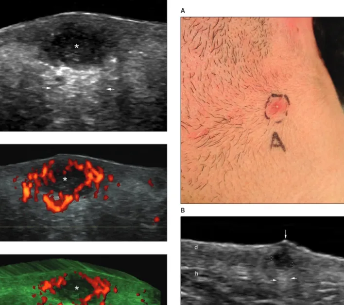
Figure 7. Clinical nodule: sonographic fistula in a 19-year-old male patient with acne vulgaris. A, Clinical image showing a nodule in the right mandibular region. B, Grayscale sonogram (longitudinal view) showing a 1.56-cm-long × 0.21-cm-deep dermal and hypodermal anechoic fistulous tract (between markers). C, Three-dimensional reconstruction (5- to 8-second sweep) of the same lesion (asterisks). Abbreviations are as in Figure 3.
A
B
C Figure 6. Clinical papule: sonographic pseudocyst in an 18-year-old male patient with acne vulgaris. A, Clinical image showing a papule on the left cheek. B, Grayscale sonogram (transverse view) showing an oval hypoechoic dermal structure (between markers) with a mild posterior acoustic reinforcement artifact, indicative of pseudocyst. Abbreviations are as in Figures 2 and 3.
A
(n = 12). Statistical analysis showed strong concor-dance, at 90% (κ= 0.8020), between the two clinicians. Regarding the sonographic scoring system classification, it showed a relative concentration of pseudocysts among the most severe cases, with average numbers of 1.3 in stage 1, 6.6 in stage 2, and 17 in stage 3. The average number of fistulas per patient in stage 3 was 1.6. Calci-nosis deposits were almost evenly distributed across the 3 stages (stage 1, 1 case; stage 2, 3 cases; and stage 3, 2 cases).
Discussion
In this first systematic pilot assessment of the sonographic morphologic characteristics of acne vulgaris, we found a
somewhat surprising discrepancy between mild clinical expressions and the much broader and severe involvement found on sonography. Moreover, clinically unsuspected components were often present deep in the skin, some-times reaching the hypodermis.
The clinical importance of the observations above may reside in the high frequency of treatment failure, reported in more than 50% of acne cases, with the risk of resistance increasing with disease severity.6In addition to the known bacterial or biological resistance, it is possible that incomplete lesion characterization, given the spatially restricted properties of clinical exploration, may contribute to the less than optimal effects of therapy. Moreover, serial histologic study, the usual gold standard, cannot be used because of its limited applicability in acne due to possible scarring of the face. Within this context, fistulous tracts, which most often fail to be recognized clinically and which
Figure 8. Clinical papule-pustule: sonographic fistulas in a 17-year-old male patient with acne vulgaris. A, Clinical image showing a mixed papule-pustule (marked with ink) on the left cheek. B, Grayscale panoramic longitudinal view (left cheek) along the same axis as the clinical lesion showing two hypoechoic fistulous tracts (asterisks) involv-ing the dermis and upper hypodermis. Abbreviations are as in Figure 3.
A
B
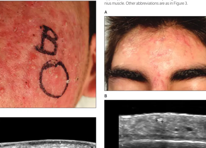
Figure 9. Subclinical calcinosis in a 17-year-old male patient with acne vulgaris. A, Clinical image. B, Grayscale sonogram (transverse view, frontal region) showing 1.0- and 1.2-mm hyperechoic spots (between markers) indicating calcinosis (calcium deposits) of the dermis and upper hypodermis; b indicates bony margin of the skull; and m, epicra-nius muscle. Other abbreviations are as in Figure 3.
A
may imply facial disfigurement, require precise diagnosis and complex treatment.10Therefore, the availability of noninvasive sonographically generated anatomic data, including lesion depth and size, can represent an impor-tant addition to the pathophysiologic management of acne. Regarding the inference from clinical features on the pathologic involvement in nodules, it is clear that, although the probabilistic odds are that sonography would report a pseudocyst, in individual cases, the same clinical finding could also mask a fistula or focal inflammation in the der-mis. Furthermore, the finding of deeper involvement in acne lesions that reach the hypodermis can clinically mean
suspending a topical treatment and starting systemic man-agement. It must be borne in mind that topical formula-tions can show limited and variable penetration in the skin, which depends on the time of exposure, the physico-chemical characteristics of the carrier and the active com-pounds used, as well as the state of the stratum corneum, which acts as a barrier, impeding the penetration of exter-nal agents.11
It is interesting that calcinosis, a presumed late sequela of severe acne vulgaris, may also be observed in cases of mild disease. In this study, calcium deposits were not rec-ognized on clinical examination, although they occasion-ally reached the hypodermis and potentioccasion-ally influenced the cosmetic outcome. Nevertheless, the true importance of calcinosis is unclear and deserving of future investigations. Interestingly, the lesions of open and closed come-dones have been clinically described as noninflammatory, but on sonography they appear as small focal inflamma-tory dermal lesions. It may be that in this setting, the inflammatory process is already under control, and this type of inflammation is self-limiting. Probably, long-term sequential and outcome-defined studies will answer the questions raised by this investigation, including the sono-graphic analysis of other less common types of acne excluded from this research.
It is also notable that sonography makes it possible to document the complete pathogenic cycle of acne, starting with the comedonal phase, continuing with formation of pseudocysts, and ending with fistulous tract production. Also, end-stage lesions (sequelae) such as scars and calci-nosis can be detected with the same imaging technique. Conversely, known limitations of skin sonography, such as lesions less than 0.1 mm thick, epidermal-only conditions, and pigment deposits, seem irrelevant in the study of acne lesions because the main target of this condition is in the dermis and upper hypodermis.
In spite of the relevant number of acne lesions, a lim-itation of our study was the small number of patients. This limitation should encourage further investigation of the topic. Even though the lack of histologic correlation could be viewed as another limitation, this modality is rarely used for diagnosing or managing acne. Moreover, it would be difficult to correlate this large number of facial lesions with histologic findings, taking into consideration the high probability of scars.
Although acne vulgaris is very common, definition of the factors responsible for its worsening, resolution, or therapeutic resistance is still incomplete. In this regard, the information generated by sonography could help in the selection of better and earlier treatments, more specifically
Figure 10. Ice pick scars in an 18-year-old female patient with acne vul-garis. A, Clinical image showing ice pick scars. B, Grayscale sonogram (transverse view, right frontal region) showing thickening and hypo e-chogenicity involving the dermis and upper hypodermis (1.7 mm deep; right, between markers). Note mild epidermal depressions on the scarred area (arrows). Abbreviations are as in Figures 3 and 9.
A
targeted to the pathologic characteristics of the individual patient. Importantly, the possibility of sonographically scoring the disease can support management and clinical trials, although this pilot scoring requires further investi-gation. Last, this imaging technique could provide a non-invasive tool to look for the anatomic reasons for the failure of a certain treatment in difficult cases and could be an instrument for supporting further investigations into this condition.
In conclusion, facial acne vulgaris often involves deeper tissues, beyond the reach of the spatially restricted clinical examination. Sonography can define the morpho-logic characteristics of acne lesions, including their type, size, and extension, while also uncovering subclinical involvement. Acne is amenable to sonographic scoring; thus, sonography potentially represents a relevant nonin-vasive diagnostic tool for managing this condition.
References
1. Williams HC, Dellavalle RP, Garner S. Acne vulgaris. Lancet2012; 379:361–372.
2. Taylor M, Gonzalez M, Porter R. Pathways to inflammation: acne patho-physiology. Eur J Dermatol2011; 21:323–333.
3. Saitta P, Keehan P, Yousif J, Way BV, Grekin S, Brancaccio R. An update on the presence of psychiatric comorbidities in acne patients, part 2: depression, anxiety, and suicide. Cutis2011; 88:92–97.
4. Witkowski JA, Parish LC. The assessment of acne: an evaluation of grad-ing and lesion countgrad-ing in the measurement of acne. Clin Dermatol2004; 22:394–397.
5. Katsambas AD, Stefanaki C, Cunliffe WJ. Guidelines for treating acne. Clin Dermatol 2004; 22:439–444.
6. Quéreux G, Volteau C, N’Guyen JM, Dréno B. Prospective study of risk factors of relapse after treatment of acne with oral isotretinoin. Dermatology 2006; 212:168–176.
7. Wortsman X. Common applications of dermatologic sonography. J Ultrasound Med2012; 31:97–111.
8. Gollnick H, Cunliffe W, Berson D, et al; Global Alliance to Improve Outcomes in Acne. Management of acne: a report from a Global Alliance to Improve Outcomes in Acne. J Am Acad Dermatol 2003; 49(suppl):S1– S37.
9. Wortsman X, Wortsman J. Clinical usefulness of variable-frequency ultra-sound in localized lesions of the skin. J Am Acad Dermatol 2010; 62:247– 256.
10. Jansen T, Romiti R, Plewig G, Altmeyer P. Disfiguring draining sinus tracts in a female acne patient. Pediatr Dermatol2000; 17:123–125. 11. Katare OP, Raza K, Singh B, Dogra S. Novel drug delivery systems in
top-ical treatment of psoriasis: rigors and vigors. Indian J Dermatol Venereol Leprol2010; 76:612–621.