IMAGING
Whole-Body Lymphoscintigraphy Using
Transmission Scans
Martha Vallejo Mar, BBA, CNMT; Sonia Gee-Johnson, BS, CNMT; E. Edmund Kim, MD; and Donald A. Podoloff, MD
Department of Nuclear Medicine, University of Texas M.D. Anderson Cancer Center, Houston, Texas
Objective: Our objective was to show the advantages of performing whole-body lymphoscintigraphy using transmis-sion sources. This technique should decrease scanning time, help locate the sentinel lymph node, and decrease radiation exposure to the technologist.
Methods:Twenty patients with proven melanoma received 18.5 MBq (0.5 mCi) filtered (0.22m)99mTc-sulfur colloid in a 0.2-mL volume, administered as multiple intradermal or subcutaneous injections around the known melanoma le-sion or scar. All 20 patients underwent serial static imaging immediately after the injection, along with whole-body scan-ning after the static imaging. The static emission images were acquired for 5 min and the transmission images for 1 min using a 256⫻256 matrix. The whole-body transmission scans were acquired after the whole-body emission scans. The transmission scans were obtained with the same pa-rameters as the emission scans, with the addition of place-ment of a57Co sheet source on one of the detectors of the large-field-of-view dual-head camera. The planar static axial images (transmission, emission) were compared with the whole-body images (transmission, emission) to determine whether the same number of lymph nodes was visualized with each technique. Posterior outlines were obtained through computer manipulation of anterior transmission im-ages.
Results:In all 20 patients, the number of lymph nodes seen on the static images was the same as that seen on the body emission and transmission images. The whole-body emission and transmission scanning time was an av-erage of 30 min less than the time required to acquire the serial static images.
Conclusion: The anatomic location of the sentinel lymph node is seen more easily on whole-body images, both an-terior transmission and posan-terior transmission, than on pla-nar static images. Whole-body emission and transmission imaging decreased scanning time and thus improved patient comfort and throughput. Technologists received less radia-tion exposure when handling the 57Co source only twice during whole-body imaging, as opposed to several times during static imaging.
Key Words: melanoma; lymphoscintigraphy; transmission J Nucl Med Technol 2002; 30:12–17
T
he lymphatic system consists of a network of vessels and lymph nodes that are dispersed throughout the body. The average adult has approximately 500 to 1,000 nodes (1). Lymphoscintigraphy is the injection of radioactive par-ticles that are then imaged as they pass through afferent lymphatic vessels to their respective lymph node drainage basins. Lymphoscintigraphy has made it easier to trace the complicated lymphatic drainage to the sentinel lymph node (SLN), which is the draining node nearest the tumor. It has been proposed, and appears to have been proven, that in melanoma the pathologic status of the SLN accurately pre-dicts the status of the entire nodal basin (2). Not only nodes of the draining basin but also in-transit lymph nodes, situ-ated between the injection site and the anatomically recog-nized regional lymph node groups, have been found to be SLNs and to accurately predict the pathologic status of the regional nodal basin as a whole. The SLN hypothesis has been strongly supported by the results of studies on both melanoma and, more recently, breast cancer (2,3). Preoper-ative lymphoscintigraphy has become an important step before surgical removal of the SLNs at risk for metastatic disease (4). The nuclear physician must track the afferent drainage channels to see whether multiple drainage chan-nels culminate in multiple SLNs or whether the lymphatic channels converge to terminate in a single SLN. Cutaneous lymphatic flow is so rich that afferent lymphatics can be traced in most cases. Therefore, dynamic or sequential imaging is important in SLN identification. The patient is imaged in multiple projections for proper SLN localization. We used whole-body lymphoscintigraphy in conjunction with a whole-body transmission scan to better locate the SLN.MATERIALS AND METHODS
Twenty patients received 18.5 MBq (0.5 mCi) filtered (0.22 m)99mTc-sulfur colloid as multiple intradermal or subcutaneous injections around a cutaneous melanoma. Im-For correspondence or reprints contact: Martha Vallejo Mar, BBA,
CNMT, Department of Nuclear Medicine, University of Texas M.D. Anderson Cancer Center, 1515 Holcombe Blvd., Box 83, Houston, TX 77030.
FIGURE 1. (A) From left to right, 3 whole-body images and 1 posterior-outline transmission image of patient with melanoma. Injection site was left foot. Continuous lymphatic vessel and inferior in-guinal node are seen. (B) Serial static emission images of same patient. (C) Respective transmis-sion images of same patient. ANT⫽anterior; Lt⫽ left; POST⫽posterior.
mediately after the injections, serial static images were obtained using a 256⫻256 matrix, acquiring the emission images for 5 min and the transmission images for 1 min. A whole-body emission scan was acquired after the static imaging, using a 1,024⫻ 256 matrix with a speed of 12 cm/min. A whole-body transmission scan was acquired
after the emission scan without moving the patient. The transmission scan was acquired at the same speed as the emission scan, with the addition of a 57Co sheet source placed on the detector that was under the scanning table. The melanoma lesions were on the back of 11 patients, shoulder of 3, forearm of 2, left foot of 1, left mid shin of
FIGURE 2. (A) Whole-body emission/transmission images of patient with melanoma. (B) Serial static images of same patient. Static imaging does not allow acquisition of both anterior and posterior transmission images unless additional images are acquired by positioning patient prone. ANT⫽anterior; POST⫽posterior.
1, and anterior chest of 2. The 18 patients with upper-body lesions, such as on the back, chest, and forearm, were scanned from the top of the shoulders to the pelvis. The 2 patients with lower-extremity lesions were scanned from the top of the shoulders to the feet.
Posterior transmission images were obtained through computer manipulation of the anterior transmission images. A copy of the anterior transmission image was placed beside the original anterior transmission image. A matrix utility program was used to flip the anterior image along the
long axis of the patient, left to right. A region of interest was drawn around the outline of the body, and the inside was masked to obtain only the body outline, without the activity seen from the anterior transmission image. Lastly, the pos-terior emission whole-body image was superimposed on this posterior body outline.
RESULTS
Whole-body imaging allowed physicians to better visu-alize the anatomic location of the sentinel node.
Interpreta-FIGURE 3. Acquisition time, shown ony-axis in minutes, for static and whole-body emission and transmission images. Trans⫽transmission.
TABLE 1
Comparison of Acquisition Time for Emission/Transmission Whole-Body Images and Serial Planar Images
Patient no.
57Co source
(MBq) Injection site
Total time (min)
Time reduced by (min) No. of nodes in WB vs. planar Emission/ transmission WB Planar
1 126.17 Upper back 16 58 42 3 WB/3 planar 2 130.61 Mid back 20 58 38 2 WB/2 planar 3 130.61 Lower back 20 53 33 4 WB/4 planar
4 130.61 L foot 36 67 31 2 WB/2 planar
5 175.75 R forearm 18 53 35 1 WB/1 planar 6 175.75 R shoulder 16 59 43 2 WB/2 planar 7 126.17 Mid back 18 50 32 1 WB/1 planar 8 126.17 Upper back 20 55 35 1 WB/1 planar 9 126.17 L mid tibia 30 65 35 1 WB/1 planar 10 121.73 Mid back 18 40 22 2 WB/2 planar 11 127.28 Upper mid back 20 50 30 3 WB/3 planar 12 122.84 L forearm 20 62 42 1 WB/1 planar 13 122.84 Upper back 20 50 30 2 WB/2 planar 14 122.84 Lower back 19 65 46 3 WB/3 planar 15 122.84 L shoulder 20 45 25 4 WB/4 planar 16 122.84 R posterior shoulder 20 40 20 1 WB/1 planar 17 114.33 R anterior chest 21 44 23 1 WB/1 planar 18 114.33 Upper mid back 20 30 50 4 WB/4 planar 19 127.28 L mid back 18 47 29 1 WB/1 planar 20 106.56 L clavicle 20 55 35 3 WB/3 planar WB⫽whole body.
tion of conventional planar static images from lymphoscin-tigraphy was confusing when several spot images were obtained. Viewing one film from whole-body emission im-aging was much easier than viewing several films from static imaging (Figs. 1 and 2). A physician trying to identify the olecranon or popliteal sentinel nodes could continuously follow the lymphatic vessels in the upper or lower extrem-ities. Locating these nodes on static images was difficult because continuous lymphatic channels could not be viewed.
The time to acquire the static images and the whole-body emission and transmission images was recorded. Acquisi-tion of both whole-body emission images and whole-body
transmission images required 20 min, whereas serial static images required 55 min (Table 1). The total imaging time was an average of 30 min less for whole-body images than for static images (Fig. 3).
The whole-body method benefited technologists by decreas-ing the number of times they directly handled the57Co source. During static imaging, technologists had to handle the 57Co source approximately 7 times, because each static planar view was acquired with and without the transmission source. During whole-body transmission lymphoscintigraphy, technologists handled the 57Co source only twice. This method allowed better adherence to the “as low as reasonably achievable” principle because the source was handled less frequently.
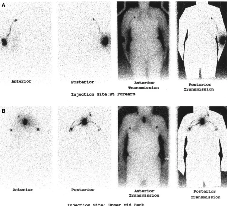
FIGURE 4. (A) Images obtained when injection site was right forearm show continuous lymphatic vessel and inferior axillary node but no olecranon node. (B) Images obtained when injection site was upper mid back show bilateral inferior axillary nodes and right jugular node. Rt⫽ right.
DISCUSSION
Whole-body lymphoscintigraphy was used to better lo-cate the sentinel node and to reduce imaging time. Viewing several static images with and without transmission is con-fusing when one must match several planar images to the transmission images to locate the sentinel node. Physicians can more easily see the anatomic location of the sentinel node on whole-body lymphoscintigrams by viewing one film containing both emission and transmission images (Fig. 4). Because whole-body lymphoscintigraphy takes 20 min, it cannot replace dynamic or cine imaging. It can only be supplementary. However, immediate whole-body lympho-scintigraphy may obviate dynamic or cine imaging if an afferent lymphatic vessel is shown. Static imaging poses a problem with obtaining both posterior and anterior trans-mission images. If a lesion is on a patient’s back, the posterior transmission image has to be obtained with the patient lying prone on the table, over the transmission source. Most patients are uncomfortable lying prone for 1 h, but if whole-body scanning is performed, the patient can lie supine while both the anterior and the posterior images are acquired. Surgeons have questioned findings of metastases to popliteal nodes in patients with lower-extremity melano-mas and metastases to olecranon nodes in patients with upper-extremity melanomas. Patients with upper-extremity melanomas will usually show SLNs in the axilla, but drain-age to olecranon nodes may occasionally occur. Patients with lower-extremity melanomas will usually show drain-age to the groin, with draindrain-age to popliteal nodes occurring occasionally (5). Whole-body lymphoscintigraphy, by showing the continuous lymphatic vessel, allows the phy-sician to detect the popliteal or olecranon nodes. Being able
to follow the lymphatic vessels can also aid in detecting 2 adjacent lymph nodes (6).
CONCLUSION
Whole-body transmission lymphoscintigraphy benefits patients, physicians, and technologists. Scanning time is 30 min less than for static imaging, and patients can lie com-fortably in the supine position during both anterior and posterior transmission imaging. Physicians need view only one film containing all pertinent images for diagnosis, can locate the sentinel node with less confusion, and, because continuous lymphatic vessels are shown, can better detect popliteal sentinel nodes in the lower extremity and olecra-non sentinel nodes in the upper extremity. Technologists benefit from decreased exposure to the57Co source used for
transmission images, handling it twice during whole-body lymphoscintigraphy as opposed to 7 times during planar static studies.
REFERENCES
1. Charman WN, Stella VJ, eds. Lymphatic Transport of Drugs. Boca Raton, FL: CRC Press;1992:3–10.
2. Krag D, Weaver D, Ashikaa T, et al. The sentinel node in breast cancer.
N Engl J Med. 1998;339:941–945.
3. Albertini JJ, Cruse CW, Rapaport D, et al. Intraoperative radiolymphoscin-tigraphy improves sentinel lymph node identification for patients with melanoma. Ann Surg. 1996;223:217–224.
4. Olmos RV, Wieweg OE. Reproducibility of cutaneous lymphoscintigraphy: same or different lymphatic routes and sentinel nodes after reinjection.
J Nucl Med. 2001;42:430 – 431.
5. Uren RF, Thompson JF, Howman-Giles RB, eds. Lymphatic Drainage of
the Skin and Breast. Amsterdam, The Netherlands: Hardwood Academic
Publishers;1999:51– 67.
6. Nieweg OE, Jansen L, Uren RF, Thompson JF. Instructive cases. In: Nieweg OE, Essner R, Reintgen DS, Thompson JF, eds. Lymphatic
Map-ping and Probe Applications in Oncology. New York, NY: Marcel Dekker,