CANINE SYSTEMIC LUPUS ERYTHEMATOSUS GENETIC ANALYSIS OF AN ESTABLISHED BREEDING COLONY* BY R O B E R T M. LEWIS, D.V.M., ANn ROBERT S. SCHWARTZ, M.D. (From the Departments of Surgery and Medicine, Tufts University School of Medicine,
Boston, Massachusetts 02111) (Received for publication 29 April 1971)
Systemic lupus erythematosus (SLE) 1 occurs in dogs and like its counterpart in man, it is a multisystem disease usually affecting young adult females and often terminating in renal failure (1, 2). The typical patient with canine SLE sequentially or simultaneously develops autoimmune hemolytic anemia, throm- bocytopenic purpura, and glomerulonephritis. In some cases symmetrical polyarthritis, dermatitis, or thyroiditis also occur. In addition to the clinical picture, the diagnosis of SLE is substantiated by laboratory findings, which include positive LE cell tests, antinuclear antibodies, complement-fixing anti- bodies to DNA-histone complexes, positive antiglobulin tests, and rheumatoid factor (3-5). There is no predilection for any particular breed to develop canine SLE, and no environmental stimuli that induce the disease have been identified. Apart from its importance in veterinary medicine, the disease in dogs provides an opportunity to study the etiology and pathogenesis of SLE in a large sub- human species. In 1965 steps were taken to acquire dogs with SLE, to breed them, and to establish a colony in which genetic and environmental factors related to the disease could be analyzed. In this paper we will describe the outcome of breeding experiments designed to test the hypothesis that SLE is genetically transmitted. Our findings cannot be fully explained by any con- ventional genetic mechanism; instead, vertical transmission of the disease by nongenetic means seems likely.
Methods
Animals.--The colony is housed in the Animal Research Center, Southborough, Mass. In this isolated country environment the dogs are kept in 9 X 5 ft cement-floor pens, and they are fed a commercial kibbled dog food and water ad tibitum. With the exception of a breeding colony of normal beagle dogs housed within this same facility, the SLE colony dogs have only intermittent and brief exposure to outside personnel or other animals.
All adult dogs in this facility are managed in a similar fashion. I n addition to routine maintenance requirements, each animal is examined visually every day, routine health care
* Supported in part by National Institutes of Health Research Grant AM09351. 1 Abbreviations used in this paper: ANA, antinuclear antibodies; ITP, idiopathic thrombo- cytopenic purpura; PBS, phosphate-buffered saline; SLE, systemic lupus erythematosus. THE JOURNAL OF EXPERIMENTAL MEDICINE • VOLUME 134, 1971 417
on May 13, 2019
jem.rupress.org
Downloaded from
http://doi.org/10.1084/jem.134.2.417
418 CANINE SYSTEMIC LUPUS ERYTHEMATOSUS
is provided by the veterinary staff, and every animal receives a biannual vermifuge and vac- cination for canine distemper and infectious canine hepatitis.
Pregnant dogs are isolated in a nursery unit and delivery is attended by personnel trained in veterinary nursing. The puppies are raised as litters, with intensive care being provided during the first 2 wk of life. They are weaned at 4 wk and returned to the boarding area when they are 6 wk old.
When possible, female dogs are bred during their first estrus, as determined by examina- tion of vaginal cytology (6). Two observed breedings are required to insure maximum pro- duction. In those instances where compatible matings cannot be achieved naturally, artificial insemination is utilized to maintain the breeding program.
Characteristics of the Parents.--The A breeding line originated from a mating between two purebred German shepherd dogs. The male was a normal registered show dog with no history of serious illness and no serologic evidence of canine SLE. The female had ragweed sensitivity from the age of 2 yr, but was otherwise in good health until the age of 4 yr, when she developed acute hemolytic anemia with a positive direct antiglobulin test the day after a family vaca- tion at the beach. The disease, which was characterized by weakness, severe anemia, reticulo- cytosis, jaundice, hemoglobinutia, and proteinuria, was treated with high doses of corticos- teroids and recovery was prompt. Positive LE cell tests were observed and antinuclear anti- body was detected in her serum during the hemolytic crisis. After recovery, her annual epi- sodes with ragweed hay fever continued and intermittently she developed eczema during the warm summer months. When 6 yr old, she delivered a litter of eleven normal puppies and these formed the nucleus of the A breeding line. 4 yr later she developed severe, unresponsive, non- bacterial colitis, and due to continuing debilitation, euthanasia was performed. Postmortem examination was not possible.
The B breeding line resulted from a mating in which both parents had SLE. The dam was a purebred German shepherd that developed acute hemolytic anemia at the age of 6 months. The disease was characterized by severe anemia, a positive direct antiglobulin test, retieulo- cytosis, thrombocytopenia, leukopenia, splenomegaly, and lymphadenopathy. Antinuclear antibody and LE cell tests were positive, and complement-fixing antibodies to DNA-histone complexes were detected in her serum. She responded well to corticosteroid therapy, and re- mained in good health for 5 yr, during which time she gave birth to five litters of puppies.
The male used in originating the B breeding line developed idiopathic thrombocytopenic purpura (ITP) at the age of 3//~ yr. Before this, the only abnormality noted by the dog's owner was a severe necrotlzing solar dermatitis, which occurred on the unpigmented portion of the muzzle each summer. Treatment of the ITP with corficosteroids was initially successful but three relapses occurred over a period of 10 months. Each of these episodes was charac- terized by severe thrombocytopenia, extensive puYpura, and epistaxis. Megakaryocytes in the femoral marrow were present in normal numbers. Although steroids were effective in overcoming the clinical signs, the repeated exacerbations led to splenectomy in November 1965. Positive LE cell tests and antinuclear antibody were noted early during the disease. Since splenectomy, bleeding has not reoccurred; however, vasculitis and thrombosis, which have plagued the dog, required amputation of three toes.
The C breeding line resulted from mating two purebred French poodle dogs. The male was unaffected and the female had the following history. When 3 ~ yr old, she was presented with muscle pain, ill-defined joint pain, and weakness. Radiographic abnormalities were not present at this time and the clinical diagnosis was that of herniated cervical disc. Despite enforced rest at home, multiple leg lameness and joint pain persisted. In addition, she developed eczema, and focal necrotizing dermatitis 4 months later. Severe, progressive, symmetrical polyarthritis affecting the distal joints of all limbs was evident 1 yr after the initial signs of her illness. Biopsy of a joint capsule revealed lesions consistent with those of rheumatoid arthritis. In
R O B E R T M. L E W I S AND R O B E R T S. SCHWARTZ 419 addition, hyperglobulinemia, positive LE cell tests, and leukopenia were found. This dog has produced three litters of puppies since developing her disease, and the symptoms of symmetri- cal polyarthritis are moderately controlled with aspirin.
Laboratory Techniques.--Samples of whole blood, serum, and urine were collected at regular intervals. Routine laboratory methods were used in performing blood counts, urine analyses total serum protein determinations, serum eleetrophoresis, and bone marrow aspirate exami- nations. In addition, automated laboratory microtechnies were used to obtain serum levels of cholesterol, calcium, phosphorus, bilirubin, uric acid, urea nitrogen, glucose, lactic dehydro- genase, alkaline phosphatase, and transaminase in selected animals.
LE cell tests were performed according to the method of Hargraves (7). Preparations con- taining two or more classical LE cells were considered positive. An indirect immunofluorescent test for antinuclear antibody was performed using frozen sections of newborn rat liver as a source of nuclei and monovalent rabbit anti-dog IgG sera as the fluorescein conjugate. A fluorescein-conjugated polyvalent rabbit anti-canine immunoglobulin serum was also used to detect antinuclear antibody in 25 sera that were negative when tested with the monovalent antiserum. In no case were the results positive. Dog sera were tested both undiluted and after 1:5 dilution in phosphate-bufered saline (PBS). Sera with positive reactions were serially diluted in PBS and the highest titer giving a positive result was determined. The pattern of nuclear staining was recorded for each positive result. Tests for rheumatoid factor were per- formed by incubating bentonite particles coated with canine IgG, previously purified by zone eleetrophoresis, with 1:20 dilutions of test sera on a rotating agglutination plate. Positive tests were recorded when 50% or more of the bentonite particles were observed as large agglu- tinations. The titer of rheumatoid factor in positive samples was then determined by doing a similar test on serial fivefold dilutions of the serum. A microimmunoelectrophoretic method (8) was used to analyze serum samples for the presence of canine immunoglobulins (9), transferrin, and ~IC globulin.
Control animals used in these studies came from four sources: (a) normal beagle dogs housed in the same facility as the SLE colony; (b) inbred beagle dogs from a colony at Harvard Medi- cal School; (c) inbred Labrador retriever dogs from a colony at the University of Oregon Med- ical School; and (d) hospitalized animals from the Angell Memorial Animal Hospital, Boston, Mass.
R E S U L T S
T o date, 480 a n i m a l s h a v e b e e n b o r n in t h e c o l o n y f r o m b r o t h e r - t o - s i s t e r m a t i n g s , a n d t h i r d g e n e r a t i o n offspring h a v e r e a c h e d b r e e d i n g age in e a c h b r e e d i n g line. Clinical signs of S L E h a v e n o t y e t a p p e a r e d in a n y of t h e c o l o n y animals. T h i s is n o t u n e x p e c t e d , h o w e v e r , since a m a j o r i t y of t h e a n i m a l s are less t h a n 3 y r old, a n age a t w h i c h S L E does n o t u s u a l l y d e v e l o p in t h e r a n d o m dog p o p u l a t i o n . D e s p i t e t h e l a c k of o v e r t signs of disease, t h r e e m a j o r a b n o r - m a l i t i e s h a v e d e v e l o p e d : excessively h i g h n e o n a t a l m o r t a l i t y , m u l t i p l e serologic a b n o r m a l i t i e s , a n d t h y m i c lesions.
Neonatal Mortalily.--Decreased f e r t i l i t y a n d i n c r e a s e d n e o n a t a l d e a t h r a t e s h a v e g e n e r a l l y b e e n a c c e p t e d as c o m p l i c a t i o n s t h a t m a y arise d u r i n g t h e de- v e l o p m e n t of i n b r e d strains of animals, b u t t h e s e findings h a v e n o t b e e n well d o c u m e n t e d in dogs (10). I t is t h o u g h t t h a t a n e o n a t a l m o r t a l i t y r a t e of a b o u t 2 0 % c a n be e x p e c t e d in dog b r e e d i n g colonies. I n o u r colony, t h e m o r t a l i t y r a t e of a n i m a l s u n d e r 6 m o n t h s of age was v e r y h i g h ( T a b l e I ) . T a k i n g 6
420 C A N I N E S Y S T E M I C L U P U S E R Y T H E M A T O S U S
m o n t h s as the age below which death might be related to congenital causes, the n e o n a t a l m o r t a l i t y rates of first generation offspring (F1) in each of the breeding lines falls within the cited figure of 20 %. However, the rate in second generation (F2) progeny was considerably higher t h a n this in each of the breeding lines (Fig. 1), a n d it continued to rise in the A a n d B lines in the third generation
(F~).
TABLE I
MortdityBetwe~ Birthand6 Months ~ Age
F1 F2 Fa
A line
No. of puppies born 11 90 68
Males born 5 36 29
Males died 0 24 29
Females born 4 51 18
Females died 0 38 16
Dead, of unknown sex 2 3 21
Total mortality 18.1% 72.2 c~ 97.0%
B line
No. of puppies born 43 145 38
Males born 17 71 14
Males died 2 32 6
Females born 26 64 22
Females died 4 27 18
Dead, of unknown sex 0 10 2
Total mortality 13.9% 47.9% 68.4%
C line
No. of puppies born 7 55 23
Males born 2 30 14
Males died 0 15 6
Females born 4 25 9
Females died 0 12 3
Dead, of unknown sex 10 0 0
Total mortality 14.3% 49.1% 34.7%
Analysis of autopsy material from 184 puppies t h a t died between b i r t h a n d 6 m o n t h s of age revealed a degree of consistency in both the distribution a n d character of the lesions present. W h e n the most significant lesions are classified b y body system (Fig. 1), the p a t t e r n of distribution is the same for all three breeding lines. W i t h i n the most commonly affected systems, evidence of bac- terial infection was present in the m a j o r i t y of cases. T h e most frequently en- countered lesions were p u r u l e n t bronchopneumonia, p u r u l e n t omphalophlebitis a n d hepatitis, a n d bacterial enterocolitis. Lesions suggestive of canine SLE were n o t found in these animals.
ROBERT M. LEWIS AND ROBERT S. SCHWARTZ 421 In addition to the rising neonatal mortality rate, there was a reduction in litter size with each successive generation (Table II). Although the average number of puppies in FI litters was within the range of expected values for the breeds composing the colony, a progressive reduction in litter size occurred in each subsequent generation of all three breeding lines. This reduction is most apparent in the F~ progeny of the A breeding line.
Minor differences in the male to female sex ratio exist within the second and third generation offspring of each breeding line, but no significant effect of inbreeding on the sex ratio was found when each breeding line was examined
Pul G I I ~\\\\\\\\\\\\\\\~ UG CV RES Other NSL % AFFECTED I0 20 .30 40 i i I i 5O i • A LINE [ ] B LINE [ ] C LINE
FIG. 1. Classification of lesions responsible for neonatal mortality by body system. Pul = pulmonary; GI = gastrointestinal; UG = urogenital; CV = cardiovascular; RES = reticulo- endothelial; NSL = no significant lesion; Other = all other systems.
individually (Table I). In summarizing overall production figures, in those cases where the sex could be determined 219 male and 223 female puppies have been born. No consistent variations have been observed in the neonatal mortality figures of male as compared to female puppies.
Serologic Abnormalities.--The
L E cell test was selected as the primary labora- tory screen to monitor the colony because of its specificity for SLE in the random dog population. Consequently, LE cell tests have been made serially over a period of years on members of the colony and a high incidence of positive tests has been found in the offspring within each breeding line (Table I I I ) . As can be seen in Fig. 2, conversion of the test from negative to positive usually occurred before 1 yr of age, and frequently when the litter was less than 6 months old. Commonly, all or most of the members of a litter developed positive422 C A N I N E SYSTEMIC LUPUS ERYTtlEMATOSUS T A B L E I I
Puppy Production in SLE Colon)"
F~ F~ F~
A line
No. of litters 1 12 12
No. of puppies 11 90 68
R a n g e - - 2-11 3-12
Average litter size - - 7 . 5 5 . 6
B line No. of litters No. of puppies R a n g e
Average litter size
5 23 27 43 145 38 4-11 3-11 1-9 8 . 6 6.3 5 . 4 C line No. of litters 1 11 5 No. of puppies 7 55 23 R a n g e - - 4 - 6 4-5
Average litter size - - 5 . 0 4 . 6
T A B L E I I I
Serologic Abnormalities and Genetic Background
Strain and generation LE cell test
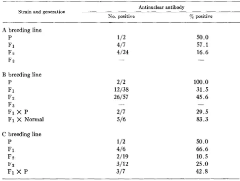
No. positive % positive
A breeding line P 1/2 50.0 F 1 7/7 100.0 F2 21/24 87.5 F;~ - - - - B breeding line P 2/2 100.0 F1 37/38 97.3 F2 46/57 80.7 F 3 - - _ F1 X P 4 / 7 57.1 F I X N o r m a l 2 / 6 3 3 . 3 C breeding line P 1/2 5O.O F 1 6 / 6 100.0 F~ 13/19 6 8 . 4 F s 9/12 75.0 F1 X P 5/7 7 1 . 4
R O B E R T M . L E W I S A N D R O B E R T S. S C H W A R T Z 423
tests at about the same time. The high percentage of positive tests first detected at 3 months of age reflects the fact that routinely, this is the age at which the animals are initially tested.
To allow genetic analysis of these data, a positive L E cell test was selected as the marker to identify the phenotype for SLE. To be included in the data, at least three members of any litter must have survived long enough to be adequately tested. An animal was considered negative only if it was older than 6 months and had had at least three consecutively negative tests.
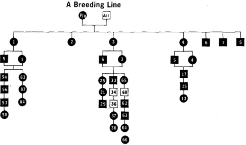
In the A and C lines, where the original female parent had SLE and the male was normal, all of the first generation offspring were positive (Figs. 3, 4). Positive LE cell tests were found in 87.5 % of the Fe progeny in the A line and
55- m 3 0 - -I 25- w 20- I- 15- m IO' m O. 0 2 4 6 8 10 12 14 16 18 20 22 24 26 28 30 39 AGE IN MONTHS
FIG. 2. Age when first positive LE cell test was detected.
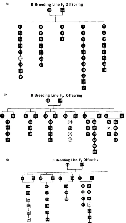
68.4% of the F2 progeny in the C line. When F2 brother-sister matings were made in the C line, 75 % of the F3 puppies developed positive L E cell tests. Only animals with a positive test were used in these matings. Similar matings of F2 offspring have been made in the A line, but we have yet to obtain three surviving members of any of these litters. Consequently, these data are not included in our genetic analysis. Backcross experiments (Fig. 5), in which a positive Fz son in the C line was twice mated to his affected mother, produced a total of seven animals, five of which (71.4%) developed positive L E cell tests. In the B line, where both original parents had SLE, 37/38 (97.3%) of the first generation offspring were found to be positive (Fig. 6 a). Sibling matings (or generationally equivalent matings) of these F1 progeny resulted in the de- velopment of positive LE cell tests in 81% of the second generation offspring (Figs. 6 b, 6 c). To date, F3 offspring have not been produced in sufficient numbers to be adequately tested within this breeding line.
424 CANINE SYSTEMIC LUPUS ERYTHEMATOSUS
When two different seropositive F1 sons of the B breeding line were crossed back to their affected mother, 57 % of the puppies produced had positive L E cell tests (Fig. 5). One of these same F1 sons was mated to a normal beagle dog, and one-third of the offspring developed positive L E cell tests. In addition,
A Breeding Line
r
y
@
Fro. 3. Pedigree of A breeding fine. The legend is the same for all breeding lines: circles =
females; squares = males; black figures = positive phenotype for SLE. Numbers within figures identify individual animals.
C Breeding Line
]
R O B E R T M. L E W I S A N D R O B E R T S, S C H W A R T Z 425 positive tests for antinuclear antibody were found in five of the six members of the litter produced by this outcross (Table IV).
A second abnormal finding, antinuclear antibody, was detected in the sera of 55/176 (31.2%) inbred dogs in the colony. 48 of these animals also had positive L E cell tests. When tested over a period of time, a rising titer of anti- nuclear antibody was observed in approximately half (27/55) of these animals. Using rat liver nuclei as the substrate, the staining pattern was usually diffuse over the entire nucleus. However, cells with more intense peripheral staining of
B Backcross
m
®
m
| |
C Backcross
FIo. 5. Pedigree of backcross and outcross experiments.
B Outcross
m
()
[]
1
E]
the nucleus (rim pattern) and cells with a speckled pattern were also seen in these preparations. (Fig. 7)
Although most (48/55) of the inbred animals with antinuclear antibodies also had positive L E cell tests, a close correlation between these two laboratory tests was not found. Serial testing of 196 inbred progeny for the presence of L E cells disclosed 160 animals to be positive (81.6%). Of these only 29.0% (55/160) also had antinuclear antibody. When the distribution and frequency of these two abnormalities are compared (Tables
III, IV),
it becomes apparent that the development of antinuclear antibody in the dog is unrelated to the L E cell phenomenon.6a
()
l l
l !
I !
()
,)
(..)
l |
(,)
B Breeding Line F I Offspring
.)
(
l
(
|
(
!
!
1
t
!
(.)
()
El
El
| t
|ll
6b B Breeding Line F 2 Offspring
6c B Breeding Line F 2 Offspring
I
~
~,~
~,~
FIG. 6. Pedigree of B breeding line. (a) First generation offspring; (b) second generation offspring; (c) second generation offspring.
ROBERT M. LEWIS AND ROBERT S. SCHWARTZ 427 13 m e m b e r s of the colony (Table VI). N o n e of these animals have signs of arthritis.
D e s p i t e extensive serial testing for variances in hemotologic and clinical chemistry values, no consistent abnormalities were detected in over 100 animals during a 2 y r period of investigation.
Thymic Abnormalities.--Postmortem examination revealed lesions in the t h y m u s of 62 of 169 (36.6%) colony dogs. All these animals were over 6 m o n t h s
TABLE IV
Serologic Abnormalities and Genetic Background
Strain and generation Antinuclear antibody
No. positive % positive A breeding line P 1/2 50.0 F1 4/7 57.1 F ~ 4/24 16.6 F3 - - - - B breeding line P 2/2 lOO.O F1 12/38 31.5 F 2 26/57 45.6 F3 - - - - F1 )< P 2/7 29.5 F1 X Normal 5/6 83.3 C breeding line P 1/2 50.0 F t 4/6 66.6 F,2 2/19 10.5 F3 3/12 25.0 F1 X P 3/7 42.8
of age and t h e y were either sacrificed as p a r t of a p o p u l a t i o n control p r o g r a m within the breeding colony or t h e y h a d died unexpectedly.
Control tissue was collected from 32 r a n d o m - b r e d hospitalized dogs t h a t re- ceived complete p o s t m o r t e m examination. These animals were u n r e l a t e d to the S L E colony dogs, t h e y ranged in age from 8 m o n t h s to 10 yr, a n d their d e a t h s were from such diverse causes as automobile accidents and congestive h e a r t failure. N o evidence of S L E was found in the pathologic e x a m i n a t i o n of these animals. A n occasional follicle was found in the t h y m u s from four of these control animals. I t consisted of homogenous aggregates of m e d i u m - s i z e d l y m p h o c y t e s located within the m e d u l l a r y p o r t i o n of the organ. N o other changes were noted.
428 C A N I N E SYSTEMIC L U P U S E R Y T H E M A T O S U S
Gross abnormalities were not observed in the thymic tissue examined from the SLE dog colony. Microscopically, four lesions were present: (a) lymphoid follicles; (b) germinal center formation;
(c)
multiple granulomas; (d) reticulum cell sarcoma.Multiple lymphoid follicles, composed of homogenous collections of medium- sized lymphocytes, were present in 37% of the animals examined (Fig. 8).
FiG. 7. Serologic abnormalities in canine SLE.
(Top)
Positive tests for antinucle~r antibody. Rat liver nuclei illustrating diffuse (a), rim (b), and speckled (c) staining pattern. X 250.(Bottom)
LE cells from third generation progeny in the C breeding llne. X 1000.These follicles occurred in the relatively hypocellular medullary portions of the organ a n d were clearly discernible from the normal thymic tissue by their uniform appearance and the concentric orientation of the cells. The follicles contained well-defined germinal centers, composed of reticulum cells, lympho- blasts, mitotic figures, and macrophages. Distinct collars of small lymphocytes surrounded the germinal centers. These follicles were identical in appearance to those found in the lymph nodes or spleen of the dog. In six animals, the germinal centers were grouped together in a portion of the thymus, sinmlating the ap-
ROBERT M. LEWIS AND ROBERT S. SCHWARTZ 429 pearance of a lymph node. In these cases, tile absence of a capsule, subcapsular sinus, and meduUary sinusoids preclude the possibility that the structures ob- served were foci of aberrant normal lymph node.
FIG. 8. (a) Lymphoid follicle with germinal center in the thymus of an F i dog in the B line. X 100. (b) Germinal center formation in the thymus of an F i dog in the C line. X 250.
Multiple, small, well-formed granulomas were present in one thymus (Fig. 9). They appeared to arise from within normal thymic lobules and had entirely replaced the normal tissue in some of these lobules. Additional granulomata were present, singly or in small dusters, throughout many parts of the affected
430 C A N I N E S Y S T E M I C L U P U S E R Y T H E M A T O S U S
Fic. 9. (a) High power view of germinal center in dog thymus, with mitotic figures, reticu- lure cells, blast cells, and nuclear debris. X 400. (b) Multiple granulomas in the thymus of a B dog. X 250.
organ. N o cause could be found for this lesion, despite extensive e x a m i n a t i o n for bacteria, fungi, and foreign material. Similar lesions were not found else- where in the b o d y a t necropsy.
R O B E R T M, L E W I S A N D R O B E R T S. S C H W A R T Z 431
FIG. 10. (a) Discrete granulomata found in the thymus of a dog from the B breeding line. X 400. (b) Reticulum cell tumor in thymic lobule (upper), germinal center formation (lower right). X 40. (c) Pleomorphic reticulum ceils replacing normal thymic tissue. X 400.
addition to l y m p h o i d follicles and active germinal center formation, an expand- ing mass of pleomorphic reticulum cells replaced normal t h y m i c tissue. T h i s mass was confined to one lobule of the affected organ and a p p e a r e d similar to the t y p e B reticulurn cell t u m o r described in mice (11) (Fig. 10).
432 C A N I N E SYSTEMIC L U P U S E R Y T H E M A T O S U S D I S C U S S I O N
The high frequency of perinatal death that occurred in conjunction with our attempt to inbreed the offspring of dogs affected with SLE cannot be adequately explained at this time. Judging by the autopsy findings, an increased suscep- tibility to infection occurred despite exceptional nursing care and environmental control. There was no histological evidence of SLE in any of these dogs; there- fore, we assume that the cause of death in these puppies was unrelated to SLE. To what degree the disease of the parents contributed to the high mortality rate is not known, and additional means of investigating this problem, such as in- breeding under gnotobiotic conditions, are currently being explored. The peri- natal mortality rate of the children of women with systemic lupus erythema- tosus is normal (12), although excessive number of spontaneous abortions have been noted (13).
Thymic abnormalities consisting of lymphoid follicles, many of which con- TABLE V
Ant~udearAnt~My
Und.* 1:5 1:25 1:125 1:625
Normal cohabitant beagles (24) 0 0 0 0 0
Random hospitalized dogs (36) 0 0 0 0 0
Inbred beagle dogs (12) 0 0 0 0 0
Inbred Labrador retriever dogs (32) l 0 0 0 0
SLE colony dogs (176) 55 22 4 1 0
* U n d . , u n d i l u t e d s e r u m .
tained germinal centers, were commonly found in the dogs of this colony. Formations like these also occur in guinea pigs and hamsters after direct injection of antigens into the thymus (14, 15), in New Zealand Black mice (16, 17), and in humans with SLE (18). Furthermore, hyperplasia of the lymphoepithelial elements of the thymus and the development of thymoma, well-recognized abnormalities associated with myasthemia gravis (19), have also been reported in human SLE (20, 21).
The inability to produce experimentally lesions of this type by the injection of antigen by any route other than intrathymically (14, 15), plus the exclusion from the thymus of intravenously injected vital dyes (22), indicate the existence of a blood-thymus barrier similar to the blood-brain barrier. The spontaneous development of lymphoid follicles and germinal centers in humans with SLE, in NZB mice, and in the dogs of this colony suggests that if such a barrier exists, the stimulus provoking the changes must either arise within the thymus, or arrive there by means other than diffusion through semipermeable mem- branes. In any case, the lesions present in the thymuses of our colony dogs have
ROBERT M. LEWIS AND ROBERT S. SCHWARTZ 433 every appearance of proliferating clones of cells arising in an apparently normal organ. The cause of these changes is unknown, but the fact that a reticulum cell tumor was found in one of the animals suggests its malignant potential.
The high frequency of thymic lesions and serologic abnormalities in the members of this colony might be explained by one or more of the following possibilities: (a) influence of environmental factor(s); (b) an effect of inbreeding per se; (c) an inordinately high rate of somatic mutation; (d) a cytoplasmic factor, defect, or abnormality of a germ cell; (e) inheritance of a genetic defect; (f) the presence of a transmissible agent.
The possibility that effects of environment or inbreeding could account for the abnormalities seems remote, since control populations with comparable inbreeding coefficients, and cohabitant normal dogs have not developed them (Tables V, VI).
Although somatic mutation of lymphocyte precursors in a genetically sus- ceptible subpopulation has been proposed as the underlying mechanism of
TABLE VI Rheumatoid Factor
Positive test
Normal cohabitant beagles 0/6
Random hospitalized dogs 0/30
Inbred beagle dogs 0/12
Inbred Labrador retriever dogs 0/32
SLE colony dogs 13/124
autoimmunization in humans by Burch and Rowell (23), it seems unlikely that a mutation rate could be achieved that would produce the high incidence of abnormalities we observed in two different purebred strains of animals and one population of mongrel (mixed breed) dogs.
Results of the outcross experiment make it unlikely that some peculiar, inherited cytoplasmic factor within the germ cell could account for the ab- normalities. Such a mechanism would most likely be effected through the ova, with its plentiful cytoplasm; yet, when a seropositive male dog was bred to a normal female, a high percentage of their offspring developed positive L E cell tests (33 %) and antinuclear antibodies (86 %) (Fig. 5).
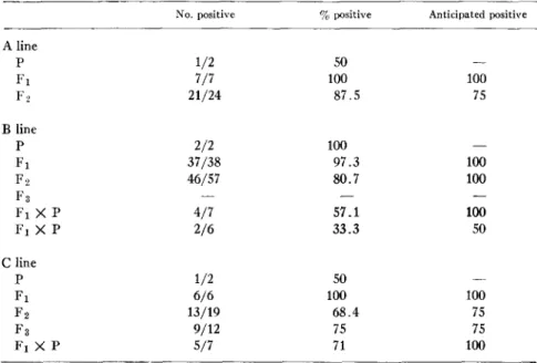
The simplest genetic explanation for the high frequency of offspring with positive L E cell tests in our colony requires that a single, dominant, fully penetrant autosomal gene is responsible for the abnormality (Table VII). Un- der these conditions, when one of the original parents is affected (A line, C line), then 100% of the F1 progeny and 75 % of the F~ progeny can be expected to be positive if the original parent is homozygous for the trait. If, however,
434 CANINE SYSTEMIC LUPUS ERYTItEMATOSUS
the affected parent is heterozygous for the trait, then only 75% of the FI progeny would be expected to be positive.
All seven F1 puppies in the A line were positive. T h e probability of this occurring is 1.0 if the dam were homozygous for the hypothetical gene; it is unlikely that she was a heterozygote, for the probability of a heterozygote X normal mating producing 7/7 positive offspring is only 0.0078. I n mating positive F1 A dogs, all of which must be heterozygous regardless of parental zygosity, 24 puppies were produced, 21 of which were positive. T h e number of positive animals follows the binomial distribution with a mode of 18 positive
TABLE VII
D~tHbution ~thePost~at~Genet~ M a ~ r C a n ~ e S L E
No. positive % positive Anticipated positive A line P 1/2 50 - - F1 7/7 100 100 F2 21/24 87.5 75 B line P 2/2 100 - -F1 37/38 97.3 100 F 2 46/57 80.7 100 F 3 - - _ _ F~ X P 4/7 57.1 100 F~ X P 2/6 33.3 50 C line V 1/2 50 - - F1 6/6 100 100 F2 13/19 68.4 75 F3 9/12 75 75 F I X P 5/7 71 100
dogs. The probability of achieving the modal number is 0.19. T h e probability of Obtaining 21 positive animals, or three dogs distant from the mode, is 0.14. Therefore, the hypothesis of a single dominant gene is supported b y this result. A more stringent test of the genetic hypothesis was carried out in the C line. I n this combination, the sire was normal, the d a m was affected, and each of the six F1 offspring was positive. T h e probability of this occurring if the d a m were homozygous is 1.0. If that were actually the case, all of the progeny from the backcross mating must have been positive. Since only 5/7 of those animals had positive L E cell tests, the dam could not have been a homozygote. The result of the backcross mating could have been obtained if the d a m were a heterozygote ( P = 0.31), b u t that seems unlikelv in view of the result obtained
R O B E R T M. L E W I S AND R O B E R T S. SCHWARTZ 435 in the F1 generation: the probability of a heterozygous X normal mating pro- ducing 6/6 positive offspring is only 0.016. Since the dam must be either homozygous or heterozygous for the hypothetical gene, and since neither case is upheld by the results, exclusively genetic mechanisms in the C line can be excluded.
Analysis of the B line also yields results in conflict with the hypothesis of a single, dominant gene. When both original parents are affected, as in this case, there must be no negative progeny if either one of the parents is homozygous for the trait. Since only 37/38 of the F1 progeny in this line were positive, both affected parents would have had to be heterozygous. But the probability of obtaining 37/38 positive progeny from mating two heterozygotes is only 0.0002. The single negative F~ dog in this breeding experiment was tested eleven times during the 2 yr of his life. In no instance was a positive L E cell test found, and repeated tests for antinuclear antibody were negative. Further- more, autopsy of this dog revealed no abnormalities.
In another experiment with the B breeding line, four of seven puppies pro- duced by crossing a seropositive F~ son to his mother are positive (57 %). These results are compatible with a genetic explanation, provided both the parent and the son are heterozygous for the hypothetical trait. However, as we have seen from the inbreeding experiment, it is impossible for the dam to be a homozygote, and it is highly unlikely that she is a heterozygote (P = 0.0002). In a third experiment with the B line, a seropositive F~ son was mated to a normal beagle dog. Two of the six offspring had positive L E cell tests. This result is consistent with the hypothesis that the positive L E cell test is trans- mitted by a single dominant gene. However, it seems unusual that 5/6 of the offspring from this outcross mating have positive tests for antinuclear antibody. In summary, six breeding experiments were carried out in these dogs. The results of two of them, the inbred A line animals and the outcross mating in the B line, are consistent with the hypothesis that these findings are due to a single dominant autosomal gene. The other four experiments, which included inbreed- ing of the B and C line and the backcrosses in these same two breeding lines, do not support the hypothesis. In our opinion, the results of the inbreeding experiments, although apparently due to a genetic mechanism, may in fact be the result of other factors, which may be transmitted by nongenetic mecha- nisms. Because the backcross experiments, which are more reliable for predicting the segregation of genes than inbreeding experiments, failed to uphold the hypothesis of a single gene, we must seek an alternative explanation of the results.
If a single gene cannot explain the results, perhaps a series of interacting genes can. Should multiple genes be located on a single chromosome and in close approximation to each other, the trait would be inherited in the same manner as a single gene defect, so this seems unlikely. If the linkage was less
436 C A N I N E SYSTEMIC L U P U S E R Y T H E M A T O S U S
complete, or if the genes were located on different chromosomes, then the frequency of the trait would be less than that of a single gene defect, due to the fact that the chance for random combination of different genes is inversely proportional to the number of genes involved. Therefore, it seems that the results obtained from our breeding experiments cannot solely be explained by any conventional genetic mechanism.
This leaves us with the sixth possibility. It is conceivable that, in addition to a genetic factor, a transmissible agent is required for the expression of the disease. Experiments have been undertaken to test this idea, and in a following paper we will present evidence that a transmissible agent, present in cell-free filtrates of tissue obtained from dogs in the SLE colony, can provoke the de- velopment of antinuclear antibodies (ANA) and positive LE cell tests when administered to normal animals.
The SLE colony is still evolving and clinical signs of SLE have not yet appeared. We therefore must stress that any relationship between positive L E cell tests, antinuclear antibodies, and the development of SLE remains un- proven in these animals. However, judging by the specific correlation between the presence of L E cells and canine SLE, as well as the large body of evidence that positive LE cell tests in humans usually coincide with SLE, there is every expectation that clinical evidence of SLE will eventually occur in the colony. As a rule, LE cell tests are done in man only when there is clinical evidence of SLE; it is entirely possible that that "marker" is present long before signs of the disease are apparent. In NZB and (NZB > ( N Z W ) F1 mice, positive anti- globulin tests and antinuclear antibodies appear months before overt disease (24); extrapolation of this to the dog suggests that clinical signs of SLE may be expected in the colony within 2 or 3 yr.
SUMMARY
Three breeding lines, originating from dogs with SLE, have been established. Two lines were initiated by mating a female with SLE with a normal male. The third line resulted from a mating of two affected dogs. Brother-to-sister matings have reached the third generation in each line. In addition, backcross and outcross matings were carried out. More than one-third of the autopsied dogs had thymic abnormalities. The commonest lesion was a lymphoid follicle; the thymus of one dog contained multiple granulomas,0 and in one animal a reticulum cell sarcoma of the thymus was found.
Multiple serological abnormalities, including positive L E cell tests, anti- nuclear antibodies (ANA), and rhenmatoid factor, were found in the progeny. The development of ANA appeared unrelated to the incidence of positive L E cell tests. About 10% of the animals had rheumatoid factor in their serum. Control populations of dogs; including house pets; two other, unrelated lines of inbred dogs; and normal dogs housed in the same facility as the SLE colony
ROBERT M. LEWIS AND ROBERT S. SCHWARTZ 437 did not have these abnormalities. The incidence of positive L E cell tests in the inbred, backcross, and outcross matings was not consistent with any conven- tional genetic mechanism of inheritance. I t is conceivable t h a t the results can be explained b y vertical transmission of an infectious agent in a genetically susceptible individual.
The authors wish to thank Drs. Park Gerald, Joseph Templeton, and Walter Tannenberg for their help in analyzing the genetic data and Miss Christina Smith for her expert technical assistance.
BIBLIOGRAPHY
1. Lewis, R. M., R. S. Schwartz, and W. B. Henry. 1965. Canine systemic lupus erythematosus. Blood J. Hematol. 25:143.
2. Lewis, R. M., R. S. Schwartz, and C. E. Gilmore. 1965. Autoimmune disease in domestic animals. Ann. N. Y. Acad. Sci. 124:178.
3. Lewis, R. M. 1965. An evaluation of the clinical usefulness of the LE cell phe- nomenon in dogs. J. Amer. Vet. Med. Ass. 137:939.
4. Lewis, R. M., W. B. Henry, Jr., G. W. Thornton, and C. E. Gilmore. 1963. A syndrome of autoimmune hemolytic anemia and throlnbocytopenia in the dog.
Sci. Proc. J. Amer. Vet. Mad. Ass. 1:140.
5. Lewis, R. M., and Y. Borel. 1971. Canine rheumatoid arthritis: a case report.
Arthritis Rheum. 14:67.
6. Hooper, B. E., A. Hall, and H. E. Dale. 1961. Characteristics of the vaginal smear.
Small Anita. Clinician. 1:355.
7. Hargraves, M. M. 1954. The LE cell phenomenon. Advan. Intern. Med. 6:133. 8. Scheidegger, J. J. 1955. Une microm6thode de l'immuno~lectrophorese. Int. Arch.
Allergy Appl. Immunol. 7:103.
9. Johnson, J. S., J. H. Vaughan, and S. N. Swisher. 1967. Canine immunoglobulins. II. Antibody activity in six iinmunoglobulin classes. J. Immunol. 98:935. 10. Harrop, A. E. 1960. Reproduction in the Dog. Bailli6re, Tindall, and Cassell,
Ltd., London. 1st edition. 191.
11. Dunn, T. B., and M. K. Deringer. 1968. Reticuluin cell neoplasms, type B, or the "Hodgkins-like lesion" of the mouse. J. Nat. Cancer Inst. 40:771.
12. Donaldson, L. B. and R. R. de Alvarez. 1962. Further observations on lupus erytheinatosus associated with pregnancy. Amer. Y. Obstet. Gynecol. 83:1461. 13. Mund, A., J. Simson, and N. Rothfield. 1963. Effect of pregnancy on course of
systemic lupus erythematosus. J. Amer. Med. Ass. 183:917.
14. Marshall, A., and R. G. White. 1961. The immunological activity of the thymus.
Brit. J. Exp. Pathol. 49.:379.
15. Sherman, J. D., M. M. Adner, and W. Dameshek. 1964. Direct injection of the thymus with antigenic substances. Proc. Soc. Exp. Biol. ]fled. 115:866.
16. Burnet, M. 1966. Implications for autoiminune disease in man of studies on NZB mice and their hybrids. II. Renal and thymic disease. Roy. Inst. Pub. Health Ityg. J. 29:95.
438 C A N I N E SYSTEMIC L U P U S E R Y T H E M A T O S U S
cells and autoimmune disease in NZB, NZW, and (NZB X NZW) F1 mice. Immundogy. 19.:179.
18. Mackay, I. R., and P. DeGail. 1963. Thymic "germinal centers" and plasma cells in systemic lupus erythematosus. Lancet. 9.:667.
19. Murray, N. A., and J. R. McDonald. 1945. Tumors of thymus in myasthenia gravis. Amer. J. Clin. Path& 15:87.
20. Galbraith, R. F., H. J. Summerskill, and J. Murray. 1964. Systemic lupus erythe- matosus, cirrhosis and ulcerative colitis after thymectomy for myasthenia gravis. N. Engl. J. Med. 9.70:229.
21. Larsson, O. 1963. Thymoma and systemic lupus erythematosus in the same pa- tient. Lancet. 9.:665.
22. Marshall, A. H. E., and R. G. White. 1961. The immunological reactivity of the thymus. Brit. J. Exp. Pathol. 4~:379.
23. Burch, P. R. J., and N. R. Rowell. 1965. Systemic lupus erythematosus: etio- logical aspects. Amer. J. Med. 38:793.
24. East, J. 1970. Immunopathology and neoplasms in New Zealand Black (NZB) and SJL/J mice. Prog. Exp. Tumor Res. 13:84.