King’s Research Portal
Document Version Peer reviewed version
Link to publication record in King's Research Portal
Citation for published version (APA):
Dorent, R. P. R., Joutard, S. R. D., Modat, M., Ourselin, S., & Vercauteren, T. (Accepted/In press). Hetero-Modal Variational Encoder-Decoder for Joint Modality Completion and Segmentation p. In Medical Image Computing and Computer Assisted Intervention – MICCAI 2019
Citing this paper
Please note that where the full-text provided on King's Research Portal is the Author Accepted Manuscript or Post-Print version this may differ from the final Published version. If citing, it is advised that you check and use the publisher's definitive version for pagination, volume/issue, and date of publication details. And where the final published version is provided on the Research Portal, if citing you are again advised to check the publisher's website for any subsequent corrections.
General rights
Copyright and moral rights for the publications made accessible in the Research Portal are retained by the authors and/or other copyright owners and it is a condition of accessing publications that users recognize and abide by the legal requirements associated with these rights. •Users may download and print one copy of any publication from the Research Portal for the purpose of private study or research.
•You may not further distribute the material or use it for any profit-making activity or commercial gain •You may freely distribute the URL identifying the publication in the Research Portal
Take down policy
If you believe that this document breaches copyright please contact [email protected] providing details, and we will remove access to the work immediately and investigate your claim.
Joint Modality Completion and Segmentation
Reuben Dorent, Samuel Joutard, Marc Modat, S´ebastien Ourselin and Tom Vercauteren
School of Biomedical Engineering and Imaging Sciences, Kings College London
Abstract. We propose a new deep learning method for tumour segmen-tation when dealing with missing imaging modalities. Instead of produc-ing one network for each possible subset of observed modalities or usproduc-ing arithmetic operations to combine feature maps, our hetero-modal varia-tional 3D encoder-decoder independently embeds all observed modalities into a shared latent representation. Missing data and tumour segmen-tation can be then generated from this embedding. In our scenario, the input is a random subset of modalities. We demonstrate that the opti-misation problem can be seen as a mixture sampling. In addition to this, we introduce a new network architecture building upon both the 3D U-Net and the Multi-Modal Variational Auto-Encoder (MVAE). Finally, we evaluate our method on BraTS2018 using subsets of the imaging modali-ties as input. Our model outperforms the current state-of-the-art method for dealing with missing modalities and achieves similar performance to the subset-specific equivalent networks.
Keywords: Tumour segmentation, Modality completion, multi-modal, missing modalities
1
Introduction
Tumour segmentation and associated volume quantification plays an essential role during the diagnosis, follow-up and surgical planning stages of primary brain tumours. Multiple imaging sequences are usually employed to distinguish and assess the key tumour components such as the whole tumour, the peritumoral edema and the enhancing region. The common sequences are T1-weighted (T1), contrast enhanced T1-weighted (T1c), T2-weighted (T2) and Fluid Attenuation Inversion Recovery (FLAIR) images. These modalities reveal different charac-teristics of brain tissues. In practice, the set of acquired modalities may vary during the clinical assessment. For this reason, we aim to automatically segment these key components given an arbitrary set of modalities.
Methods based on deep learning currently achieve the best performance in brain tumour segmentation. Most of them require the full set ofnmodalities as input [4,9], while a scenario of missing modalities is common in practice. Seg-mentation with missing data can be achieved by: 1/ Training a model for each
possible subset of modalities; 2/ Synthesising missing modalities [6] in order to then perform full modality segmentation; 3/ Creating a common feature space which encodes the shared information from which the segmentation is created [3,12]. The two first options involve training and handling a different network for each of the 2n−1 combinations. These two solutions are cumbersome and com-putationally sub-optimal since duplicate information is extracted 2n−1 times.
In contrast, encoding the modalities into a common feature space produces a single model that shares feature extraction.
The current state-of-the-art network architecture which allows for missing modalities is HeMIS [3] and related extensions [12]. Feature maps are first ex-tracted independently for each modality, then their first and second moments are computed across the modalities and used for predicting the final segmen-tation. However, using these arithmetic operations does not force the network to learn a shared latent representation. In contrast, Multi-modal Variational Auto-Encoders (MVAE) [13] provide a principled formulation to create a com-mon representation: thenmodalities and the segmentation map are considered conditionally independent given the common latent variablez.
While our goal to segment the tumour with missing modalities, auto-encoding and modality completion promote informativeness of the latent space and can be seen as regularizers, similarly to [9]. Ideally, all the modality-specific information should be encoded in the common latent space, meaning that the model should be able to reconstruct all the observed modalities. Additionally, the information loss related to any missing modality should be minimal (modality completion).
In this paper, we introduce a hetero-modal variational encoder-decoder for tumour segmentation and missing modalities completion. The contribution of this work is four-fold. First, we extend the MVAE for 3D tumour segmenta-tion from multimodal datasets with missing modalities. Secondly, we propose a principled formulation of the optimisation process based on a mixture sampling procedure. Thirdly, we adapt the 3D U-Net in a variational framework for this task. Finally, we show that our model outperforms HeMIS in terms of tumour segmentation while comparing favourably with equivalent subset-specific models.
2
Method
2.1 Multi-modal Variational Auto-Encoders (MVAE)
The MVAE [13] aims at identifying a model in whichnmodalitiesx= (x1, .., xn)
are conditionally independent given a hidden latent variablez. We consider the directed latent-variable model parameterised by θ (typically the weights of a decoding networkfθ(·) going from the latent space to the image space):
pθ(z, x1, ..., xn) =p(z) n Y
i=1
pθ(xi|z) (1)
wherep(z) is a prior on the latent space, which we classically choose as a standard normal distributionz∼ N(0, I). The goal is then to maximise the marginal log-likelihood L(x;θ) = log(pθ(x1, ..., xn)) with respect toθ. However, the integral
pθ(x1, ..., xn) = R pθ(x|z)p(z) is computationally intractable. [5] proposed to
optimise, with respect to (φ, θ), the evidence lower-bound (ELBO):
L(x;θ)≥ELBO(x;θ, φ),Eqφ(z|x)[log(pθ(x|z))]−KL[qφ(z|x)||p(z)] (2) where qφ(z|x) is a tractable variational posterior that aims to approximate the
intractable true posteriorpθ(z|x). For this purpose,qφ(z|x) is typically modelled
as a Gaussian after an encoding ofxinto a mean and diagonal covariance by a neural network,hφ(x) = µφ(x), Σφ(x)
, such that:
qφ(z|x) =N(z;µφ(x), Σφ(x)) (3)
The KL divergence between the two Gaussiansqφ(z|x) andp(z) can be computed
in closed form given by their means and covariances. In contrast, estimating Eqφ(z|x)[log(pθ(x|z))] is done by sampling the hidden variablezaccording to the Gaussianqφ(·|x) and then decoding it asfθ(z) in image space to evaluatepθ(x|z).
To make sampling fromz|xamenable to back-propagation, reparametrisation is used [5]:µφ(x) +Σφ(x)×where∼ N(0, I).
Wu,et al.[13] extended this variational formulation to a multi-modal setting.
The authors remarked thatpθ(z|x)∝p(z)Qni=1pθp(z(z|x)i). This expression shows
thatpθ(z|x) can be decomposed intonmodality-specific terms. For this reason,
the authors approximate each pθ(z|xi)
p(z) with a modality-specific variational
pos-teriorqφi(z|xi). Similarly to (3),qφi(z|xi) is modelled as a Gaussian distribution after an encoding ofxi into a mean and a diagonal covariance by a neural
net-work,hφi(xi) = µφi(xi), Σφi(xi)
, such that qi(z|xi) =N(z;µφi(xi), Σφi(xi)). Finally, [1] demonstrates that qφ(z|x) ∝ p(z)Qin=1qφi(z|xi) is Gaussian with meanµφ and covarianceΣφ defined by:
Σφ= (I+ X i Σφ−1 i ) −1 andµ φ=Σφ−1( X i Σφ−1 i µφi) (4)
This formulation allows for encoding each modality independently and fusing their encoding using a closed-form formula.
However, from this well-posed multimodal extension of the ELBO, [13] resort to a ad hoc training sampling procedure. At each training iteration, the extremes cases (one modality and all the modalities) and random modality subsets are used concurrently. This option is highly memory consuming, not suitable for 3D images and not adapted to the clinical scenarios where some imaging subsets are clinically more frequent than others. The next section proposes to include this prior information in our principled training procedure via ancestral sampling.
2.2 Mixture Sampling for Modality Completion and Segmentation
In our scenario, the clinician provides a subset of n = 4 imaging modalities with some subsets of input modalities being more likely to be provided than others. We use an encoder-decoder to produce the missing modalities as well
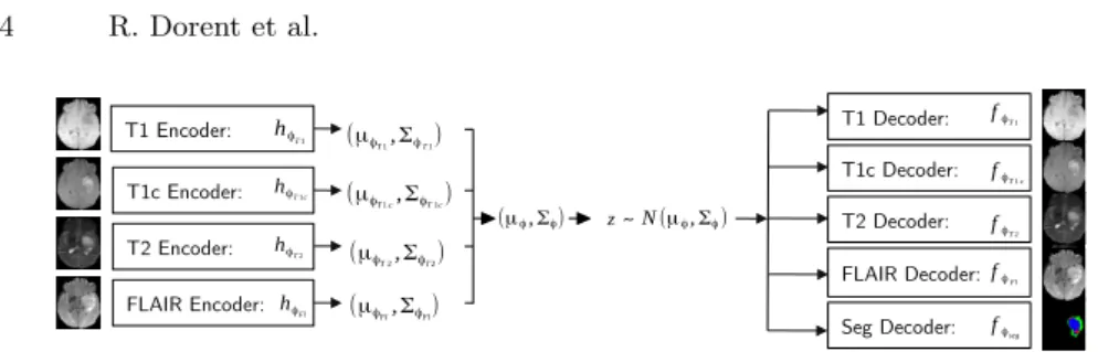
T1 Encoder: T1c Encoder: T2 Encoder: FLAIR Encoder: (μϕT1,ΣϕT1) hϕT1 hϕFl N(μϕ,Σϕ) ~ z T1 Decoder: fϕT1 T1c Decoder: fϕT1c T2 Decoder: fϕT2 FLAIR Decoder: fϕFl
Seg Decoder: fϕseg
hϕT1c hϕT2 (μϕT1c,ΣϕT1c) (μϕT2,ΣϕT2) (μϕFl,ΣϕFl) (μϕ,Σϕ)
Fig. 1.MVAE architecture. Each imaging modality is encoded independently, the mean and covariance of eachq(z|xi) are fused using the closed-form formula (4). A samplez is randomly drawn and is decoded into imaging modalities and the segmentation map.
as the tumour segmentation. Although segmentation could be considered as a missing modality, we chose not to encode it as it is not observed in practice. Consequently, our model is composed of 4 encoders and 5 decoders (see Fig. 1). Without loss of generality, we consider a training set providing the complete n modalities per subject. Consequently, during training, we can artificially re-move some modalities as input yet evaluate the reconstruction error on all the modalities. When the training set is incomplete, the reconstruction error is only evaluated on the available data.
LetP denote the set of all possible non-empty combinations of then modal-ities. Our goal is to maximise (2) whenzhas been encoded via a random subset π ∈ P drawn with probability απ. This is exactly the ancestral sampling of a
mixture model: we first draw the class label (here the subset) and then we draw a sample from the distribution associated to this class. For this reason, we model qφ(z|x) as a mixture where the probabilities απ are chosen to be representative
of the clinical scenario:
qφ(z|x) = X
π∈P
απqφπ(z|xπ)
We chooseqπ
φ(z|xπ) as Gaussian. Given the convexity of the KL divergence and
the fact thatP
π∈Pαπ= 1, we obtain:
KL[qφ(z|x)||p(z)]≤ X
π
απKL[qφπ(z|xπ)||p(z)]
Finally, our lower-bound is a weighted sum of the subset-specific lower-bound:
L(x;θ)≥X π∈P απ(Eqπ φ(z|xπ)[log(pθ(x|z))]−KL[q π φ(z|xπ)||p(z)] | {z } ELBOπ(x) ) (5)
The single Gaussian prior model forp(z) promotes consistency of the embedding z across the subsets of modalitiesπ(qπφ(z|xπ)) and in turn across the full set of
modalities (qφ(z|x)). In our optimisation procedure, at each iteration, we propose
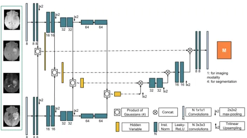
M x2 x2 x2 x2 2x2x2 max-pooling Trilinear Upsampling Product of Gaussians (4) Hidden Variable Concat. N 3x3x3 convolutions Inst. Norm Leaky-ReLU N N 1x1x1 Convolutions 8 8 32 64 N 1: for imaging modality 4: for segmentation 32 8 16 32 64 32 x2 x2 x2 8 8 16 32 64 8 16 32 64 ~ ~ ~ ~ 16 16 16 8 8 x2 x2 x2 x2
Fig. 2. Our 3D variational encoder-decoder (U-HVED). Only two encoders and one decoders are shown. Product of Gaussian is defined in (4).
optimise ELBOπ(x). Classical modelling ofpθ(.|z) includes Gaussian distribution
for image reconstruction and Bernoulli distribution for classification.
2.3 Network architecture: 3D Variational Encoder-Decoder
To exploit our framework we propose a novel network architecture: a 3D encoder-decoder with variational skip-connections. Our model is a mix between a 3D U-Net [10] and the MVAE [13].
In the U-net architecture, context information is extracted via the contract-ing path (encoder) and precise localisation is produced by the expandcontract-ing part (decoder). In addition, information is captured at different levels via the skip-connections. To avoid a trivial identity function, existing auto-encoder architec-tures do not use skip-connections. In our case, the encoding of the latent variable is multi-modal and the imposed consistency of the latent representation creates a bottleneck. Skip-connections therefore do not allow for trivial identity mapping and can be included in our architecture.
We propose to use a multi-level latent variable to generate them. Figure 2 shows our network architecture. Unlike the existing hierarchical VAE models [11,14], we propose a fully convolutional network. Each modality i is indepen-dently encoded which produces 4 multi-scale means and variances (µk
i, Σki)k∈[1,..,4].
At each level, the means and the variances of the modalities present in the input subset xπ are combined via the product of Gaussian defined in (4). We then
decode the multi-scale latent variable for each of the modalities and the segmen-tation. Consequently, we havenencoders andn+ 1 decoders. We assert that it is the first deep network which allows for missing modalities and performs 3D imaging reconstruction and segmentation in a variational manner.
3
Data and implementation details.
Data. We evaluate our method on the training set of BRATS18 [7]. The training set contains the scans of 285 patients, 210 with high grade glioma and 75 with low grade glioma. Each patient was scanned with four sequences (T1, T1c, T2 and FLAIR) and pre-processed by the organisers: scans have been skull-striped and re-sampled to an isotropic 1mm resolution, and the four sequences of the each patient have been co-registered. The ground truth was obtained by manual segmentation results given by experts. The segmentation classes include the following tumour tissue labels: 1) necrotic core and non-enhancing tumour, 2) oedema, 3) enhancing core.
Implementation details. As pre-processing step, we used histogram-based scale standardisation method [8] followed by a zero mean and unit-variance nor-malisation. As a data augmentation, we randomly flip the axes and include a rotation with a random angle in [−10◦,10◦]. The networks were implemented in Tensorflow using NiftyNet [2]. We used Adam as optimiser with initial learning rate 10−3 divided by 4 every 104 iterations, batch size 1 and maximal iteration
60k. Early stopping is performed if a plateau of performance is reached on the validation data set. At each iteration, a 112×112×112 random patch is fed to the network. We did a 3-fold validation by random split of the data set a training (70%), validation (10%) and testing (20%) sets. We regularize with aL2 weight decay of 10−5. During training, we uniformly draw a number of modalities i between 1 and 4 and uniformly draw a subsetπof sizei. During inference, given a subset of modalities, we randomly draw 10 hidden variablezfromq(.|xπ) and
decode them and average the outputs. Implementation is publicly available1.
Choices of the losses. The reconstruction loss follows frompθ(xi|z). For the
segmentation we use the sum of the cross-entropyLcross and the dice loss
func-tion Ldice [4]. For the imaging reconstruction loss, we used the classic L2 loss.
Additionally, given a drawn subsetπ, our loss includes the closed-form KL diver-gence between the Gaussiansqφ(z|xπ) andp(z). For weighting the regularization
losses (KL divergence and reconstruction loss), we did a grid search over weights in [0,0.1,1]. Finally, the loss associated to maximising the ELBO (5) is:
L=Ldice+Lcross+ 0.1∗L2+ 0.1∗KL
4
Experiments and results
Model comparison. To evaluate the performance of our model (U-HVED), we compare it to three different approaches: The first, HeMIS is the model de-scribed in [3] and is the current state-of-the-art for segmentation with missing modalities. The second,U-HeMIS, is a particular case of our method where the
1
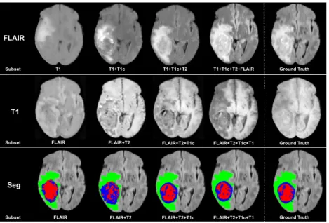
T1
FLAIR FLAIR+T2 FLAIR+T2+T1c FLAIR+T2+T1c+T1 Ground Truth
Seg
Subset
FLAIR FLAIR+T2 FLAIR+T2+T1c FLAIR+T2+T1c+T1 Ground Truth Subset
T1 T1+T1c T1+T1c+T2 T1+T1c+T2+FLAIR Ground Truth
FLAIR
Subset
Fig. 3.Example of FLAIR and T1 completion and tumour segmentation given a subset of modalities as input. Green: edema; Red: non-enhancing core; Blue: enhancing core.
modalities are encoded asU-HVEDand the skip-connection are the first and sec-ond moments of the modality-specific feature maps such as inHeMIS.U-HeMIS
has only one decoder for tumour segmentation. The third approach, Single, is the ”brute-force” method in which for each possible subset of modalities, we train a U-Net network where the observed modalities are concatenated as input. The encoder and decoder are those of our model. Given the 3-fold validation, we consequently trained 45Single networks.
Missing modalities completion. Unlike these three approaches, U-HVED
(Ours) generates missing modalities. Since image completion is a means rather than an end, we only provided a qualitative evaluation (Fig. 3) of T1 and FLAIR reconstruction examples. We find the reconstruction to be good quality, given that VAEs classically suffer of blurriness. Interestingly, our model tries to recon-struct the tumour information even when the tumour information is missing or not clear, such as in T1 scans. Moreover, comparable reconstructions are per-formed using 3 modalities and 4 modalities. This suggests that our network can effectively learn a common representation of the imaging modalities.
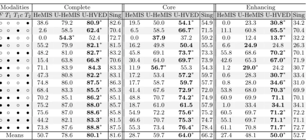
Tumour segmentation. In order to evaluate the robustness of our model, we present qualitative results in Fig. 3 and comparative results with other methods in Table 1 for all the possible input subsets. We used the Dice Similarity as metric. First, the U-Net architecture in U-HeMIS always achieves better
per-Table 1.Comparison of the different models (Dice %) for the different combinations of available modalities. Modalities present are denoted by•, the missing ones by ◦.∗ denotes significant improvement provided by a Wilcoxon test (p <0.05).
Modalities Complete Core Enhancing
F T1T1c T2HeMIS U-HeMIS U-HVED Sing HeMIS U-HeMIS U-HVED Sing HeMIS U-HeMIS U-HVED Sing
◦ ◦ ◦ • 38.6 79.2 80.9∗ 82.6 19.5 50.0 54.1∗ 54.9 0.0 23.3 30.8∗ 34.2 ◦ ◦ • ◦ 2.6 58.5 62.4∗ 70.4 6.5 58.5 66.7∗ 71.5 11.1 60.8 65.5∗ 70.4 ◦ • ◦ ◦ 0.0 54.3∗ 52.4 72.7 0.0 37.9 37.2 59.2 0.0 12.4 13.7∗ 32.2 • ◦ ◦ ◦ 55.2 79.9 82.1∗ 81.5 16.2 49.8 50.4 55.5 6.6 24.9 24.8 26.3 ◦ ◦ • • 48.2 81.0 82.7∗ 83.2 45.8 69.1 73.7∗ 73.3 55.8 68.6 70.2∗ 70.1 ◦ • • ◦ 15.4 63.8 66.8∗ 70.6 30.4 64.0 69.7∗ 73.9 42.6 65.3 67.0∗ 71.9 • • ◦ ◦ 71.1 83.9 84.3 83.3 11.9 56.7∗ 55.3 54.3 1.2 29.0∗ 24.2 30.7 ◦ • ◦ • 47.3 80.8 82.2∗ 83.1 17.2 53.4 57.2∗ 59.7 0.6 28.3 30.7∗ 33.4 • ◦ ◦ • 74.8 86.0 87.5∗ 86.3 17.7 58.7 59.7 57.7 0.8 28.0 34.6∗ 31.0 • ◦ • ◦ 68.4 83.3 85.5∗ 85.3 41.4 67.6 72.9∗ 72.0 53.8 68.0 70.3∗ 69.9 • • • ◦ 70.2 85.1 86.2∗ 85.1 48.8 70.7 74.2∗ 74.9 60.9 69.9 71.1 70.1 • • ◦ • 75.2 87.0 88.0∗ 85.7 18.7 61.0 61.5 57.9 1.0 33.4 34.1 34.1 • ◦ • • 75.6 87.0 88.6∗ 85.8 54.9 72.2 75.6∗ 75.2 60.5 69.7 71.2∗ 72.2 ◦ • • • 44.2 82.1 83.3∗ 81.5 46.6 70.7 75.3∗ 74.7 55.1 69.7 71.1∗ 71.1 • • • • 73.8 87.6 88.8∗ 87.5 55.3 73.4 76.4∗ 78.4 61.1 70.8 71.7∗ 72.7 Means 50.7 78.6 80.1∗ 81.6 28.7 59.7 64.0∗ 66.2 27.4 48.1 50.0∗ 52.7
formance than the original 2D fully-convolutionnal HeMIS. This highlights the efficiency of the 3D U-net architecture. Secondly,U-HVED (Ours) outperforms significantly U-HeMIS in most of the cases: 13 out of 15 cases for the com-plete tumour, 10 out of 15 cases for the core tumour; 11 out 15 cases for the enhancing tumour. This demonstrates that auto-encoding and modality comple-tion improves the segmentacomple-tion performance. Finally,U-HVED achieves similar performance to the 15 subset-specific models (Single). Again, this suggests that the imaging modalities are efficiently embedded in the latent space.
5
Discussion and conclusion
In this work, we demonstrate the efficacy of a multi-modal variational approach for segmentation with missing modalities. Our model outperforms the state-of-the-art approach HeMIS [3]. In fact, HeMIS could be seen as the non-variational version of our method where: 1/one does not sample but uses the mean of the latent variable instead; 2/the modality-specific covariances are set up to the identity, Σi = I; 3/only the segmentation is reconstructed from the hidden
variable. In this case, each modality are independently encoded and averaged such as HeMIS. Finally, our method (U-HVED) offers promising insight for leveraging large but incomplete data sets. For future work, we want to provide an analysis of the the learned embedding. This task is particularly challenging due to the multi-scale representation of the hidden variable.
Acknowledgement We thank C. Sudre, W. Li, B. Murray, Z. Eaton-Rosen, F. Bragman, L. Fidon and T. Varsavsky for their useful comments. This work was supported by the Wellcome Trust [203148/Z/16/Z] and EPSRC [NS/A000049/1]. TV is supported by a Medtronic/RAEng Research Chair [RCSRF1819/7/34].
References
1. Cao, Y., Fleet, D.J.: Generalized product of experts for automatic and principled fusion of Gaussian Process Predictions. CoRRabs/1410.7827(2014)
2. Gibson, E., Li, W.,et al.: NiftyNet: a deep-learning platform for medical imaging. Computer Methods and Programs in Biomedicine158, 113 – 122 (2018)
3. Havaei, M., Guizard, N., Chapados, N., Bengio, Y.: Hemis: Hetero-modal image segmentation. In: MICCAI 2016. pp. 469–477. Springer, Cham (2016)
4. Isensee, F.,et al.: No new-net. In: Brainlesion: Glioma, Multiple Sclerosis, Stroke and Traumatic Brain Injuries. pp. 234–244. Springer, Cham (2019)
5. Kingma, D.P., Welling, M.: Auto-encoding variational bayes. In: ICLR (2014) 6. Li, R., Zhang, W., Suk, H.I., Wang, L., Li, J., Shen, D., Ji, S.: Deep learning based
imaging data completion for improved brain disease diagnosis. In: MICCAI 2014. pp. 305–312. Springer, Cham (2014)
7. Menze, B.H., et al.: The multimodal brain tumor image segmentation benchmark BRATS. In: IEEE Transactions on Medical Imaging. vol. 34, pp. 1993–2024 (2015) 8. Milletari, F., N.N., Ahmadi, S.: V-net: Fully convolutional neural networks for volumetric medical image segmentation. In: Inter-national Conference on 3D Vision (3DV),. pp. 565–571 (2016)
9. Myronenko, A.: 3d MRI brain tumor segmentation using autoencoder regular-ization. In: Brainlesion: Glioma, Multiple Sclerosis, Stroke and Traumatic Brain Injuries. pp. 311–320. Springer, Cham (2019)
10. Ronneberger, O., Fischer, P., Brox, T.: U-net: Convolutional networks for biomed-ical image segmentation. In: MICCAI 2015. pp. 234–241. Springer, Cham (2015) 11. Sø nderby, C.K., Raiko, T., Maalø e, L., Sø nderby, S.r.K., Winther, O.: Ladder
variational autoencoders. In: NeurIPS. pp. 3738–3746 (2016)
12. Varsavsky, T., et al.: PIMMS: permutation invariant multi-modal segmentation. In: DLMIA 2018, MICCAI 2018. pp. 201–209 (2018)
13. Wu, M., Goodman, N.: Multimodal generative models for scalable weakly-supervised learning. In: NeurIPS. pp. 5580–5590 (2018)
14. Zhao, S., Song, J., Ermon, S.: Learning hierarchical features from deep generative models. In: ICML. pp. 4091–4099 (2017)