R E V I E W A R T I C L E
O p e n A c c e s s
The hypoxic tumour microenvironment
Varvara Petrova
1, Margherita Annicchiarico-Petruzzelli
2, Gerry Melino
1,3and Ivano Amelio
1Abstract
Cancer progression often bene
fi
ts from the selective conditions present in the tumour microenvironment, such as the
presence of cancer-associated
fi
broblasts (CAFs), deregulated ECM deposition, expanded vascularisation and
repression of the immune response. Generation of a hypoxic environment and activation of its main effector,
hypoxia-inducible factor-1 (HIF-1), are common features of advanced cancers. In addition to the impact on tumour cell biology,
the in
fl
uence that hypoxia exerts on the surrounding cells represents a critical step in the tumorigenic process.
Hypoxia indeed enables a number of events in the tumour microenvironment that lead to the expansion of aggressive
clones from heterogeneous tumour cells and promote a lethal phenotype. In this article, we review the most relevant
fi
ndings describing the in
fl
uence of hypoxia and the contribution of HIF activation on the major components of the
tumour microenvironment, and we summarise their role in cancer development and progression.
Hypoxia and hypoxia-inducible factors
The major components of the tumour
microenviron-ment (TME) are blood vessels, lymphatic vessels,
fi
bro-blasts, immune cells and chemico-physical components
such as the extracellular matrix (ECM)
1. The functional
and physical interaction of these elements with cancer
cells determines clinical outcomes. During tumour
development and progression, cancer and stromal cells
often have restricted access to nutrients and oxygen. Most
solid tumours indeed have regions permanently or
tran-siently subjected to hypoxia because of aberrant
vascu-larisation and a poor blood supply
2. The hypoxic response
is mainly ascribed to hypoxia-inducible factors (HIFs).
HIF-dependent signalling can promote the adaptation and
selection of both cancer and stromal cells to the
sur-rounding conditions, thus promoting changes that favour
cancer progression. The HIF family of transcription
fac-tors includes HIF1, HIF2 and HIF3. These facfac-tors all
contain an oxygen-sensitive HIF-
α
subunit (HIF1-
α
,
HIF2-
α
or HIF3-
α
, respectively), which dimerises with the
constitutively expressed HIF1-
β
subunit
3. HIF1-
α
and
HIF2-
α
proteins are the best studied among HIF-
α
subunits. Each of these subunits contains two proline
residues (HIF1-
α
: P402/P564 and HIF2-
α
: P405/P531),
which are hydroxylated in the presence of oxygen by
prolyl hydroxylase domain-containing proteins (PHDs).
Hydroxylation of the proline residues promotes binding
to von Hippel-Lindau tumour suppressor (pVHL), leading
to HIF-
α
ubiquitination and degradation
4,5. Another
fac-tor regulating HIF-
α
in an oxygen-dependent manner is
factor inhibiting HIF1 (FIH1). Asparagine hydroxylation
of HIF1-
α
(and to a lesser extent, of HIF2-
α
) driven by
FIH1 impedes HIF1 interaction with its cofactors, histone
acetylases p300 and CBP, and hence impairs HIF1
tran-scriptional activity
6–8. The hypoxic tumour
micro-environment
(TME)
is
subjected
to
HIF-driven
transcriptional responses in cancer and stromal cells. In
addition, HIF activity switches the cell metabolism into
glycolytic mode, increasing glucose consumption and
pyruvate, lactate and H
+
production. In this review
arti-cle, we summarise and discuss the in
fl
uence of hypoxia
and HIFs on TME components and how this impacts
cancer progression.
Cancer-associated
fi
broblasts (CAFs)
It is widely accepted that
fi
broblasts in
fi
ltrating tumour
tissue acquire very different features from normal
fi
bro-blasts, leading to the de
fi
nition of CAF. CAFs often
represent the major component of tumour stroma,
© The Author(s) 2018
Open AccessThis article is licensed under a Creative Commons Attribution 4.0 International License, which permits use, sharing, adaptation, distribution and reproduction in any medium or format, as long as you give appropriate credit to the original author(s) and the source, provide a link to the Creative Commons license, and indicate if changes were made. The images or other third party material in this article are included in the article’s Creative Commons license, unless indicated otherwise in a credit line to the material. If material is not included in the article’s Creative Commons license and your intended use is not permitted by statutory regulation or exceeds the permitted use, you will need to obtain permission directly from the copyright holder. To view a copy of this license, visithttp://creativecommons.org/licenses/by/4.0/.
Correspondence: Ivano Amelio ([email protected])
1Medical Research Council, Toxicology Unit, Leicester University, Hodgkin Building, Lancaster RoadP.O. Box 138, Leicester LE1 9HN, UK
2Biochemistry Laboratory, IDI-IRCCS, Rome, Italy
Full list of author information is available at the end of the article
1234567890()
:,;
1234567890(
sometimes constituting up to the 80% of the entire
tumour
9. The population of CAFs can be quite
hetero-geneous, as several progenitor cell types can be
repro-grammed into CAFs. Although most CAFs are considered
to arise from resident
fi
broblasts, bone marrow cells,
adipocytes, endothelial cells and epithelial cells can also
turn into CAFs
10–17.
Reciprocal paracrine signalling between murine cancer
cells and
fi
broblasts was described by Olaso et al.
Mela-noma cells could induce proliferation and expression of
CAF marker
α
-SMA in adjacent
fi
broblasts. These
fi
bro-blasts excessively produced glucosaminoglycans and
MMP-2, promoting the migration of melanoma cells
18.
Following this initial study, the ability of CAFs to favour
tumour progression was shown in a prostate cancer
xenograft model when CAFs were co-injected with
initi-ated (tumorigenic) prostatic epithelial cells and promoted
their tumorigenic potential, in contrast to co-injection
with normal
fi
broblasts
19. A study by Bhomwick and
Colleagues demonstrated that TGF-beta type II receptor
de
fi
ciency in mouse
fi
broblasts led to increased HGF
secretion and initiation of tumour formation in adjacent
prostate and forestomach epithelium
20, suggesting one
possible mechanism of
fi
broblast transformation. Other
examples of paracrine signalling that is deregulated by
CAFs include the secretion of chemokine CXCL12 with
subsequent tumour growth facilitation and the expression
of intra-cellular and extracellular Ca2
+
-binding protein
S100A4 with subsequent tumour progression and
meta-static spread
21,22. Except for paracrine signalling, the
Fig. 1 Tumour stroma and extracellular matrix in hypoxia. A rapidly growing tumour leads to a reduction in the oxygen supply of the cancer and in tumour stromal cells that are far from the blood vessels. In hypoxia, these cells switch to glycolytic metabolism, which contributes to the acidification of the tumour microenvironment. Produced glycolytic metabolites such as lactate can be utilised by cancer cells and promote tumour growth. The hypoxic microenvironment is also enriched in diverse types of immune cells, and many of them are recruited from the circulation. Cytokine expression by tumour and stromal cells is altered by hypoxia. In particular, hypoxic cancer cells produce signalling molecules that promote the transformation offibroblasts into CAFs. Together with cancer cells, in hypoxia, CAFs produce an ECM that is stiff and aligned, different from a normoxic ECM, and support cell migration. CAF cancer-associatedfibroblasts, ECM extracellular matrixoncogenic functions of CAFs are partially mediated by
altered ECM production. In a breast cancer study, ECM
deposited by CAFs was organised differently (aligned)
than ECM produced by normal
fi
broblasts and could
in
fl
uence premalignant human mammary epithelial cells,
assigning them a mesenchymal phenotype and increasing
their
tumorigenic
and
metastatic
potential.
The
mesenchymal phenotype transition in epithelial cells is
dependent on the TGF-
β
-dependent Smad, Erk, Jun and
Rho signalling pathways. As TGF-
β
is stored in the ECM
before activation, the function of CAFs in that model
could consist of increasing TGF-
β
availability as well as
building an ECM framework with a metastasis-promoting
spatial structure
23–26. In addition to the direct effect of
CAFs on cancer cells, they can promote angiogenesis via
vascular endothelial growth factor-C (VEGF), CXCL12a
and FGF-2 factor production and modulate the immune
response by inducing macrophage in
fi
ltration and
tumour-promoting cell polarisation, reducing T-cell
in
fi
ltration and interfering with natural killer cell
function
27.
Hypoxia can in
fl
uence both
fi
broblast reprogramming
and tumour-promoting functions (Fig.
1
). Oxygen de
fi
-ciency in
fl
uences paracrine signalling between cancer
cells and
fi
broblasts. Hypoxia was shown to stimulate
cytokine CXCL13 secretion by cancer-associated myo
fi
-broblasts in prostate cancer progression
28. While CAFs
secrete chemokine CXCL12, facilitating cancer growth
22,
hypoxia was shown to stimulate CXCR4 (CXCL12
receptor) expression in many cell types, therefore
sug-gesting a feed-forward loop between cancer cells and
CAFs
29. Hypoxic cancer cells can secrete paracrine
sig-nalling molecules, which promote reprogramming of
progenitor cells into CAFs
30, and HIF1 was shown to
regulate some of these signalling molecules, such as
TGF-β
, bFGF and PDGF-B
31–33.
It has been shown that the hypoxia-inducible factor-1
(HIF-1)
α
level is often upregulated in CAFs. In a model in
which CAF formation is stimulated by TGF-
β
and PDGF
treatment, the rate of aerobic glycolysis in primary CAFs
was increased compared to that in normal
fi
broblasts, and
this effect was associated with HIF-1
α
protein
stabilisa-tion
34,35. The lactate produced by highly glycolytic CAFs
can be consumed by adjacent cancer cells and lead to
induced tumour growth, which suggests a negative
out-come of HIF-1 upregulation in
fi
broblasts
34. The role of
the increase in HIF-1
α
levels during CAF formation is still
poorly understood, that is, whether it is a driving force for
reprogramming or a consequence. Chiavarina and
col-leagues observed that stable HIF-1
α
overexpression
endowed
fi
broblasts with oncogenic functions, as they
increased tumour growth after ectopic co-injection with
breast cancer cells. The proposed mechanism included
HIF-1
α
-promoted
autophagy,
mitophagy,
and
the
production of lactate and recyclable nutrients, which
could fuel cancer cells and provide them with building
blocks
36–39. However contradicting evidence supports a
negative regulatory role for HIF-1a signalling in stromal
fi
broblast. Kim and Colleagues indeed showed that
selective deletion of HIF-1a (or VEGFA) in
fi
broblast was
enhancing tumour growth in murine mammary cancer
models
40. Additionally after 80 h of hypoxia normal
fi
broblasts were shown to produce a stiff, aligned matrix,
and that this matrix supported the migration of breast
cancer cells
41. Con
fi
rming the abovementioned
experi-ments hypoxia was inducing secretion of protumorigenic
factors, such as hepatocyte growth factor (HGF), in
human
fi
broblast cell line MRC5 due to HIF-1 activity.
Conditioned media from hypoxic MRC5 could promote
invasiveness of pancreatic cancer cell line PK8
42. Thus
several studies have shown gain of oncogenic functions by
fi
broblasts in response to HIF-1 activation, and one can
suggest that HIF-1 is able to drive
fi
broblast
reprogram-ming to CAFs. Seereprogram-ming controversially to the described
studies, Madsen et al. suggest that long-term hypoxia
dampens CAF function in a HIF-1-alfa-dependent way.
These authors showed that 72 h of hypoxic treatment or
72 h of PHD2 silencing impeded the ability of head and
neck CAFs and vulval CAFs to remodel ECM in vivo,
which was accompanied by reduced expression of the
activated
fi
broblast marker
α
-SMA. HIF-1
α
silencing in
these conditions reverted the phenotype. In vivo
treat-ment of breast cancer-bearing mice with PHD-inhibitor
DMOG reduced tumour resilience and metastatic
potential, while it had no effect on tumour size
43. This
controversy could arise from the fact that CAFs indeed
are not identical to normal
fi
broblasts, originate from
different cell types and can develop distinct response. It
might also be possible that HIF-1 promotes some of the
oncogenic functions of CAFs, such as increasing tumour
growth, while inhibiting other oncogenic functions, i.e.,
metastasis promotion. HIF-2 is known to accumulate and
mediate long-term hypoxia responses when HIF-1
α
is
downregulated. Therefore, metastasis inhibition in
long-term hypoxia and some other hypoxic effects of CAFs that
are currently thought to be HIF-1 dependent indeed may
be regulated by HIF-2. HIF-2 functions in CAFs have been
poorly assessed and need further exploration. It is worth
mentioning that after 80 h of hypoxia, normal
fi
broblasts
produce a stiff, aligned matrix and that this matrix
sup-ported the migration of breast cancer cells
41. Taken
together, these studies also raise the possibility of different
hypoxic responses in CAFs compared to normal
fi
broblasts.
Extracellular matrix
The ECM primarily consists of
fi
brillar proteins and
proteoglycans, which together form a net that serves as a
framework for most tissues
44. Collagens are the dominant
component of the ECM and account for approximately
90% of its mass
45. The physical properties of tumour ECM
differ from healthy tissue and continuously change
46–49.
In many cases, solid tumours are characterised by
exces-sive deposition of ECM proteins (
fi
brosis)
50–55, and
especially by collagen deposition
56–61. They are the main
source for synthesis of ECM proteins, namely collagen,
fi
bronectin and hyaluronan, and on the other hand CAFs
are an important source for ECM-remodelling enzymes
62.
CAFs share several features with normal activated
fi
bro-blasts, including the ability to produce ECM components,
which, on contrary of physiologic microenvironment,
results in an abnormal ECM that supports tumour
dis-semination
63. ECM and
fi
broblasts in TME are tightly
reciprocally regulated. Modi
fi
cations of ECM structure or
composition induce cytoskeleton reorganisation and
sig-nalling cascades in CAFs, further regulating synthesis of
ECM components and extracellular remodelling enzymes.
Most of these changes in ECM are often supportive for
formation
of
pro-tumorigenic
microenvironment
64.
Besides CAFs, cancer cells themselves signi
fi
cantly
con-tribute to ECM remodelling
65. In breast cancer,
localisa-tion of
fi
brotic areas often coincides with localisation of
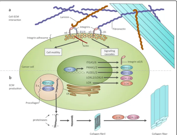
Fig. 2 HIF regulates interactions of cancer cells with ECM and ECM biosynthesis.aRegulation of cell–ECM interactions by HIF. HIF was shown to transcriptionally induce ITGA5 and ITGA6 genes encoding integrinsα5 andα6. Each integrinαsubunit together with aβsubunit forms a specific ECM receptor. Integrinα5β1 bindsfibronectin and integrinα6β1, orα6β4 binds integrin. In the cell, integrins bind with a multi-component complex named the integrin adhesome. Some proteins of this complex can be involved in signalling cascades, and others interact with the cytoskeleton. As a result of interactions with the ECM, cells undergo alteration of their signalling networks and their motility.bHIF contributes to collagen production. P4HA1, P4HA2, PLOD1, PLOD2, LOX, LOXL2 and LOXL4 are transcriptional targets of HIF that are involved in collagen posttranslational modification. P4HA1/2 and PLOD1/2 catalyse thefirst step of procollagen molecule modification, which occurs in the ER and allows the formation of the triple-stranded procollagen molecule. Triple-triple-stranded procollagens are exported from the cell and into the extracellular space, where they are modified by proteinases and assembled in collagenfibrils. Subsequently, LOX, LOXL2 and LOXL4 catalyse the crosslinking of collagenfibrils and the formation of a functional collagenfibre. ER endoplasmic reticulum
hypoxic regions
66,67. HIF1 was shown to directly in
fl
uence
ECM remodelling and promote
fi
brosis in kidney, liver
and adipose tissue
68–70. After hypoxic treatment, rats have
increased mRNA levels of procollagens I, II, and IV
71.
Skin, heart and kidney
fi
broblasts contain elevated mRNA
levels of procollagen I
α
1 chain when cultivated under
hypoxic conditions
72–74. In
fi
broblasts,
fi
bulin-5 was
shown to be transcriptionally induced by hypoxia or in a
PHD inhibitor-induced hypoxic environment in a TGF-
β
/
PI3K/Akt-dependent way, although the role of HIFs in
that process was not assessed
75. Thus, hypoxia may be
able to potentiate ECM protein deposition in tumours in a
HIF-dependent and HIF-independent manner (Fig.
1
).
The metastatic potential of cancer cells depends on
their interactions with the ECM. Cells receive mechanistic
signals from the ECM by means of focal adhesions.
For-mation of focal adhesions requires the binding of ECM
proteins with integrins and cell-surface ECM receptors
and subsequent signal transduction through intermediate
molecules to the cytoskeleton. Silencing of the
ITGA5
gene encoding integrin
α
5, a subunit of
fi
bronectin
receptor
α
5
β
1, reduced breast cancer cell motility,
migration and invasion capacity. These cells had
decreased metastatic potential after orthotopic injection
in mice
76. The same study showed that both HIF-1 and
HIF-2 could induce the transcription of integrin subunits
α
5 and
β
1. Integrin
α
6 was demonstrated to be a direct
transcriptional target of HIF-1 and HIF-2 and to increase
breast cancer cell invasion potential
77. In addition to
integrins, another
fi
bronectin receptor, syndecan-4, is
transcriptionally induced in hypoxia by an unknown
mechanism
78. Thus, hypoxia is implicated in the
regula-tion of cell
–
ECM interactions, partially through HIF (Fig.
2
). In addition to integrin expression, hypoxia regulates
ECM proteins synthesis. HIFs can regulate collagen
pro-duction at several stages (Fig.
2
). Posttranslational
mod-i
fi
cation
of
procollagen
chains
requires
prolyl-4-hydroxylases and procollagen lysyl-prolyl-4-hydroxylases. HIF-1
can induce the expression of prolyl-4-hydroxylase
alfa-subunits P4HA1 and P4HA2 and procollagen
lysyl-hydroxylases PLOD1 and PLOD2 in different cancer
and non-cancer cell lines
41,79–85. Expression of P4HA1,
P4HA2, PLOD1 and PLOD2 is necessary for the
pro-duction of stiff and aligned collagen
fi
brils. In this
microenvironment, cancer cells were shown to take on an
elongated, adhesive, motile phenotype and have an
ele-vated capacity for invasion and migration
41,81,83,84,86.
Another point in the collagen deposition process that is
regulated by HIFs is the deamination of secreted collagen
fi
brils on lysine and hydroxylysine residues by
lysyl-oxidases. This deamination is required for
fi
bril
cross-linking and collagen
fi
bre formation
87. HIF-1 was shown
to induce the expression of lysyl-oxidases LOX
88,
LOXL2
89and LOXL4
90. The LOX family is intimately
linked to the metastatic process via several aspects,
involving ECM remodelling and potentially
ECM-independent EMT regulation
91–93. The role of LOX
expression can be crucial, as lung metastases in a breast
cancer model were shown to be dependent on
LOX-driven
fi
brosis
94. LOX family proteins can mediate several
oncogenic HIF-1 functions. LOX knockdown prevented
focal adhesion formation and reduced cell motility,
abrogating HIF-1-dependent cell migration and
inva-sion
95. In another study, the transformation of cancer cells
to an invasive mesenchymal phenotype was dependent on
HIF-1-driven LOX and LOXL2 expression
89. Hypoxic
LOX, LOXL2, and LOXL4 secretion by breast cancer cells
results in collagen remodelling in lungs, which allows the
recruitment of bone-marrow derived cells (BMDCs) to
the area. ECM remodelling performed by LOX and
BMDC-derived matrix metalloproteinases favours the
formation of pre-metastatic niches, showing that the
oncogenic ECM remodelling and tumour cell recruitment
driven
by
hypoxia
can
also
have
long-distance
effects
88,90,96.
Simultaneous with collagen synthesis, hypoxia promotes
the degradation of extracellular matrix. HIF1 is known to
induce the expression of matrix metalloproteinases
MMP2
97, MMP9
98and MMP15
99and the expression of
urokinase receptor uPAR
97. HIF-2 can increase MMP14
levels
100. Hypoxia-driven expression of these MMPs can
promote invasion and correlates with poor patient
prog-nosis
97,99,101. Downregulation of tissue inhibitors of
metalloproteinases TIMP2 and TIMP3 by hypoxia
represents an additional level of hypoxic control over
ECM remodelling
98,102. The simultaneous increases in
ECM biosynthesis and degradation, which are driven by
HIFs, and the hypoxic regulation of focal adhesion
for-mation creates a mechanism for the metastatic spread of
cancer cells.
Blood vessels
Vascularisation is one of the main outcomes of HIF
signalling. HIF-1
α
and HIF-2
α
inactivation led to
devel-opmental lethality in mice, which was linked to defects in
blood vessel formation
103,104. A connection between
excessive vascularisation and cancer was shown in several
models. Redundant vessel formation is considered a
fea-ture of cancer, and it promotes cancer progression. Some
therapeutic approaches aiming to suppress vascularisation
have been developed
105–108. New vessel development was
shown to be important for the transition from hyperplasia
to neoplasia
109. Although vessels transport oxygen, they
can be affected by hypoxia. The two major components of
blood vessels are endothelial cells and pericytes. Those
endothelial cells that reside at the end of growing
capil-laries and direct their branching are located far from
functional vessels, and they can become hypoxic and thus
develop a hypoxic cellular response
110. HIF-1
α
and
HIF-2
α
presence in endothelial cells differentially affects
vas-cularisation. While HIF-1
α
deletion reduces tumour
vas-cularisation and tumour growth, HIF2-
α
deletion, on the
contrary, is able to augment angiogenesis with the
for-mation of a more disorganised vascular system and more
hypoxic tumours
111–113.
Tumours become hypoxic when they grow too large in
size apparently because the blood supply is insuf
fi
cient. At
this point, tumour growth decelerates, and HIF1
pro-motes the secretion of factors inducing vascularisation
from tumour and stromal cells; for example, VEGF
in
fl
uences endothelial cells, pericytes and BMDC to
induce vessel growth
114–116. The ECM produced by
can-cer cells cultured under hypoxic conditions was also
shown to support angiogenic growth
117,118. HIF-induced
neovascularization attempts a compensation of the
oxy-gen de
fi
ciency in the tumour tissue. But the rate of
uncontrollable proliferation of cancer cells exceeds the
speed of organised capillary net formation. Indeed
sometimes new blood vessels are able to transiently
restore the oxygenation, and in this case cancer tissues
have interchanging hypoxic areas with a combination of
both acute and chronic hypoxic regions. Fluctuations in
red cell
fl
ux in tumour microvessels can lead to transient
hypoxia and reoxygenation in tumour parenchyma
119.
This uncontrolled activation of hypoxia signalling in
tumour mass often results in an aberrant, disorganised
vascularisation that fails to compensate oxygen de
fi
ciency.
It is noteworthy that endothelial cells play an important
role in cancer cell migration, as they are the major
structural component of blood vessels and serve as a
barrier in extravasation and intravasation processes
120.
HIF1-
α
depletion in endothelial cells was shown to
sup-press the migration of tumour cells through endothelial
cells, but HIF2-
α
depletion was shown to stimulate
metastatic spread. These opposite effects of HIF1-
α
and
HIF2-
α
on vessel formation and metastasis can be
explained by the ability of these factors to differentially
regulate the nitric oxide level, which regulates endothelial
cell function
121.
Pericytes are cells embedded in basement membrane of
blood microvessels and cover the endothelial cells. They
regulate angiogenesis, but also participate to other
func-tions, such as formation of blood-brain barrier. Hypoxic
stress of brain pericytes leads to their migration out of the
blood vessels, but at the same time HIF-1-signalling leads
to VEGF level upregulation and vascularisation
stimula-tion
122. During the process of kidney
fi
brosis detachment
of pericytes from the capillaries in response to VEGF and
PDGF is followed by their transformation to
mesenchy-mal
fi
broblasts, actively producing ECM
123. As both of
these factors can be produced in epithelial cells due to
HIF-1 signalling, presumably hypoxia in TME could also
lead to detachment of pericytes and their subsequent
transformation, but this formal experimental evidence in
support of this assumption are currently missing.
Lymphatic vessels
In addition to dissemination in the blood circulation,
cancer cells can disseminate through lymphatic vessels.
Lymphatic vessel density is increased in breast cancer
tissues and correlates with positive lymph node
metas-tases and worsened prognosis
124. An increased HIF1-
α
level in primary malignant neoplasias is tightly associated
with the density of the surrounding tumour lymphatic
vessels and with breast cancer patient mortality
125,126. In
oesophageal cancer, HIF1-
α
levels correlate with the
colonisation of lymph nodes
127. Different studies suggest
a direct contribution of HIF1-
α
in the regulation of
lym-phangiogenesis
128,129. In particular, HIF1-
α
was proven to
induce lymphatic metastases through the activation of
different growth factors, including VEGF-c a and
PDGF-B
33.
Expression
of
VEGF-c,
one
of
the
major
lymphangiogenesis-driving factors, was shown to
corre-late with HIF1-
α
expression
130; however, it remains
unclear whether VEGF-c induction requires HIF1-
α
, as
the data are controversial
131,132. HIF1 contribution to
lymphatic vessel formation can be mediated by
VEGF-a
133. In contrast, there is evidence for the opposite role of
HIF2-
α
in lymphangiogenesis, as HIF2-
α
knockdown
increased lymphatic vessel formation in vivo in a
xeno-graft model
134. The in
fl
uence of hypoxia on lymphatic
vessel formation is a poorly studied topic that needs
fur-ther investigation, as lymphatic vessel formation
con-tributes to metastatic tumour spread.
Immune cells
Adaptive immune cells can potentially inhibit tumour
growth by the recognition of tumour-speci
fi
c antigens on
the surface of cancer cells and the elimination of those
cells. Innate immune cells can promote the antitumour
activity of in
fi
ltrating lymphocytes and lead to signi
fi
cant
tumour regression. HIFs have been shown to be tightly
connected with the in
fl
ammatory processes
135,136, and
hypoxia can directly or indirectly in
fl
uence the function of
almost all immune cell types, thereby in
fl
uencing tumour
development
137.
Innate tumour immunity
Myeloid cell-speci
fi
c PHD2 depletion was shown to
suppress tumour growth and metastases, underlining the
importance of oxygen sensitivity for myeloid cells
138. A
hypoxic TME is known to promote neutrophil
engage-ment in tumours by regulation of their adherence to
epithelial cells
139,140. HIF1-
α
and HIF2-
α
were separately
shown to increase the survival and function of
neu-trophils
141–144. The outcome of this hypoxic effect is still
unknown, as cancer-associated neutrophils can induce
both tumour suppression and tumour progression
145.
An increase in cancer-associated macrophage density
correlates with poor patient prognosis in different types of
cancer
146,147. Macrophage polarisation plays a signi
fi
cant
role in tumorigenesis. Indeed, macrophages polarised to
the M1 type (classical activation) counteract cancer
pro-gression and metastases, while M2-polarised
macro-phages (alternative activation) can promote it
148. Hypoxia
induces tumour cells to secrete chemoattractants, such as
Sema3A, EMAPII, ET-1 and ET-2, promoting the
che-motaxis of macrophages from the circulation
149–151.
HIF1-
α
was shown to be necessary for macrophage
maturation, function
141and glycolytic reprogramming
and when associated with HIF1-
α
-induced PDK1 activity,
it increases the migratory capacity of macrophages,
representing a possible mechanism of macrophage in
fi
l-tration regulation by HIF1-
α
152. In addition, hypoxia
determines macrophage polarisation through the
induc-tion of M2 polarisainduc-tion-related genes
153and promotes
lactic-acid-induced M2 polarisation
154. In vivo
experi-ments show that HIF1-
α
and HIF2-
α
are both crucial for
macrophage in
fi
ltration and immune suppression in
tumours, as their separate ablation led to reduced tumour
growth
155,156.
Another noteworthy innate immune cell is the
myeloid-derived suppressor cell (MDSC). These cells originate
from bone marrow cells, and their numbers are
sig-ni
fi
cantly increased in cancer. Their main feature is the
ability to suppress the activity of other immune cells and
therefore suppress the antitumour immune response
157.
The role of hypoxia in MDSC regulation has been
con-sidered mostly oncogenic. Hypoxia-treated MDSCs show
activation of HIF-signalling and of those HIF targets that
enhance MDSC function
158. Hypoxia can also augment
MDSC function through a mechanism partially
depen-dent on HIF: by miR-210 regulation and ArgI
expres-sion
158,159. However, some studies suggest that MDSCs
are able to gain immunostimulatory properties
160. Liu and
Colleagues described MDSCs
’
differentiation into the
tumour-suppressing M1 subtype after SIRT1 induction,
and this differentiation occurred as a result of mTOR/
HIF-1
α
-dependent glycolytic reprogramming
161. This
observation suggests that HIF1 may also endow MDSCs
with tumour-suppressive functions and that the role of
HIFs in MDSC regulation needs to be further investigated.
Adaptive tumour immunity
Although T-cells are able to in
fi
ltrate into the tumour,
anticancer immunity is often limited due to
character-istics of the TME and a hypoxic environment
162.
Increased glycolysis in the tumour, which is partially
mediated by HIF activity, is a reason for so-called
“
metabolic competition
”
between cancer cells and
T-cells. Lack of nutrients suppresses T-cell function and the
antitumour response
163–165. Hypoxia induces the
differ-entiation of non-speci
fi
c CD4
+
cells into regulatory
T-cells (CD4
+
CD25
HighFOXP3
+
) or T-helpers (
Тн
17), and
expression of transcriptional factors FOXP3 and ROR
γ
t,
which are crucial for differentiation, is regulated by
HIF-1
166–168. While regulatory T-cells have
immunosuppres-sive functions, the contribution of
Тн
17 cells to the
immune response is unclear
169. In addition, under
hypoxic conditions, cancer cells and macrophages
syn-thesise chemokines and cytokines, which attract
reg-ulatory T-cells from the circulation and repress the
antitumour response of other T-cells
170–172. Regulatory
T-cells in hypoxia produce extracellular adenosine, which
represses
effector
T-cell
function
173,174.
Another
mechanism of effector T-cell suppression involves a
hypoxia-dependent increase in lysyl oxidase secretion,
which causes the formation of premetastatic niches.
MDSCs can migrate into those niches and suppress the
anti-tumour response of T-killers
95,175,176.
HIF1 is involved in the regulation of T-cell immune
checkpoints (Fig.
3
). In cancer cells, macrophages,
den-dritic cells and MDSCs, HIF1-
α
directly induces PD-L1
expression
177–179. Binding of PD-L1 expressed on the cell
surface to the PD-1 receptor on T-cells leads to their
dysfunction, and hence, this mechanism is a target for
different pharmaceutical approaches, such as the
devel-opment of anti-PD-L1 antibodies and PD-1/PD-L1
interaction inhibitors
180,181. Another checkpoint
regu-lated by hypoxia is the CTLA-4 receptor, which is
upre-gulated on CD8
+
T-cells in hypoxia potentially via
HIF1
182. Binding of CTLA-4 on T-cells to ligands CD80
and CD86 on the surface of antigen-presenting cells
results in effector T-cell inhibition and regulatory T-cell
activation
183. As with PD-1, anti-cancer treatments using
4 blocking antibodies have been developed.
CTLA-4 and PD-1/PD-L1-targeted therapies showed positive
responses in clinical trials for several types of cancer
184– 186. However, the outcome of the therapies is dependent
on many parameters, including the frequency of
tumour-in
fi
ltrating lymphocytes, which inversely correlates with
the glycolytic rate and HIF1-
α
expression
187,188.
In contrast to the described effector T-cell suppression
by hypoxia, some data indicate that HIF function is
important for the activity of effector cytotoxic T-cells. It
was shown by Doedens and colleagues in 2013 that
ele-vated HIF-1 and HIF-2 support the function of cytotoxic
CD8
+
T-cells
182. In agreement with these
fi
ndings, a
recent study by Tyrakis et al. has suggested a role for
HIF1-
α
in CD8
+
T-cell proliferation, differentiation and
antitumour activity through the regulation of L-2HG
189.
Apart from cytotoxic T-cells, HIF-1 was also shown to
contribute to natural killer cell priming and activation via
regulation of the glycolytic rate
190, thus demonstrating a
stimulatory role in immune activation.
Conclusions
The role of TME in cancer progression is currently
attracting impressive interest in the
fi
eld. Hypoxia is a
condition that often occurs at late stages of cancer, and
even before that, HIFs can be upregulated due to
envir-onmental acidi
fi
cation and the presence of glycolytic
metabolites
191–193. The HIF-mediated hypoxic impact on
TME in most cases is mediated by transcriptional activity
of HIFs, secretion of signalling molecules by cancer cells
and tumour stromal cells, and metabolic changes
asso-ciated with the switch from oxygen-dependent catabolism
to glycolysis. Non-tumorous cells in the TME are all
affected by hypoxia and HIFs (Table
1
). Often, hypoxia
leads to their dysregulation in a way that supports cancer
growth:
fi
broblasts can be transformed into tumour-prone
CAFs, ECM remodelling supports metastases,
vascular-isation process facilitates cancer progression, and
anti-tumour immune function becomes generally repressed.
Nevertheless, the hypoxic response can also be
detri-mental for tumorigenesis (Table
1
). This
fi
nding can be
partially explained by the differences in HIF-1 and HIF-2
stability and function, as acute hypoxic response is mainly
mediated by HIF1 activity, and chronic hypoxia by
HIF2
194. Dual roles can also arise from the presence of
HIF interactors, among which the p53 family and MDM2
can play important roles, as they can affect HIF stability
and function
195,196. In this context, the interplay between
Fig. 3 Immune checkpoints in the tumour microenvironment. Effector T cells infiltrating the tumour can become repressed due to the activation of immune checkpoints. The targeting of PD-1 and CTLA-4 checkpoint pathways with specific antibodies is a promising therapeutic approach. In the tumour microenvironment, these pathways can be activated by the following mechanisms.aPD-1 receptor binding to its ligand PD-L1 leads to effector T-cell repression. PD-L1 can be expressed on the surface of cancer cells, MDSCs, DCs, and macrophages, and in these cells, it is directly transactivated by HIF in hypoxia.bBinding of interferonγsecreted by active effector T cells to its receptor on cancer cells results in activation of PD-L1 gene expression and subsequent T-cell repression.cPD-L1 gene expression can be constantly upregulated in cancer cells because of oncogenic mutations and signalling alteration, which leads to effector T cell repression upon interaction with this type of tumour cell.dAntigen-presenting cells express CD80 and CD86 ligands. Upon CD80/86 binding to CTLA-4 receptor on T cells, they become functionally repressed. Hypoxia was shown to induce CTLA-4 expression in cells, which potentially could contribute to their repression in the hypoxic tumour microenvironment. Teff effector T-cell, MDSC myeloid-derived suppressor T-cell, DC dendritic T-cell, IFNγinterferon gamma, IFN-γR interferon gamma receptor, APC antigen-presenting cellHIF and the p53 family can in
fl
uence a wide range of
cellular processes speci
fi
cally associated with complexity
of p53 family members in controlling cell death
197–201,
metabolism
202–207,
reproduction
208,209,
and
develop-ment
210–213.
Considering the global impact of HIF on cancer cells
and the TME, therapies targeting HIF activity, such as the
usage of small molecules preventing the interactions of
the HIF-
α
and HIF1-
β
subunits
214, can be bene
fi
cial for
some groups of patients.
Author details
1Medical Research Council, Toxicology Unit, Leicester University, Hodgkin Building, Lancaster RoadP.O. Box 138, Leicester LE1 9HN, UK.2Biochemistry Laboratory, IDI-IRCCS, Rome, Italy.3Department of Experimental Medicine and Surgery, University of Rome“Tor Vergata”, 00133 Rome, Italy
Conflict of interest
The authors declare that they have no competing interests.
Received: 31 July 2017 Accepted: 4 October 2017
References
1. Balkwill, F. R., Capasso, M. & Hagemann, T. The tumor microenvironment at a glance.J. Cell. Sci.125, 5591–5596 (2012).
2. Pouyssegur, J., Dayan, F. & Mazure, N. M. Hypoxia signalling in cancer and approaches to enforce tumour regression.Nature441, 437–443 (2006). 3. Wang, G. L., Jiang, B. H., Rue, E. A. & Semenza, G. L. Hypoxia-inducible factor 1
is a basic-helix-loop-helix-PAS heterodimer regulated by cellular O2 tension.
Proc. Natl. Acad. Sci. USA92, 5510–5514 (1995).
4. Jaakkola, P. et al. Targeting of HIF-alpha to the von Hippel-Lindau ubiqui-tylation complex by O2-regulated prolyl hydroxylation.Science292, 468–472 (2001).
5. Ohh, M. et al. Ubiquitination of hypoxia-inducible factor requires direct binding to the beta-domain of the von Hippel-Lindau protein.Nat. Cell. Biol. 2, 423–427 (2000).
6. Koivunen, P., Hirsila, M., Gunzler, V., Kivirikko, K. I. & Myllyharju, J. Catalytic properties of the asparaginyl hydroxylase (FIH) in the oxygen sensing pathway are distinct from those of its prolyl 4-hydroxylases.J. Biol. Chem. 279, 9899–9904 (2004).
7. Mahon, P. C., Hirota, K. & Semenza, G. L. FIH-1: a novel protein that interacts with HIF-1alpha and VHL to mediate repression of HIF-1 transcriptional activity.Genes. Dev.15, 2675–2686 (2001).
Table 1 In
fl
uence of hypoxia on TME components
TME component Influence of hypoxic environment Cancer suppression (+)/promotion (−)
CAF progenitors Recruitment, activation and transformation into CAFs −
CAFs Secretion of cytokines, promoting tumour growth −
Deactivation by chronic hypoxia +
ECM Increase of ECM deposition (fibrosis) and remodelling −
Increase of interaction with cancer cells −
Blood vessels Increase in vascularisation by HIF-1 −
Decrease in vascularisation by HIF-2 +
Lymphatic vessels Increase in lymphangiogenesis by HIF-1 −
Decrease in lymphangiogenesis by HIF-2 ?
Neutrophils Recruitment to tumour ?
Increase in survival and function ?
Macrophage Maturation and functioning are dependent on HIF1 ?
Recruitment from bloodflow −
Increase in migratory capacity −
M2 polarisation −
MDSCs Enhancement of immune-suppressive function −
Polarisation to M1 type, driven by SIRT1 +
Non-specific CD4+ cells Differentiation into regulatory T-cells and T-helpers −/?
Regulatory T-cells Recruitment from bloodflow −
Enhancement of immune suppressive function −
Effector T-cells Immune checkpoints activation −
Repression due to“metabolic competition”for nutrients − CD8+ T-cells Support in proliferation, differentiation and activation +
8. Mandl, M., Lieberum, M. K. & Depping, R. A HIF-1alpha-driven feed-forward loop augments HIF signalling in Hep3B cells by upregulation of ARNT.Cell.
Death Dis.7, e2284 (2016).
9. Olive, K. P. et al. Inhibition of Hedgehog signaling enhances delivery of chemotherapy in a mouse model of pancreatic cancer. Science 324, 1457–1461 (2009).
10. Baroni, S. et al. Exosome-mediated delivery of miR-9 induces cancer-associatedfibroblast-like properties in human breastfibroblasts.Cell. Death Dis.7, e2312 (2016).
11. Bochet, L. et al. Adipocyte-derivedfibroblasts promote tumor progression and contribute to the desmoplastic reaction in breast cancer.Cancer Res.73, 5657–5668 (2013).
12. Lecomte, J. et al. Bone marrow-derived myofibroblasts are the providers of pro-invasive matrix metalloproteinase 13 in primary tumor.Neoplasia14, 943–951 (2012).
13. McDonald, L. T. et al. Hematopoietic stem cell-derived cancer-associated fibroblasts are novel contributors to the pro-tumorigenic microenvironment.
Neoplasia17, 434–448 (2015).
14. Quante, M. et al. Bone marrow-derived myofibroblasts contribute to the mesenchymal stem cell niche and promote tumor growth.Cancer Cell.19, 257–272 (2011).
15. Radisky, D. C., Kenny, P. A. & Bissell, M. J. Fibrosis and cancer: do myofi bro-blasts come also from epithelial cells via EMT?J. Cell. Biochem.101, 830–839 (2007).
16. Tang, X. et al. Stromal miR-200s contribute to breast cancer cell invasion through CAF activation and ECM remodeling.Cell. Death. Differ.23, 132–145 (2016).
17. Zeisberg, E. M., Potenta, S., Xie, L., Zeisberg, M. & Kalluri, R. Discovery of endothelial to mesenchymal transition as a source for carcinoma-associated fibroblasts.Cancer Res.67, 10123–10128 (2007).
18. Olaso, E. et al. Tumor-dependent activation of rodent hepatic stellate cells during experimental melanoma metastasis.Hepatology26, 634–642 (1997). 19. Olumi, A. F. et al. Carcinoma-associatedfibroblasts direct tumor progression of initiated human prostatic epithelium.Cancer Res.59, 5002–5011 (1999). 20. Bhowmick, N. A. et al. TGF-beta signaling infibroblasts modulates the
oncogenic potential of adjacent epithelia.Science303, 848–851 (2004). 21. Grum-Schwensen, B. et al. Suppression of tumor development and
metas-tasis formation in mice lacking the S100A4(mts1) gene.Cancer Res. 65, 3772–3780 (2005).
22. Orimo, A. et al. Stromalfibroblasts present in invasive human breast carci-nomas promote tumor growth and angiogenesis through elevated SDF-1/ CXCL12 secretion.Cell121, 335–348 (2005).
23. Dumont, N. et al. Breastfibroblasts modulate early dissemination, tumor-igenesis, and metastasis through alteration of extracellular matrix char-acteristics.Neoplasia15, 249–262 (2013).
24. Fornetti, J. et al. Mammary epithelial cell phagocytosis downstream of TGF-beta3 is characterized by adherens junction reorganization.Cell. Death. Differ. 23, 185–196 (2016).
25. Ghavami, S. et al.Autophagy is aregulator of TGF-beta1-inducedfibrogenesis in primary human atrial myofibroblasts.Cell. Death Dis.6, e1696 (2015). 26. Wang, X. et al.Upregulation ofMiR-205 under hypoxia promotes
epithelial-mesenchymal transition by targeting ASPP2.Cell. Death Dis.7, e2517 (2016). 27. Pietras, K. & Ostman, A. Hallmarks of cancer: interactions with the tumor
stroma.Exp. Cell. Res.316, 1324–1331 (2010).
28. Ammirante, M., Shalapour, S., Kang, Y., Jamieson, C. A. & Karin, M. Tissue injury and hypoxia promote malignant progression of prostate cancer by inducing CXCL13 expression in tumor myofibroblasts.Proc. Natl. Acad. Sci. USA111, 14776–14781 (2014).
29. Schioppa, T. et al. Regulation of the chemokine receptor CXCR4 by hypoxia.
J. Exp. Med.198, 1391–1402 (2003).
30. Gilkes, D. M., Semenza, G. L. & Wirtz, D. Hypoxia and the extracellular matrix: drivers of tumour metastasis.Nat. Rev. Cancer14, 430–439 (2014). 31. Caniggia, I. et al. Hypoxia-inducible factor-1 mediates the biological effects of
oxygen on human trophoblast differentiation through TGFbeta(3).J. Clin.
Invest.105, 577–587 (2000).
32. Moeller, B. J., Cao, Y., Li, C. Y. & Dewhirst, M. W. Radiation activates HIF-1 to regulate vascular radiosensitivity in tumors: role of reoxygenation, free radi-cals, and stress granules.Cancer Cell.5, 429–441 (2004).
33. Schito, L. et al. Hypoxia-inducible factor 1-dependent expression of platelet-derived growth factor B promotes lymphatic metastasis of hypoxic breast cancer cells.Proc. Natl. Acad. Sci. USA109, E2707–2716 (2012).
34. Fiaschi, T. et al. Reciprocal metabolic reprogramming through lactate shuttle coordinately influences tumor-stroma interplay.Cancer Res.72, 5130–5140 (2012).
35. Zhang, D. et al. Metabolic reprogramming of cancer-associatedfibroblasts by IDH3alpha downregulation.Cell. Rep.10, 1335–1348 (2015).
36. Chiavarina, B. et al. HIF1-alpha functions as a tumor promoter in cancer associatedfibroblasts, and as a tumor suppressor in breast cancer cells: Autophagy drives compartment-specific oncogenesis. Cell. Cycle 9, 3534–3551 (2010).
37. Jawhari, S., Ratinaud, M. H. & Verdier, M. Glioblastoma, hypoxia and autop-hagy: a survival-prone‘menage-a-trois’.Cell. Death Dis.7, e2434 (2016). 38. Qi, Y. et al. PTEN induces apoptosis and cavitation via HIF-2-dependent Bnip3
upregulation during epithelial lumen formation. Cell. Death. Differ. 22, 875–884 (2015).
39. Sukumaran, P., Sun, Y., Vyas, M. & Singh, B. B. TRPC1-mediated Ca(2)(+) entry is essential for the regulation of hypoxia and nutrient depletion-dependent autophagy.Cell. Death Dis.6, e1674 (2015).
40. Kim, J. W. et al. Loss offibroblast HIF-1alpha accelerates tumorigenesis.
Cancer Res.72, 3187–3195 (2012).
41. Gilkes, D. M., Bajpai, S., Chaturvedi, P., Wirtz, D. & Semenza, G. L. Hypoxia-inducible factor 1 (HIF-1) promotes extracellular matrix remodeling under hypoxic conditions by inducing P4HA1, P4HA2, and PLOD2 expression in fibroblasts.J. Biol. Chem.288, 10819–10829 (2013).
42. Ide, T. et al. Tumor-stromal cell interaction under hypoxia increases the invasiveness of pancreatic cancer cells through the hepatocyte growth factor/c-Met pathway.Int. J. Cancer119, 2750–2759 (2006).
43. Madsen, C. D. et al. Hypoxia and loss of PHD2 inactivate stromalfibroblasts to decrease tumour stiffness and metastasis.Embo. Rep.16, 1394–1408 (2015). 44. Frantz, C., Stewart, K. M. & Weaver, V. M. The extracellular matrix at a glance.J.
Cell. Sci.123, 4195–4200 (2010).
45. van der Rest, M. & Garrone, R. Collagen family of proteins.Faseb J.5, 2814–2823 (1991).
46. Chen, S. Z. et al. The miR-181d-regulated metalloproteinase Adamts1 enzy-matically impairs adipogenesis via ECM remodeling.Cell. Death Differ.23, 1778–1791 (2016).
47. Clarijs, R., Ruiter, D. J. & De Waal, R. M. Pathophysiological implications of stroma pattern formation in uveal melanoma.J. Cell. Physiol.194, 267–271 (2003).
48. Oh, S. Y., Lee, S. J., Jung, Y. H., Lee, H. J. & Han, H. J. Arachidonic acid promotes skin wound healing through induction of human MSC migration by MT3-MMP-mediatedfibronectin degradation.Cell. Death Dis.6, e1750 (2015). 49. van Kempen, L. C., Ruiter, D. J., van Muijen, G. N. & Coussens, L. M. The tumor
microenvironment: a critical determinant of neoplastic evolution.Eur. J. Cell.
Biol.82, 539–548 (2003).
50. Artinian, V. & Kvale, P. A. Cancer and interstitial lung disease.Curr. Opin. Pulm. Med.10, 425–434 (2004).
51. Bissell, D. M. Chronic liver injury, TGF-beta, and cancer.Exp. Mol. Med.33, 179–190 (2001).
52. Boyd, N. F., Jensen, H. M., Cooke, G. & Han, H. L. Relationship between mammographic and histological risk factors for breast cancer.J. Natl. Cancer
Inst.84, 1170–1179 (1992).
53. Boyd, N. F. et al. Mammographic densities and the prevalence and incidence of histological types of benign breast disease. Reference Pathologists of the Canadian National Breast Screening Study.Eur. J. Cancer Prev.9, 15–24 (2000). 54. Boyd, N. F. et al. Heritability of mammographic density, a risk factor for breast
cancer.N. Engl. J. Med.347, 886–894 (2002).
55. Zhao, J., Du, F., Shen, G., Zheng, F. & Xu, B. The role of hypoxia-inducible factor-2 in digestive system cancers.Cell. Death Dis.6, e1600 (2015). 56. Coussens, L. M. et al. Inflammatory mast cells up-regulate angiogenesis
during squamous epithelial carcinogenesis.Genes. Dev.13, 1382–1397 (1999). 57. Gould, V. E., Koukoulis, G. K. & Virtanen, I. Extracellular matrix proteins and their receptors in the normal, hyperplastic and neoplastic breast.Cell. Differ. Dev.32, 409–416 (1990).
58. Huijbers, I. J. et al. A role forfibrillar collagen deposition and the collagen internalization receptor endo180 in glioma invasion.PLoS. One 5, e9808 (2010).
59. Kauppila, S., Stenback, F., Risteli, J., Jukkola, A. & Risteli, L. Aberrant type I and type III collagen gene expression in human breast cancer in vivo.J. Pathol. 186, 262–268 (1998).
60. Shapiro, F. D. & Eyre, D. R. Collagen polymorphism in extracellular matrix of human osteosarcoma.J. Natl. Cancer Inst.69, 1009–1016 (1982).
61. Zhu, G. G. et al. Immunohistochemical study of type I collagen and type I pN-collagen in benign and malignant ovarian neoplasms.Cancer75, 1010–1017 (1995).
62. Cirri, P. & Chiarugi, P. Cancer associatedfibroblasts: the dark side of the coin.
Am. J. Cancer Res.1, 482–497 (2011).
63. Kalluri, R. The biology and function offibroblasts in cancer.Nat. Rev. Cancer 16, 582–598 (2016).
64. Alexander, J. & Cukierman, E. Stromal dynamic reciprocity in cancer: intri-cacies offibroblastic-ECM interactions.Curr. Opin. Cell. Biol.42, 80–93 (2016). 65. Ungefroren, H., Sebens, S., Seidl, D., Lehnert, H. & Hass, R. Interaction of tumor
cells with the microenvironment.Cell. Commun. Signal.9, 18 (2011). 66. Colpaert, C. G. et al. The presence of afibrotic focus in invasive breast
carcinoma correlates with the expression of carbonic anhydrase IX and is a marker of hypoxia and poor prognosis.Breast Cancer Res. Treat.81, 137–147 (2003).
67. Trastour, C. et al. HIF-1alpha and CA IX staining in invasive breast carcinomas: prognosis and treatment outcome.Int. J. Cancer120, 1451–1458 (2007). 68. Halberg, N. et al. Hypoxia-inducible factor 1alpha inducesfibrosis and insulin
resistance in white adipose tissue.Mol. Cell. Biol.29, 4467–4483 (2009). 69. Higgins, D. F. et al. Hypoxia promotesfibrogenesis in vivo via HIF-1
stimu-lation of epithelial-to-mesenchymal transition.J. Clin. Invest.117, 3810–3820 (2007).
70. Moon, J. O., Welch, T. P., Gonzalez, F. J. & Copple, B. L. Reduced liverfibrosis in hypoxia-inducible factor-1alpha-deficient mice.Am. J. Physiol. Gastrointest.
Liver Physiol.296, G582–592 (2009).
71. Berg, J. T., Breen, E. C., Fu, Z., Mathieu-Costello, O. & West, J. B. Alveolar hypoxia increases gene expression of extracellular matrix proteins and platelet-derived growth factor-B in lung parenchyma.Am. J. Respir. Crit. Care Med.158, 1920–1928 (1998).
72. Falanga, V. et al. Low oxygen tension increases mRNA levels of alpha 1 (I) procollagen in human dermalfibroblasts.J. Cell. Physiol.157, 408–412 (1993). 73. Norman, J. T., Clark, I. M. & Garcia, P. L. Hypoxia promotesfibrogenesis in
human renalfibroblasts.Kidney Int.58, 2351–2366 (2000).
74. Tamamori, M., Ito, H., Hiroe, M., Marumo, F. & Hata, R. I. Stimulation of collagen synthesis in rat cardiacfibroblasts by exposure to hypoxic culture conditions and suppression of the effect by natriuretic peptides.Cell. Biol. Int. 21, 175–180 (1997).
75. Topalovski, M., Hagopian, M., Wang, M. & Brekken, R. A. Hypoxia and trans-forming growth factor beta cooperate to inducefibulin-5 expression in pancreatic cancer.J. Biol. Chem.291, 22244–22252 (2016).
76. Ju, J. A. et al. Hypoxia selectively enhances integrin receptor expression to promote metastasis.Mol Cancer Res.15, 723–734 (2017).
77. Brooks, D. L. et al. ITGA6 is directly regulated by hypoxia-inducible factors and enriches for cancer stem cell activity and invasion in metastatic breast cancer models.Mol. Cancer15, 26 (2016).
78. Koike, T. et al. Hypoxia induces adhesion molecules on cancer cells: a missing link between Warburg effect and induction of selectin-ligand carbohydrates.
Proc. Natl. Acad. Sci. USA101, 8132–8137 (2004).
79. Aro, E. et al. Hypoxia-inducible factor-1 (HIF-1) but not HIF-2 is essential for hypoxic induction of collagen prolyl 4-hydroxylases in primary newborn mouse epiphyseal growth plate chondrocytes. J. Biol. Chem. 287, 37134–37144 (2012).
80. Bentovim, L., Amarilio, R. & Zelzer, E. HIF1alpha is a central regulator of collagen hydroxylation and secretion under hypoxia during bone develop-ment.Development139, 4473–4483 (2012).
81. Eisinger-Mathason, T. S. et al. Hypoxia-dependent modification of collagen networks promotes sarcoma metastasis.Cancer Discov.3, 1190–1205 (2013). 82. Elvidge, G. P. et al. Concordant regulation of gene expression by hypoxia and 2-oxoglutarate-dependent dioxygenase inhibition: the role of HIF-1alpha, HIF-2alpha, and other pathways.J. Biol. Chem.281, 15215–15226 (2006). 83. Gilkes, D. M. et al. Procollagen lysyl hydroxylase 2 is essential for
hypoxia-induced breast cancer metastasis.Mol. Cancer Res.11, 456–466 (2013). 84. Gilkes, D. M. et al. Collagen prolyl hydroxylases are essential for breast cancer
metastasis.Cancer Res.73, 3285–3296 (2013).
85. Hofbauer, K. H. et al. Oxygen tension regulates the expression of a group of procollagen hydroxylases.Eur. J. Biochem.270, 4515–4522 (2003). 86. Xiong, G., Deng, L., Zhu, J., Rychahou, P. G. & Xu, R. Prolyl-4-hydroxylase alpha
subunit 2 promotes breast cancer progression and metastasis by regulating collagen deposition.BMC Cancer14, 1 (2014).
87. Gordon, M. K. & Hahn, R. A. Collagens.Cell. Tissue Res.339, 247–257 (2010).
88. Erler, J. T. et al. Hypoxia-induced lysyl oxidase is a critical mediator of bone marrow cell recruitment to form the premetastatic niche.Cancer Cell.15, 35–44 (2009).
89. Schietke, R. et al. The lysyl oxidases LOX and LOXL2 are necessary and sufficient to repress E-cadherin in hypoxia: insights into cellular transforma-tion processes mediated by HIF-1.J. Biol. Chem.285, 6658–6669 (2010). 90. Wong, C. C. et al. Hypoxia-inducible factor 1 is a master regulator of breast
cancer metastatic niche formation. Proc. Natl. Acad. Sci. USA 108, 16369–16374 (2011).
91. Barker, H. E., Cox, T. R. & Erler, J. T. The rationale for targeting the LOX family in cancer.Nat. Rev. Cancer12, 540–552 (2012).
92. Li, X. et al.Negative feedback loop between p66Shc and ZEB1 regulates fibrotic EMT response in lung cancer cells.Cell. Death Dis.6, e1708 (2015). 93. Yun-Ju Huang, R. & Yo-Yan Huang, T. A new dimension in drug discovery:
reversing epithelial-mesenchymal transition (EMT).Cell. Death Dis.7, e2417 (2016).
94. Cox, T. R. et al. LOX-mediated collagen crosslinking is responsible forfi brosis-enhanced metastasis.Cancer Res.73, 1721–1732 (2013).
95. Erler, J. T. et al. Lysyl oxidase is essential for hypoxia-induced metastasis.
Nature440, 1222–1226 (2006).
96. Wong, C. C. et al. Inhibitors of hypoxia-inducible factor 1 block breast cancer metastatic niche formation and lung metastasis.J. Mol. Med.90, 803–815 (2012).
97. Krishnamachary, B. et al. Regulation of colon carcinoma cell invasion by hypoxia-inducible factor 1.Cancer Res.63, 1138–1143 (2003).
98. Choi, J. Y., Jang, Y. S., Min, S. Y. & Song, J. Y. Overexpression of MMP-9 and HIF-1alpha in breast cancer cells under hypoxic conditions.J. Breast Cancer 14, 88–95 (2011).
99. Zhu, S. K. et al. Overexpression of membrane-type 2 matrix metalloproteinase induced by hypoxia-inducible factor-1alpha in pancreatic cancer: Implica-tions for tumor progression and prognosis.Mol. Clin. Oncol.2, 973–981 (2014).
100. Petrella, B. L., Lohi, J. & Brinckerhoff, C. E. Identification of membrane type-1 matrix metalloproteinase as a target of hypoxia-inducible factor-2 alpha in von Hippel-Lindau renal cell carcinoma.Oncogene24, 1043–1052 (2005). 101. Munoz-Najar, U. M., Neurath, K. M., Vumbaca, F. & Claffey, K. P. Hypoxia
stimulates breast carcinoma cell invasion through MT1-MMP and MMP-2 activation.Oncogene25, 2379–2392 (2006).
102. Kai, A. K. et al. Down-regulation of TIMP2 by HIF-1alpha/miR-210/HIF-3alpha regulatory feedback circuit enhances cancer metastasis in hepatocellular carcinoma.Hepatology64, 473–487 (2016).
103. Iyer, N. V. et al. Cellular and developmental control of O2 homeostasis by hypoxia-inducible factor 1 alpha.Genes. Dev.12, 149–162 (1998). 104. Peng, J., Zhang, L., Drysdale, L. & Fong, G. H. The transcription factor EPAS-1/
hypoxia-inducible factor 2alpha plays an important role in vascular remo-deling.Proc. Natl. Acad. Sci. USA97, 8386–8391 (2000).
105. Bergers, G. & Benjamin, L. E. Tumorigenesis and the angiogenic switch.Nat.
Rev. Cancer3, 401–410 (2003).
106. Carmeliet, P. & Jain, R. K. Molecular mechanisms and clinical applications of angiogenesis.Nature473, 298–307 (2011).
107. Ferrara, N. & Kerbel, R. S. Angiogenesis as a therapeutic target.Nature438, 967–974 (2005).
108. Watson, E. C., Whitehead, L., Adams, R. H., Dewson, G. & Coultas, L. Endothelial cell survival during angiogenesis requires the pro-survival protein MCL1.Cell.
Death. Differ.23, 1371–1379 (2016).
109. Folkman, J., Watson, K., Ingber, D. & Hanahan, D. Induction of angiogenesis during the transition from hyperplasia to neoplasia.Nature339, 58–61 (1989). 110. Coulon, C. et al. From vessel sprouting to normalization: role of the prolyl hydroxylase domain protein/hypoxia-inducible factor oxygen-sensing machinery.Arterioscler. Thromb. Vasc. Biol.30, 2331–2336 (2010).
111. Skuli, N. et al. Endothelial deletion of hypoxia-inducible factor-2alpha (HIF-2alpha) alters vascular function and tumor angiogenesis.Blood114, 469–477 (2009).
112. Skuli, N. et al. Endothelial HIF-2alpha regulates murine pathological angio-genesis and revascularization processes.J. Clin. Invest.122, 1427–1443 (2012). 113. Tang, N. et al. Loss of HIF-1alpha in endothelial cells disrupts a hypoxia-driven VEGF autocrine loop necessary for tumorigenesis.Cancer Cell.6, 485–495 (2004).
114. Du, R. et al. HIF1alpha induces the recruitment of bone marrow-derived vascular modulatory cells to regulate tumor angiogenesis and invasion.