Notch Signaling Balances Adult Neural
Stem Cell Quiescence and Heterogeneity
Inauguraldissertation
zur
Erlangung der Würde eines Doktors der Philosophie
vorgelegt der
Philosophisch Naturwissenschaftlichen Fakultät
der Universität Basel
Von
Anna Elisabeth Engler
aus Rankweil, Österreich
Basel, 2016
Originaldokument gespeichert auf dem Dokumentenserver der Universität Basel edoc.unibas.ch
Genehmigt von der Philosophisch-Naturwissenschaftlichen Fakultät
auf Antrag von
Prof. Dr. Verdon Taylor, Prof. Dr. Sebastian Jessberger
Basel, den 20. September 2016
Unterschrift des Fakultätsverantwortlichen
Acknowledgement
Acknowledgement
Over the course of my PhD I have got to know great scientists and have made great friends. I would like to spend these few paragraphs thanking the people directly involved in this great time.
A big thank you to my supervisor Prof. Dr. Verdon Taylor for the opportunity to work on the exciting projects presented in this thesis. Thank you for giving me freedom to explore, opportunities to grow as a scientist, your patience when failing, your excitement when succeeding and your permanent guidance, making these projects competitive on a global level.
A heartfelt thank you goes to Dr. Chiara Rolando. Over the course of the last years you have helped me technically and experimentally making me a better scientist. You always had an open ear for my problems and an open eye for my drafts. You always knew what to say to ground me when I was flying high, and lift me up, when I was down.
Thank you to Andrea Erni. Over the course of the last years you have always supported me with valuable scientific inputs, experimental help and fantastic coffee breaks. Being in the lab was so much more fun with you there.
Furthermore, I would like to thank Frank Sager, for excellent technical support and cakes. Dr. Claudio Giachino for technical and experimental support and his enormous patience. Dr. Miriam Vogt for welcoming me into the laboratory with open arms and supporting me scientifically even after she has moved on into another lab.
Last but not least, I would like to thank the entire Taylor lab for amazing scientific support, great input during the labmeetings and over a coffee and fruitful discussions throughout my PhD. Furthermore I would also like to thank the members of the neighboring Zeller laboratory for providing a different perspective. Thanks to all the proofreaders for your additional sets of eyes and the great input you have given me!
Danksagung
Danksagung
Liebe Mama! Lieber Papa! Lieber Clemens!
Ich danke euch für die Unterstützung und Kraft die ihr mir immer gebt. Danke für die wunderschönen Heimaturlaube mit lustigen Gesprächen, vielseitigen Diskussionen und leckerem Essen. Danke für die schönen Feste und Erinnerungen die ich mit euch teilen darf. Danke für all eure Liebe! Ich liebe euch von ganzem Herzen!
Liebster Bernhard!
Ich danke dir für all die Kraft und Liebe die du mir gibst. Danke dafür, dass du mich daran erinnerst, dass es auch ein Leben außerhalb des Labors gibt. Danke für all die schöne Zeit im Garten, auf der Piste, unter Wasser und in der Luft. Mit dir an meiner Seite ist alles möglich. Ich liebe dich über alles!
Table of Content
Table of Content
Acknowledgement 3 Danksagung 4 Table of Content 5 Summary 7Lay Summary (German) 9
Publication List and Contributions 10
Introduction 11
Neurogenesis 11
Embryonic neurogenesis 12
Development of the Neocortex 12
Development of the Hippocampal Dentate Gyrus 14
Adult Neurogenesis 16
Neural Stem Cell Hierarchy 16
Cytoarchitecture of the Adult Subventricular Zone 18
Organization of the Adult Subgranular Zone 21
Adult Neurogenesis Contributes During Aging and Pathologies 22
Age-Related Decrease of Adult Neurogenesis 23
Neurogenesis and Mood Related Disorders 23
Aberrant Neurogenesis and Epilepsy 24
Adult NSCs and Tumor Biology 24
Stem Cell Maintenance 25
Notch Signaling: a Summary of History 27
Notch Receptors and Ligands 27
The Notch Signaling Cascade 29
Notch signaling in Neural Stem Cells 31
Questions and Aims 33
Results 35
Neurogenic Stem Cells in a Dormant Niche are Activated by Antidepressant
Fluoxetine and Suppressed by Notch2 Signaling 35
Summary 35
Manuscript 36
Notch2 Maintains Adult Neural Stem Cell Quiescence in the Hippocampal
Subgranular Zone 73
Summary 73
Table of Content
Adult Hippocampal Heterogeneity and its Modulation Under Physiological and
Pathological Conditions 75
Summary 75
Manuscript 76
Thesis Discussion 98
Neurogenic Stem Cells in a Dormant Niche are activated by antidepressant
Fluoxetine and suppressed by Notch2 signaling 98
Notch2 signaling keeps quiescent NSCs in check 98
The dorsal medial wall is a vestigial niche of the SVZ 100
Notch2 Maintains Adult Neural Stem Cell Quiescence in the Hippocampal
Subgranular Zone 101
Adult Hippocampal Heterogeneity and its Modulation Under Physiological and
Pathological Conditions 102
Outlook 105
Identification of distinct molecular targets of Notch signaling 105
Conclusion 106
References 107
Materials and Methods 124
Animals and Husbandry 124
Administration of Chemicals 125
Tissue Generation 125
Immunohistochemistry 126
Westernblot 126
Fluorescent Activated Cell Sorting (FACS) 129
Microarray analysis 130
Quantitative PCR 130
Generation of adeno-gfap::Cre virus particles 131
Stereotactic injection of adeno-gfap::Cre virus particles 131
Quantification and statistical analysis 132
Abbreviations 132
Summary
Summary
Adult neurogenesis continues throughout life in the subventricular zone (SVZ) and the subgranular zone (SGZ) of the hippocampal dentate gyrus (DG) of mammals. At the base of adult neurogenesis lie adult neural stem cells (NSCs). These cells can either be found in a dormant, non-dividing state (quiescent) or in a proliferating state (active). Over the last three decades the field of neurogenesis has expanded, but there are still open questions with regards to adult NSC maintenance and potential capacity. Over the course of my PhD studies I addressed three major questions of adult NSC maintenance.
(1) What are the differences between active and quiescent NSCs? (2) Do NSCs have similar maintenance factors in the SVZ and the SGZ?
(3) What are the capacities of distinct subtypes of NSCs and progenitors to respond to external stimuli?
I was able to show that in the adult mouse brain, Notch2 is the gatekeeper of quiescent NSCs in both neurogenic niches, the SVZ and the SGZ. The loss of this Notch paralogue led to the activation of quiescent NSCS and a prolonged and abnormal activation, followed by NSCs exhaustion in the long term. If Notch1 was deleted in addition to Notch2, quiescent and active NSCs are no longer maintained properly and will differentiate to a neural fate. Thus an intricate interplay between Notch1 and Notch2 is needed for adult NSC maintenance in both neurogenic niches. In the SVZ the receptors Notch1 and Notch2 are coexpressed on NSCs. We addressed NSC identity also in the second neurogenic niche, the SGZ, where the receptors are also coexpressed by NSCs. The loss of Notch2 led to the activation of quiescent NSCs and an increased production of neuroblasts.
The differential signal requirement for the maintenance of quiescent and active NSCs raises the question, whether these distinct cell populations might have unique functions in response to external physiological and/or pathological stimuli. In order to address this question we characterized the SGZ in great detail at different ages. In the geriatric SGZ active NSCs were lost and the NSCs that remained were quiescent. These quiescent NSCs have the capacity to replenish the active NSC pool upon induction of epileptic seizures. On the other hand, administration of
Summary
antidepressants left the NSCs unaffected initially. It was the amplifying progenitor pool that responded. In long-chase experiments the NSCs were then reactivated by either the resulting induced changes from the amplifying progenitors or a delay in NSC response.
Figure 1: Graphical Summary; Cells of the neurogenic lineage express Notch receptors Notch1 and Notch2 and the Notch signaling mediator Rbpj. However, only NSCs exhibit active Notch signaling characterized by expression of Notch effector genes such as Hes5 and BLBP. Hes5 and BLBP allow for the discrimination between quiescent (Hes5+BLBP-) and active (Hes5+BLBP+) NSCs as well as transient amplifying progenitors (TAPs/IPs) (Hes5-BLBP+). The distinct cell populations in the early neurogenic lineage are maintained by different signals. Notch2 maintains quiescent NSCs, whereas Notch1 maintains active NSCs. Furthermore, the transition from quiescence to activity is fostered by induction of seizures, whereas ageing leads to a loss of active NSCs. The administration of antidepressants (namely Fluoxetine) is affecting the TAP cells, however not the NSCs.
NSC maintenance in the adult murine brain is an intricate mechanism highly dependent on the proper internal and external mechanisms. In the work presented here, I will illustrate the importance of Notch signaling in NSC maintenance and the high level of heterogeneity within the NSC pool and the NSC niche.
Lay Summary (German)
Lay Summary (German)
Das Gehirn von Säugetieren enthält bis ins hohe Alter Stammzellen, welche die Fähigkeit haben, die unterschiedlichsten neuronalen Zelltypen zu bilden. Die neuronalen Stammzellen (nSZ) liegen in zwei Zuständen vor: Die nSZ, welche sich selten teilen und in einem Ruhezustand befinden und die zweite Art nSZ, welche mehrere Zellteilungen durchläuft und schnell teilende Tochterzellen generiert. Diese Tochterzellen produzieren die Vorläuferzellen für vollständig entwickelte Neuronen und Glia, welche neu integriert werden können. Die nSZ werden in zwei klar definierten Regionen des Hirns gefunden, in der subventrikulären Zone zwischen Striatum und Seitenventrikel (SVZ) und in der subgranulären Zone (SGZ) des Gyrus Dentatus des Hippocampus (DG).
Die natürliche Balance zwischen ruhenden und aktiven Stammzellen, sowie deren direkten Nachkommen, ist essentiell für den Erhalt dieser Zellen bis ins hohe Alter. Während meines Doktorats habe ich mich mit den Mechanismen beschäftigt, welche Stammzellen regulieren und zur Generation neuer Zellen führen. Ich habe mich speziell mit drei Fragen beschäftigt:
(1) Wie werden nSZ im Ruhezustand und aktiven Zustand korrekt erhalten? (2) Sind nSZ in den neurogenen Zonen SVZ und DG vergleichbar?
(3) Wie reagieren nSZ im Ruhezustand bis zuweilen aktiven Zustand auf externe Stimuli?
Wir konnten zeigen, dass der Ruhezustand durch einen speziellen Zellrezeptor, Notch2, aufrecht erhalten wird. Ein weiterer Verwandter in dieser Zellrezeptorfamilie, Notch1, ist essentiell für den Erhalt des aktiven Zustands. Das Fehlen von Notch1 und Notch2 führt zum Verlust der nSZ. Dieses Verhalten konnten wir sowohl in der SVZ als auch im DG beobachten.
Um die nSZ zu testen, wurden verschiedene Stimuli verabreicht und wir konnten feststellen, dass die einzelnen nSZ Typen unterschiedlich reagierten. Während die nSZ im Ruhezustand ein Reservoir darstellten, sind die aktiven Zellen die funktionale Einheit. Die hier präsentierten Erkenntnisse sind wichtig für die Entwicklung von neuen, gezielten Therapiemöglichkeiten.
Publication List and Contributions
Publication List and Contributions
(1) Neurogenic Stem Cells in a Dormant Niche are Activated by Antidepressant Fluoxetine and Suppressed by Notch2 signaling;
Anna Engler, Chiara Rolando, Claudio Giachino, Andrea Erni, Ichiko Saotome, Runrui Zhang, Philipp Berninger, Erik van Nimwegen, Ursula Zimber-Strobl, Freddy Radtke, Spyros Artavanis-Tsakonas, Angeliki Louvi and Verdon Taylor; submitted Cell Stem Cell (2016)
Contribution: I planned, conducted and analyzed all the experiments, prepared the figures and the manuscript.
(2) Notch2 Maintains Adult Neural Stem Cell Quiescence in the
Hippocampal Subgranular Zone; Runrui Zhang, Anna Engler, Claudio Giachino, Ichiko Saotome, Angeliki Louvi, Ursula Zimber-Strobl, Verdon Taylor; in preparation
Contribution: I planned, conducted and analyzed the Notch levels in the DG and prepared the corresponding figure. I planned and conducted the Tamoxifen experiments as well as the FACS experiments.
(3) Adult Hippocampal Heterogeneity and its Modulation in Physiological and Pathological Conditions; Anna Engler*, Chiara Rolando*, Claudio Giachino, Andrea Erni, Onur Basak, Verdon Taylor; prepared Glia (2016) Contribution: I planned and analyzed all the experiments and prepared the figures and manuscript. CR conducted the Fluoxetine experiments and edited the figures and manuscript.
(4) Multipotency of Adult Hippocampal NSCs In Vivo Is Restricted by Drosha/NFIB; Chiara Rolando*, Andrea Erni*, Alice Grison, Robert Beattie,
Anna Engler, Paul J. Gokhale, Marta Milo, Thomas Wegleiter, Sebastian Jessberger and Verdon Taylor; Cell Stem Cell (2016), In Press Corrected Proof; DOI: http://dx.doi.org/10.1016/j.stem.2016.07.003
Contribution: I assisted with the FACS and animal experiments and edited the manuscript.
Introduction
Introduction
Cell diversification in the body is largely completed by birth, or shortly thereafter, but organs possess mechanisms to replace lost cells throughout life. To be able to maintain this repairing capacity many developing organs set aside somatic stem cells (SC). These adult stem cells (aSC) maintain some of the features of embryonic stem cells (eSC), such as the capacity to self-renew. aSC remain within specific regions of the organ and are able to differentiate into one (unipotent) but more typically many (multipotent) lineages (Fuchs, 2004; Schofield, 1978). As organs differ in size, architecture and function they are subject to different biological and physical challenges and therefore have different regenerative needs. Thus different ways to restore cell numbers have evolved. Today it is known that aSC are not only found in high turnover organs, such as the bone marrow, which harbors hematopoietic stem cells (HSCs). They are also present in organs where cell replacement is slower, including the brain where neural stem cells (NSCs) generate restricted neuronal cell types (Gage, 2000; Kempermann et al., 2015).
Neurogenesis
The development of the central nervous system (CNS) is an intricate process precisely regulated in time and space. In rodents, the majority of the cells present in the adult brain are produced during embryogenesis. The SCs responsible for building the brain are retained in the ventricular zone (VZ) These SCs give rise to all cells of the developing and mature CNS, including NSCs (Fuentealba et al., 2015; Kazanis et al., 2008). The process by which new neurons are formed from NSCs is termed neurogenesis.
Neurogenesis in mammals is a complex process, which needs to be controlled and regulated properly as it is an energetically expensive process that bears risks (Kempermann, 2015). In the last years various populations of stem and progenitor cells have been identified in the developing and the adult brain. These distinct stem cells in turn can be found within specific regions of the brain. The regional specificity and the distinct intrinsic properties of NSCs illustrate the high complexity and heterogeneity of neurogenesis. Improper regulation, due to extrinsic injury or intrinsic genetics, can lead to aberrant wiring of newborn neurons both in the embryo and the adult, contributing to pathologies (Dietrich and Kempermann, 2006).
Introduction
Embryonic neurogenesis
Around embryonic day 8 (E8) neurulation is initiated through a combination of released growth factors and inhibitory signals secreted by the notochord, the dorsal ectoderm and the Spemann organizer (Tam and Loebel, 2007). During neurulation the neural plate folds and forms the neural tube, the very early precursor of the CNS. The neural tube is lined exteriorly by neural crest cells (NCCs) and interiorly by neuroepithelial progenitors (NEPs). NCCs give rise to the majority of the peripheral nervous system (PNS); they also generate smooth muscle cells, pigment cells and the cranium bone (Bhatt et al., 2013; Sauka-Spengler and Bronner, 2010). While NCCs give rise to the PNS, it is the NEPs in the neuroepithelium of the neural tube that are essential in the formation of the CNS. The neural tube follows sequential, competing patterning steps during brain development. An interplay of morphogen gradients and signaling pathways, including sonic hedgehog (Shh), retinoic acid (RA), fibroblast growth factor (FGF), wingless (Wnt) and bone morphogenic protein (BMP) (Lupo et al., 2006) regionalize the neural tube. Due to this patterning the NEPS of the neural tube become more specified and defined structural domains appear.
The four most important segments of the regionalized tube are the forebrain, the midbrain, the hindbrain and the spinal cord. The forebrain contains two cortical structures – the neocortex and the hippocampus. Both of these structures are derived embryonically and early postnatally. The neocortex starts to be formed by E11.5 and is finished by birth whereas the hippocampus starts to be formed by E17.5 and is finished around postnatal day 14 (P14). These two regions harbor the NSC niches in the adult brain.
Development of the Neocortex
At E9 the neuroepithelium is a single layer of NEPs. As the progenitors proliferate and increase in number, some will become radial glia cells (RGC), that constitute the ventricular zone (VZ) and will function as NSCs (Noctor et al., 2004) (Figure 2, adapted from Greig et al., 2013). The precursor and progenitor populations have distinct features. RGCs span the thickness of the cortex from the apical to the basal surface with their radial processes whereas intermediate progenitor cells (IPCs) are not connected to the surfaces. RGCs have a polarized organization, initially they undergo more symmetric division to increase their numbers but switch to an asymmetric division mode later on, with a horizontal cleavage plane, thus dividing
Introduction
apical and basal positioning, to self-renew and produce IPCs. The IPCs are multipolar and lack the apical and basal process. They rapidly divide and amplify the precursor pool (Noctor et al., 2007). The number of divisions IPCs can undergo is limited and they will mostly divide symmetrically to produce neurons (Noctor et al., 2004). Alternatively the produced daughter cell of the RGCs is differentiated, will no longer divide and migrate out of the ventricular zone, along the RGC process (Greig et al., 2013).
Neurogenesis starts at E10.5 in the dorsal telencephalon and from the beginning excitatory neurons are being produced. These neurons are produced in a sequential manner, whether this is happening from one common RGC or whether distinct subtypes of RGCs mediate the generation of the individual layers is currently debated (Franco and Muller, 2013; Guo et al., 2013). It is accepted that the first neurons produced migrate away from the progenitors to form the preplate, which will form a boundary for neurogenesis (Marin-Padilla, 1978). Subsequently the newly born neurons migrate into the cortical plate in an “inside-out” fashion – early born Figure 2: Development of the Neocortex; This scheme shows the sequential generation of neocortical neuron subtypes and their migration to the appropriate layers during embryonic neurogenesis. Around E11.5 radial glia cells start to give rise to intermediate progenitors or directly to migrating neurons. Shortly after this initiation of embryonic neurogenesis NSCs are set aside for adult neurogenesis that will not divide until the animal reaches adulthood. The distinct projection neuron subtypes are born in sequential waves. During embryonic neurogenesis the newly generated neurons migrate to their dedicated layer where they will integrate. Neocortical layering is complete around E16.5. After this the remaining NSCs are thought to take on a more gliogenic fate, giving rise to astrocytes and oligodendrocytes.
Introduction
neurons will be found in the deep layer, late born neurons migrate pass them and will be found in the superficial layers. At E17.5 cortical development is largely finished, the VZ will disappear and the SVZ will remain as neurogenic zone postnatally (Figure 2, adapted from Greig et al., 2013).
Much like neurogenesis, gliogenesis is a complex mechanism crucially depending on the right temporospatial input. Glial cells carry out a diverse range of critical functions in the brain, including nutrient supply, removal of cellular debris, providing a scaffold and axonal insulation (Auld and Robitaille, 2003). The two major types of glial cells are oligodendrocytes and astrocytes. The production of astrocytes, a process termed astrogliogenesis, occurs most likely from the same pool of stem cells that gives rise to neurons. Astrogliogenesis is thought to be a default mode of differentiation of the IPCs if they do not obtain the proper proneural input (Kanski et al., 2014). The production of oligodendrocytes, a process termed oligodendrogenesis occurs in two sequential, competitive waves beginning in the embryo around E12.5, continuing into the early postnatal brain. Whether any of the oligodendrocytes produced in the first embryonic waves, survive is unclear, the ones from the postnatal wave however are maintained (Kessaris et al., 2006). Both, oligodendrocytes and astrocytes produced early in life are retained into adulthood.
Besides neurons, astrocytes and oligodendrocytes that are retained from embryonic neocortical development also the postnatal NSCs become regionally specified and put aside. These set aside NSCs remain largely quiescent until reactivation in the adult. The remaining embryonic NSCs diverge their lineage during their development. The set aside adult NSCs share a common origin with the embryonic NSCs (Fuentealba et al., 2015; Furutachi et al., 2015; Greig et al., 2013; Gridley, 1996). Similar mechanisms can be observed in the second developing niche, the SGZ of the hippocampal DG.
Development of the Hippocampal Dentate Gyrus
In hippocampal development, RGCs detach from the embryonic ventricular wall and move into the subgranular zone (SGZ) where they transform into elongated cells, similar to the RGCs in cortical development, which generate neurons of the granule layer (Seri, 2001; Seri et al., 2004). The granule cell layer and subgranular layer of the DG of the hippocampus are only fully established late into postnatal development around P14 (Nicola et al., 2015). The formation of the dentate gyrus occurs in two stages – migration to the future DG and formation of the neurogenic zone of the adult SGZ (Figure 3, adapted from Rolando and Taylor, 2014).
Introduction
The first stage occurs during embryonic development. The precursor cells are led from the hippocampal hem to the area of the future DG. Around E17.5-E19.5 GFAP+
precursors cells, originating from the VZ, migrate and accumulate in the hippocampal hilus and future SGZ. The granule cell migration is marked by Tbr2 and is broadly distributed in the developing DG. At this time point, the newly settled cells do not have a radial orientation (Rolando and Taylor, 2014).
The second stage occurs postnatally. The embryonically formed scaffold transforms into the neurogenic zone of the adult SGZ. The cells start to obtain their typical NSC characteristics around P7 and present their typical radial type morphology at P14. At this time point, the expression of Tbr2 becomes more restricted to the SGZ (Nicola et al., 2015). The development of the DG is completed just in time when young mice start to open their eyes and explore freely (Rakic,
Figure 3: Development of the SGZ; Around E17.5 precursor cells from the VZ migrate into the hippocampal hilus (A). Migration, integration and maturation continue in the postnatal brain, the designated NSCs (green) do not display radial type morphology, the granule cell layer (GCL, red) and molecular layer (ML, blue) is being formed (B). Only around P14 the formation of the SGZ is finalized with NSCs (green) present in the SGZ, projecting through the GCL into the ML (C).
Introduction
2002). The remaining stem/progenitor cells in the SGZ are retained life-long and continue to produce new neurons throughout adulthood (Altman and Bayer, 1990; Gage, 2000; Kempermann et al., 2015).
From both the SVZ and the SGZ, developmental NSCs endure into adulthood. These spatially restricted zones are the ones where, under physiological conditions, new neurons can be formed even in the adult. The production of functioning, new neurons and proper integration into the adult CNS is termed as adult neurogenesis. Adult neurogenesis recapitulates many aspects of embryonic neurogenesis and is conserved among mammalian species (Faigle and Song, 2013).
Adult Neurogenesis
At the base of adult neurogenesis are adult NSCs, which are a rare population of cells that divide infrequently. The maintenance of NSCs in the adult is a life-long process, ensured by highly regulated control mechanisms that keep proliferation and differentiation in check (Faigle and Song, 2013). In order to assure a life-long reservoir of NSCs, the cells can be found as two distinct populations, quiescent and actively proliferating. This way the NSC pool renews itself while an adequate number of differentiated cells can be provided (Fuchs, 2009). In the adult brain we can find quiescent NSCs, giving rise to active NSCs, which in turn give rise to dividing daughter progenitors that become progressively postmitotic, and eventually provide various cell types such as neurons, astrocytes and oligodendrocytes to the adult brain (Bonaguidi et al., 2011).
Neural Stem Cell Hierarchy
In order to avoid stem cell depletion, an intricate hierarchy can be found in NSC lineage. At the beginning of the lineage are the NSCs. These can either be found as quiescent NSCs, potentially functioning as reserve pool or in an active, more frequently dividing form. The active NSCs can divide symmetrically to give rise to two NSCs or asymmetrically, to give rise to a stem cell and an amplifying progenitor. The amplifying progenitors will give rise to rarely dividing fate committed progenitors which in turn will give rise to the differentiated cells, either neurons or glia cells. It is proposed that there is an initial bias in the stem cell pools (Bonaguidi et al., 2012), meaning, the precursors and the amplifying progenitors will already have an intrinsic mechanism for either a glial fate or a neuronal fate.
Introduction
At the beginning of the lineage are the NSCs, which are rarely dividing and exhibit either longer (10 - 28 days) and shorter (48 hours) cell cycle times (Encinas et al., 2011; Ihrie and Alvarez-Buylla, 2011). It is assumed that the stem cells with longer cell cycle (quiescent) are functioning as a reservoir. The more actively dividing cells have shorter cell cycles however maintain their stemness. Whether the active cells have the capacity to go back to a quiescent state or will eventually deplete is currently debated (Cavallucci et al., 2016; Urban et al., 2016). It is accepted that active NSCs give rise to transient amplifying progenitors (TAPs, called IPs in the DG SGZ), that are dividing faster, with a cell-cycle time of about 12 hours (Morshead, 1994). This shorter cell cycle allows them to amplify the cell pool of the early progenitor state. It is under investigation whether the TAPs are already biased towards a fate commitment or whether they only commit at a later progenitor stage (Taylor, 2011). The committed progenitors, called neuroblasts in the neuronal lineage, will rarely divide, become postmitotic and will develop into mature neurons (van Praag et al., 2005). The newly integrated, matured cells are morphologically and physiologically indistinguishable from the embryonically developed cells (Figure 4).
Figure 4: NSC Hierarchy; At the beginning of neurogenesis are the NSCs. NSCs can be divided in quiescent and active NSCs. NSCs give rise to transient amplifying progenitor cells (TAPs) which amplify the pool and give rise to fate committed neuroblasts. Neuroblasts undergo maximally one more division and become postmitotic hereafter. If they obtain the correct signals they can become mature neurons and integrate into existing circuits. The newly born neurons are indistinguishable from the embryonically generated neurons. The lineage can be analysed using distinct markers for the cell stages. Hes5 marks quiescent and active NSCs, BLBP marks active NSCs and TAPs. Nestin marks quiescent and active NSCs as well as TAPs. The proliferation marker PCNA is found in all dividing cells, namely the active NSCs, TAPs and few neuroblasts. Ascl1 is a marker for active NSCs and TAPs. Doublecortin (Dcx) labels late TAPs, neuroblasts and goes into the early neuron lineage. NeuN is a nuclear antigen for neurons.
Introduction
Although adult NSCs have an intrinsic property to provide new cells throughout life, it is a delicate balance that ought to be tightly controlled. The SVZ and SGZ are stem cell niches with a defined microenvironment to avoid NSC exhaustion (Conover and Notti, 2008). NSC fate is regulated through cues provided by the niche, such as cell-cell contacts and secreted factors (Schofield, 1978; Voog and Jones, 2010). The specific cytoarchitectural properties found in the SVZ and the SGZ (Figure 5) maintain the NSC population, guide cell fate decisions and ultimately regulate the regenerative potential of the niche (Fuchs, 2004).
Cytoarchitecture of the Adult Subventricular Zone
The NSCs in the SVZ are found between the lateral ventricle (LV) and the striatum. A single layer of ependymal cells separates the SVZ from the cerebral spinal fluid (CSF) in the LV (Ihrie and Alvarez-Buylla, 2011). New neurons originating in the SVZ will migrate along the rostral migratory stream (RMS) to the olfactory bulb (OB) (Figure 5). Under physiological conditions, the OB is provided continuously with new interneurons from the SVZ (Lois, 1996). The OB is the terminal location of the newborn neurons and thus an interesting region to look at the fate commitment of the cells originating in the SVZ niche.
Figure 5: Adult neurogenic niches of the murine brain; Schematic representation of a sagittal mouse brain section. Neurogenesis occurs in the SGZ of the DG (red) and the LW of the SVZ (green). The SGZ is a stationary niche with NSCs and progeny found in the DG. The LW of the SVZ contains NSCs, however the daughter cells will migrate out of the SVZ along the rostral migratory stream (RMS; orange) into the olfactory bulb (OB; blue). If the cells obtain the correct signals they can functionally integrate into the OB.
Introduction
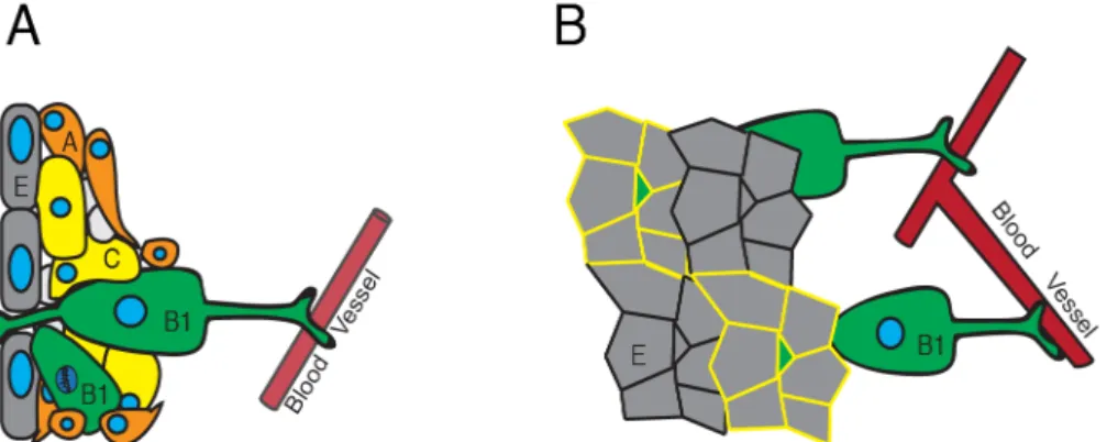
The NSCs found in the SVZ project bidirectionally through the ependyma into the CSF and radially to blood vessels (BV) to obtain systemic inputs (Merkle et al., 2007) (Figure 6A). Electron microscopy has revealed that the SVZ niche consists of four major cell types, E- (ependymal), B- (SVZ astrocytes and NSCs), C- (transitory amplifying) and A-cells (neuroblasts) (Figure 6A). Using whole mount techniques it was observed that B-cells of the lateral wall with NSC properties, defined as B1 cells (Ihrie and Alvarez-Buylla, 2011), can be found in a typical pinwheel structure (Mirzadeh et al., 2008). The core of the pinwheel contains the apical ending of a radial B1-cells and in its periphery are ependymal cells (Figure 6B). This typical embedding of B-cells within ependymal wall, blood vessels, immediate surrounding and own progeny allows for distinct response mechanisms of the stem cells. Comprised, these response mechanisms can be put in four categories.
First, the apical ending in the core of the pinwheel contains sensory cilia that respond to signals in the CSF and flow of the CSF. The CSF, for example, contains gradients of Slit2. These gradients are partially regulated by the movement of the mechanocilia on the ependymal cells (Sawamoto et al., 2006). Additionally, a cellular response might be triggered mechanically via the flow of the CSF passing the cilia (Banizs et al., 2005), due to shear forces activating ion channels and Ca2+ influx
(Yamamoto et al., 2000). The role of cilia in neurogenesis is proposed to be crucial, as misregulation of this dual response system potentially has severe implications for NSC maintenance and progenitor migration (Goetz and Stricker, 2006).
Figure 6: NSCs of the SVZ are organized in pinwheels; NSCs of the SVZ are divided into B-cells (NSCs, green), C-cells (TAPs, yellow) and A-cells (neuroblasts, orange). They are in a tight scaffold with each other. The A-cells will migrate out of the SVZ, along the RMS into the OB. The radial B1 cells, projecting to blood vessels and through the ependyma (E, grey) are the quiescent NSCs (A). The radial B1-cells are characterized by their typical pinwheel morphology. In whole mount preparations the process projecting through the ependyma is generating this NSC typical morphology (yellow trace) (B).
Introduction
Second, the B-cells are connected to another supply of extrinsic signals the BVs, which have a dual function. On one hand, the blood-brain barrier (BBB) in close proximity to the SVZ is more permeable than in the rest of the brain (Cheung and Rando, 2013; Shen et al., 2008), thus releasing blood-born factors including pigment epithelium-derived factor (Andreu-Agullo et al., 2009) and β−cellulin (Gomez-Gaviro
et al., 2012) that are proposed to be involved in maintenance, proliferation and differentiation. On the other hand NSCs directly contact the epithelial cells of the BVs with their processes, getting input from surface receptors such as Delta-like (Dll) and Jagged ligands (Temple, 2001). These juxtacrine signals play a pivotal role in maintenance of NSCs in a quiescent and undifferentiated state (Ottone et al., 2014).
Third, the NSCs are located in regionalized portions of the SVZ innervated by distinct nuclei. Distinct OB interneuron subtypes are produced in finely patterned progenitor domains of the SVZ. These microdomains of the SVZ correlate with expression domains of distinct transcription factors such as Nkx6.2 and Zic-family members. These domains are potentially defined by the nuclei they are innervated by (Merkle et al., 2014). Axons from defined nuclei, such as the raphe or the pons can form an extensive plexus in close proximity to adult NSCs. NSCs express different receptors of neurotransmitters, which makes them susceptible to neuronal stimuli (Tong et al., 2014b). These microdomains, potentially regulated by innervation from CNS nuclei, exemplify the interconnectivity of NSCs and the niche.
Fourth, the immediate progenitors are in direct contact with the NSCs, allowing for direct cell-cell interactions. In the SVZ, mother and daughter cells are in close proximity. It is presumed that this direct interaction balances the populations of NSCs and TAPs in the niche. Both NSCs and TAPs present and secrete a vast array of proteins involved in regulating neurogenesis (Drago et al., 2013; Hermann et al., 2014). Some of the presented receptors are endodermal growth factor receptor (Doetsch, 2003) and Notch receptors (Aguirre et al., 2010) Notch ligands Jagged (Basak et al., 2012; Nyfeler et al., 2005). In parallel some of the secreted soluble growth factors are FGF and EGF (Deleyrolle et al., 2006; Türeyen et al., 2005). These paracrine mechanisms provide a feedback loop to keep neurogenesis and stem cell maintenance in tight control.
Thus, the NSCs in the SVZ are controlled on a niche and hierarchical level by extrinsic (CSF and BVs) and intrinsic (axons and feedback loops) factors. A similar system can be found in the SGZ of the DG the second neurogenic niche.
Introduction
Organization of the Adult Subgranular Zone
The DG of the hippocampus is part of the limbic system and plays a key role in memory consolidation and spatial navigation. Neurogenesis in the DG is found in the SGZ of adult rodents, primates as well as humans (Spalding et al., 2013) and ongoing neurogenesis in the adult SGZ has been proposed to be important in learning and memory (Zhao et al., 2008).
The nomenclature in the SGZ is different from the SVZ. One distinguishes Type-1, Type-2 and Type-3 cells (Figure 7, adapted from Kempermann 2004), and these types in turn are divided in further subtypes. Type-1 cells are divided into radial, quiescent, and horizontal, active, NSC (Lugert et al., 2010). The Type1 cells give rise to the Type-2 cells, which are divided into Type-2a (early progenitors) and Type-2b (late progenitors). Type-2 cells are intermediate precursor cells (IPs). The Type-2 cells give rise to Type-3 cells, which are fate-committed neuroblasts (Ehninger and Kempermann, 2008). Upon maturation they become neurons that potentially integrate into the DG circuits. In contrast to the SVZ, a single neuron-type – DG granule neurons - are produced (Seri et al., 2004). This population makes up 10% of the murine neural circuits (Kempermann et al., 2015) and 35% of the human neural circuits (Spalding et al., 2013). Newly generated neurons in the hippocampus integrate into established networks, making neurogenesis a unique form of neuronal plasticity. Although the neurogenic niches have distinct architectures and exhibit high levels of heterogeneity, the stem cells found in the SGZ and SVZ have, besides their differences also commonalities.
Figure 7: NSCs of the SGZ are in close proximity to their progeny; NSCs in the SGZ can be found as radial or horizontal cells (Type-1, green). The radial Type-1 cells, projecting through the granule cell layer (GCL) divide less frequently than the horizontal Type-1 cells. The radial cells are characterized as quiescent, the horizontal as active. They give rise to 2 cells, which are intermediate progenitors (IPs, yellow). The Type-3 cells are characterized as neuroblasts (orange), which are fate committed. They give rise to immature neurons that can mature and stably integrate into the DG circuits.
Introduction
Due to lack of contact the SGZ NSCs do not obtain input from the CSF, however they are provided, just as in the SVZ with external stimuli via the vasculature. In the SGZ the NSCs are positioned close to endothelial cells of blood vessels. As in the SVZ it is presumed that the BBB in the SGZ might be more permeable, thus providing extrinsic signals (Cheung and Rando, 2013). Furthermore the endothelial cells might provide paracrine signals themselves that play into the signaling of direct or proximal cell-cell contact.
Similar to the patterned SVZ (Merkle et al., 2014), there are implications that there is a longitudinal regionalization of the SGZ, topographically separating dorsal and ventral blade of the DG (Kheirbek and Hen, 2011). Although no significant differences in dividing cells can be observed in the dorsal and ventral blade, the number of Type-1 cells seems to be less in the ventral blade as compared to the dorsal. Alongside the number of Doublecortin+ (Dcx+) neuroblasts in the dorsal blade is increased. Furthermore, the neuroblasts present in the ventral blade express less Calretinin (CR), a marker of immature granule cells (Jinno, 2011). This asymmetric density in hippocampal neurogenesis might affect the strength of the feedback loops generated by the nearby progeny (Snyder et al., 2009).
In the SGZ, just as the SVZ, the NSCs are in close contact with their progeny. One major difference of the two niches is that in the SGZ the neuroblasts and newborn neurons do not migrate out of the niche area. Thus, the regulation of NSCs by axonal inputs will be impacted additionally by a feedback of the newly generated neurons. Interneurons in the DG are critical niche components, coupling neuronal circuit activity to quiescent NSCs. The activation of NSCs is increased when the local circuit activity is low. Upon increase in activity of the circuit NSCs are maintained quiescent (Song et al., 2012). This implicates that neurogenesis can be impacted long-lasting if newly generated neurons are integrated wrongly, potentially causing pathological changes to the system.
Adult Neurogenesis Contributes During Aging and Pathologies
Various pathological conditions are associated with either an upregulation or a downregulation of adult neurogenesis (Abrous et al., 2005; Kempermann et al., 2015). A few pathologies associated with the downregulation of neurogenesis are depression (Bremner et al., 1995; Gurvits et al., 1996; MacQueen et al., 2003), schizophrenia (Heckers, 2001; Schmajuk, 2001), drug addiction (Koob and Le Moal, 2001; Nestler, 1997) as well as ageing and dementia (Ben Abdallah et al., 2010;
Introduction
Lugert et al., 2010). On the other hand, diseases associated with up regulation of neurogenesis are epilepsy (Parent et al., 1997; Scott et al., 1998), ischemia (Kee et al., 2001; Liu et al., 1998; Zhang et al., 2004), Huntington’s disease (Curtis et al., 2003; Eriksson et al., 1998), various traumatic brain injuries (Dash et al., 2001; Lu et al., 2003; Rice et al., 2003) as well as specific types of tumors (Giachino et al., 2015). Whether the change in neurogenesis is causative or a consequence of the pathologies depends on the individual disorder and very often it is not known.
Age-Related Decrease of Adult Neurogenesis
Neurogenesis diminishes with age. The age-related decline in neurogenesis might be a result of decreased activity of NSCs, and potentially quiescence. Decreased levels of proliferating stem cells in the hippocampus are associated with impaired aspects of learning and memory. Ageing is associated with a 6-fold decrease in the number of neurons generated in the adult murine brain. Conversely, exercise elicits beneficial effects on the aged brain, and affects NSC function increasing the number of newborn neurons some 3-fold (van Praag et al., 1999b). Exercise-induced increases in neurogenesis correlate with a better performance of mice in spatial learning (Creer et al., 2010) and memory tasks (van Praag et al., 1999a). These results are supported by studies in humans.
In a large-scale investigation, 631 individuals between the ages of 60 and 77 years underwent a 2-year multi-domain intervention, consisting of a change in diet, physical exercise, cognitive training and vascular risk monitoring. The physically active participants in the study performed significantly better than controls (n=629) with regards to working memory, task flexibility, problem solving and planning as well as processing speed (Hawkins et al., 1992; Ngandu et al., 2015). Thus, the neuroplasticity caused by neurogenesis itself is crucial for certain forms of learning and memory in the murine brain (Zhao et al., 2008) as well as the human brain (Ngandu et al., 2015).
Neurogenesis and Mood Related Disorders
In the brains of depressed patients monoamines, such as 5-hydroxytryptamine (5-HT, Serotonin), have a tendency to be reduced. Conventional antidepressants enhance the 5-HT transmission, for example by inhibiting the reuptake of the neurotransmitter. Problematically, a decrease in 5-HT does not immediately cause major depression (Mahar et al., 2014) and the administration of drugs, which increase 5-HT levels rapidly after administration, are not sufficient for depressive
Introduction
amelioration immediately after intake (Hasler, 2010). These observations indicate that long-term mechanisms are involved in major depression. The cause for 5-HT impairment in patients suffering from depression has intrinsic, for example genetics and gender, but also extrinsic, for example drug use or stress, factors. Stress is being viewed as one of the most potent factors for developing major depression. Chronic stress has been shown to negatively regulate adult neurogenesis in the DG. Brain images of patients with major depression have shown hippocampal atrophy (Bremner et al., 1995). Decreased neurogenesis seems to underlie symptoms of depression (Kempermann, 2002). The high neuronal turnover in humans in the hippocampus supports the possibility that hippocampal neurogenesis can be causative in depression and/or the response to stress or antidepressants (Spalding et al., 2013). Hippocampal neurogenesis is regulated by monoamines (Diaz et al., 2012) and neurotrophic factors (Waterhouse et al., 2012) and chronic antidepressant treatment increases neurogenesis (Dranovsky and Hen, 2006). The selective 5-HT reuptake inhibitor Fluoxetine has been tested for its effects on both the SVZ (Tong et al., 2014b) and the SGZ (Encinas et al., 2006). NSCs seem to be in close proximity to serotonergic axons. In both neurogenic niches the antidepressant causes an increase in symmetric divisions of early progenitor cells.
Aberrant Neurogenesis and Epilepsy
While exercise is associated with a healthy increase in DG neurons, epileptic seizures (SE) are associated with a pathological increase. It is known that epilepsy stimulates proliferation in the DG (Parent, 2007). The DG responds shortly after SE with an increased cell proliferation in the subgranular zone (Parent et al., 1997). Seizures increase the activation of quiescent cells, recruiting them into an active state (Lugert et al., 2010). Upon SE abnormal mossy fiber sprouting and abnormal basal dendrite development, as well as migration of dentate granule cells are observed (Jessberger et al., 2007a). This abnormal integration might cause an imbalance in inhibition. Making abnormal neurogenesis the potential cause for epileptogenesis, leading to reoccurring, acute seizures (Di Maio, 2014; Pierce et al., 2005). In acute seizures this precocious NSC activation comes at an expense of long-term exhaustion for short-term plasticity (Sierra et al., 2015).
Adult NSCs and Tumor Biology
The idea that tumors contain a rare subset of stem-like cells capable of self-renewal, indefinite division and differentiation is gaining acceptance (Pierfelice et al., 2008) – this hypothesis is called the cancer-stem-cell theory. Adult neurogenesis
Introduction
implicates the presence of undifferentiated, active stem and progenitor cells. Disruption of the regulatory mechanism either of the SCs or the rapidly dividing daughter cells is probably one cause for the formation of cancer initiating stem-like cells (Reya et al., 2001) also in the brain. This was underlined when neurosphere forming precursors with characteristic NSCs genes, such as Sox2, Musashi (Hemmati et al., 2003) were obtained from a human glioblastoma biopsy (Ignatova et al., 2002) and a human medulloblastoma (Singh et al., 2003). It appears as though the tumor initiating cells with neural precursor features respond to the same mitogens, possess some of the molecular features and seem to express similar markers as adult NSCs (Tamaki et al., 2002). Many tumors develop near the neurogenic SVZ indicating that they might derive from transformed undifferentiated precursor cells (Sanai et al., 2005).
Recently it was shown that NSCs in the SVZ with deleted p53, a cell cycle control gene, and deleted Rbpj, the Notch signaling mediator, form tumors in the brain. Loss of proper NSC maintenance and additionally the cell cycle disturbance leads to the formation of brain tumors (Giachino et al., 2015), highlighting the essentiality of temporospatial proper NSC maintenance.
Stem Cell Maintenance
Deregulation of NSC maintenance can lead to an early exhaustion of the NSC pool or worse, as previously highlighted, in various pathologies. Thus, adult NSCs are tightly regulated and controlled in order to achieve proper physiological functioning and maintenance. Three crucial features characterize proper maintenance: proper self-renewal, controlled fate determination and preservation of stemness.
Self-renewal depends on a cells capacity to undergo either symmetric or asymmetric cell division. While a symmetric cell division gives rise to two identical daughter cells, asymmetric division produces an exact copy of itself and a distinct daughter cell that will eventually terminally differentiate (Gotz and Huttner, 2005). Fate determination of stem and progenitor cells is subject to intrinsic and extrinsic factors. Besides the presence of a receptors on the cell surface (intrinsic) also the presence, timing and concentration of the extrinsic ligand will influence the cellular response (Fuchs, 2004). Stemness is preserved by the specific factors provided by the niche. These are local and environmental factors such as cytokines, growth factors, adhesion and signaling molecules, which are crucial for proper NSC
Introduction
functioning and maintenance (Conover and Notti, 2008). There are multiple factors known to orchestrate these maintenance tasks. The best studied in terms of adult neurogenesis and adult NSCs maintenance are Shh, (Fuccillo et al., 2006), Wnt, (Zechner et al., 2003) and Notch signaling (Ables et al., 2011). These three pathways are implicated in regulating adult neurogenesis and potentially even crosstalk.
Shh has been implicated in adult neurogenesis and is important in stem cell proliferation and progenitor specification (Alvarez-Buylla and Ihrie, 2014). Shh signaling functions via a surface receptor complex consisting of Patched (Ptc) and its G-protein-coupled co-receptor Smoothened (Smo). Ptc inhibits signal transduction of Smo in the absence of Shh. Once Shh binds Ptc, Smo is disinhibited, leading to the activation of the Shh signaling cascade. This results in the disinhibition of Gli2/3. Gli 2/3 then function as transcription factors whose nuclear-cytoplasmic distribution is regulated via a protein-protein interaction with suppressor of fused (Su(Fu)) (Kogerman et al., 1999). Activation of proper Shh cascade leads to the transcription of further Gli-proteins (Gli1/7) and other Shh target genes (Philipp and Caron, 2009). Some known target genes of Shh signaling in the brain are Nkx2.2, Pax6 and Ptc1 (Shahi et al., 2010). Genetic manipulation of the Shh signaling cascade via deletion of Ptc leads to an increase of NSC divisions and symmetric NSC divisions in adult neurogenesis in the SVZ (Ferent et al., 2014).
Wnt signaling is highly conserved and has been implicated in CNS development and NSC differentiation (Zechner et al., 2003). In the absence of Wnt, Glycogen-Synthetase-kinase-3 (GSK3) is forming a complex with Axin and other cofactors. This complex ultimately phosphorylates and ubiquitinates β-catenin, thus keeping a
low β-catenin level in the cell. Once Wnt is binding Frizzled receptor a tertiary
complex with Lrp6 is formed. Axin is recruited to the intracellular domain of Lrp6, sequestering GSK-3 away and β-catenin is no longer tagged for degradation, thus
accumulates and can migrate into the nucleus where it is acting as a transcription factor (Komiya and Habas, 2008) regulating for example the expression of Neurogenin1 (Hirabayashi et al., 2004), Six3 (Braun et al., 2003) and NeuroD1 (Kuwabara et al., 2009). Wnt signaling has mostly been proposed in proliferation and differentiation of neuronal progenitor cells. It has been shown that NeuroD1, a proneurogenic transcription factor, is a downstream mediator of Wnt-induced neurogenesis (Kuwabara et al., 2009). Inhibition of Wnt signaling via the secretion of Dickkopf or Secreted Frizzled-related Protein 3 in the adult SGZ has been implicated in downregulation of adult NSC proliferation and neuronal maturation. Interestingly,
Introduction
Dickkopf expression is naturally increased with age, implicating a role of Wnt signaling in stem cell quiescence with progressed age (Wu and Hen, 2013)
The third, crucial signaling pathway is Notch signaling. The remainder of this work will be focusing on the Notch signaling pathway and the role of Notch in NSC maintenance in the adult murine brain.
Notch Signaling: a Summary of History
In 1914 John S. Dexter noticed a “notched” phenotype in the wings of Drosophila melanogaster. The responsible allele was then found by T.H. Morgan’s group in 1917 and through to the cloning of the gene in the 1980s (Artavanis-Tsakonas, 1983), the Notch family members are now recognized as essential signaling molecules that control a diverse array of cellular responses ranging from normal development to the maintenance of homeostasis in metazoans. Notch signaling components are evolutionarily conserved in all metazoan organisms - with a single receptor present in Drosophila, two in C. elegans and four in mammals (Kopan and Ilagan, 2009).
The Notch signaling pathway, compared to Shh and Wnt signaling, is highly dependent on direct cell-cell interactions and niche architecture. Notch signaling affects a wide range of cellular processes (Andersson et al., 2011) both during development (Artavanis-Tsakonas et al., 1999; Harper et al., 2003) and adulthood including stem cell maintenance (Borggrefe and Oswald, 2009; Koch et al., 2013), cell proliferation (Androutsellis-Theotokis et al., 2006), differentiation (Bigas and Espinosa, 2012; Gaiano and Fishell, 2002) and apoptosis (Gotte et al., 2011).
Notch Receptors and Ligands
The four mammalian Notch receptors (Notch1-Notch4) reside on the cell surface as non-covalently linked heterodimers (HD) and are Type I transmembrane receptors (Figure 8A, adapted from Mumm and Kopan 2000). They are comprised of an extracellular domain, which functions as receiver and an intracellular domain which functions as sender of signal information. The Notch extracellular part is comprised of numerous EGF-like repeats. The extracellular EGF-like repeats contain Thr/Ser amino-acid residues that prone for Fringe-mediated O-glycosylation. These sugar modifications are proposed to modulate signaling outcome influencing the interactions of different ligands (Takeuchi and Haltiwanger, 2014).
The extracellular and intracellular parts of Notch are combined at the heterodimerization (HD) domain. Two cleavage sites (S1 and S2) are found within
Introduction
the HD domain (Mumm and Kopan, 2000). In order for all four Notch receptors to become mature, they need to be cleaved at the S1 site in the Golgi before integration into the membrane. The S2 extracellular cleavage, mediated by the metalloprotease Adam10 under physiological conditions (Alabi et al., 2016), and the intracellular S3, mediated by γ-secretase, cleavages are needed for proper signaling (Figure 8B,
adapted from Mumm and Kopan 2000).
The intracellular domains of all Notch paralogues contain an Rbpj associated molecule (RAM) domain, the nuclear localization signal (NLS), multiple Ankyrin (ANK) domains and the Proline-Glutamate-Serine-Threonine rich domain (PEST). Notch1 and Notch2 additionally contain a carboxy-terminal transactivation domain (TAD). The RAM domain is crucial for interaction with several cytosolic and nuclear proteins, including Rbpj, the transcriptional mediator of Notch signaling. The Ankyrin domain is important for further protein-protein interactions. The composition of Figure 8 Notch receptors and ligands; Notch receptors are heterodimers with an extracellular and an intracellular domain. There are four Notch paralogues (Notch1-4) (A). Notch receptors undergo three cleavages (S1-S3). S1 is a non-activating maturation cleavage occurring in the Golgi. S2 and S3 are activating cleavages necessary for canonical Notch signaling (B). Notch ligands are composed of a large extracellular domain rich in EGF-repeats that interact with the extracellular domain of the Notch receptor. Upon interaction of ligand and receptor, the receptor undergoes a conformational change making the S2 cleavage site available. There are five Notch ligands, Jagged1/Jagged2, Dll1, Dll4 and Dll3 (C).
Introduction
modulator proteins bound at the RAM and ANK domains lead to the formation of the Notch nuclear transcription complex. The TAD domain is important for transcriptional activation. The PEST domain, the most C-terminal component of the Notch protein, is essential in the regulation of Notch degradation (Kurooka et al., 1998) (Figure 8, adapted from Mumm and Kopan, 2000). Mutations in the HD, RAM, ANK or PEST domain can lead to severe phenotypes. Mutations in the HD domain can cause a ligand-independent activation of Notch receptors. Mutations in the RAM or ANK domain can cause improper or block of binding to the interaction partners (Mumm and Kopan, 2000). Mutations in the PEST domain can lead to an incorrect inactivation, and thus Notch signaling is prolonged (Chillakuri et al., 2012).
The two most related mammalian Notch paralogues are Notch1 and Notch2. These two are well characterized both genetically and functionally and share many structural features (Weinmaster et al., 1992). The extracellular domains of Notch1 and Notch2 have 57% amino acid conservation the intracellular 53%. It is worth to mention that the intracellular PEST and TAD domains are only 37%, while the ANK domain is 85% conserved at the amino acid level (Liu et al., 2015b).
The ligands of Notch signaling are receptors on the juxtapose cells. There are five mammalian Notch ligands: Jagged1, Jagged2, Delta-like 1 (DLL1), DLL3 and DLL4. These are Type I transmembrane ligands, and they provide short-range signals between directly opposed cells. The ligands possess a Delta/Serrate/LAG-2 (DSL) motif on their N-terminus as well as tandem EGF-repeats (Figure 8C, adapted from Mumm and Kopan 2000). The EGF-repeat regions mediate the short-range interaction of Notch and its ligands. The specificity is then ensured by O-Glycosylation, mediated by POFUT1 and Fringe, and by regulation of the availability of ligand and receptor in a temporospatial manner on the cell surfaces. Once short-range interaction of Notch and of its ligands, Delta, or Jagged occurs, the canonical Notch signaling pathway is activated.
The Notch Signaling Cascade
In the absence of Notch ligands, the receptor is not cleaved at the S2 and S3 sites and Rbpj, the nuclear mediator of Notch signaling in the nucleus is bound to Corepressors (CoR) and histone deacetylases (HDAc) at target genes (Figure 9-1). The transcriptional program in NSCs, in the absence of active Notch signaling, can be described as proneural, the NSCs are not maintained and potentially differentiate. In order to maintain NSCs, the Notch receptor needs to interact with one of its ligands, be activated and transduce a transcriptional signal to the nucleus.
Introduction
Canonical Notch signaling is initiated by short-range signals between directly opposed cells (Figure 9–2a). Notch proteins and cell bound Notch ligands (DLL, Jagged) interact causing a conformational change, exposing the S2 cleavage site to a metalloprotease family (ADAM) (Figure 9-2b). The proteolytic release of the Notch extracellular domain leaves the Notch receptor truncated and exposed to intracellular
γ-secretase mediated S3-cleavage (Figure 9-2c), which releases the Notch
intracellular domain into the cytoplasm (Figure 9-2d). This active intracellular domain traverses to the nucleus and interacts with Rbpj. Upon interaction the nuclear Rbpj-complex the Rbpj-complex composition is changed (Figure 9-2e). CoR and HDAc are exchanged for Coactivators (CoA) and Histone acetyltransferases (HAcT). This change of the complex leads to the transcription of downstream Notch target genes, switching the function of Rbpj from a repressor to a transcriptional activator. Rbpj/NICD transcriptional complex activates a set of basic helix-loop-helix transcriptional repressors (Mumm and Kopan 2000).
Figure 9: Canonical Notch signaling cascade; In the absence of ligand the Notch receptor is integrated as Type-I receptor in the membrane. In the absence of ligand, the nuclear Rbpj complex is bound to Corepressors (CoR) and Histone deacetylases (HDAc) (1). In the presence of ligand the Notch extracellular domain and the ligand extracellular domain interact, leading to a conformational change of the Notch receptor (2a). This conformational change leads to the exposure of the S2 site and a consecutive cleavage by a metalloprotease (2b). After the S2 cleavage the S3 cleavage site becomes available to a γ-secretase (2c). This cleavage releases the Notch intracellular domain (NICD) into the lumen (2d). The NICD will migrate into the nucleus where it can interact with Rbpj. The binding of NICD leads to the recruitment of members of the activated complex, exchanging the CoR through a Coactivator (CoA) and the HDAc with a Histone acetyltransferase (HAcT) resulting in transcription of Notch effector genes (2e).
Introduction
This conserved cascade is repeatedly used in multiple developmental processes. The pathway appears simple, without any second messengers or apparent cytosolic interactions with a binary decision. However, Notch receptors and ligands are influenced by a broad spectrum of posttranslational modifications. Therefore, Notch signaling can drive numerous mechanisms, such as stem cell differentiation and maintenance both in the embryo and the adult (Koch et al., 2013).
Notch signaling is context and tissue dependent. In muscle stem cells, Notch has been implicated in maintenance of stem cells, self-renewal of progenitor cells and inhibition of terminal differentiation (Brack and Rando, 2012). In the intestine, Notch signaling is active in the intestinal stem cells and regulates their proliferation and the terminal differentiation (Barker et al., 2007). In the bone marrow HSCs, Notch does not seem to be essential for physiological HSC maintenance, however constitutive expression can lead to an expansion of these cells (Bigas and Espinosa, 2012). In NSCs Notch has been implicated in NSCs maintenance (Basak et al., 2012; Ehm et al., 2010; Imayoshi et al., 2010), inhibition of neuronal differentiation and even terminal differentiation into an astrocyte lineage (Gaiano and Fishell, 2002).
Notch signaling in Neural Stem Cells
Accumulating evidence underlines the importance of Notch signaling in NSC maintenance, differentiation and fate choice (Artavanis-Tsakonas et al., 1999). The dependence of NSCs on Notch signaling becomes evident when Rbpj, the downstream mediator of Notch signaling, is deleted specifically from NSCs in the adult murine brain. The NSCs are no longer maintained properly, this leads to an initial activation of the stem cell pool and an expansion of the progenitor population, however in the long run caused a depletion of the quiescent and active NSCs from the SVZ (Imayoshi et al., 2010). Interestingly when Notch1 was deleted from the same SC population only the active NSCs were affected (Basak et al., 2012). In Zebrafish, a similar observation was made - Notch signaling levels are crucial for maintenance of quiescent NSCs and recruitment to activity (Chapouton et al., 2010b). In a follow-up analysis it was shown that Notch1 is dispensable in the maintenance of quiescent NSCs also in Zebrafish, however Notch3 is required (Alunni et al., 2013).
When looking at the expression levels of Notch on NSCs in the SVZ, it appears as though the Notch paralogues Notch1 and Notch2 are coexpressed on all cells of the neurogenic lineage (Basak et al., 2012). Interestingly, Notch signaling is only
Introduction
active, as determined by expression of Notch effector genes, in NSCs and TAPs (Giachino et al., 2014b). Two very prominent direct Notch target and effector genes, crucial for NSC maintenance are hairy-enhancer-of-split (Hes) and brain lipid binding protein (BLBP) genes. Hes genes are basic helix-loop-helix (bHLH) genes and essential effectors of Notch signaling for maintaining undifferentiated cells (Artavanis-Tsakonas et al., 1999; Gaiano and Fishell, 2002). The single deletion of either Hes1 or Hes5 has no apparent defects in embryonic development, thus illustrating a compensatory mechanism. Parts of this compensation might come from different upstream regulators, such as BMP4 (Kageyama et al., 2007). However, the double deletion causes severe phenotypes leading to disorganization of the neural tube, premature neuronal differentiation and loss of radial glia in the embryo (Hatakeyama et al., 2004).
Another well-known direct Notch target gene is BLBP. BLBP is broadly expressed throughout the brain of the embryo to the adult. It is proposed to be involved in neuronal–glial signaling. Antibody blocking experiments have shown that BLBP is required for NSC morphological changes in response to neuronal cues in the embryo (Anton et al., 1997) and loss of BLBP in the adult leads to precocious differentiation and loss of the adult NSCs (Matsumata et al., 2012).
Although the role of Notch signaling in NSCs is widely accepted as crucial, the distinct role of the Notch paralogues in maintenance of quiescent and active NSCs is only poorly understood. The goal of this thesis thus was to investigate the role of Notch signaling in balancing between adult NSC quiescence and heterogeneity.