The Effect of Forefoot and Arch Posting
Orthotic Designs on First
Metatarsophalangeal Joint Kinematics
During Gait
Deborah A. Nawoczenski, PT, PhD
1Paula M. Ludewig, PT, PhD
2Study Design: Repeated-measures analysis of variance.
Objective: To examine the effect of 2 different orthotic posting designs on first metatarsophalangeal (first MTP) joint kinematics during gait.
Background: Common orthotic designs used to control abnormal pronation incorporate the use of a medial post in the forefoot and/or rearfoot locations. Although this design may favorably alter rearfoot and lower-limb kinematics, the incorporation of a forefoot post has been theorized to negatively impact first MTP joint function by limiting hallux dorsiflexion during push off. An alternative design that has been proposed to be more favorable for function of the hallux and first metatarsal is the medial arch support.
Methods and Measures: Eighteen subjects with a mean age of 28.2 years (SD, 8.3 years) completed the study. All subjects were judged to have excessive pronation based on a clinical orthopaedic examination. Two different pairs of orthoses were custom molded for each subject. One design incorporated an extrinsic rearfoot and forefoot post and the second design had a high medial longitudinal arch in combination with an extrinsic rearfoot post. The ‘‘Flock of Birds’’ electromagnetic tracking device was used to collect 3-dimensional position and orientation data of 3 body segments (hallux, first metatarsal, and calcaneus) during the stance phase of walking for 3 conditions (no orthosis and each of the 2 different orthotic designs). A repeated-measures analysis of variance was used to assess differences in first MTP joint dorsiflexion at midstance and during the push-off period of gait, as well as metatarsal declination angle changes during relaxed stance. An exploratory regression analysis was used to investigate factors that related to the change in peak dorsiflexion for the orthotic conditions.
Results: Peak first MTP joint dorsiflexion averaged between 38° and 40° across all conditions. Although slight increases in first MTP joint dorsiflexion values were noted with both types of orthotic designs, these differences were not significant at either phase of the stance cycle (P = .50). The metatarsal declination angle in relaxed stance significantly increased (P = .001) under both orthotic conditions. Considerable individual variability was present. For the rearfoot-forefoot posted orthosis, a change in the declination angle of the first metatarsal during relaxed stance with the orthosis was a significant nonlinear predictor of change in peak dorsiflexion during push off. 1Professor, Department of Physical Therapy, Ithaca College, University of Rochester Campus, Rochester,
NY.
2Associate Professor, Program in Physical Therapy, Department of Physical Medicine and Rehabilitation,
University of Minnesota, Minneapolis, MN.
This study was conducted in the Movement Analysis Laboratory at Ithaca College’s Rochester campus facility, and supported, in part, by a grant from the Orthopaedic Section of the American Physical Therapy Association. The study was approved by the Research Subjects Review Board at the University of Rochester, Rochester, NY.
Address correspondence to Deborah A. Nawoczenski, Professor, Ithaca College, University of Rochester Campus, 300 East River Road 1-102, Rochester, NY 14623. E-mail: dnawoczenski@ithaca.edu
Conclusions: Foot orthoses that incorporate a medial forefoot post do not have a consistent negative effect of reducing first MTP joint dorsiflexion during walking. J Orthop Sports Phys Ther 2004;34:317-327.
Key Words: arch support, first
meta-tarsal joint, hallux, medial orthotic
posts
F
oot orthoses are widelyused as part of the
man-agement plan for a
broad range of foot, ankle, and lower extrem-ity pathologies. One foot type that commonly receives orthotic inter-vention is the pes planus, or exces-sively pronated foot. Orthoses are intended to control the potentially excessive or prolonged subtalar and midtarsal joint motions during gait that frequently accompany this
foot type.15,23,29,34,48
Regardless of the underlying eti-ology, pronation that is excessive or prolonged throughout stance is believed to limit the ability of the first metatarsal and first cuneiform bones (first ray) to provide a
stable structure for
propul-sion.21,39,43 Failure of the fibularis
longus to stabilize the first metatar-sal may result in a dormetatar-sal transla-tion or elevatransla-tion of this structure, particularly during the later part
RESEARCH
FIGURE 1. Disallowing first metatarsal plantar flexion will also cause first metatarsophalangeal (MTP) joint dorsiflexion to be restricted (A). Unrestricted first metatarsal plantar flexion will allow normal first MTP joint dorsiflexion (B).
of the stance phase of gait. Further, in static analysis investigations, an elevated first metatarsal has been shown to result in a significant decrease in hallux or
first metatarsophalangeal (MTP) joint
dorsi-flexion22,44 (Figure 1). Restrictions of first MTP joint
dorsiflexion may lead to abnormal compression of the dorsal surfaces of the metatarsal and proximal phalanx, resulting in proliferative and degenerative
changes of the articular cartilage.27,46,47 Over time,
the range of motion available to the first MTP joint will gradually decrease, resulting in altered gait me-chanics, decreased stride length, and abnormal tibial
rotation.13,27,46
Relief of pain and the ability to return to previous levels of activity are major outcome measures used to
assess the success of orthotic intervention.2,10,12,19 In
addition to the subjective changes reported with their use, foot orthoses have been shown to modify se-lected aspects of lower extremity kinematic behavior during the stance phase of gait. To date, the majority of investigations have assessed the effectiveness of
orthoses on rearfoot motion parameters,2,5,23,36,48
rearfoot-tibia coupling behavior,15,34,49 or tibial
rota-tion.34,49 The findings from these studies have shown
varied responses to orthotic intervention, ranging from no significant differences to an assortment of changes that include a reduction in maximum prona-tion (calcaneal eversion), maximum pronaprona-tion veloc-ity, time to maximal pronation, total rearfoot motion, alterations in the displacement and velocity coupling between the rearfoot and tibia, and tibial
rota-tion.2,5,23,34,36,48,49 Differences in reported findings
may be due, in part, to the variability in experimental designs, such as the choice of orthotic material and rigidity, the amount and location of posting, the testing surface, selected walking or running speeds, footwear variations, and variations in individual sub-ject responses to the orthotic intervention. Well-defined subject inclusion criteria are also of considerable importance when comparing the effects of foot orthoses and kinematic responses, and may account for the differences among investigations.
Although the forefoot has been implicated in foot pathologies, investigations of alterations of forefoot function with the use of orthoses have primarily focused on plantar pressure and center-of-pressure
changes.4,8,36,42 Little is known about how forefoot
kinematics are directly influenced, if at all, by the use of orthoses. Yet, practitioners are required to make decisions regarding the design characteristics of foot orthoses, such as posting or positioning of the first metatarsal, that may positively or adversely impact foot and, in particular, hallux and first metatarsal function.
One type of design frequently utilized in orthotic therapy for the abnormally pronated foot incorpo-rates the use of a build-up or post under the medial rearfoot and forefoot regions of the foot. The pur-ported goal of this design is to ‘‘bring the ground up
to the foot’’ during gait.43 However, the potential
restriction of first metatarsal plantar flexion believed inherent in this forefoot post design (FF POST) may also hinder the normal first MTP joint dorsiflexion during the latter part of stance or the push-off period of gait (Figure 2A). An alternative design option proposed for the pronated foot is the high medial longitudinal arch (ARCH). The nature of the high-arch design is theorized to promote first metatarsal plantar flexion, thus restoring the normal arch and windlass mechanism of the foot. Additionally, if first metatarsal plantar flexion is not restricted, then normal first MTP joint motion can occur (Figure 2B).21,44Although the ultimate goals of each orthotic design are to restore a normal and pain-free gait pattern that allows the foot accommodate to varia-tions in the ground surface and to provide adequate stability for propulsion, the design characteristics are distinct.
The purpose of this study was to examine the effect of 2 different orthotic designs on the kinematic behavior of the hallux-first metatarsal complex at
A
FIGURE 2. Rearfoot and forefoot posted (FF POST) orthotic design with extrinsic medial posts at both rearfoot and forefoot locations (A) and arch (ARCH) design with an extrinsic rearfoot post and the apex of the arch lying inferior to the navicular (B).
midstance and during the push-off period of gait in individuals with abnormal pronation. We hypoth-esized that the FF POST orthotic design would result in reduced first MTP joint dorsiflexion when com-pared to the ARCH and no orthotic condition (NO).
METHODS
Subjects
Twenty subjects were enrolled in the study. Two of these subjects were lost to follow-up. Of the 18 subjects that completed the study, there were 11 females and 7 males with a mean age of 28.2 years (SD, 8.3 years; range, 22-45 years). Average (±SD) height and mass were 1.67 (±0.1) m and 73.5 (±16.0) kg, respectively. These individuals were referred to the study by local orthopaedic specialists, podiatrists, and physical therapists for activity-related foot or lower-limb musculoskeletal pain of at least 1 month in duration. The subjects were considered appropri-ate candidappropri-ates for orthotic intervention based on
their histor y of foot or lower extremity
musculoskeletal symptoms, the presentation of a pes planus foot, and the results of a clinical lower-limb
screening exam14,17,25,39 to establish whether subjects
met all the following criteria: a forefoot varus exceed-ing 10° in the non–weight-bearexceed-ing exam; calcaneal eversion beyond vertical during the standing weight-bearing exam; a minimum navicular drop difference of 10 mm or greater between subtalar joint neutral
and relaxed stance;31 and no limitations in first MTP
joint range of motion, or first metatarsal mobility in either the dorsal or plantar direction with respect to
the remaining metatarsal heads.18 The presence of
intrinsic frontal plane deviations has been linked to
abnormal pronation in previous
investiga-tions.5,9,15,20,23,34 Exclusion criteria included a history
of foot or ankle fracture, hallux rigidus or valgus deformity, neuromuscular disease, or previous orthotic use. A single examiner with more than 20 years of clinical experience with the management of foot and ankle injuries (D.A.N.) conducted all clin-ical measurements and performed subsequent casting
FF Post Arch A B
RESEARCH
REPORT
techniques. Intrarater reliability of many of these clinical measures has been addressed in previous
literature.14,17,25,28,31,39,45 A priori power analyses
de-termined 80% power to detect first MTP joint dorsiflexion differences of 4° or larger with a sample size of 20, alpha level of .05, and variance of 64° (SD, 8°). The estimated variance was based on available
literature on first MTP joint motion.33,51
All participants read and signed an informed consent document approved by the Human Subjects Review Board of the University of Rochester. Testing was done in the Movement Analysis Laboratory at Ithaca College, Department of Physical Therapy at the Rochester campus.
Materials
Two different pairs of semirigid orthoses were fabricated for each subject. Each pair of orthoses was constructed of a 3-, 4-, or 5-mm polypropylene/ polyethylene shell material, depending on the sub-ject’s mass, and covered with a 1/8-in (0.3175-cm) poron or PPT full-length cover. The FF POST orthoses were fabricated from slipper cast impressions taken in the non–weight-bearing position as
de-scribed in previous reports.29,42,52 These orthoses
were fabricated from a neutral cast of the foot, whereby the subject was in a prone position, the talonavicular joint was palpated for maximum con-gruency, and the forefoot was stabilized against the rearfoot. The positive cast was balanced with the calcaneal bisection in a vertically oriented
posi-tion.21,36,42,52 A minimum of 5 mm of high-density
material (extrinsic post) was applied to the orthotic shell under the medial aspect of the rearfoot (calcaneus) and the medial aspect of the forefoot/ first metatarsal, ending just proximal to the first metatarsal head. The orthoses were fabricated so that the natural contour of the foot was captured in the non–weight-bearing cast position and maintained in the fabrication process.
The ARCH orthoses were fabricated from a foam cast impression taken with the subject in a seated, partial weight-bearing position. During this fabrica-tion technique, the first metatarsal was placed in a relative plantar flexed position with respect to meta-tarsal heads 2 through 4. The height and location of the apex of the arch of this orthotic design were fabricated to lie immediately inferior to the navicular.
Similar to the technique for the FF POST,
talonavicular congruency was maintained during cast-ing and an extrinsic post was added to the rearfoot. Subjects were also issued a pair of TEVA (Deckers Outdoor Corporation, Goleta, CA) sport sandals that were used during the orthotic break-in period and testing sessions. These sandals had straps at both forefoot and rearfoot locations that enabled the orthoses to be held onto the foot. The strap design also allowed for unrestricted placement of the ‘‘Flock
of Birds’’ sensors (Ascension Technology Corpora-tion, Burlington, VT) on the skin directly over the bony segments of interest (Figure 3). These commer-cially available sandals were designed to provide shock absorption but did not offer any intrinsic motion control, thereby minimizing the confounding effects of footwear on outcome measurements. This footwear has been used successfully in previous
orthotic intervention studies.34,35
Prior to testing, subjects were given written instruc-tions for a 4-week progressive wearing schedule. To minimize any effect of orthotic preference on out-come measures, half of the subjects were asked to wear the first pair of orthoses (FF POST) with the sandals during the first 2 weeks and the second pair of orthoses (ARCH) with the sandals during weeks 3 and 4. The other half of the subjects followed a reverse schedule over the 4-week period. If unable to wear the sandals during the day, subjects were en-couraged to wear them at night. All subjects were instructed to discontinue wearing their orthoses if
they experienced any discomfort or new
musculoskeletal pain that continued beyond 48 hours and to contact the principal investigator immediately. Three subjects were initially unable to tolerate the height of the medial longitudinal arch in the ARCH orthosis and returned for modifications. All subjects returned to the Movement Analysis Laboratory for testing after 4 weeks.
Instrumentation
Three-dimensional position and orientation of the subject’s hallux, first metatarsal, and calcaneus were collected using the Flock of Birds magnetic tracking device. Operating within 1 m of its standard transmit-ter, the system was configured to simultaneously track segmental motion using 3 receivers, each sampling at a rate of 100 Hz. By attaching a sensor to a stylus with known tip offsets from the sensor, the system was also used to manually digitize anatomical landmarks
FIGURE 3. Magnetic tracking sensors, orthoses, and footwear used in the study.
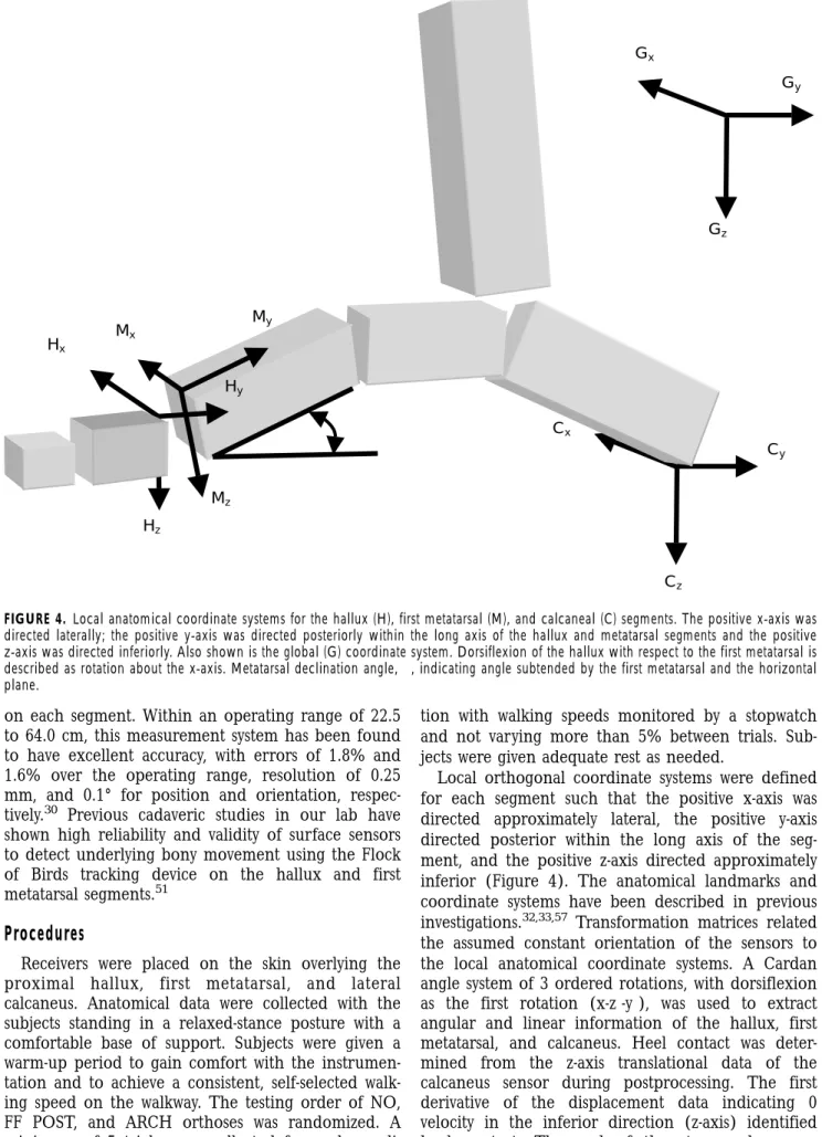
FIGURE 4. Local anatomical coordinate systems for the hallux (H), first metatarsal (M), and calcaneal (C) segments. The positive x-axis was directed laterally; the positive y-axis was directed posteriorly within the long axis of the hallux and metatarsal segments and the positive z-axis was directed inferiorly. Also shown is the global (G) coordinate system. Dorsiflexion of the hallux with respect to the first metatarsal is described as rotation about the x-axis. Metatarsal declination angle,␣, indicating angle subtended by the first metatarsal and the horizontal plane.
on each segment. Within an operating range of 22.5 to 64.0 cm, this measurement system has been found to have excellent accuracy, with errors of 1.8% and 1.6% over the operating range, resolution of 0.25 mm, and 0.1° for position and orientation,
respec-tively.30 Previous cadaveric studies in our lab have
shown high reliability and validity of surface sensors to detect underlying bony movement using the Flock of Birds tracking device on the hallux and first
metatarsal segments.51
Procedures
Receivers were placed on the skin overlying the
proximal hallux, first metatarsal, and lateral
calcaneus. Anatomical data were collected with the subjects standing in a relaxed-stance posture with a comfortable base of support. Subjects were given a warm-up period to gain comfort with the instrumen-tation and to achieve a consistent, self-selected walk-ing speed on the walkway. The testwalk-ing order of NO, FF POST, and ARCH orthoses was randomized. A minimum of 5 trials were collected for each
condi-tion with walking speeds monitored by a stopwatch and not varying more than 5% between trials. Sub-jects were given adequate rest as needed.
Local orthogonal coordinate systems were defined for each segment such that the positive x-axis was directed approximately lateral, the positive y-axis directed posterior within the long axis of the seg-ment, and the positive z-axis directed approximately inferior (Figure 4). The anatomical landmarks and coordinate systems have been described in previous
investigations.32,33,57 Transformation matrices related
the assumed constant orientation of the sensors to the local anatomical coordinate systems. A Cardan angle system of 3 ordered rotations, with dorsiflexion
as the first rotation (x-z⬘-y⬙), was used to extract
angular and linear information of the hallux, first metatarsal, and calcaneus. Heel contact was deter-mined from the z-axis translational data of the calcaneus sensor during postprocessing. The first derivative of the displacement data indicating 0 velocity in the inferior direction (z-axis) identified heel contact. The end of the stance phase was
Cz Hx Hz Hy Mz My Mx Cy Cx Gx Gz Gy
␣
RESEARCH
REPORT
similarly determined using the y-axis translational data of the hallux sensor. The initiation of a positive velocity in the anterior direction was operationally defined as the end-of-stance phase. Due to the nature of hallux-first metatarsal motion in gait, the primary focus of this investigation was directed to the hallux dorsiflexion/plantar flexion component of rotation (x-axis rotation).
Intraclass correlation coefficients (ICC3,1)40 and
standard error of measurements (SEM)16 were used
to determine the within-day trial-to-trial variability of kinematic measures. Kinematic data were analyzed using a 1-factor, repeated-measures ANOVA with the factor of orthotic condition having 3 levels (NO, FF POST, and ARCH). Data were analyzed at midstance and during the push-off period of gait. Midstance was identified as the midpoint between heel contact and toe off of the reference extremity. Push off was defined as the period of gait between heel off and toe off (40%-60% of stance), which is the time when peak first MTP joint dorsiflexion occurred. The dependent variables were peak first MTP joint dorsiflexion during gait and metatarsal declination angle during relaxed stance. The metatarsal declina-tion angle was defined as the magnitude of x-axis
rotation of the first metatarsal relative to the floor22
(or global coordinate system) and represents the anterior component of the medial longitudinal arch. In addition to changes in arch height, this angle also provides an indication of the relative amount of metatarsal plantar flexion with respect to the floor. For all walking conditions, the mean of the 3 intermediate trials (out of a total of 5 trials) were entered into the analysis. In the presence of
signifi-cant condition effects (P⬍.05), Tukey follow-up tests
were planned to adjust for multiple pairwise compari-sons of conditions.
Additionally, an exploratory analysis was completed to determine factors that may help to predict changes in peak first MTP joint dorsiflexion during push off. The change in peak dorsiflexion during push off for the respective orthotic condition (orthotic condition peak dorsiflexion minus no orthotic condition peak dorsiflexion) was the dependent variable. Baseline measurements of the metatarsal declination angle in relaxed stance, changes in baseline metatarsal
decli-FIGURE 5. Means and standard errors for peak first metatarsophalangeal (MTP) joint dorsiflexion for each orthotic con-dition at midstance and during push-off period of gait. No signifi-cant differences were found across the 3 conditions (P⬎.50) for either part of the gait cycle.
nation angle for the orthotic conditions during re-laxed stance (orthotic condition declination angle minus no orthotic condition declination angle), and the peak dorsiflexion for NO orthotic condition were entered as predictor variables in a multiple polyno-mial regression. Higher-order (nonlinear) effects were assessed by using squared terms for the vari-ables.
RESULTS
Within-day trial-to-trial ICC and SEM values for each orthotic condition are provided in the Table. ICC values for peak dorsiflexion ranged from 0.92 to 0.98 and SEM values from 1.2° to 2.8°.
Figure 5 represents the mean peak values and standard errors (SE) for first MTP joint dorsiflexion at midstance and during the push-off period of gait for each condition. Although slight increases (3°) in peak dorsiflexion values were noted with orthotic wear compared to the no orthotic condition, these
TABLE. Within-day trial-to-trial reliability: intraclass correlation coefficient (ICC)* and standard error of measurement (SEM)† for peak first metatarsophalangeal joint dorsiflexion at midstance and the push-off period of gait.
Orthotic Condition
Gait Phase NO FF POST ARCH
Midstance 0.98 (1.2) 0.97 (1.6) 0.97 (1.6)
Push-off 0.92 (2.8) 0.95 (2.1) 0.96 (1.9)
Abbreviations: NO, no orthoses; FF POST, rearfoot and forefoot post orthoses; ARCH, arch orthoses.
* Formula for ICC3,1: (BMS-EMS)⫼(BMS + [k – 1] EMS), where BMS = between-subject mean square, EMS = within-subject mean square, and k = the number of replicate measures per subject.
†
Values expressed in degrees.
0 5 10 15 20 25 30 35 40 45 Midstance Push-off
Peak First MTP Dorsiflexion (degrees)
None FF Post Arch
FIGURE 6. Change in peak first metatarsophalangeal (MTP) joint dorsiflexion with the rearfoot and forefoot posted (FF POST) orthotic (A), measured during push off. Change in peak first MTP joint dorsiflexion with the arch (ARCH) orthotic (B). In both graphs, a positive value indicates an increase in first MTP joint dorsiflexion and negative value indicates a reduction in dorsiflexion motion when compared to the NO orthotic condition (0°).
differences were not significant when compared across conditions for either midstance (df = 2,34; F = 0.71; P = .50) or push off (df = 2,34; F = 0.64; P = .53) of gait. Metatarsal declination angles in relaxed stance significantly increased from an average (±SE) of –20.4° (±1.4°) for the NO to –23.6° (±1.4°) and –23.5° (±1.2°) for the ARCH and FF POST orthotic conditions, respectively (df = 2,34; F = 9.29; P = .001). In pairwise follow-up comparisons, the no orthotic condition was different than either orthotic condi-tion, but the orthotic conditions were not different from each another.
Individual subject data are presented in Figure 6 for each orthotic condition for the push-off period of gait. The values for each subject indicate the change (ie, a positive change indicates an increase in peak first MTP joint dorsiflexion, whereas a negative
change indicates a reduction in peak first MTP joint dorsiflexion) from the sandal to the respective orthotic condition (ie, orthotic condition peak dorsiflexion during push off minus no orthotic condi-tion peak dorsiflexion during push off). These data describe the individual variation in response to the different posting conditions.
The exploratory multiple regression analysis re-sulted in a poorer ability to predict individual subject responses for change in first MTP joint peak
dorsiflexion with the ARCH orthosis (r2 = 0.16, P =
.10) as compared to the FF POST orthosis (r2= .58, P
= .02). For the FF POST orthosis, peak first MTP joint dorsiflexion for the NO orthotic condition and change in metatarsal declination angle in relaxed stance were retained as significant nonlinear predic-tors of the change in peak first MTP joint dorsiflexion during push off.
-15 -10 -5 0 5 10 15 20 1 2 3 4 5 6 7 8 9 10 11 12 13 14 15 16 17 18 Subject Number
Change in Peak First MTP Joint
Dorsiflexion (degrees) -15 -10 -5 0 5 10 15 20 25 1 2 3 4 5 6 7 8 9 10 11 12 13 14 15 16 17 18 Subject Number
Change in Peak First MTP Joint
Dorsiflexion (degrees)
A
B
RESEARCH
DISCUSSION
The orthotic modifications used in this study are commonly recommended for people who have symp-toms associated with abnormal pronation; however, the fabrication designs of each orthotic are uniquely different and have the potential to negatively alter motion of the first metatarsal and hallux. During normal walking, first metatarsal plantar flexion is requisite for unrestricted first MTP joint dorsiflexion
to take place during the push-off period of gait.31
The hypothesis for this study was based on the assumption that an extrinsic post under the medial forefoot and first metatarsal regions of the foot would limit normal first metatarsal plantar flexion, thus creating a restriction or block to first MTP joint dorsiflexion. For the subjects in the current investiga-tion, maintaining or increasing first MTP joint dorsiflexion was considered a favorable kinematic response with orthotic wear.
When averaged across all subjects, the results do not support our original hypothesis that the FF POST orthosis would result in a reduction in peak first MTP joint dorsiflexion. Both orthotic conditions had a similar effect on peak dorsiflexion during walking. Using the sample of 18 subjects who completed the study, a mean difference of interest of 4° for first MTP joint dorsiflexion, and the actual variability from the study data, our resulting post hoc power was 78%, confirming sufficient power to feel confident in our results. Although the sample size was decreased from the proposed 20 subjects, the measured variance was also slightly less than estimated a priori.
While all subjects were similar with regards to the minimum criteria for inclusion into the study and the orthotic design and footwear features were kept consistent, there were highly variable individual re-sponses to the different orthotic posting conditions. The variability in individual subject responses to orthotic use is not unique to our investiga-tion2,5,15,22,34,36,42,49 and there are excellent review papers that have summarized the wide-ranging effects
of orthotic intervention.1,24,41
To further investigate individual subject responses, we used the smallest real difference (SRD) to indicate the magnitude of change for an individual across conditions that is beyond the expected trial-to-trial variability. Based on an average SEM across condi-tions during push off of 2.3°, the SRD or minimally
detectable change can be calculated as SEM × 公2 ×
1.96 = 6.4°.3 This value indicates the individual
subject change that would be expected (with 95% confidence) to be a true change from the NO orthotic condition. Subsequently evaluating the indi-vidual subject responses using the SRD as the criteria for change, 89% of subjects were able to utilize the FF POST orthosis without any detrimental effects on first MTP motion (Figure 6). In this investigation,
‘detrimental’ would be considered a reduction of first MTP joint dorsiflexion. Twenty-two percent actually showed a positive change (increased first MTP joint dorsiflexion exceeding 6.4° threshold), while only 11% showed a negative change.
When wearing the ARCH orthosis, 83% of the subjects were able to use the orthosis without signifi-cant reduction in first MTP joint dorsiflexion.
Seven-teen percent demonstrated an increase in
dorsiflexion, while another 17% showed a decrease in first MTP joint dorsiflexion. These individual subject data, as well as the average group data, do not support a preferential advantage of using the ARCH orthosis over the FF POST with regard to maintaining or increasing available first MTP joint dorsiflexion. There are multiple factors related to foot structure, alignment, and motion that may influence which subjects respond positively or negatively in terms of first MTP joint motion for specific orthotic designs. Understanding some of the factors that may help to predict the subjects’ response would be beneficial when making decisions regarding orthotic modifica-tions. For this investigation, we considered the peak first MTP joint dorsiflexion value under the NO condition, the relaxed standing metatarsal declination angle, and the change in metatarsal declination angle with orthotic use as possible predictor variables in an exploratory multiple regression analysis of the changes in peak first MTP joint dorsiflexion with orthotic use. For example, if peak first MTP dorsiflexion without orthoses was lower than
norma-tive values during gait,33 a positive outcome with the
orthotic intervention would be increased dorsiflexion. Conversely, for a subject with normal values for peak first MTP joint dorsiflexion under the no orthotic condition, maintenance of the normal dorsiflexion range of motion would be desired.
Another factor that was considered a potential contributor to altering first MTP joint dorsiflexion was the metatarsal declination angle in relaxed stand-ing. This angle reflects the anterior component of the arch height measurement, such that an increase in the metatarsal declination angle corresponds to an elevation of the anterior portion of the medial
longitudinal arch.6,22,26 An increase in the metatarsal
declination angle also places the first metatarsal in a position of relative plantar flexion, which is
consid-ered favorable for the propulsive phase of gait.43,44,47
We selected the metatarsal declination angle to assess whether a change, if it did occur with orthotic use during relaxed standing, would parallel the direction of change in first MTP joint dorsiflexion during gait. For the ARCH condition, only 16% of the variance for the change in peak first MTP dorsiflexion could be explained by a combination of the above factors. Although the average metatarsal declination angle across subjects showed an increase of 3° for the ARCH orthosis, this declination angle change,
whether positive or negative, was poorly predictive of individual subject responses. However, for the FF POST condition, a similar average change in metatar-sal declination angle (3° increase) in relaxed stand-ing was predictive of the subject’s change in peak first MTP joint dorsiflexion during gait. Increasing the metatarsal declination angle in relaxed stance gener-ally was associated with an improvement in peak first MTP joint dorsiflexion in push off. For the FF POST condition, approximately 60% of the variance for the change in peak dorsiflexion could be explained from the combination of tested variables. These data sug-gest that a FF POST design resulting in an increase in the metatarsal declination angle in relaxed stance would be associated with a similar increase for peak first MTP joint dorsiflexion during the push-off period of gait. Conversely, if the declination angle is reduced, peak first MTP dorsiflexion would be simi-larly reduced. FF POST orthoses posted too far distally under the first metatarsal head (Figure 2A) or too thick under the distal region of the medial forefoot would potentially contribute to this reduc-tion in the metatarsal angle. Similarly, FF POST designs that do not restore or maintain the arch of the subjects, thereby disallowing metatarsal plantar flexion, are also likely to result in a reduction in the metatarsal declination angle.
Given that the metatarsal declination angle in relaxed stance was increased under both orthotic conditions, differences among individuals may be related to other features of the orthotic design that influence rearfoot and/or lower-leg kinematics. For example, differences in fabrication design related to rearfoot posting and arch height have been shown to alter selected aspects of rearfoot and lower leg
rotations.5,15,34 However, the linkage between the
changed rearfoot/leg movement patterns with orthotic intervention and hallux/forefoot kinematics has not been studied to date. Future studies are planned to assess this relationship in kinematic cou-pling with orthotic use.
It should be noted that when averaged across all subjects, first MTP joint dorsiflexion remained only slightly lower (mean values ranging between 38° and 40°) when compared to normative data of similar in
vivo investigations during walking.33 Average values
for hallux dorsiflexion have been reported to be 42°
± 2.3° during walking11,33; however, the data in these
previous studies were acquired under barefoot condi-tions. Though enabling direct access of the sensors to skin overlying the bony segments of the hallux, first metatarsal, and calcaneus, the use of the sandals may have minimized the magnitude of motion changes associated with both nonorthotic and orthotic use. Previous investigations of orthotic effectiveness
as-sessed while wearing sandals34,35 have found the
magnitude of rearfoot and lower-leg kinematic changes to be similar to those reported by other
investigators who have assessed subjects while wearing footwear. While shoes may afford increased rearfoot motion control, they do not allow access to the forefoot and hallux, the main focus of this study. It is also possible that certain shoes may provide addi-tional support under the medial longitudinal arch, thus enhancing the proposed benefit of the ARCH orthoses on first metatarsal and hallux function. Both types of orthoses were constructed of the same semirigid material that did not collapse during re-laxed standing in the sandals. Although we did not directly measure changes in the navicular or dorsal foot height, the indirect assessment of medial longitu-dinal arch height using the metatarsal declination angle demonstrated changes between the NO, ARCH, and FF POST orthotic conditions. These changes were maintained under dynamic conditions.
The subjects in this study had first MTP joint motion that approximated normative values during gait. The results of this study can be applied to those individuals who have musculoskeletal symptoms re-lated to abnormal foot pronation and in whom semirigid orthoses are appropriate. In individuals with a nonfixed deformity, alignment of the first metatarsal with a resultant increase in pain-free first MTP joint dorsiflexion would be highly beneficial. The results may have been different if rigid orthoses were used, subjects had abnormal supination, or faulty mechanics were associated with hallux rigidus or valgus deformities. Additionally, in some cases, first MTP joint dorsiflexion may be excessive and equally detrimental to predisposing the joint to abnormal loading and early arthritic changes. For these indi-viduals, a favorable response with orthotic wear would be a reduction in first MTP joint dorsiflexion.
Perhaps equally important short-term findings of orthotic intervention for the tested subjects were the effect on the subjects’ musculoskeletal symptoms and their comfort. As mentioned previously, 3 subjects were initially unable to tolerate the height of the medial longitudinal arch in the ARCH orthotic and required modifications. Anecdotally, subjects were asked to report changes with their symptoms and specify if they preferred using one orthosis over the other. It was interesting to note that all subjects reported improvement in their musculoskeletal symp-toms with orthotic use, but they were evenly divided on their personal wearing preference of ARCH versus FF POST design.
CONCLUSIONS
This study examined 2 common clinically recom-mended orthotic designs on the kinematics of the first MTP joint in persons with abnormal pronation. Although the FF POST was originally hypothesized to reduce first MTP joint dorsiflexion during gait when compared to the ARCH and NO orthotic conditions,
RESEARCH
the FF POST design did not demonstrate a consistent negative effect on kinematics of the first MTP joint during walking.
ACKNOWLEDGEMENTS
The authors would like to acknowledge Moy Labs, Inc. (Stow, OH), Sole Supports, Inc. (Bon Aqua, TN), Deckers Outdoor Corporation (Goleta, CA), and our illustrator, Chris McKee for their assistance in various phases of the project.
REFERENCES
1. Ball KA, Afheldt MJ. Evolution of foot orthotics--part 1: coherent theory or coherent practice? J Manipulative Physiol Ther. 2002;25:116-124.
2. Bates BT, Osternig LR, Mason B, James LS. Foot orthotic devices to modify selected aspects of lower extremity mechanics. Am J Sports Med. 1979;7:338-342.
3. Beckerman H, Roebroeck ME, Lankhorst GJ, Becher JG, Bezemer PD, Verbeek AL. Smallest real difference, a link between reproducibility and responsiveness. Qual Life Res. 2001;10:571-578.
4. Bennett PJ, Miskewitch V, Duplock LR. Quantitative analysis of the effects of custom-molded orthoses. J Am Podiatr Med Assoc. 1996;86:307-310.
5. Brown GP, Donatelli R, Catlin PA, Wooden MJ. The effect of two types of foot orthoses on rearfoot mechan-ics. J Orthop Sports Phys Ther. 1995;21:258-267. 6. Bryant A, Tinley P, Singer K. A comparison of
radio-graphic measurements in normal, hallux valgus, and hallux limitus feet. J Foot Ankle Surg. 2000;39:39-43. 7. Budiman-Mak E, Conrad KJ, Roach KE. The Foot Function Index: a measure of foot pain and disability. J Clin Epidemiol. 1991;44:561-570.
8. Cornwall MW, McPoil TG. Effect of rearfoot posts in reducing forefoot forces. A single-subject design. J Am Podiatr Med Assoc. 1992;82:371-374.
9. Cowan DN, Jones BH, Robinson JR. Foot morphologic characteristics and risk of exercise-related injury. Arch Fam Med. 1993;2:773-777.
10. D’Ambrosia RD. Orthotic devices in running injuries. Clin Sports Med. 1985;4:611-618.
11. Darter B, Hovland K, Nawoczenski DA. Relationship between clinical measurements and motion of the first metatarsophalangeal joint during functional activities [abstract]. J Orthop Sports Phys Ther. 2000;30:A4. 12. Donatelli RA. Abnormal biomechanics. In: Donatelli R.
eds. The Biomechanics of the Foot and Ankle. Philadel-phia, PA: FA Davis Company; 1995:34-72.
13. Easley ME, Anderson RB. Hallux rigidus in the adult and adolescent. In: Disorders of the Great Toe, AAOS Monograph Series. Rosemont, IL: American Academy of Orthopaedic Surgeons; 1997.
14. Elveru RA, Rothstein JM, Lamb RL. Goniometric reliabil-ity in a clinical setting. Subtalar and ankle joint measurements. Phys Ther. 1988;68:672-677.
15. Eng JJ, Pierrynowski MR. The effect of soft foot orthotics on three-dimensional lower-limb kinematics during walking and running. Phys Ther. 1994;74:836-844. 16. Fleiss J. Reliability of measurement period. In: Fleiss J.
ed. Design and Analysis of Clinical Experiments. New York, NY: John Wiley and Sons; 1986:1-32.
17. Garbalosa JC, McClure MH, Catlin PA, Wooden M. The frontal plane relationship of the forefoot to the rearfoot in an asymptomatic population. J Orthop Sports Phys Ther. 1994;20:200-206.
18. Glasoe WM, Yack HJ, Saltzman CL. Anatomy and biomechanics of the first ray. Phys Ther. 1999;79:854-859.
19. Gross ML, Davlin LB, Evanski PM. Effectiveness of orthotic shoe inserts in the long-distance runner. Am J Sports Med. 1991;19:409-412.
20. Hamill J, Bates BT, Knutzen KM, Kirkpatrick GM. Relationship between selected static and dynamic lower extremity measures. Clin Biomech. 1989;4:217-225. 21. Hicks JH. The mechanics of the foot. I. The joints.
J Anat. 1953;87:345-357.
22. Horton GA, Park YW, Myerson MS. Role of metatarsus primus elevatus in the pathogenesis of hallux rigidus. Foot Ankle Int. 1999;20:777-780.
23. Johanson MA, Donatelli R, Wooden MJ, Andrew PD, Cummings GS. Effects of three different posting methods on controlling abnormal subtalar pronation. Phys Ther. 1994;74:149-158; discussion 158-161.
24. Landorf KB, Keenan AM. Efficacy of foot orthoses. What does the literature tell us? J Am Podiatr Med Assoc. 2000;90:149-158.
25. Lattanza L, Gray G, Kantner R. Closed versus open kinematic chain measurements of subtalar joint ever-sion: implications for clinical practice. J Orthop Sports Phys Ther. 1988;9:310-314.
26. Lombardi CM, Silhanek AD, Connolly FG, Dennis LN. The effect of first metatarsophalangeal joint arthrodesis on the first ray and the medial longitudinal arch: a radiographic study. J Foot Ankle Surg. 2002;41:96-103. 27. McMaster MJ. The pathogenesis of hallux rigidus.
J Bone Joint Surg Br. 1978;60:82-87.
28. McPoil T, Cornwall MW. Relationship between neutral subtalar joint position and pattern of rearfoot motion during walking. Foot Ankle Int. 1994;15:141-145. 29. Michaud TM. Foot Orthoses and Other Forms of
Conservative Foot Care. Baltimore, MD: Williams and Wilkins; 1997.
30. Milne AD, Chess DG, Johnson JA, King GJ. Accuracy of an electromagnetic tracking device: a study of the optimal range and metal interference. J Biomech. 1996;29:791-793.
31. Mueller MJ, Host JV, Norton BJ. Navicular drop as a composite measure of excessive pronation. J Am Podiatr Med Assoc. 1993;83:198-202.
32. Nawoczenski DA. Nonoperative and operative interven-tion for hallux rigidus. J Orthop Sports Phys Ther. 1999;29:727-735.
33. Nawoczenski DA, Baumhauer JF, Umberger BR. Rela-tionship between clinical measurements and motion of the first metatarsophalangeal joint during gait. J Bone Joint Surg Am. 1999;81:370-376.
34. Nawoczenski DA, Cook TM, Saltzman CL. The effect of foot orthotics on three-dimensional kinematics of the leg and rearfoot during running. J Orthop Sports Phys Ther. 1995;21:317-327.
35. Nawoczenski DA, Ludewig PM. Electromyographic ef-fects of foot orthotics on selected lower extremity muscles during running. Arch Phys Med Rehabil. 1999;80:540-544.
36. Novick A, Kelley D. Position and movement changes of the foot with orthotic intervention during the loading response of gait. J Orthop Sports Phys Ther. 1990;11:301-312.
37. Novick A, Stone J, Birke JA, et al. Reduction of plantar pressure with the rigid relief orthosis. J Am Podiatr Med Assoc. 1993;83:115-122.
38. Payne C, Aquino A. Correlation between different measures of the flat or pronated foot [abstract]. J Orthop Sports Phys Ther. 2001;31:159.
39. Picciano AM, Rowlands MS, Worrell T. Reliability of open and closed kinetic chain subtalar joint neutral positions and navicular drop test. J Orthop Sports Phys Ther. 1993;18:553-558.
40. Portney LG, Watkins MP. Foundations of Clinical Re-search: Application to Practice. Norwalk, CT: Appleton and Lange; 1995.
41. Razeghi M, Batt ME. Biomechanical analysis of the effect of orthotic shoe inserts: a review of the literature. Sports Med. 2000;29:425-438.
42. Redmond A, Lumb PS, Landorf K. Effect of cast and noncast foot orthoses on plantar pressure and force during normal gait. J Am Podiatr Med Assoc. 2000;90:441-449.
43. Root MC, Orien WP, Weed JH. Normal and Abnormal Function of the Foot. Vol II. Los Angeles, CA: Clinical Biomechanics Corp.; 1977.
44. Roukis TS, Scherer PR, Anderson CF. Position of the first ray and motion of the first metatarsophalangeal joint. J Am Podiatr Med Assoc. 1996;86:538-546.
45. Saltzman CL, Nawoczenski DA, Talbot KD. Measure-ment of the medial longitudinal arch. Arch Phys Med Rehabil. 1995;76:45-49.
46. Shereff MJ, Baumhauer JF. Hallux rigidus and osteoarthrosis of the first metatarsophalangeal joint. J Bone Joint Surg Am. 1998;80:898-908.
47. Shereff MJ, Bejjani FJ, Kummer FJ. Kinematics of the first metatarsophalangeal joint. J Bone Joint Surg Am. 1986;68:392-398.
48. Smith LS, Clarke TE, Hamill CL, Santopietro F. The effects of soft and semi-rigid orthoses upon rearfoot movement in running. J Am Podiatr Med Assoc. 1986;76:227-233.
49. Stacoff A, Reinschmidt C, Nigg BM, et al. Effects of foot orthoses on skeletal motion during running. Clin Biomech (Bristol, Avon). 2000;15:54-64.
50. Stratford PW, Goldsmith CH. Use of the standard error as a reliability index of interest: an applied example
using elbow flexor strength data. Phys Ther.
1997;77:745-750.
51. Umberger BR, Nawoczenski DA, Baumhauer JF. Reli-ability and validity of first metatarsophalangeal joint orientation measured with an electromagnetic tracking device. Clin Biomech (Bristol, Avon). 1999;14:74-76. 52. Wooden MJ. Biomechanical evaluation for functional orthotics. In: Donatelli R. ed. The Biomechanics of the Foot and Ankle. Philadelphia, PA: FA Davis Company; 1995:168-183.