Inflammation in Equine
Articular Cartilage
The Effect of Cytokines in Chondrocyte Pellets and
Explants: Two
in vitro
Models
Emilia Svala
Faculty of Veterinary Medicine and Animal Science Department of Biomedical Sciences and Veterinary Public Health
Uppsala
Doctoral Thesis
Acta Universitatis Agriculturae Sueciae
2014:78
ISSN 1652-6880
ISBN (print version) 978-91-576-8104-1 ISBN (electronic version) 978-91-576-8105-8 © 2014 Emilia Svala, Uppsala
Print: SLU Service/Repro, Uppsala 2014
Cover: Photomicrograph of a section of microscopically normal articular cartilage obtained from the third carpal bone of a 2-year-old horse. The section has been immunohistochemically stained for growth differentiation factor -5. Reprinted from American Journal of Veterinary Research, 2014;75(2), 132-140 by permission of the American Veterinary Medical Association.
Inflammation in Equine Articular Cartilage. The Effect of
Cytokines in Chondrocyte Pellets and Explants: Two in vitro
Models
Abstract
Osteoarthritis (OA) is the main reason for lameness in racehorses. OA is currently clinically diagnosed late in the disease process, when irreversible damages to the articular cartilage have become evident. Diagnostic biomarkers that can detect the disease before irreversible tissue damage and prior to the onset of clinical signs would be very desirable. These biomarkers could be used to monitor the progression of articular cartilage destruction, repair, and inflammatory status. In order to develop biomarkers, it is necessary to further elucidate the pathogenesis of OA. The initiation and development of OA involves inflammatory processes mediated by pro-inflammatory cytokines. The objective of this thesis was to investigate the influence of cytokines, known to be involved in OA development, on equine articular chondrocytes in vitro. The aim was to increase the knowledge of the complex molecular mechanisms of the extracellular matrix (ECM), which may be responsible for the development, and progression of OA.
Healthy equine articular cartilage was stimulated with cytokines (interleukin (IL)-1β, HMGB-1, and IL-6) in two in vitro models (explants and three-dimensional pellet cultures). Analyses were performed by: immunohistochemistry, immunoassays (ELISAs, Western blot), biochemical assays (glycosaminoglycan content), quantification of gene expression, and quantitative proteomics. Additionally, synovial fluid collected from horses with healthy or OA joints was analysed with regard to content and glycosylation profile of lubricin.
Our studies showed that IL-1β induced a catabolic response in ECM-related genes and proteins. A time-dependent release of ECM proteins from equine explants was also detected. HMGB-1 stimulation of chondrocyte pellets indicated a promotion of chondrocyte differentiation or increased metabolic activity of chondrocytes. IL-6 stimulation of chondrocyte pellets inhibited the canonical Wnt-signalling pathway and upregulated the gene expression of growth differentiation factor (GDF)-5. The O-glycosylation profile of lubricin in synovial fluid was different for equine joints with OA compared to the
normal joints/controls. Additionally, an endogenous cleavage site of lubricin was found both in vitro and in vivo.
The results from this thesis confirm IL-1β as a master cytokine in equine articular cartilage destruction. Furthermore the results indicate that IL-6 has a regulatory or protective role on articular cartilage metabolism. The results from the in vitro studies of equine articular cartilage render novel findings regarding the detailed and time-dependent ECM protein release caused by cytokines involved in OA. This knowledge can be used for the development of diagnostic biomarkers of early OA in vivo.
Keywords: articular chondrocytes, in vitro, cytokines, osteoarthritis, horse, IL-1β, IL-6, inflammation, extra cellular cartilage matrix
Author’s address: Emilia Svala, SLU, Department of Biomedical Sciences and Veterinary Public Health, P.O. Box 7028, 750 07 Uppsala, Sweden
Dedication
In loving memory of my grandmother Linnéa.
Nog finns det mål och mening i vår färd - men det är vägen, som är mödan värd.
Contents
List of Publications 9Abbreviations 12
1
Introduction 14
1.1
General background 14
1.2
Articular cartilage 15
1.2.1
Molecular organization of articular cartilage 17
Collagens 18
Proteoglycans 19
Non-collagenous matrix proteins 20
1.3
Joint structure changes in osteoarthritis 23
1.3.1
Osteochondral fragments 26
1.4
Factors involved in the development of OA 26
1.5
Inflammation 27
1.5.1
Interleukin-1β and Tumor necrosis factor-α 28
1.5.2
Interleukin-6 29
1.5.3
High-mobility group box protein-1 (HMGB-1) 29
2
Aims of the thesis 31
3
Hypotheses 33
4
Material and methods 34
4.1
Animals and sample collection (papers I-IV) 34
4.2
Isolation and expansion of chondrocytes (papers I-II) 34
4.3
In vitro chondrocyte culture models (papers I,II,III,IV) 35
4.3.1
Three-dimensional chondrocyte pellet cultures (papers I-II) 35
4.3.2
Explant cultures (paper III-IV) 36
4.3.3
Cytokine stimulation (paper I-IV) 36
4.4
Histological methods (papers I,II,III) 37
4.4.1
Fixation 37
4.4.2
Histological stainings (papers I,II,III) 37
4.4.3
Immunohistochemical stainings (paper I-II) 37
4.5
Gene expression analyses 38
4.5.1
RNA isolation and quantification (papers I,II) 38
4.5.2
Complementary DNA synthesis 38
4.5.3
Quantitative real time polymerase chain reaction (qRT-PCR) 38
qRT-PCR protocol 39
Primers and probes 39
4.6
Biochemical analyses (paper III) 41
4.6.1
Measurement of glycosaminoglycans. 41
4.7
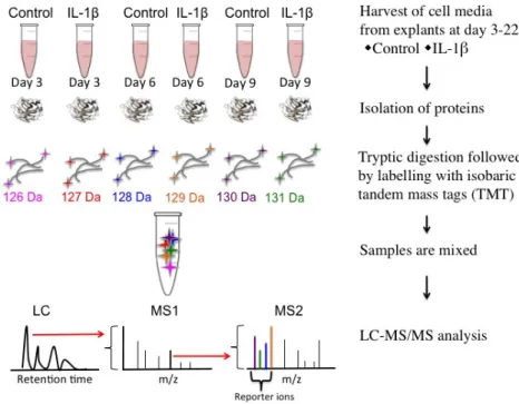
Protein expression analyses (papers III-IV) 41
4.7.1
Proteomic analyses 41
Sample preparation 43
Labelling of culture media with isobaric mass tags 43
Liquid chromatography-tandem mass spectrometry (LC-MS/MS) analysis
43
Database search and TMT quantification 43
Characterization of endogenous proteolytic peptides of lubricin in synovial
fluid 44
4.7.2
Western Blot 44
COMP 44
Lubricin 45
4.7.3
ELISAs 45
Chondroitin sulphate 846 (CS846) 45
MMP-13 45
C1, 2C 45
Lubricin 46
4.8
Glycomic analysis 46
4.9
Statistics 46
5
Results 48
5.1
Paper I 48
5.2
Paper II 49
5.3
Paper III 51
5.4
Paper IV 53
6
General discussion 57
6.1
In vitro methods: chondrocyte pellets and cartilage explants 57
6.2
Effects of the cytokines IL-1β, IL-6 and HMHB-1 on chondrocyte pellets in vitro (papers I and II) 58
6.3
Effects of IL-1β stimulation on the secretome of articular cartilage
explants cultured in vitro (paper III) 61
6.4
Presence of GDF-5 in normal articular cartilage and OCFs (paper II) 63
6.5
Characterization of lubricin (paper IV) 63
6.6
Horse as a model system for translational OA research 65
6.7
Change in diagnostic procedure of osteoarthritis 66
7
Conclusions 67
9
References 69
List of Publications
This thesis is based on the work contained in the following papers, referred to by Roman numerals in the text:
I Ley, C., Svala, E., Nilton, A., Lindahl, A., Eloranta, ML., Ekman, S. and Skiöldebrand, E. (2011). Effects of high mobility group box protein-1, interleukin-1β, and interleukin-6 on cartilage matrix metabolism in three-dimensional equine chondrocyte cultures. Connective Tissue Research
52(4), 290-300.
II Svala, E., Thorfve, A., Ley, C., Henriksson, HB., Synnergren, J., Lindahl, A., Ekman, S. and Skiöldebrand, E. (2014). Effects of interleukin-6 and interleukin-1β on expression of growth differentiation factor-5 and Wnt signaling pathway genes in equine chondrocytes. American Journal of Veterinary Research 75(2), 132-140.
III Svala, E., Löfgren, M., Sihlbom, C., Rüetschi, U., Lindahl, A., Ekman, S. and Skiöldebrand, E. (2014). An inflammatory equine model demonstrates dynamic changes of immune response and cartilage matrix molecule degradation in vitro. (Submitted)
IV Svala, E*., Jin, C*., Rüetschi, U., Ekman, S., Lindahl, A., Karlsson, N., and Skiöldebrand, E. (2014). Characterization of lubricin in synovial fluid from horses with osteoarthritis. (In manuscript)
Papers I-II are kindly reproduced with the permission of the publishers.
*Authors contributed equally to this work and should both be considered as main authors.
The contribution of ES to the papers included in this thesis was as follows:
I Second author. Active part in the planning and design of the study.
Performed the in vitro chondrocyte pellet culture, gene expression analysis and immunoassays. Active in presenting data from these experiments, and active in the drafting and revising the manuscript.
II Main author. Active part in the formation of hypothesis and study design. Major responsibility for the planning and organization of the study. Performed the majority of the experiments and analyses, such as in vitro
cultures, gene expression analyses and immunohistochemistry. Interpreted and summarized the microarray data. Performed the presentation of the data and statistical analyses. Drafted and edited the manuscript.
III Main author. Active part in the formation of hypothesis and study design. Major responsibility for the planning and organization of the study. Performed all experiments (in vitro cultures, immunoassays, biochemical analysis, Western Blot) except the mass spectrometry analyses. Interpreted and summarized the mass spectrometry data. Performed the presentation of the data and drafted/edited the manuscript.
IV Main author. Active part in the planning and design of the study, together with the second author. Performed the in vitro cultures. Interpreted and summarized the results. Active in presenting the data from these experiments and drafted/edited the manuscript
Abbreviations
3D three dimensional
ADAMTs a disintegrin and metalloproteinase with thrombospondin motifs AgPAGE agarose polyacrylamide composite gel
BSA bovine serum albumin
C1, 2C carboxy terminus neoepitope of the ¾ piece of collagen type I and II
C3 third carpal bone cDNA complementary DNA
COMP cartilage oligomeric matrix protein CS846 chondroitin sulphate 846
DAPI 4',6-diamidino-2-phenylindole dihydrochloride
DMEM/F12 Dulbecco’s modified eagle medium: nutrient mixture F-12 DRF dorsal radial facet
ECM extra cellular matrix
EDTA ethylenediaminetetraacetic acid GAG glycosaminoglycan
Gal galactose
GalNAcol N-acetylgalactosaminitol GDF-5 growth differentiation factor-5 GlcNAc N-acetylglucosamine
GUSB beta glucoronidase
HMGB-1 high mobility group box protein -1 HRP horseradish peroxidase
IL interleukin
LC/MS-MS liquid chromatography-tandem mass spectrometry LTQ linear trap quadrupole
MCP metacarpophalangeal MMP metalloproteinases
mRNA messenger ribonucleic acid
NC4 N-terminal non-collagenous domain Neu5Ac N-acetylneuraminic acid
Neu5Gc N-glycolylneuraminic acid OA osteoarthritis
OCF osteochondral fragment
PAGE polyacrylamide gel electrophoresis PBS phosphate buffered saline
PC palmar condyle PEST pencillin-streptomysin PVDF polyvinylidene fluoride
qRT-PCR quantitative real time polymerase chain reaction RA rheumatoid arthritis
SDS sodium dodecyl sulphate
SLRPs small leucine rich proteins/proteoglycans
SOX-9 sex determing region on the Y chromosome–related high mobility group box-9
TGF-β transforming growth factor beta TMT tandem mass tag
1 Introduction
1.1 General background
Osteoarthritis (OA) is a highly prevalent, chronic, and disabling disease (Johnson & Hunter, 2014) leading not only to articular cartilage destruction but also involving the entire joint and its structures: articular cartilage, subchondral bone, synovial membrane/capsule, and ligaments (Lane et al., 2011). The full pathogenesis for OA is unknown but factors suggested to contribute to the development of the disease are joint injury, mechanical loading, age, obesity, and genetics (Johnson & Hunter, 2014). Inflammation is always involved (Sokolove & Lepus, 2013; Goldring & Goldring, 2007; Saxne et al., 2003; Pelletier et al., 2001).
It is estimated that 12% of the adult human population aged ≥ 18 years have symptomatic OA (Hunter et al., 2014b) and its prevalence increases with age to encompass nearly 34% of those over 65 (Neogi, 2013). The joint most often affected by OA in humans is the knee, and the lifetime risk of developing the disease in this joint is as high as 45% (Neogi, 2013). Considering how many patients that never seek medical advice, the number is probably much higher. The individual burdens, to name a few, include pain, stiffness, and activity limitation leading to a lower quality of life. The socioeconomic cost that follows is tremendously high (Hunter et al., 2014b). Currently there is no disease-modifying drug for structural progression of OA (Jotanovic et al., 2014) and therefore the available treatments aim to relieve pain.
In horses, OA is a main reason for lameness (McIlwraith et al., 2012). The disease can develop at a young age in racehorses and is also a naturally occurring disease in older horses (McIlwraith et al., 2012; Bjornsdottir et al., 2004). The metacarpophalangeal (MCP) (Neundorf et al., 2010), followed by the carpal, are the most commonly OA-afflicted joints in horses (McIlwraith et
al., 2012). Racehorses develop OA at a young age due to the heavy and repetitive mechanical load from high-speed training and racing (Poole, 1996). Horseracing is a multibillion industry in Sweden and the economic importance of the horse sector is enormous. It has a direct and indirect total annual turnover of 46 billion Swedish crowns (Remmerth, 2008; Johansson & Andersson, 2004), greater than that for the metal and steel industries.
OA is currently clinically diagnosed late in the disease process, when irreversible damages to the cartilage have become evident. Therefore, there is a need for the development of reliable molecular markers (also called biomarkers) for the early stages of the disease. Optimal biomarkers could be used not only for early diagnosis but also for monitoring progression of the disease and evaluating treatment effects. To develop these biomarkers, the pathogenesis of OA needs to be further elucidated for an understanding of the complex molecular mechanisms responsible for the onset, development, and advancement of the disease.
The work included in this thesis focus on the changes of the extra cellular matrix in articular cartilage caused by inflammation.
1.2 Articular cartilage
Cartilage is a connective tissue consisting of chondrocytes, water, and the extra cellular matrix (ECM) synthesised by chondrocytes. Cartilage can be divided into hyaline cartilage, fibrocartilage, and elastic cartilage. This thesis examines the hyaline articular cartilage outlining the articular surface of bones.
Adult articular cartilage is an avascular, hypocellular, aneural, and alymphatic tissue with limited regenerative capacity (Iwamoto et al., 2013). This complex structure, found in all synovial joints allows pain-free, low-friction joint movement and disperses high mechanical load. Articular cartilage is organized in different zones (Figure 1) on the basis of its morphological features (Pritzker & Aigner, 2010). The superficial zone is immediately subjacent to the joint surface and chondrocytes of this zone are elongated and flattened. Underneath the superficial zone is the middle zone where the morphology of chondrocytes changes to rounder cells. The deep zone is in between the middle zone and the calcified cartilage; here chondrocytes exist as round cells in columns. The boundary between uncalcified and calcified cartilage is called the tidemark and beneath the calcified cartilage is the borderline of subchondral bone called the cement line. The thickness of equine articular cartilage varies in different joints; the average thickness of the articular cartilage in the MCP and carpal joints is similar (0.86 mm) while the
stifle has a thicker articular cartilage (2 mm) (Lee et al., 2014), comparable to that in a human knee (Frisbie et al., 2006).
Figure 1. Morphological structure of articular cartilage, organized in superficial, middle, and deep zones. The orientation of the collagen fibres as well as chondrocyte morphology vary in these zones. Originally published in “Surgical Alternatives for Treatment of Articular Cartilage Lesions”, Journal of the American Academy of Orthopaedic Surgeons, (2000;8(3)-180-189), by Browne, E. Jon and Branch, P. Thomas. Reused with permission and license from Lippincott Williams and Wilkins/Wolters Kluwer Health, Inc. © 2000, American Academy of Orthopaedic Surgeons.
1.2.1 Molecular organization of articular cartilage
Chondrocytes synthesize the ECM as well as produce enzymes responsible for degradation of the ECM. Water is the major constituent (approx. 75%) of ECM and the remainder is composed of several molecular components interacting to form the specialised network – collagens, proteoglycans and non-collagenous proteins. The ECM, which embeds the chondrocytes, is organized into zones based on their distance from the chondrocyte. Closest to the chondrocyte is the pericellular matrix, where molecules from the ECM bind to surface receptors. The territorial and interterritorial zones are found at a further distance from the chondrocyte (Las Heras et al., 2012; Kvist et al., 2008). The distribution of the components in the ECM varies within these zones (Heinegård & Saxne, 2011; Heinegård, 2009) (Figure 2).
Figure 2. Illustration of molecular components and their organization in the extracellular matrix (ECM) of articular cartilage. The ECM surrounding chondrocytes is arranged into zones described by their distance to the cell. The interterritorial matrix closer to the cells and the interritorial matrix at a further distance from the cell are indicated. Originally published in “Proteoglycans and more - from molecules to biology”, International Journal of Experimental Pathology (2009),90,575-586 by Heinegård, D. Reused with permission and license from John Wiley & Sons, Inc. ©Dick Heinegård © 2009 Blackwell Publishing Ltd.
Collagens
The collagen network gives cartilage its tensile strength and contributes to approximately 60% of the dry weight of articular cartilage. Articular cartilage comprises of collagen types II, III, VI, IX, XI and XII, and the organization of collagen varies in the different zones of the articular cartilage. In the superficial zone, collagens are arranged parallel to the articular joint surface, in contrast to the deep zone where the collagen fibres are organized perpendicular to the surface. In between these zones (middle zone), the collagen is organized in a random way (Figure 1). The superficial zone has the highest content of collagen and it decreases with the distance from the surface (Eyre et al., 2006). Collagen type II is the most common form of the collagen network in cartilage, representing approximately 90% of the total collagen content. Collagen type II, which has a fibrillar structure, is synthesized and secreted as procollagen α chains containing amino- and carboxy propeptides that are cleaved away by amino- and carboxy-terminal proteinases in the ECM (Peltonen et al., 1985). Other enzymes, metalloproteinases, (MMPs), cleave the mature collagen type II molecule, leading to a decreased content of collagen type II (Poole et al., 2002). In OA there is evidence of an attempt to repair damage by an increased synthesis (Nelson et al., 1998) of collagen type II, but meanwhile there is also an increased degradation of collagen type II (Hollander
et al., 1994) mediated mainly by MMP -1 and -13 (Dahlberg et al., 2000). Fragments resulting from the degradation of collagen type II have been shown to induce cartilage degradation by enhancing release of proteolytic enzymes (Guo et al., 2009) and thereby increasing the cleavage of both collagen type II and aggrecan (Yasuda et al., 2006).
Collagen type III is a trace component of articular cartilage, but can be increased in OA (Tanaka et al., 2013; Eyre et al., 2006).
Collagen type VI, primarily localized in the pericellular matrix (Zhang et al., 2011b), is composed of α-chains (α-1, α-2, α-3) and forms a highly cross-linked microfibrillar network (Gelse et al., 2003). This type of collagen has been found to interact with the chondrocyte by binding to integrins (Marcelino & McDevitt, 1995) of the cell membrane, as well as by interacting with other ECM molecules, such as collagen type II and small leucine-rich proteins/proteoglycans (SLRPs), decorin (Bidanset et al., 1992), and biglycan (Wiberg et al., 2002). Collagen type VI is postulated to strengthen the complete collagen network by binding to other members of the ECM. In OA, the turnover of collagen type VI is increased (Arican et al., 1996) and there is evidence of alteration in the molecular distribution (Wilusz et al., 2013) compared to normal cartilage. The increased concentration seen in the
interterritorial matrix could be the result of degradation of collagen type VI, leading to diffusion of fragments from the pericellular area (Soder et al., 2002).
Collagen type IX can decorate collagen type II fibril surfaces by covalent binding (Vaughan et al., 1988), suggesting that it has a role in improving mechanical restraint. It is hypothesized to act as a glue for the collagen type II network. Collagen type IX consists of three collagenous domains separated by four noncollagenous domains and the N-terminal noncollagenous domain (NC4) that projects out from the fibril surface can interact with other ECM molecules such as cartilage oligomeric matrix protein (COMP) (Holden et al., 2001). In vivo studies of collagen IX null mouse cartilage have suggested that proteins such as matrilin-1, matrilin-4, epiphycan, and thrombospondin-4 interact with collagen type IX, (Brachvogel et al., 2013). Degradation of collagen type IX is seen in the early stages of OA (Diab, 1993).
Collagen type XI can covalently crosslink to collagen type II and thereby support the collagen network, but the collagen type XI molecules can also crosslink to each other (Gelse et al., 2003; Eyre, 2002). The importance of this type of collagen is suggested by the recent findings of single nucleotide polymorphism in the collagen type XIA1 gene that is associated with OA (Rodriguez-Fontenla et al., 2014).
Collagen type XII is a fibril-associated collagen with interrupted triple helices, which can connect to other components in the ECM and is thought to stabilize the organization of the collagen network (Gregory et al., 2001).
Proteoglycans
Proteoglycans contribute to approximately 25-35% of the dry weight of articular cartilage and consist of a core protein with attached glycosaminoglycan (GAG) chains.
Aggrecan is the most common proteoglycan in articular cartilage. Through its negatively charged chondroitin sulphate chains, it has the potential to bind a large amount of water, resulting in a swelling and expansion of the ECM, and thereby enabling the cartilage to sustain high compressive load (Heinegård, 2009).
Aggrecan is a large (>2500 kDa), aggregating proteoglycan residing in the fibrillar collagen network. The superficial zone of articular cartilage has the lowest content of aggrecan and the content increases with the distance from the surface. The structure of the core protein in aggrecan consists of three globular domains called G1, G2 and G3. G1 has the ability to bind to link proteins or hyaluronic acid (Hascall & Heinegård, 1974), thereby producing multi-molecular aggregates. The region between G1 and G2 is called the interglobular domain, which contain sites subject to proteolytic cleavage
(Sandy et al., 1991) by MMPs or aggrecanases (a disintegrin and metalloproteinase with thrombospondin motifs (ADAMTS)), resulting in several protease-mediated catabolic epitopes (Struglics et al., 2006). MMP-3 and -13, as well as other MMPs, cleave aggrecan in the interglobular domain between Asn 341 and Phe 342, (Flannery et al., 1992) while ADAMTS4 and -5 also cleave aggrecan in the interglobular domain, but between Glu 373 and Ala 374 (Tortorella et al., 2002; Tortorella et al., 2000b).
Between the G2 and G3 domains, glycosaminoglycan side chains can attach (first about 30 keratan sulphate chains followed by approximately 100 chondroitin sulphate chains). Moreover, aggrecan also contains a variable number of O- and N-linked oligosaccharides. There are several ADAMTS-4 and -5 mediated cleavage sites in between the G2 and G3 domains (Heinegård, 2009).
Non-collagenous matrix proteins
Non-collagenous proteins contribute to approximately 15-20% of the dry weight of articular cartilage.
The SLRPs family has 18 members to date, including: biglycan, chondroadherin, decorin, epiphycan, fibromodulin, lumican and proline/arginine-rich-end leucine-rich repeat (PRELP) found in cartilage. These horseshoe-shaped proteins have a molecular structure that consists of an N-terminal variable domain and a conserved domain of tandem leucine-rich repeats (Ni et al., 2014). This distinctive structure benefits protein-protein interactions, enabling binding to various growth factors, cytokines, cell surface receptors, and other ECM components, thereby affecting various signalling pathways (Schaefer & Iozzo, 2008). SLRPs exhibit many biological roles; key features of SLRPs in joints are their involvement in tissue development and assembly. The function of SLRPs in collagen fibrillogenesis has been studied in developing mice tendons (Reed & Iozzo, 2002; Ezura et al., 2000). SLRPs are also involved in modulation of fibril formation by interacting with collagens (Kalamajski & Oldberg, 2010), to construct a specific collagen matrix for a functioning ECM.
The involvement of SLRPs in the pathogenesis of OA is hypothesized to occur by several mechanisms. A loss of SLRPs can lead to undesirable changes in the ECM collagen network. The SLRPs can also modulate transforming growth factor (TGF)-β signalling and thereby affect proliferation/differentiation of chondrocytes, or they can interact with the innate immune system by activating or inhibiting the complement cascade. As an example, fibromodulin can bind to C1q and activate the classical component
system (Sjoberg et al., 2005) while the binding of biglycan to C1q inhibits the activation of the classical complement system (Groeneveld et al., 2005)
Matrilins, another type of noncollagenous proteins found in cartilage, are considered to be adaptor proteins, which support the ECM assembly by binding to aggrecan, SLRPs, and collagen fibrils (Klatt et al., 2011).
COMP, also called thrombospondin-5, is a fundamental part of the ECM in articular cartilage. This 524 kDa multidomain glycoprotein consists of five identical units (Oldberg et al., 1992) forming a pentameric structure (Figure 3). COMP can bind to collagen types I and II (Rosenberg et al., 1998) and catalyze fibril formation by interacting with free collagen I and II molecules, bringing these molecules into close proximity with each other to promote further assembly (Halasz et al., 2007). COMP can also interact and bind to collagen type IX (Holden et al., 2001), aggrecan (Chen et al., 2007), matrilins (Mann et al., 2004) and fibronectin (Di Cesare et al., 2002). COMP is found in other structures of the joint such as tendon (Sodersten et al., 2013; Smith et al., 1997). Mutations in COMP can lead to chondrodysplasias, leading to early onset OA and short-limb dwarfism (Briggs et al., 1998). COMP is considered to be a marker of cartilage metabolism. It is studied as a potential diagnostic and prognostic biomarker for OA because serum levels of COMP are elevated in human OA (Valdes et al., 2014; Verma & Dalal, 2013; Clark et al., 1999). COMP has an active role in inflammation by activating the alternative pathway and inhibiting the lectin- and classical-complement pathways in rheumatoid arthritis (RA) patients but not in OA patients (Happonen et al., 2010). This suggests that the specific and likely disease-dependent fragmentation of COMP activates or inhibits inflammation cascades.
Figure 3. Schematic structure of one subunit of COMP and the interaction between pentameric COMP and collagen type II. COMP consists of five identical subunits, each with epidermal growth factor (EGF) repeats and type 3 repeats (calcium binding domains/calmodulin type units). The coiled-coil pentamerization domain is held together by disulphide bonds to form a pentameric structure. The function of the globular domain is to bind to other members of the ECM. The high-affinity binding between COMP (via the carboxyterminal domain) and native triple-helical collagen type II affects collagen fibril assembly. The COMP molecule can bind to five collagen molecules and facilitate their interaction in accelerating collagen fibril formation (Halasz et al., 2007). COMP can also interact with other collagens of the ECM such as collagen IX, further stabilizing the collagen network. (Illustration by E.Svala)
The mucin-like glycoprotein lubricin, also known as proteoglycan-4, is found in synovial fluid where it acts as a boundary lubricator (Swann et al., 1977) and protector (Rhee et al., 2005b) of articular cartilage. The molecular structure of the protein consists of somatomedin B-like-domains at the N-terminus, followed by several mucin-like repeats which are heavily glycosylated. It ends with a hemopexin-domain at the C-terminus (Rhee et al., 2005b). It is synthesized and secreted by the chondrocytes of the superficial zone of articular cartilage (Schumacher et al., 1994), but is also found in several other structures of the joints such as tendons (Funakoshi et al., 2008) and menisci (Zhang et al., 2011a). The reduction of coefficient-of-friction is associated with inhibition of chondrocyte apoptosis (Waller et al., 2013). Lubricin attaches to denaturated, amorphous and fibrillar collagen at the articular cartilage surface (Chang et al., 2014). Lubricin can present various O -glycan structures, which are proposed not only to have a role in lubrication
(Jay et al., 2001), but also in inflammation. They contribute to inflammation by carrying inflammatory oligosaccharide epitopes (Estrella et al., 2010) and binding to peripheral and synovial polymorphonuclear granulocytes (Jin et al., 2012).
1.3 Joint structure changes in osteoarthritis
In racehorses, the MCP joint is the one most commonly affected by OA, followed by the carpal joint (Figure 4) (McIlwraith et al., 2012).
The equine MCP joint is a hinge joint consisting of the distal metacarpal bone III and the proximal phalanx bone (Fails & Kainer, 2011). The MCP has a close-fitting articular surface that can quickly develop linear erosions and wear lines in association with osteochondral fragmentation (McIlwraith et al., 2012). This is possibly due to the small joint surface, wide range of motion, and weight transmission (Pool & Meagher, 1990).
The equine carpal joint is comprised of three joint compartments; the radiocarpal, midcarpal, and carpometacarpal. The middle carpal joint is overextended during the weight-bearing phase of the stride (Bramlage et al., 1988) where it is exposed to high load, particularly on the radial facet of the third carpal bone. During trotting and galloping, it is exposed to forces in both longitudinal and transversal directions (Johnston et al., 1997; Bramlage et al., 1988). The dorsal radial facet (DRF) of the third carpal bone (C3) is a contact area with higher mechanical load compared to the palmar condyle (PC) of this bone which is a noncontact area (Palmer et al., 1994) (Figure 4).
Figure 4. The location of the equine metacarpophalangeal (MCP) and carpal joints, which are the joints most commonly affected by OA (McIlwraith et al., 2012). The dorsal radial facet (DRF) of the third carpal bone (C3) is a contact area with higher mechanical load compared to the palmar
condyle (PC) of this bone which is a noncontact area (Palmer et al., 1994). Drawings by Alexandra Leijon.
OA affects not only articular cartilage, but also the other components of the joint (McIlwraith et al., 2010), where it leads to formation of subchondral bone
sclerosis (Grynpas et al., 1991), formation of new bone at the joint margins (osteophyte formation) (Olive et al., 2009), increased bone remodeling (for review see (Li et al., 2013), and synovitis (Myers et al., 1990). These joint tissue changes are similar among species, including man.
Chondrocyte necrosis with adjacent chondrocyte cluster formations (Lotz et al., 2010), superficial fibrillation/fraying and cleft formations are seen in the pathological destruction of articular cartilage (McIlwraith et al., 2010). The mechanism behind the increased number of cell clusters (Poole et al., 1991), seen in OA cartilage, is thought to be proliferation (Pfander et al., 2001) and/or migration (Kouri et al., 1996). There is also a change of phenotype of chondrocytes in OA cartilage, resulting in hypertrophic cells (von der Mark et al., 1992). This hypertrophy is characterized by collagen X and MMP-13 expression (Nurminskaya & Linsenmayer, 1996).
A hallmark of OA is the degradation of components in the ECM (Heinegård & Saxne, 2011). Normal articular cartilage has a low metabolic activity with a low-turnover rate of ECM, delicately regulated through anabolic (production of ECM molecules) and catabolic events (degradation of ECM) (Henrotin & Reginster, 1999). In early OA, there is an initial attempt at repair, reflected by an elevated anabolic activity of the chondrocytes, with cell proliferation and synthesis of ECM molecules and growth factors (Poole et al., 2007). However this activity is later disrupted and overrun by the catabolic events, ultimately leading to a net loss of ECM.
Degradation of collagen type I, II and III molecules occurs through an initial cleavage into ¾- and ¼-length fragments by proteolytic enzymes called collagenases. MMP-13 is an important collagenase in the breakdown of collagen type II (Billinghurst et al., 1997), but chondrocytes are capable of producing and secreting several other proteins in the MMP family. Chondrocytes of human OA cartilage produce MMP-1, -2, -3, -8, -9 and -13 (Tetlow et al., 2001). The general MMP activity is increased in synovial fluid from equine OA joints (van den Boom et al., 2005; Brama et al., 2004; Brama
et al., 2000) and messenger ribonucleic acid (mRNA) gene expression of MMP-13 is elevated in synovial tissues and cartilage from equine OA joints compared to normal joints (Kamm et al., 2010). In vitro studies have demonstrated this, since cytokine-stimulated bovine (Sondergaard et al., 2006) and equine (Clutterbuck et al., 2011; Clegg & Carter, 1999) articular cartilage cause an increase in the secretion of proteolytic enzymes (MMP1, 2, 3, and -9). This in turn leads to the degradation and secretion of collagen and proteoglycan fragments into the cell media. Other proteolytic enzymes, ADAMTS-5 (Little et al., 2007; Stanton et al., 2005) and ADAMTS-4 (Tortorella et al., 2000a), also have an important role in the degradation of
equine cartilage. Several in vitro studies of articular cartilage explants have revealed a distinct degradation pattern due to inflammatory stimuli, where aggrecan is released early, followed by COMP, fibromodulin and finally, collagen type II (Williams et al., 2013; Heathfield et al., 2004; Dickinson et al., 2003; Sztrolovics et al., 1999).
1.3.1 Osteochondral fragments
Osteochondral fragments (OCFs), also known as chip fractures, are found at the dorsal aspects of the articular margins and facets of bones in the carpal joints and the proximal aspect of the first phalanx. They are thought to arise from fractured osteophytes, degenerated articular cartilage or necrotic subchondral bone in racehorses with OA (Pool & Meagher, 1990). The metabolic activity in the OCFs of the carpal joints appears elevated, as indicated by increased levels of native COMP in synovial fluid from racehorses with OCFs (Arai et al., 2008; Skiöldebrand et al., 2005) and an increased synthesis of aggrecan in serum and synovial fluid (Frisbie et al., 1999) . COMP synthesis is also seen in the chondrocytes, by in situ hybridisation, in these OCFs (Skiöldebrand et al., 2005). High levels of interleukin (IL)-6, but not TNF-α are found in the synovial fluid of carpal joints with OCFs (Ley et al., 2007), with the chondrocytes being the potential source of IL-6 (Ley et al., 2009).
1.4 Factors involved in the development of OA
OA is thought to be a multifactorial disease with mechanical loading being one of the factors that promotes it. Normal healthy cartilage has specialized biomechanical properties which allow it to spread force between joints while providing nearly friction-free movement (Kerin et al., 2002). Excessive mechanical load or a major traumatic event alters the physiological biomechanical environment by activating factors involved in the pathogenesis of OA. These factors include inflammatory mediators and proteolytic enzymes, which induce ECM degradation and chondrocyte death (Buckwalter et al., 2013). The extensive training that racehorses undergo provokes metabolic changes in the composition of the ECM, with decreased synthesis and release of COMP (Skiöldebrand et al., 2006). These changes are seen in in vitro
studies where static compression stimulates a degradation of proteoglycans and collagens in bovine cartilage (Loening et al., 2000) and decreases the synthesis of cartilage matrix proteins. On the other hand, dynamic compression increases the synthetic activity of chondrocytes (Tsuang et al., 2008; Kim et al., 1994)
OA can occur early in equine athletes; however, it can also develop later in older horses (McIlwraith et al., 2012). A prominent risk factor associated with OA is aging; the prevalence of human OA also rises with age. Age-related changes of ECM are degradation of collagen with an increased fibrillation of the articular surface (Hollander et al., 1995) and extensive heterogeneity in the molecular structure of the ECM (Dudhia, 2005). Furthermore, the chondrocyte anabolic activity decreases with age (Aigner et al., 2007). Nevertheless, since not all older horses/humans develop OA, the link between OA and ageing is not fully elucidated.
Another risk factor associated with OA is genetics. Proposed susceptibility candidate genes for human OA are linked to ECM or signalling molecules thereof, e.g., RUNX2, SMAD3 and growth differentiation factor-5 (GDF-5) (Reynard & Loughlin, 2013). There is emerging evidence of a link between obesity and the initiation and progression of human OA with possible contributions from both increased mechanical forces across joints, leading to cartilage destruction, and from systemic factors such as altered adipokine levels (Poonpet & Honsawek, 2014; Zhou et al., 2014; Spector et al., 1994).
1.5 Inflammation
Inflammation is strongly associated with the pathophysiology of both human (Saxne et al., 2003; Pelletier et al., 2001) and equine OA (Kamm et al., 2010); a diversity of inflammatory mediators, such as cytokines, control the inflammatory process. Inflammatory signs seen in the development and progression of OA are clinical local signs such as joint pain, swelling, stiffness, an increased level of C-reactive protein (Jin et al., 2013; Pearle et al., 2007; Sharif et al., 2000), and synovitis (Sellam & Berenbaum, 2010; Sutton et al., 2009) with increased synovial mononuclear cell infiltration (Benito et al., 2005). An inflamed synovial membrane can produce various inflammatory mediators, such as IL-1β and tumor necrosis factor (TNF)-α; the chondrocytes respond by producing proteolytic enzymes. However, OA chondrocytes also produce and secrete several inflammatory mediators themselves including:
IL-β, TNF-α, (Tetlow et al., 2001), IL-6 (Ley et al., 2009), IL-7 (Long et al., 2008), IL-18 (Olee et al., 1999), and HMGB-1 (Terada et al., 2011). The inflammatory cytokines are the most important compounds participating in the pathogenesis of OA; their involvement disrupts the normal metabolic homoeostasis of cartilage by favouring catabolic processes, with MMP-activation leading to destruction of cartilage (Wojdasiewicz et al., 2014).
1.5.1 Interleukin-1β and Tumor necrosis factor-α
IL-1β and TNF-α are often regarded as the master cytokines involved in the pathogenesis of OA. IL-1β is synthesized in a pro-form (pro-IL-1β) that is cleaved by casphase-1 (Piccioli & Rubartelli, 2013) to generate an active form before it is secreted by chondrocytes and synovial cells into the joint cavity. Pro-IL-1β can also be found extracellular where it can be cleaved by serine proteases (Wittmann et al., 2011).
Enhanced levels of IL-1β are found in cartilage, bone, synovial fluid and synovial membrane in human OA (Massicotte et al., 2002; Melchiorri et al., 1998; Kubota et al., 1997; Farahat et al., 1993), as well as in articular cartilage (Kamm et al., 2010) and synovial fluid (Morris et al., 1990) in equine OA joints. Increased levels of the membrane bound receptor, IL-1 receptor type I, which activates the intracellular signalling cascade upon binding to IL-1β, is found in human OA chondrocytes (Silvestri et al., 2006). The IL-1 receptor accessory protein is also necessary for signalling (Casadio et al., 2001). IL-1β
can also bind to a decoy receptor called IL-1 receptor type II; however, this binding does not generate active signalling, and the abundance of this receptor is reduced in human OA chondrocytes (Wang et al., 2003). Another counterproductive molecule, the receptor antagonist IL-1Ra, can bind to both receptors of IL-1β to inhibit the binding of IL-1β and subsequent signalling. In an established equine OA model, gene transfer of IL-1Ra displays favourable effects with upregulation of IL-1Ra protein expression, improvement in pain and disease activity, and positive effects on histologic parameters in the articular cartilage (Frisbie & McIlwraith, 2000).
There are several effects of active IL-1β signalling in cartilage. The activation of numerous signal transduction pathways leads to an increase in intracellular Ca2+, with activation of protein kinase C, p38, c-JUN N-terminal kinase, and extracellular signal regulated kinase -1 and -2. Active IL-1β
signalling also leads to nuclear translocation of nuclear factor-κB (NF-κB), activating transcription factor, and activator protein 1 (Daheshia & Yao, 2008). Activation of these transcription factors results in expression of genes leading to the production of MMPs and ADAMTs (Tetlow et al., 2001), prostaglandin, and nitric oxide. There is also an induction of the production of other cytokines such as IL-6 and to an even greater degree IL-1β, suggesting an autocrine loop (Fan et al., 2007).
Taken together, the effects of IL-1β on chondrocytes induce the expression of catabolic genes. The effects also include downregulation of the expression of anabolic genes involved in the metabolism of chondrocytes and induction of chondrocyte apoptosis (Wojdasiewicz et al., 2014; Daheshia & Yao, 2008).
TNF-α is synthesized as a homotrimeric tramsmembrane protein type II that needs to be cleaved by metallopeptidase TNF-α converting enzyme (also called ADAM-17), for activation (Black et al., 1997). The secretion and presence of TNF-α in human OA is very similar to that of IL-1β, as they are secreted by the same cells in the joint and are elevated in human OA cartilage, subchondral bone, synovial membrane, and synovial fluid (Wojdasiewicz et al., 2014). This suggests that there is a synergistic interaction between them (Henderson & Pettipher, 1989). The concentration of TNF-α is increased in synovial tissue and articular cartilage of equine OA (Kamm et al., 2010). Other studies report that TNF-α concentrations in synovial fluid are not related to equine OA (Ley
et al., 2007; Jouglin et al., 2000) suggesting that TNF-α is not a master cytokine in the disease. TNF-α can bind to two receptors, called TNFR-1 and TNFR-II, (Westacott et al., 1994) and active TNF-α signalling can inhibit proteoglycan (Saklatvala, 1986) and collagen synthesis (Seguin & Bernier, 2003) as well as produce MMPs (Wojdasiewicz et al., 2014).
1.5.2 Interleukin-6
IL-1β induces a massive production of IL-6 (Nietfeld et al., 1990). Although IL-6 is called a pro-inflammatory cytokine, this is somewhat misleading because IL-6 has several anti-inflammatory, protective effects such as inhibiting the synthesis of IL-1β and TNF-α and enhancing the production of cortisol (Mölne & Wold, 2007). Classical signalling acts through binding of the membrane-bound IL-6 receptor. The membrane-bound IL-6 receptor system consists of two membrane proteins: a ligand-binding chain (IL-6R) and a membrane-bound β-receptor glycoprotein, gp130. Binding of IL-6 to IL-6R triggers the association of IL-6R and gp130, and gp130 in turn transduces the signal (Kishimoto, 1992). IL-6 can also bind to another receptor, the soluble IL-6 receptor, and the signalling through this receptor is called trans-signalling. In trans-signalling, IL-6 binds to soluble forms of the IL-6R (sIL-6R) and this complex activate cells due to the uniform expression of gp130 (Wolf et al., 2014). A protective role for IL-6 against the pathogenesis of OA has been suggested because IL-6 gene-knockout mice develop more advanced osteoarthritis (de Hooge et al., 2005). Also, high levels of IL-6 have been detected in synovial fluid in canine OA (Venn et al., 1993) and in equine OA synovium (Ley et al., 2009).
1.5.3 High-mobility group box protein-1 (HMGB-1)
HMGB-1 is a 25 kDa chromosomal protein, consisting of two basic boxes (called A and B) responsible for DNA binding, and a highly acidic C-terminus
(Zhang & Wang, 2010). The protein has different functions depending on the intracellular or extracellular location. In the nucleus it facilitates binding of regulatory proteins, with a role as a minor-groove binding enhancer (Lotze & Tracey, 2005). Extracellularly, HMGB-1 acts as a danger-associated molecular pattern (DAMP) molecule, released by necrotic cells or secreted in response to inflammatory stimuli, thereby mediating activation of innate immune response (Castiglioni et al., 2011). HMGB-1 is expressed in synovial tissue from RA patients and from rats with experimental arthritis (Kokkola et al., 2002). HMGB-1 levels in synovial fluid of knee-OA patients are associated with radiographic disease severity (Li et al., 2011) and HMGB-1 is overexpressed in human (Garcia-Arnandis et al., 2010) and equine OA synovium (Ley et al., 2009). Additionally in vitro stimulation with IL-1β upregulates the mRNA gene expression of HMGB-1 and promotes the translocation of HMGB-1 from nuclei to cytoplasm in human OA chondrocytes (Terada et al., 2011).
2 Aims of the thesis
The overall aim of the thesis was to contribute to knowledge about the influence of inflammatory cytokines on equine articular cartilage.
The specific objectives of this thesis were to:
1) Establish a three-dimensional (3D) culture system (pellets) for equine chondrocytes as an in vitro model for OA.
2) Evaluate and compare the effect of the cytokines interleukin (IL) –1β, IL-6 and HMGB-1 on equine articular chondrocytes cultured in an in vitro 3D model.
3) Examine if IL-6 could induce an upregulation of GDF-5 in equine chondrocytes and to identify the possible signalling pathways.
4) Investigate the presence of GDF-5 expressing chondrocytes in normal equine articular cartilage and OCFs.
5) Investigate the secretome of equine articular cartilage explants stimulated with IL-1β and cultured in vitro with the aimof describing the longitudinal release pattern of ECM molecules.
6) Study the presence and glycosylation profile of lubricin in synovial fluid from horses with OA and normal joints and to examine the secretion pattern of lubricin, after IL-1β stimulation, in equine articular cartilage explants in vitro.
Two different in vitro models were used with the purpose of mimicking OA in vivo. Chondrocyte pellets and explants derived from healthy equine articular cartilage were stimulated with cytokines to induce an inflammation and the analyses were done by: immunohistochemistry, immunoassays (ELISAs, Western blot), biochemical assays (glycosaminoglycan content), quantification of gene expression, and quantitative proteomics.
3 Hypotheses
Hypotheses for the studies on equine chondrocyte pellets in vitro were:
! Paper I: Chondrocyte metabolism is affected by exposure to IL-1β, IL-6 and HMGB-1. The response can be different in chondrocytes cultured from differently loaded anatomical sites in vivo from the equine third carpal bone.
! Paper II: IL-6 induces an upregulation of GDF-5 in equine OCFs explaining the anabolic processes found in the fragments in vivo. Expression of GDF-5 is affected by short and long term stimulation of IL-1β and IL-6 in chondrocytes from the third carpal bone.
Hypotheses for studies on equine cartilage explants in vitro were:
! Paper III: Molecular changes in equine cartilage after IL-1β stimulation are time dependent.
! Paper IV: Inflammation in OA influences lubricating properties by altering the level and glycosylation profile of lubricin in equine articular cartilage.
4 Material and methods
This section summarizes the material and methods of the studies for this thesis. Detailed descriptions of the procedures are presented in each of the papers.
4.1 Animals and sample collection (papers I-IV)
Macroscopically normal articular cartilage was aseptically obtained from the DRF (paper I and II) and PC (paper I) of the C3 from euthanized horses without any clinical history of joint disease. Additional osteochondral fragments (OCF) (dorsoproximally of the radial facet of C3, dorsodistally of the radial carpal bone, or dorsoproximally of the first phalanx) were used from three horses that underwent arthroscopy because of lameness (paper II). Full-thickness articular cartilage explants from the weight-bearing part of the distal metacarpal bone III in the MCP joint from three slaughtered horses without any clinical history of joint disease, were used in papers III-IV. All collected samples were transported chilled to the cell culture lab in a sterile saline solution (0.9% sodium chloride) with 50 mg/l gentamin sulphate and 250 µg/ml amphotericin B.
Synovial fluid (paper IV) was aseptically collected from middle carpal joints with macroscopically normal articular cartilage (n=7), structural OA lesions (n=7), and OCF (n=3).
4.2 Isolation and expansion of chondrocytes (papers I-II)
Protocols for isolation and expansion of chondrocytes from articular cartilage were performed according to previously described procedures (Brittberg et al., 1994). In summary, equine articular cartilage shavings were washed with cell culture media and mechanically minced with a scalpel. Enzymatic digestion with 0.8 mg/ml collagenase type 2 was performed for 20-24 h at 37°C in 7%
CO2/93% air, and a viable count of the single cell solution was performed with
0.4% tryptan blue dye in a Bürker chamber. Cells were seeded into culture flasks at a density of 16,000 cells/cm2 in complete chondrocyte media (Dulbecco’s Modified Eagle medium, i.e., nutrient mixture F-12 (DMEM/F12) supplemented with 1x penicillin-streptomycin (PEST), 2 mM L-glutamine, 0.1 mg/ml ascorbic acid and 10% equine serum) and incubated at 37°C in 7% CO2/93% air.
At approximately 80% confluence, the chondrocytes were treated with 0.05 % trypsin with 1x ethylenediaminetetraacetic acid (EDTA) at 37°C in 7% CO2/93 % air until the cells detached from the cell culture flasks. Chondrocytes
were seeded into sterile polystyrene cell-culture flasks at a density of 3000 cells/cm2 in complete chondrocyte media at 37°C in 7% CO2/93 % air until
80% confluence. Medium was changed twice a week.
4.3 In vitro chondrocyte culture models (papers I,II,III,IV)
4.3.1 Three-dimensional chondrocyte pellet cultures (papers I-II)
Figure 5. Experimental setup for three-dimensional (3D) pellets cultured in vitro. Normal equine articular cartilage was harvested from the third carpal bone in the middle carpal joint. Chondrocytes were isolated and expanded in monolayer until 80% confluence. Chondrocytes were cultured in 3D pellets (200,000 cells/pellet) for 14 days before cytokine stimulation for 1, 2, 12 or 48 hours. Cell media and 3D pellets were analysed for gene and protein expression. Illustration by E.Svala.
The protocol for 3D chondrocyte pellet cultures was slightly modified from previously described procedures (Stenhamre et al., 2008; Tallheden et al., 2004). Expanded chondrocytes from the first subculture (passage 1) were cultured in 3D pellets in a 96-well ultralow attachment plate, for 14 days at 37°C in 7% CO2/93% air. Pellet medium was changed daily after 3D
chondrocyte pellet formation was established (Figure 5).
4.3.2 Explant cultures (paper III-IV)
Cartilage explants (5 mm in diameter) were allocated to groups of 4 explants/well into polystyrene plates. The explants were pre-incubated before cytokine stimulation for 24 h, in 2 ml DMEM/F12 supplemented with 0.1 mg/ml cell-culture-tested bovine serum albumin (BSA), 0.1 mg/ml ascorbic acid and 4% v/v PEST at 37°C in 7% CO2/93% air (Figure 6).
Figure 6. Methodological setup for in vitro culture of equine articular cartilage explants. Macroscopically normal cartilage from three horses was harvested from the weight-bearing part of the distal metacarpal bone III in the metacarpophalangeal joints. Cartilage explants (5 mm in diameter) were cultured in culture dishes with or without IL-1β during 25 days. Culture medium was changed and harvested every third day. Analyses performed on cell culture media were: quantitative proteomics, GAG, Western Blot (for COMP and Lubricin) and Elisa (for MMP-13 , C1, 2C and Lubricin). At day 25, the cartilage explants were harvested for histological evaluation (hematoxylin and eosin (H&E), toluidine blue and safranin O). Illustration by E.Svala.
4.3.3 Cytokine stimulation (paper I-IV)
At day 14 of chondrocyte culture, the pellets (papers I, II) were allocated with a randomized procedure to treatment groups and either stimulated with
recombinant equine IL-1β (5 ng/ml), recombinant equine IL-6 (5 ng/ml), recombinant human HMGB-1 (1 µg/ml) or left unstimulated. Medium change was performed daily and pellets were stimulated for 1, 2, 12 or 48 hours.
After the pre-incubation of cartilage explants (papers III, IV) for 24 h, the medium was harvested and the explants were cultured in 2 ml medium in the presence or absence of 10 ng/ml equine recombinant IL-1β (10 ng/ml) at 37°C in 7% CO2/93 % air. The medium was changed and harvested at 3, 6, 9, 12, 15, 18, 22 and 25 days and immediately frozen at -80°C for later analysis.
Pellets or explants were harvested and washed in phosphate buffered saline (PBS) or snap frozen in liquid nitrogen before storage at -80°C or fixed in buffered formalin.
4.4 Histological methods (papers I,II,III)
4.4.1 Fixation
Chondrocyte pellets were fixed in 6% v/v neutral buffered formalin, for a minimum of 24 h. Normal articular cartilage, OCFs, and cartilage explants were immersed in 10 % v/v neutral-buffered formalin and OCFs were decalcified in formic acid before embedment.
4.4.2 Histological stainings (papers I,II,III)
Sections from central parts of pellets or explants, normal articular cartilage and OCFs were dehydrated, embedded in paraffin blocks, cut into 3-6 µm thick sections, and stained with hematoxylin and eosin (H&E), safranin-O or Massons trichrome. A minimum of two sections/samples was evaluated.
4.4.3 Immunohistochemical stainings (paper I-II)
Sections were deparaffinised, rehydrated and immersed in 0.1% v/v detergent solution (GDF-5 in paper II) or immersed in 3% v/v hydrogen peroxide to quench endogenous peroxidase activity. Samples were then incubated with hyaluronidase (COMP in paper I) or subjected to heat-mediated antigen retrieval steps (GSK3β and β-catenin staining in paper II).
Subsequently, non-specific antibody binding was blocked using species-appropriate serum or BSA before incubation with primary antibodies (rabbit-anti-bovine COMP, polyclonal goat GDF-5, rabbit polyclonal GSK3β, mouse monoclonal nuclear-dephosphorylated β-catenin) at 4°C overnight. Washing was performed with PBS or 0.1% v/v detergent solution and an additional blocking were performed before incubation with secondary antibodies at room temperature. Antibodies were either conjugated with horseradish-peroxidase (HRP) (for GSK3β and β-catenin), biotinylated (for COMP) or labelled with
photostable orange fluorescent dye (for GDF-5). Avidin–biotin–peroxidase complexes were visualized using 3,3´diaminobenzidine. HRP conjugated antibodies were detected with a signal amplification kit using fluorophore labelling. Nuclei were stained with the fluorescent dye 4', 6-diamidino-2-phenylindole dihydrochloride (DAPI), or counterstained with Mayer’s hematoxylin. Immunostaining was qualitatively assessed using light microscopy or fluorescent microscopy with appropriate filters. Isotype control antibodies, which have no relevant specificity, were used to distinguish non-specific background binding from antigen-non-specific antibody binding.
4.5 Gene expression analyses
4.5.1 RNA isolation and quantification (papers I,II)
Frozen chondrocyte pellets were disrupted through high-speed shaking for 8 min in 2 ml plastic tubes with tungsten carbide beads. Lysis reagent was added to each sample and the shaking was performed again for 2 min. The homogenate was incubated at 5 min at room temperature before adding chloroform, then shaken vigorously for 15 sec. Samples were incubated at room temperature for 3 min and thereafter centrifuged for 15 min at 4°C. The upper aqueous phase of the sample was transferred into a new sample tube and total RNA was further purified using a commercially available kit and a fully automated sample preparation robot, in accordance with the manufacturer’s protocol for animal cells. DNase was added to the isolated total RNA. After quantity and quality assessments, the recovered total RNA was stored at -80°C.
4.5.2 Complementary DNA synthesis
A commercially available reverse-transcription kit was used to prepare complementary DNA (cDNA) from 100 ng of total RNA from each chondrocyte pellet sample. A thermal cycler program of 25°C (10 min), 37°C (120 min), 85°C (5 min), 4°C (∞) was performed and the cDNA (concentration 5 ng/µl) was stored at -20°C.
4.5.3 Quantitative real time polymerase chain reaction (qRT-PCR)
The polymerase chain reaction (Saiki et al., 1985) theoretically amplifies DNA exponentially, doubling the number of target molecules with each amplification cycle. The thermal cycler protocol for the qRT-PCR reaction starts by activating the enzyme uracil-DNA glycosylase at 50°C for 2 min followed by activation of DNA polymerase at 95°C for 10 minutes.
After these initial steps the qRT-PCR consists of 40 cycles of the following steps:
1) Denaturation (95°C for 10 seconds): Double-stranded DNA denatures to single-stranded by disrupting hydrogen bonds between complementary bases.
2) Annealing/extension (60°C for 1 minute): The primers anneal (hybridize) to the complementary DNA sequences. After annealing, DNA polymerase synthesizes a new DNA strand with deoxyribonucleotide triphosphates (dNTPs). The new strand will be complementary to the DNA template strand.
The amount of DNA is measured after each cycle by attaching the template to a fluorescent probe (consisting of an oligonucleotide with a fluorophore (6-carboxyfluorescein FAM)) at the 5´end and a quencher (dihydrocyclopyrroloindole tripeptide minor groove binder, MGB) at the 3´end. As long as the fluorophore and the quencher are in proximity of each other by being attached to the probe, the reporter dye is quenched. The DNA polymerase will cleave this reporter dye off the probe (Holland et al., 1991) during each extension. This increases the fluorescent signal in direct proportion to the number of the number of PCR products (also called amplicons) generated. The qRT-PCR reagent solution contains a passive reference dye (ROX), used to normalize the reporter fluorescence.
qRT-PCR protocol
The quantitative real-time polymerase chain reaction (qRT-PCR) was analysed on 2.5 ng cDNA/sample in duplicate with a solution of polymerase, primers, probes, nucleotides and buffer in a 96-well reaction plate. A negative control without any sample was used to detect contaminating DNA from the reagents. The relative gene expression was determined by the 2 –(ΔΔCT) method (Livak & Schmittgen, 2001).
Primers and probes
A commercially available qRT-PCR assay mix of primers and probes intended for the detection of human genes was used for detection of GDF-5, Sry-related high-mobility group box 9 (SOX-9) and collagen type II. A basic local alignment search tool was used to verify homology between the human and equine genes. A database and genome browser were used to select equine-specific primer and probe sequences for collagen type I, versican, aggrecan, COMP, MMP-9, MMP-13, ADAMTS-5, TIMP-1, and beta glucuronidase (GUSB). A validation experiment was performed for each equine-specific primer and probe set with a dilution series of cDNA template for the target
genes. Several potential reference genes were validated (unpublished data) and equine GUSB was used as a reference gene.
4.5.4 Microarray (paper II)
High-throughput gene expression microarrays have been developed during the last twenty years and the technology is based on the ability of DNA (on an array) to specifically bind/hybridize to complementary sequences (RNA or DNA target molecules) from samples (Forster et al., 2003). In paper II we used Affymetrix® human gene 1.1 ST microarrays on which millions of short strands (approximately 25 bases) of DNA (called oligonucleotide probes) are built onto a glass chip, one base at a time. A solution of RNA or DNA from samples, labelled with biotin, and a fluorescent stain is distributed over the array. When RNA or DNA from the samples binds to the probe on the array this results in a fluorescence signal; screening for this hybridization shows if a gene is expressed. Highly expressed genes give stronger fluorescence signals than genes with lower expression.
At the time of the experiments in paper II, no equine specific arrays were commercially available. Human microarrays had previously been used to study the transcriptome of equine chondrocytes in monolayer (Graham et al., 2010) and therefore a similar approach was used in paper II.
Total RNA (250 ng) from chondrocyte pellets was processed and analysed with a human microarray for global transcriptome quantification at the Nottingham Arabidopsis Stock Centre (University of Nottingham, United Kingdom). Briefly, target preparation from total RNA used T7 primers in the cDNA synthesis step, followed by in vitro transcription to produce cRNA. Random primers were then used to synthesize cDNA from the cRNA. The cDNA was fragmented, biotin-labeled, and mixed into a hybridization cocktail before application to the array. The high-throughput array was scanned (Genechip 3000 7G scanner) and expression signals were extracted and normalized by applying the Robust Multichip Average (RMA) normalization method. A web-based probe-match tool was used to compare equine nucleotide sequences against individual probes on the human array, with the purpose of confirming that the human probe sets matched the equine query sequence of the corresponding gene. The data set was used to screen for novel putative target genes that displayed a change in expression when IL-6 stimulated samples were compared to their unstimulated controls. The data were only analysed on the gene-level and not on the exon-level.