Research Article
Clinicopathologic Characteristics and Long-Term Outcome of
Gastric Cancer Patients with Family History: Seven-Year
Follow-Up Study for Korean Health Check-Follow-Up Subjects
Jooyoung Lee,
1Su Jin Chung
,
1Ji Min Choi,
1Yoo Min Han,
1and Joo Sung Kim
1,2 1Department of Internal Medicine and Healthcare Research Institute, Seoul National University Hospital Healthcare SystemGangnam Center, Seoul, Republic of Korea
2Department of International Medicine and Liver Research Institute, Seoul National University College of Medicine,
Seoul, Republic of Korea
Correspondence should be addressed to Su Jin Chung; medjsj7@snuh.org
Received 22 June 2020; Revised 27 August 2020; Accepted 29 September 2020; Published 22 October 2020
Academic Editor: Greger Lindberg
Copyright © 2020 Jooyoung Lee et al. This is an open access article distributed under the Creative Commons Attribution License, which permits unrestricted use, distribution, and reproduction in any medium, provided the original work is properly cited.
Background/Aims. Family history (FHx) has been reported to be a risk factor for gastric cancer (GC). However, the long-term prognosis of GC with FHx remains controversial. We aimed to investigate the clinicopathologic characteristics and long-term outcomes of GC according to the presence or absence of GC FHx.Methods. This study was conducted on asymptomatic healthy individuals who underwent upper gastrointestinal endoscopy for the purpose of GC screening. Patients who were diagnosed with GC between October 2003 and December 2013 at Seoul National University Hospital Healthcare System Gangnam Center were identified. Demographic and clinicopathologic characteristics were compared between the groups with and without FHx of GC. Overall survival (OS) and recurrence-free survival (RFS) were assessed as primary outcomes. Results. There were no significant differences in tumor characteristics according to FHx of GC. However, preexisting adenoma was more frequent in patients with FHx than in those without FHx (14.5% vs. 6.3%, p= 0:035). The proportion of patients with microsatellite instability (MSI) was also higher in groups with FHx of GC (43.2% vs. 13.2%,p= 0:006).Helicobacter pyloriinfection rates of patients with FHx of GC tended to be higher although not significant (70.5% vs. 61.3%,p= 0:188). However, OS and RFS at 5 years of the GC patients with FHx were not significantly different from those of patients without FHx.Conclusion. Preexisting adenoma and GC with MSI are more common in patients with FHx of GC than in those without. There were no significant differences in the survival rate according to FHx.
1. Introduction
The incidence of gastric cancer (GC) has declined significantly worldwide over the past half-century and is possibly attribut-able to economic development, improved sanitation, and decreased Helicobacter pylori infection rates because of the expansion of eradication [1]. Nevertheless, GC remains a global health problem as thefifth leading cancer and the third most common cause of cancer-related deaths worldwide. Newly diagnosed GC cases were estimated at 1,034,000 in 2018, representing 5.7% of all newly diagnosed malignancies
and accounting for 8.2% of cancer-related deaths [2]. South Korea is one of the high prevalence regions of GC and approx-imately 26,000 new GC cases and about 7,100 GC-related deaths were reported in 2018 [3]. Therefore, identifying risk factors for GC and managing high-risk individuals through active prevention and early detection strategies may be important in areas with high prevalence of GC, to reduce the socioeconomic burden caused by the disease.
Environmental factors such as H. pylori infection, cigarette smoking, and excessive salt intake are known to be related to the development of GC [4, 5]. In addition, family Volume 2020, Article ID 4028136, 7 pages
history (FHx) of GC has been reported to be a strong risk factor of GC; approximately 10%–20% of GC patients have FHx of the disease [6, 7]. Genetic predisposition, intrafamilial transmission ofH.pyloriinfection, and shared dietary habits within family members may result in the increased familial aggregation of GC.
Several studies have investigated the clinicopathologic differences between GC patients with and without FHx of GC; however, conflicting results have been reported. There are also debates regarding the impact of FHx on disease progression and prognosis. Therefore, we aimed to investi-gate the clinicopathologic characteristics of GC according to the FHx of GC and to analyze the effect of FHx on long-term outcomes.
2. Material and Methods
2.1. Study Population. Approximately 20,000 people every year visit the Seoul National University Hospital (SNUH) Healthcare System Gangnam Center in Seoul, South Korea, for comprehensive medical check-ups. The Health and Prevention Enhancement (H-PEACE) study is a cohort that summarizes and integrates all results of these health check-ups [8]. The H-PEACE study includes not only data widely used in medical check-ups but also data from high-quality advanced examinations for prediction of preclinical stages of noncommunicable diseases, including malignancies and metabolic disease in an average-risk healthy population in Korea. The GC cohort is a subcohort of the H-PEACE study, and we have obtained all medical records through this study. Subjects who underwent upper gastrointestinal (GI) endos-copy as a screening test for GC between October 2003 and December 2013 were potentially eligible for our retrospective cohort. The inclusion criteria for enrollment of this study were as follows: (1) diagnosis histologically confirmed as GC; (2) complete staging evaluation for GC, including radiologic imaging and pathologic examinations; (3) complete compre-hensive questionnaire regarding risk factors of GC, including FHx of GC; and (4) at least four visits according to the follow-up schedule after GC treatment.
This study protocol was approved by the Institutional Review Board (IRB) of SNUH (H-1803-049-928) and was conducted in accordance with the Declaration of Helsinki. Informed consent was waived by the SNUH IRB because of the retrospective study design.
2.2. Demographic and Clinicopathologic Findings.All subjects were requested to complete a self-administered, structured questionnaire including FHx of GC, cigarette smoking, and alcohol consumption status. FHx of GC was considered to be present when there was at least one first-degree relative diagnosed with GC. Current smoker was defined when the subject smoked at least one cigarette per day for the previous 12 months. When the subject drank>140 g/week of alcohol, we regarded this as excessive alcohol consumption. The interval between the time of upper GI endoscopy most recently confirming normalfindings and the time of cancer diagnosis was also analyzed.
Tumor size, location, differentiation, Lauren’s classifica-tion, depth of invasion, lymph node metastasis, distant organ metastasis, and multiplicity of tumor and stage of diagnosis were recorded. Preexisting adenoma was regarded as present when adenomatous components at the margin of GCs were observed on pathologic examination. Microsatellite instabil-ity (MSI) was defined at the following loci according to the National Institutes of Health guidelines: BAT25, BAT26, D2S123, D5S346, and D1S250. We assigned tumors as MSI-high (MSI-H) when two or more markers showed instability and as MSI-low (MSI-L) when one marker showed instability. Microsatellite stable status (MSS) was assigned when none of the markers was unstable [9]. H. pylori infection status was determined by histologic evaluation, rapid urease test, and serumH.pyloriIgG antibody test. Serologic tests forH.pylori infection were performed on the day of the endoscopic examination using a commercially available enzyme-linked immunosorbent assay [10]. Atrophic gastritis (AG) was diag-nosed endoscopically if a pale gastric mucosa with prominent submucosal vascularity was noted. Endoscopic features with mucosal nodularity and multiple whitish plaques were regarded as intestinal metaplasia (IM).
Curative treatment methods were also reviewed and classified as endoscopic submucosal dissection (ESD) and surgery including subtotal gastrectomy and total gastrec-tomy. The indications for ESD were as follows: (1) differen-tiated adenocarcinoma, (2) lesions≤2 cm in diameter on endoscopic estimation, and (3) no evidence of submucosal invasion and lymph node/distant organ metastasis on endo-scopic ultrasonography and/or abdominal computed tomog-raphy (CT). Surgery was performed in all cases outside of this indication.
2.3. Follow-Up Schedule. Patients who were treated by ESD were scheduled to follow-up by endoscopic examination at 3, 6, and 12 months after ESD and annually thereafter [11, 12]. Postoperative follow-up was performed every 3 months for thefirst two years and then every 6 months from 2 to 5 years after surgery. Ultrasonography, abdominal CT, and endoscopy were performed once or twice a year until 2 years postoperatively and annually thereafter [13].
2.4. Statistical Analysis.Continuous variables were presented as means and standard deviations. Categorical variables were presented as numbers and percentages. Comparison of demographic and clinicopathologicfindings were performed using the Studentt-test for continuous variables and the chi-square test for categorical variables. The primary outcomes for investigating long-term prognosis were overall survival (OS) and recurrence-free survival (RFS). OS was defined as the time from primary curative treatment to death resulting from any cause. RFS was defined as the time from primary curative treatment to tumor recurrence and death with evidence of GC recurrence. These survival rates were calculated using the Kaplan–Meier method with the log-rank test. Data analysis was performed using SPSS version 25.0 (SPSS Inc., Chicago, IL, USA), and ap value of<0.05 was considered statistically significant.
3. Results
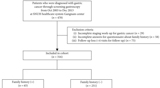
3.1. Study Population. A total of 478 GC patients were screened from our cohort. Of these, 29 patients who had not completed the cancer staging evaluation, 58 patients who had no information regarding FHx of GC, and 75 patients who were lost to follow-up were excluded. There-fore, 316 patients were included in thefinal analysis. Of these patients, 65 (20.6%) had a family history of GC (Figure 1). Fifty-six patients had a family history infirst-degree relatives, and 9 patients had both first- and second-degree family history of GC. A total of 263 patients (83.2%) underwent follow-up evaluation according to their planned follow-up schedule and visited outpatient clinic at SNUH regularly at least 5 years after the diagnosis. The proportion of patients with FHx who followed up for more than 5 years was 86.1%, and 82.4% of the patients without family history were followed up for more than 5 years.
3.2. Demographic and Clinicopathologic Characteristics.The demographicfindings of the study population are summa-rized in Table 1. The proportion of patients aged 50–59 was the highest in both groups regardless of FHx of GC. Although the difference was statistically insignificant, the age of patients with FHx of GC at the cancer diagnosis was higher than that of patients without FHx of GC (58:6 ± 9:3 years vs. 55:9 ± 10:7 years, p= 0:059). Males were 71.8% of the total cohort, and there was no significant difference in gender between groups. Health-related behaviors, including cigarette smoking, alcohol consumption, and salt intake were not significantly different between groups. When the interval of upper GI endoscopy was compared, patients with family history of GC were more likely to have intervals of less than two years between the time of upper GI endoscopy confirm-ing normalfindings and the time of cancer diagnosis (33.8% vs 21.9%, p =0.046).
Comparison of the clinicopathologic findings between the groups are presented in Table 2. Pathologic examinations
Patients who were diagnosed with gastric cancer through screening gastroscopy
from Oct 2003 to Dec 2013 at SNUH healthcare system Gangnam center
(n = 478)
Exclusion criteria
(i) Incomplete staging work-up for gastric cancer (n = 29)
(ii) Incomplete answers for questionnaire about family history (n = 58) (iii) Follow-up loss (<4 visits for follow-up) (n = 75)
Included in cohort (n = 316) Family history (+) (n = 65) Family history (–) (n = 251)
Figure1: Flow chart of enrollment of the study population.
Table1: Demographicfindings of study population. Family history of gastric
cancer pvalue Absent (n= 251) Present (n= 65) No. % No. % Age (yr) ≤39 20 8 2 3.1 0.439 40-49 42 16.7 8 12.3 50-59 101 40.2 27 41.5 60-69 62 24.7 18 27.7 ≥70 26 10.4 10 15.4 Mean + SD 5510:9 ±:7 58:6 ± 9:3 0.059 Gender 0.306 Male 177 70.5 50 76.9 Female 74 29.5 15 23.1 Smoking 0.432 Current 67 28.5 17 28.3 Ex-smoker 83 35.3 26 43.3 None 85 36.2 17 28.3
Excess alcohol (>140 g/day) 0.167
Yes 112 47.5 35 57.4
No 124 52.5 26 42.6
Excess salt intake 0.839
Yes 148 63 38 64.4
No 87 37 21 35.6
EGD interval∗ 0.046
<2 yr 55 21.9 22 33.8
≥2 yr 196 78.1 43 66.2
EGD: esophagogastroduodenoscopy; SD: standard deviation. ∗Interval between the time of upper GI endoscopy confirming normalfindings and the time of cancer diagnosis.
showed no significant differences in histopathological findings, including tumor location and size, differentiation, Lauren’s classification, depth of invasion, lymph node metastasis, and multiplicity of tumors between the groups. However, preexisting adenoma was found more frequently in patients with FHx of GC than in those without FHx (14.5% vs. 6.3%,p= 0:035). MSI was evaluated in 136 of the patients who underwent ESD or surgery. The proportion of patients with MSI was significantly higher in groups with family history of GC (43.2% vs. 13.2%,p= 0:006). Although statistically insignificant,H.pyloriinfection rate of patients with FHx of GC tended to be higher than that of patients without FHx (70.5% vs. 61.3%, p= 0:188). Presence of endoscopic AG/IM did not show any significant difference between the groups; however, IM was more common in patients with FHx of GC. Overall, most GC patients (N= 260, 82.3%) had stage I GC, and fourteen (4.4%) had stage IV at diagnosis. There was no significant difference in tumor stage between groups. Ninety-six (30.3%) patients received ESD for treatment of GC, and surgery was performed in 206 patients (65.2%). The remaining 14 patients received palliative chemotherapy as thefirst treatment without ESD or surgery.
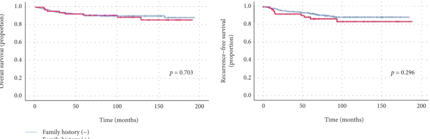
3.3. Comparison between Gastric Cancer-Related Survival Rates.During the median follow-up duration of 80.8 months (range, 4.8–187.4 months), 34 patients (10.8%) died. Of these, 27 patients died of GC. Twenty-three patients (7.2%) had recurrence of GC, of which 13 had metachronous GC recurrence. The OS and RFS at 5 years of total study popula-tion were 90.6% and 91.1%, respectively. The OS at 5 years for patients with FHx of GC was 90.6%, not significantly different from that of patients without FHx of GC (p= 0:703, Figure 2). The RFS at 5 years of patients without FHx were numerically higher than those of patients with FHx of GC; however, there were no significant differences between the groups (92.2% vs 86.4%,p= 0:296(Figure 2)).
Table2: Clinicopathologicfindings of gastric cancer patients. Family history of gastric
cancer pvalue Absent (n= 251) Present (n= 65) No. % No. % Tumor location 0.17 Upper third 29 11.6 8 12.3 Middle third 79 31.5 13 20 Lower third 139 55.4 41 63.1 All 4 1.6 3 4.6 Tumor size (cm) 0.78 <1.0 46 18.3 11 16.9 1.0-1.9 73 29.1 19 29.2 2.0-2.9 41 16.3 14 21.5 ≥3.0 91 36.3 21 32.3 Mean + SD 2:8 ± 2:3 2:8 ± 2:5 0.991 Differentiation 0.197 Differentiated 132 52.6 40 61.5 Undifferentiated 119 47.4 25 38.5
Laruen classification 0.491
Intestinal 133 57.1 39 61.9 Diffuse or mixed 100 42.9 24 38.1 Depth of invasion 0.756 pT1 193 76.9 49 75.4 pT2 32 12.7 11 16.9 pT3 13 5.2 3 4.6 pT4 13 5.2 2 3.1
Lymph node metastasis 0.235
pN0 199 79.3 56 86.2 pN1 26 10.4 6 9.2 pN2 15 6 0 0 pN3 11 4.4 3 4.6 Multiplicity of tumor 0.653 Solitary 239 95.2 61 93.8 Multiple 12 4.8 4 6.2 Stage 0.698 I 204 81.3 56 86.2 II 28 11.2 4 6.2 III 8 3.2 2 3.1 IV 11 4.4 3 4.6 Preexisting adenoma 0.035 Yes 15 6.3 9 14.5 No 222 93.7 53 85.5 Microsatellite instability 0.006 Stable 86 86.8 21 56.8
Unstable, low (MSI-L) 6 6.1 7 18.9
Unstable, high (MSI-H) 7 7.1 9 24.3
Curative treatment method 0.538
ESD 78 32.6 18 28.6
Surgery (STG+TG) 161 67.4 45 71.4
Table2: Continued.
Family history of gastric cancer pvalue Absent (n= 251) Present (n= 65) No. % No. %
H. pylori infection status 0.188
Yes 138 61.3 43 70.5 No 87 38.7 18 29.5 Atrophic gastritis 0.354 Yes 231 92 62 95.4 No 20 8 3 4.6 Intestinal metaplasia 0.113 Yes 137 53.8 44 67.7 No 114 45.4 21 32.3
ESD: endoscopic submucosal dissection;H.pylori: Helicobacter pylori; SD: standard deviation; STG: subtotal gastrectomy; TG: total gastrectomy. Microsatellite instability was evaluated in 136 of the patients (43% of total study population) who underwent ESD or surgery.
The OS and RFS according to MSI status were assessed for the study population surveyed for MSI (Figure 3). When the subjects were divided into the MSI group and microstallite stable (MSS) group, the OS at 5 years of the MSI group was numerically higher than that of the MSS group regardless of GC FHx (96.3% vs. 89.8%, p= 0:453). The RFS at 5 years of the MSI group was also higher than that of the MSS group, though it was not statistically significant (95.0% vs. 89.8%, p= 0:795).
4. Discussion
In the present study, we investigated the clinicopathologic characteristics and long-term outcomes of GC according to the FHx of GC in a health screening cohort in a high-GC-prevalence region of Korea. There were no significant differ-ences in the tumor characteristics or overall prognosis between GC patients with and without FHx of GC. However, the proportion of patients with preexisting adenoma and MSI were significantly higher in groups with FHx of GC.
Choi et al. detected preexisting adenoma in 15.6% of endoscopically resected early gastric cancer (EGC) cases and reported that EGC cases with preexisting adenoma showed a greater association withH. pylori-related chronic inflammation than those without preexisting adenoma [14].
According to the hypothesis of gastric carcinogenesis by Correa et al., it is widely accepted that chronic inflammation triggered byH.pyloriinfection develops into AG, IM, dyspla-sia, andfinally adenocarcinoma [15]. The higher presence of preexisting adenoma in patients with FHx of GC could be explained by the higherH.pyloriinfection rate in the same group. Gastric adenoma is thought to be a precancerous lesion; the annual incidence of GC is 0.6% among patients with mild to moderate dysplasia and 6% among those with severe dysplasia [16]. Our results suggest that early detection of precancerous lesions through active screening for high-risk groups including those with FHx of GC may help prevent the development of GC.
The prevalence of MSI-high GC in Asians is commonly <10% of all GC cases and is lower than that shown in Western studies [17, 18]. Higher presentation of MSI in patients with FHx of GC is known based on several prior studies, and this was consistent with our findings [19–21]. MSI-high GC is known to be associated with intestinal-type histology, infre-quent lymph node metastasis, and better overall prognosis [22–24]. Although statistically insignificant, the OS of patients with MSI was higher in our study than those without. However, since most of our study subjects were diagnosed with EGC with favorable prognosis, there is a limitation to clearly revealing the survival difference according to MSI 200
Overall survival (proportion)
0 50 100 150 Time (months) 0.0 0.2 0.4 0.6 0.8 1.0 p= 0.703 0 50 100 150 200 Time (months) 0.0 0.2 0.4 0.6 0.8 1.0 p= 0.296 Family history (–) Family history (+) Recurrence – free survival (proportion) 0 8 1.0 al
Figure2: Survival curves of patients with family history and without family history.
Overall survival (proportion)
200 0 50 100 150 Time (months) 0.0 0.2 0.4 0.6 0.8 1.0 p= 0.453 MSI stable (–) MSI unstable (+) Time (months) 0 50 100 150 200 0.0 0.2 0.4 0.6 0.8 1.0 Recurrence – free survival (proportion) p= 0.795 0 8 1.0 va l
status. The relationship between survival in GC patients and MSI has yet to be determined, and there was heterogeneity in the recent studies [25]. Further studies with a larger number of patients are needed.
There are some studies regarding the effect of FHx of GC on the long-term outcome of GC. A Korean study including 1,265 GC patients reported that survival rates of GC patients with FHx of GC did not significantly differ from those of patients without FHx of GC [26]. Another large-scale Korean study showed that first-degree FHx of GC was associated with improved survival after curative surgery in patients with stage III or IV GC [27]. A meta-analysis suggested that FHx of GC is associated with better survival of GC patients after curative surgery [28]. We found that there were no signifi-cant differences in OS and RFS at 5 years between the groups. This study was conducted on asymptomatic healthy individ-uals who underwent screening endoscopy in a single center. Therefore, most of the diagnosed tumors were early-stage GC (EGC of stage I), presenting a limitation in terms of the interpretation and generalization of our results, considering the characteristics of the study population.
The interval between the time of upper GI endoscopy confirming normalfindings and the time of cancer diagnosis of patients with FHx of GC was less than 2 years, and this was much shorter than that of patients without FHx of GC. This result may reflect the health-related behaviors of individuals with risk factors for GC. A large cohort study using the Korean National Health and Nutrition Examination Survey 2005 database showed that GC relatives were significantly more likely to undergo GC screening than were non-GC relatives [29]. Considering the high compliance with screening tests of relatives of GC patients and the high prevalence of GC fol-lowing the adenomacarcinoma sequence in patients with FHx of GC, it is advisable to recommend active endoscopy screen-ing with a shorter interval for relatives of GC patients.
This study has several strengths. First, to the best of our knowledge, this is the first study to examine associations between FHx of GC and preexisting adenoma as a precancer-ous lesion. These results can be used to support the necessity of active surveillance in a high-risk subgroup of FHx of GC for the early detection of precancerous lesions and preven-tion of GC development. Our prior study also showed that intensive screening and surveillance may be useful for high-risk subpopulations with epidemiologic high-risk factors or premalignant lesions such as IM [30]. Second, we controlled for referral bias by enrolling individuals from a health check-up cohort representing the general population with an average risk. Third, we collected, organized, and qualified medical records from our H-PEACE study.
Several factors should be considered when interpreting the results of this study. First, because of the retrospective study design,H.pylorieradication history was not thoroughly inves-tigated. It cannot be ruled out that the possible association betweenH.pyloriinfection status and FHx of GC was under-estimated. Second, there is some possibility that information about the FHx of GC is inaccurate because it was based on a self-administered questionnaire. Nevertheless, consistency and reliability of the self-reported family history was validated byfindings of previous studies [31, 32].
In conclusion, preexisting adenoma is more common in patients with FHx of GC than those without FHx of GC; GC with MSI is associated with FHx of GC. The survival of patients with FHx of GC did not significantly differ from that of those without family history. Early detection of precancer-ous lesions through active screening for high-risk groups with FHx of GC may help prevent the development of GC.
Abbreviations
GC: Gastric cancer FHx: Family history
MSI: Microsatellite instability OS: Overall survival
RFS: Recurrence-free survival.
Data Availability
The data used to support thefindings of this study are avail-able within the manuscript.
Conflicts of Interest
The authors declare that they have no conflict of interest.
Authors
’
Contributions
Jooyoung Lee and Su Jin Chung contributed to the concept and design of the study. Ji Min Choi and Yoo Min Han acquired the data. Jooyoung Lee and Su Jin Chung analyzed and interpreted the data. Jooyoung Lee and Su Jin Chung did the draft of the article. Ji Min Choi, Yoo Min Han, and Joo Sung Kim did the critical revision of the article for impor-tant intellectual content. All authors approved thefinal ver-sion of the article.
Acknowledgments
This research is supported by grant No. 04-2018-0420180880 from the Seoul National University Hospital Research Fund.
References
[1] M. Balakrishnan, R. George, A. Sharma, and D. Y. Graham,
“Changing trends in stomach cancer throughout the world,”
Current Gastroenterology Reports, vol. 19, no. 8, p. 36, 2017. [2] F. Bray, J. Ferlay, I. Soerjomataram, R. L. Siegel, L. A. Torre,
and A. Jemal,“Global cancer statistics 2018: GLOBOCAN esti-mates of incidence and mortality worldwide for 36 cancers in 185 countries,”CA: a Cancer Journal for Clinicians, vol. 68, no. 6, pp. 394–424, 2018.
[3] K. W. Jung, Y. J. Won, H. J. Kong, and E. S. Lee,“Prediction of cancer incidence and mortality in Korea, 2018,” Cancer Research and Treatment, vol. 50, no. 2, pp. 317–323, 2018. [4] H. Brenner, D. Rothenbacher, and V. Arndt,“Epidemiology of
stomach cancer,” Methods in Molecular Biology, vol. 472, pp. 467–477, 2009.
[5] E. Van Cutsem, X. Sagaert, B. Topal, K. Haustermans, and H. Prenen, “Gastric cancer,” Lancet, vol. 388, no. 10060, pp. 2654–2664, 2016.
[6] H. Yatsuya, H. Toyoshima, T. Mizoue et al.,“Family history and the risk of stomach cancer death in Japan: differences by age and gender,” International Journal of Cancer, vol. 97, no. 5, pp. 688–694, 2002.
[7] C. Oliveira, H. Pinheiro, J. Figueiredo, R. Seruca, and F. Carneiro, “Familial gastric cancer: genetic susceptibility, pathology, and implications for management,” The Lancet Oncology, vol. 16, no. 2, pp. e60–e70, 2015.
[8] C. Lee, E. K. Choe, J. M. Choi et al.,“Health and Prevention Enhancement (H-PEACE): a retrospective, population-based cohort study conducted at the Seoul National University Hos-pital Gangnam Center, Korea,”BMJ Open, vol. 8, no. 4, article e019327, 2018.
[9] K. J. Kim, H. K. Yang, W. H. Kim, and G. H. Kang,“Combined prognostic effect of PD-L1 expression and immunoscore in microsatellite-unstable advanced gastric cancers,”Oncotarget, vol. 8, no. 35, pp. 58887–58902, 2017.
[10] J. Y. Yim, N. Kim, S. H. Choi et al.,“Seroprevalence of Helico-bacter pylori in South Korea,” Helicobacter, vol. 12, no. 4, pp. 333–340, 2007.
[11] J. M. Choi, S. G. Kim, H. J. Yang et al.,“Clinical outcomes of no residual disease in the specimen after endoscopic resection for gastric neoplasms,”Surgical Endoscopy, vol. 30, no. 2, pp. 610– 618, 2016.
[12] J. Y. Park, S. G. Kim, J. Kim et al.,“Risk factors for early meta-chronous tumor development after endoscopic resection for early gastric cancer,” PLoS One, vol. 12, no. 9, article e0185501, 2017.
[13] J. H. Lee, C. M. Lee, S. Y. Son, S. H. Ahn, D. J. Park, and H. H. Kim,“Laparoscopic versus open gastrectomy for gastric can-cer: long-term oncologic results,” Surgery, vol. 155, no. 1, pp. 154–164, 2014.
[14] J. M. Choi, S. G. Kim, J. Kim et al.,“Clinical implications of pre-existing adenoma in endoscopically resected early gastric cancers,”PLoS One, vol. 12, no. 5, article e0178419, 2017. [15] P. Correa, “Human gastric carcinogenesis: a multistep and
multifactorial process–first American Cancer Society award lecture on cancer epidemiology and prevention,” Cancer Research, vol. 52, no. 24, pp. 6735–6740, 1992.
[16] A. C. de Vries, N. C. T. van Grieken, C. W. N. Looman et al.,
“Gastric cancer risk in patients with premalignant gastric lesions: a nationwide cohort study in the Netherlands,” Gastro-enterology, vol. 134, no. 4, pp. 945–952, 2008.
[17] J. Y. Kim, N. R. Shin, A. Kim et al.,“Microsatellite instability status in gastric cancer: a reappraisal of its clinical significance and relationship with mucin phenotypes,”Korean Journal of Pathology, vol. 47, no. 1, pp. 28–35, 2013.
[18] M. Ratti, A. Lampis, J. C. Hahne, R. Passalacqua, and N. Valeri,
“Microsatellite instability in gastric cancer: molecular bases, clinical perspectives, and new treatment approaches,” Cellular and Molecular Life Sciences, vol. 75, no. 22, pp. 4151–4162, 2018.
[19] L. Ottini, D. Palli, M. Falchetti et al.,“Microsatellite instability in gastric cancer is associated with tumor location and family history in a high-risk population from Tuscany,” Cancer Research, vol. 57, no. 20, pp. 4523–4529, 1997.
[20] D. Palli, A. Russo, L. Ottini et al.,“Red meat, family history, and increased risk of gastric cancer with microsatellite instabil-ity,”Cancer Research, vol. 61, no. 14, pp. 5415–5419, 2001. [21] K. Kanemitsu, K. Kawasaki, M. Nakamura et al.,“MSI is
fre-quently recognized among gastric cancer patients with a family
history of cancer,” Hepato-Gastroenterology, vol. 54, no. 80, pp. 2410–2414, 2007.
[22] K. Polom, D. Marrelli, V. Pascale et al.,“The pattern of lymph node metastases in microsatellite unstable gastric cancer,”
European Journal of Surgical Oncology, vol. 43, no. 12, pp. 2341–2348, 2017.
[23] H. Nakashima, M. Honda, H. Inoue et al., “Microsatellite instability in multiple gastric cancers,” International Journal of Cancer, vol. 64, no. 4, pp. 239–242, 1995.
[24] K. Polom, L. Marano, D. Marrelli et al., “Meta-analysis of microsatellite instability in relation to clinicopathological characteristics and overall survival in gastric cancer,”The Brit-ish Journal of Surgery, vol. 105, no. 3, pp. 159–167, 2018. [25] Y. Y. Choi, J. M. Bae, J. Y. An et al.,“Is microsatellite instability
a prognostic marker in gastric cancer? A systematic review with meta-analysis,” Journal of Surgical Oncology, vol. 110, no. 2, pp. 129–135, 2014.
[26] O. Jeong, M. R. Jung, Y. K. Park, and S. Y. Ryu,“ Clinicopath-ological features and survival of patients with gastric cancer with a family history: a large analysis of 2,736 patients with gastric cancer,” Journal of Gastric Cancer, vol. 17, no. 2, pp. 162–172, 2017.
[27] M. A. Han, M. G. Oh, I. J. Choi et al.,“Association of family history with cancer recurrence and survival in patients with gastric cancer,” Journal of Clinical Oncology, vol. 30, no. 7, pp. 701–708, 2012.
[28] M. G. Oh, J. H. Kim, M. A. Han, J. Park, S. Y. Ryu, and S. W. Choi,“Family history and survival of patients with gastric can-cer: a meta-analysis,”Asian Pacific Journal of Cancer Preven-tion, vol. 15, no. 8, pp. 3465–3470, 2014.
[29] J. M. Kang, D. W. Shin, Y. M. Kwon et al.,“Stomach cancer screening and preventive behaviors in relatives of gastric can-cer patients,” World Journal of Gastroenterology, vol. 17, no. 30, pp. 3518–3525, 2011.
[30] S. J. Chung, M. J. Park, S. J. Kang et al.,“Effect of annual endo-scopic screening on clinicopathologic characteristics and treat-ment modality of gastric cancer in a high-incidence region of Korea,” International Journal of Cancer, vol. 131, no. 10, pp. 2376–2384, 2012.
[31] R. A. Kerber and M. L. Slattery,“Comparison of self-reported and database-linked family history of cancer data in a case-control study,” American Journal of Epidemiology, vol. 146, no. 3, pp. 244–248, 1997.
[32] F. L. Roth, S. A. Camey, M. Caleffiet al.,“Consistency of self-reportedfirst-degree family history of cancer in a population-based study,”Familial Cancer, vol. 8, no. 3, pp. 195–202, 2009.