Texas Medical Center Library
DigitalCommons@The Texas Medical Center
UT GSBS Dissertations and Theses (Open Access)
Graduate School of Biomedical Sciences
5-2014
Functional Analysis of Cytosolic Hsp70 Nucleotide
Exchange Factor Networks in Yeast
Jennifer Lynn Abrams
Follow this and additional works at:
http://digitalcommons.library.tmc.edu/utgsbs_dissertations
Part of the
Biochemistry Commons,
Cell Biology Commons,
Microbiology Commons, and the
Molecular Biology Commons
This Dissertation (PhD) is brought to you for free and open access by the Graduate School of Biomedical Sciences at DigitalCommons@The Texas Medical Center. It has been accepted for inclusion in UT GSBS
Dissertations and Theses (Open Access) by an authorized administrator of DigitalCommons@The Texas Medical Center. For more information,
Recommended Citation
Abrams, Jennifer Lynn, "Functional Analysis of Cytosolic Hsp70 Nucleotide Exchange Factor Networks in Yeast" (2014).UT GSBS Dissertations and Theses (Open Access).Paper 447.
FUNCTIONAL ANALYSIS OF CYTOSOLIC HSP70
NUCLEOTIDE EXCHANGE FACTOR
NETWORKS IN YEAST
by
Jennifer Lynn Abrams
APPROVED:
_____________________________________________
Supervisory Professor, Kevin A. Morano, Ph.D.
_____________________________________________
Heidi B. Kaplan, Ph.D.
_____________________________________________Michael C. Lorenz, Ph.D.
_____________________________________________Eric J. Wagner, Ph.D.
_____________________________________________Ambro van Hoof, Ph.D.
___________________________________________________
FUNCTIONAL ANALYSIS OF CYTOSOLIC HSP70
NUCLEOTIDE EXCHANGE FACTOR
NETWORKS IN YEAST
A
DISSERTATION
Presented to the Faculty of
The University of Texas Health Science Center at Houston and
The University of Texas MD Anderson Cancer Center
Graduate School of Biomedical Sciences
in Partial Fulfillment
of the Requirements
for the Degree of
DOCTOR OF PHILOSOPHY
by
Jennifer Lynn Abrams, B.S.
Houston, Texas
DEDICATION
I would like to dedicate this thesis to my late Aunt Sally A. Malter. She was always supportive of me advancing my education and encouraged me to keep pushing forward when life was difficult. She will be greatly missed.
ACKNOWLEDGMENTS
The completion of my thesis has occurred only with the help and support of many people. First I would like to express my gratitude to my wonderful mentor, Dr. Kevin Morano, who has supported me and taught me so much over the years. His ability to keep me motivated, while simultaneously identifying ways I could improve as a scientist, is unmatched. He has made my time here enjoyable, but challenging, which has helped me to develop the skills necessary to move on to the next step in my career.
I would also like to thank all of the members of the Morano lab past and present including Dr. Patrick Gibney, Dr. Hugo Tapia, Dr. Jacob Verghese, Dr. Yanyu Wang, Kim Cope, Veronica Garcia, and Sara Peffer. I could not have asked to work with a better group of people. All of the lab outings were a lot of fun in the company of these awesome people. In addition, engaging lab meetings and scientific discussions kept me on my toes. Not to mention all of the impromptu question/answer sessions prior to my candidacy exam that made facing my committee much easier. A special thanks to Sara Peffer, who I have know for the shortest time, but has always provided me with a smiling face and a clever gif just when I needed it. In addition, I would specifically like to thank Veronica Garcia, also known as “my other half”, for supporting me through good times and bad and for being an incredible friend inside and outside of the lab. I would also like to give a special thanks to Patrick Gibney, who mentored me through my rotation and has continued to be an extremely encouraging, wonderful person that I would not have made it this far without.
I would like to acknowledge my committees members throughout the years that have provided me with the feedback and knowledge that was necessary for me to advance to this stage of my career. These members include: Dr. Heidi Kaplan, Dr. Michael Lorenz, Dr. Michael Blackburn, Dr. Ambro van Hoof, Dr. Hung Ton-That, and Dr. Eric Wagner. This engaging group of people guided me through my project and through graduate school regardless of their status on my committee.
In my time here, I was lucky enough to receive awards that afforded me opportunities I may not have had otherwise. I would like to thank the American Society for Microbiology, who awarded me with the Robert. D. Watkins Graduate Research Fellowship. With this fellowship, I was able to attend the ASM General Meetings as well as the ASM minority conferences, which has helped build a network of people that I will keep in touch with throughout my career. In addition, I would like to thank the Graduate School for Biomedical Sciences (GSBS) for choosing me to receive the esteemed R.W. Butcher award that recognized my work for being a citizen’s scientist, which is an important aspect of who I am.
My sincerest thanks goes to the Department of Microbiology and Molecular Genetics office staff, with special recognition for Lyz Morrison, Vicci Sanders, and Karen Oandasan, who all helped me work through various issues I faced throughout Graduate School. In addition, these ladies were always friendly and helpful even if I just needed to talk. I appreciate all the effort and care that went into maintaining a well-organized program and department. In addition, the faculty and staff at GSBS including: Joy Lademora, Bunny Perez, Dr. Andrew Bean, Brenda Gaughan, Eric Solberg, and Karen Weinberg have all helped me at different stages of my time here.
The Microbiology and Molecular Genetics Program provided me with a very unique experience in that I have always felt so much support and love from everyone making it feel like a home away from home. I have made so many friends in this program from the very beginning. The ladies that I started classes with in 2007, Dr. Ale Klauer-King and Jennifer Herricks, were my study partners, my friends, and my counselors when I needed it, so I would like to thank them for that. In addition, Dr. Jenny Dale was a dedicated friend that helped me through every step of my candidacy exam. I have also met many lifelong friends here including Alex Marshall, Jill Losh, and Kim Busiek, who I have spent a lot of time getting to know and love. I met Katie “Squirt” McCallum in her rotation in my laboratory and she has become one of my best friends and is one of the most caring, supportive, and loving friends that I have. I would also like to thank the Valhalla crew including Katie McCallum, Veronica Garcia, George
Tiller, and Amy Ford; many difficult days were made better with the support of these people. Finally I would like to thank my tech support and good friend Doug Litwin for being there when I needed him and sometimes when I did not.
I also have a number of great friends who have supported me outside of work that always listened to me no matter how much I wanted to talk about science; these people include Reisha Beaty, Joeseph Edmonson, both of whom I have known and have been incredibly supportive since high school as well as some new friends Matt Madeiro, Carissa Ara, Clint Stengel, Lauren Chapman, and Ambri Garcia who have all been accepting, loving friends. Finally, I would like to give a special thanks to Carlos Calderon, who has not only been supportive as a friend for many years, but has also provided a very conducive and encouraging environment for me to work and study, so this thesis could come to fruition.
Aside from work I would like to thank my family who helped me overcome many obstacles and for whom none of this would be possible. I would first like to thank my parents Marion and James Abrams, who have always been my biggest supporters and have never doubted that I could do anything I wanted to with my life. I would also like to thank my brother Christopher Abrams, who always kept me grounded through the best times and the worst times. All of my family’s love and support has made me the person I am today and without them I could not have been successful in a career that I love.
FUNCTIONAL ANALYSIS OF CYTOSOLIC HSP70 NUCLEOTIDE EXCHANGE
FACTOR NETWORKS IN YEAST
Jennifer Lynn Abrams, B.S.
Advisory Professor: Kevin A. Morano, Ph. D.
The Hsp70 class of molecular chaperones play critical roles in protein homeostasis via an ATP-dependent folding cycle. Cytosolic Hsp70s in the budding yeast Saccharomyces cerevisiae, Ssa and Ssb, interact with up to three distinct nucleotide exchange factors (NEFs) homologous to human counterparts; Sse1/Sse2/HSP110, Fes1/HspBP1, and Snl1/Bag1. In an effort to understand the differential functional contributions of the cytosolic NEFs to protein homeostasis (“proteostasis”), I carried out comparative genetic, biochemical and cell biological analyses. For these studies, I developed protocols to monitor protein disaggregation and reactivation in a near real-time coupled assay that revealed the importance of aggregate dynamics in the solubilization of proteins for their refolding. This coupled experimental approach, represents an important step toward developing tools necessary to monitor in vivo mechanisms of proteostasis.
This work determined that the Hsp110, Sse1, is the primary NEF contributing to most Hsp70 functions and also uncovered a unique role for Fes1 in Ssa-mediated regulation of the cytosolic heat shock response, while revealing no significant contributions from Snl1 and Sse2. These findings suggest that NEFs do have overlapping functions, but their distinct associations with the Hsp70s as well as unique structural components could contribute to differential roles in proteostasis.
Additionally, this study uncovered that relative levels of Snl1 and Sse1 are important for optimal growth. To probe this relationship, I exploited the Snl1 overexpression toxicity phenotype exhibited in the absence of Sse1 to examine which unique characteristics of Snl1 are important for its function. I discovered that Snl1 localization to the ER membrane is required for toxicity and that Sse1-mediated alleviation of this phenotype is Ssb-dependent. These results demonstrate a network of interactions that supports a hypothesis where Snl1 plays a role in translation regulation.
This investigation was conducted to gain a better understanding of NEF roles within the Hsp70 chaperone network. Understanding these dynamics is critical to obtain successful treatments that can reverse the debilitating effects of neurodegenerative disorders such as Alzheimer’s and Parkinson’s diseases. Pharmacological targeting of molecular chaperones and their co-factors, such as the NEFs, is an attractive therapeutic goal that may contribute to improving human health, most notably in the aging population.
TABLE OF CONTENTS
Approval sheet……….………i Title page……….………ii Dedication………..iii Acknowledgements………..iv Abstract………..iiv Table of contents………..ix List of figures……….xii List of tables……….xiv Chapter1: Introduction……….…….….1INTRODUCTION AND BACKGROUND.………..2
Stress and the maintenance of proteostasis………...…2
The heat shock response………...………3
Molecular chaperones……….5
The Hsp70s………..………6
The Hsp40 co-chaperones………...16
Cytosolic Hsp70 nucleotide exchange factors………..17
SIGNIFICANCE OF THIS STUDY………...……….………..24
Understanding cytosolic Hsp70 NEFs………24
Human diseases related to proteostasis………24
Treatments and therapies………25
Chapter2: Materials and methods………..…….……….27
Strains, plasmids and growth conditions………..…..28
Protein isolation and visualization……….……..30 Fluorescence microscopy……….………31 FFL-GFP activity assay…….………...……….31 Hsf1 de-repression assay………...………..31 Immunoprecipitation………...32 Statistical analysis………..32
Chapter3: Coupled assays for monitoring protein refolding in Saccharomyces cerevisiae………..……….….37
INTRODUCTION……….38
METHODOLOGY.………...40
RESULTS.………46
DISCUSSION………...55
Chapter 4: Hierarchical functional specificity of cytosolic Hsp70 nucleotide exchange factors in Saccharomyces cerevisiae……….……….57
INTRODUCTION……….58
RESULTS……….61
DISCUSSION………..81
Chapter 5: Snl1 function in proteostasis………..……….88
INTRODUCTION……….89
RESULTS……….91
DISCUSSION……….116
SUMMARY……….119
DISCUSSION……….121
The role of NEFs in regulating Hsp70 activity……….121
Understanding differential NEF contributions to Hsp70-mediated processes…………...123
The role of Fes1 in the heat shock response………..126
Understanding functional contribution of Snl1 to proteostasis……….129
The chaperone networks in process specificity………..132
Disease implications and relevance……….134
Future directions and new technologies targeting chaperones for disease treatment….135 Bibliography………...139
LIST OF FIGURES
Fig. 1.1. Model of Hsp70-mediated folding………...8
Fig. 1.2. Hsp70 Cycle……….10
Fig. 1.3. Hsp70 mediated functions………..15
Fig. 1.4. Cytosolic Hsp70 nucleotide exchange factor (NEF) architectures and interaction interfaces………..19
Fig. 3.1. Schematic of FFL-GFP heat-shock recovery assay………..48
Fig. 3.2. FFL-GFP enzymatic refolding/recovery assay………50
Fig. 3.3. FFL-GFP refolding microscopy………..52
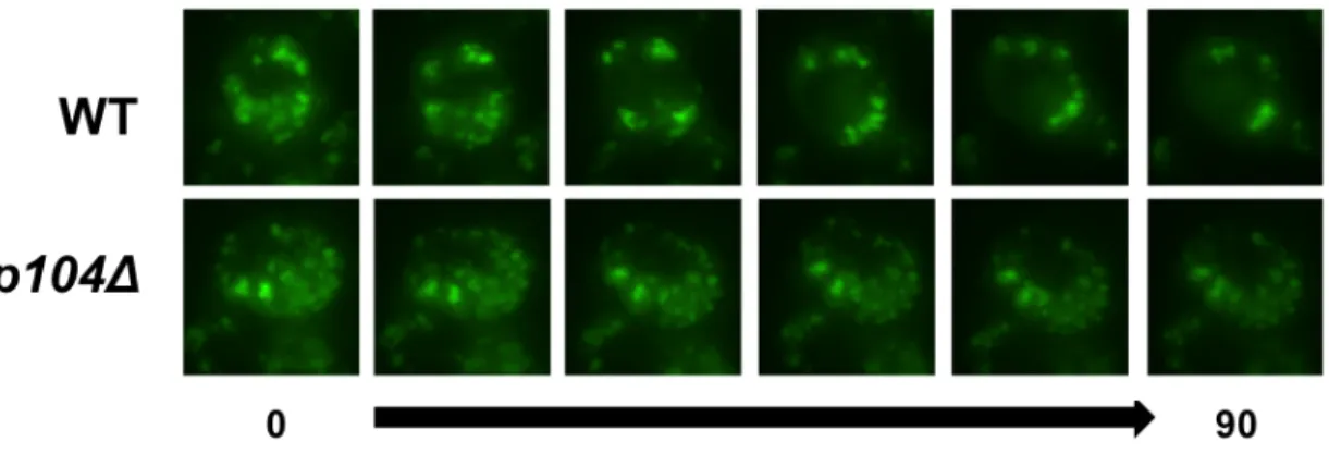
Fig. 3.4. Single cell refolding microscopy time course of FFL-GFP………54
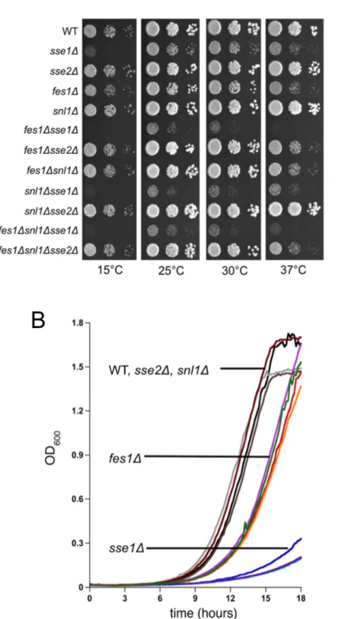
Fig. 4.1. Growth analysis of wild-type and nucleotide exchange factor deletion strains………..63
Fig. 4.2. Nucleotide exchange factor deletions differentially affect firefly luciferase GFP biogenesis………...66
Fig. 4.3. Cytosolic nucleotide exchange factors are not required for luciferase refolding in vivo………...70
Fig. 4.4. Sse1 and Fes1 contribute to regulation of the heat shock response through Hsf1…..73
Fig. 4.5. Sse1 uniquely contributes to Hsp70-mediated protein degradation………76
Fig. 4.6. Fes1 specifically interacts with Ssa chaperone in vivo………..79
Fig. 4.7. Model of nucleotide exchange factor roles in Hsp70-mediated protein biogenesis and quality control……….83
Fig. 5.1. Cells lacking Sse1 are hypersensitive to Snl1 overexpression………93
Fig. 5.2. Soluble truncation mutants of Snl1 lose toxicity and partially rescue the sse1Δ growth defect………...95
Fig. 5.3. Snl1 overexpression toxicity is dependent on ER localization but independent of Hsp70 interaction………..98
Fig. 5.5. Snl1 overexpression toxicity is independent of ribosomal association……….103 Fig. 5.6. The sse1-N281A mutant that interacts with SSA only, does not cause
overexpression toxicity associated with wild-type Sse1………....105 Fig. 5.7. The sse1-N281A increases Snl1 overexpression toxicity in the sse1Δ strain……….109 Fig. 5.8. Loss of the SSB proteins alleviates Snl1 overexpression toxicity in an
sse1Δstrain………..111
LIST OF TABLES
Table 1.1. Yeast Cytosolic Chaperones………..12 Table 2.1. Strains used in these studies………..33 Table 2.2. Plasmids used in these studies………..35
INTRODUCTION AND BACKGROUND
Stress and the maintenance of proteostasis - Cellular viability is at the mercy of the protein homeostasis (“proteostasis”) machinery, which includes the protein complexes responsible for maintaining the balance between polypeptide synthesis, transport, modification, and ultimately degradation. Aggregation and loss of function resulting from improper inter- and intramolecular interactions of misfolded and partially folded proteins can lead to a deleterious loss of proteostasis [1]. Challenges in the external environment as well as internal stressors can negatively impact the balance of protein maintenance. For example, temperature, pH, and osmolyte concentration are external factors that once altered beyond physiological range can increase the probability of misfolding and aggregation. In addition, internal factors such as proteome complexity and protein synthetic load can heighten the need for proteome stabilizers such as molecular chaperones. In support of this idea, there is a positive correlation between the complexity of the proteome and the number of conserved chaperone families maintained in an organism [2].
Nascent chain folding is particularly sensitive to stress conditions. Co-translational protein folding is important for longer, more complex, proteins because larger regions of the unfolded polypeptide have to be released from the ribosome in order to reach their native state. The ribosome exit tunnel is 100 Å long x 20 Å in diameter, which is only enough space for smaller intramolecular interactions to take place, limiting folding to simple secondary structures such as α-helices [3-7]. Upon release from the ribosome, the highly crowded, aqueous environment of the cytosol poses a folding challenge to the polypeptide. Exposed hydrophobic stretches will attempt to bury themselves adjacent to other hydrophobic residues, which can lead to improper structural associations [8]. In eukaryotes this is a significant problem because in addition to exposed hydrophobic regions that need to be protected, 30% of the proteins are classified as intrinsically unstructured [9]. It is plausible that high levels of unstructured proteins would be evolutionarily disfavored, but protein function requires a balance of flexibility and thermodynamic potential energy [10,11]. Due to this delicate balance, proteins that fold without
assistance can fall into what is termed a kinetic trap, which is an alternative non-functional fold that has a lower potential energy then the next intermediate fold on the journey to the native active state.
The rate of translation is another factor that can affect nascent protein folding efficiency. The average speed of eukaryotic translation is four amino acids per second compared to 20 amino acids per second in prokaryotes [12]. The mistranslation rate is 1 out of every 10-4 residues in eukaryotes, so the rate of translation allows time to correct or eliminate misfolded proteins [13-17]. One way that the cell deals with terminally misfolded proteins is through the ubiquitin proteasome pathway in which 5-15% of nascent chains are ubiquitinated. This number increases upon loss of the chaperone complexes in contact with ribosomes and nascent chains and these complexes include TRiC/CCT and prefoldin, the yeast Hsp70, Ssb, with the ribosome-associated complex (RAC) and the nascent chain associated complex (NAC) and will be discussed in more detail later [18]. All of the above mentioned chaperones are coregulated with the ribosome. As a result their expression is suppressed during stress conditions [19]. These complexes maintain proteostasis at the ribosome. In addition, proteins that have already been folded can be damaged during stress conditions, such as heat shock, and this requires repair mechanisms that involve chaperones, but also usually require global transcriptional responses.
The heat shock response - When the cell senses stress, it activates protective measures in order to prepare for future threats. In yeast, multiple transcription programs including the environmental stress response (ESR), the heat shock response (HSR), the oxidative stress response (OSR), and the unfolded protein response (UPR) allow cells to quickly adapt to changing environments. Stress responses can cross-protect against different stressors, but they each primarily respond to specific stressors. The most general stress response is the ESR and genes upregulated in the HSR are a subset of the ESR-regulated genes [20]. The HSR mounts a global cellular response upon exposure to temperatures greater than 36-37°C
characterized by cell cycle arrest and upregulation of heat shock proteins (HSPs) and other housekeeping proteins.
The master regulator of the HSR is the heat shock transcription factor, Hsf. In mammals there are four different isoforms of this master regulator, HSF1-4, but HSF1 controls the HSR [21]. In yeast there is only one isoform, Hsf1, which is essential for growth at optimal temperatures [22]. Hsf1 consists of three domains: the winged helix-turn-helix DNA binding domain (DBD), which is important for activating gene targets upon initiation of the HSR [23, 24]; the leucine zipper oligomerization domain is important for trimerization, which is necessary prior to binding DNA [25, 26]; and a carboxyl-terminal transactivation domain (CTD), which is important for initial and prolonged transcription activation by the transcription factor [27, 28]. Yeast Hsf1 has an additional amino terminal transactivation domain (NTA), which can initiate transcriptional activity independently or act as a negative regulator modulating CTD initiated activation [28, 29].
In mammals, activation of Hsf1 requires several steps including nuclear localization, trimerization, post-translational modifications, and DNA binding. In yeast, regulation is much simpler in that the Hsf1 is constitutively trimerized, localized to the nucleus, and bound to promoters of many HS genes [30-32]. This is more similar to Drosophila than mammalian HSF1 [33]. Activation also results in Hsf1 hyperphosphorylation, which is mediated by multiple kinases that respond to specific stressors. In yeast, it was shown that Snf1 phosphorylates Hsf1 in response to glucose starvation [32], but the kinase responsible for heat shock associated phosphorylation is unknown. In addition to the above-mentioned regulatory mechanisms, Hsf1 is also bound and repressed by the molecular chaperones Hsp70 and Hsp90. This has been shown via genetic evidence in yeast and mammals, and biochemical evidence in humans [34]. Hsp70 cannot completely repress Hsf1 alone; Hsp90 and co-chaperones are also necessary for complete repression[35]. Exposure to heat shock is thought to titrate the HSPs away from Hsf1 allowing activation. Once the HSPs have restored
proteostasis, they reestablish interaction with Hsf1 to attenuate the response. In yeast, there is currently genetic evidence, but no biochemical evidence to support this hypothesis.
Molecular chaperones - In order to maintain proteostasis, cells employ the help of molecular chaperones. All organisms maintain multiple families of chaperones, but not every family. Chaperones are named based on their molecular weights, and many of them are referred to as heat shock proteins (HSPs) due to their induction during heat shock. One of the most highly conserved families is the 70 kilodalton (kd) heat shock protein or Hsp70, which is present in all eukaryotes and bacteria currently identified, but not in all Archaea [36]. There is a roughly linear relationship between the number of genes maintained by an organism and the number of canonical HSPs, with exception of the Hsp100s. There is approximately 1 Hsp70 gene, 5-6 Hsp40 genes, 1 sHsp gene, and 1 Hsp60 gene for every 2000 genes and 1 Hsp90 gene per 6000 genes [2]. It is unclear why this relationship exists since many of these chaperones do not have a high level of specificity, but it appears that throughout evolution mutations have resulted in slight adaptations improving survivability of the organisms that code for them in the above mentioned ratios. These minor differences result in high levels of redundancy with the development of some distinct functions. The variation provides more regulatory opportunities to respond to changing environments. A number of molecular chaperones are classified as HSPs because many of them were originally identified in pulse chase experiments, where protein abundance was monitored by incorporation of [35S]-methionine after heat treatments
followed by SDS-PAGE [37]. Although many chaperones including some of the Hsp70s and Hsp90s are expressed constitutively, they are also upregulated upon HS and additional stress responsive isoforms are expressed in order to prepare for any additional assaults or to ultimately attenuate the active response. A subset of these chaperones are referred to as housekeeping proteins or “generalists.” The generalists are involved in ensuring a protein successfully transitions from “birth” as nascent nonfunctional polypepetide to a localized, modified, active protein, and then upon reaching the limit of its lifetime, chaperones target it to
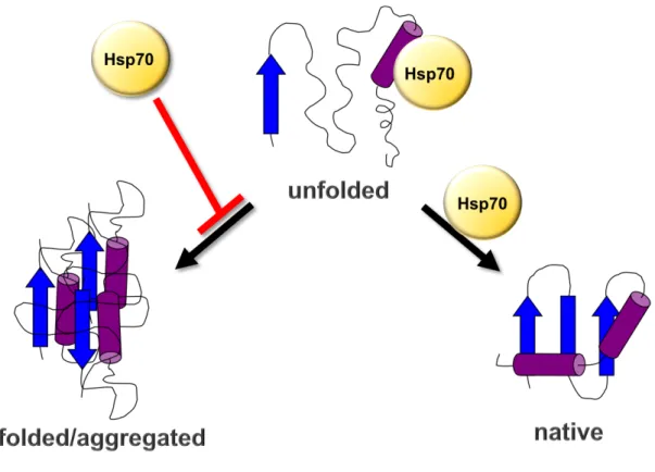
one of the degradation pathways for “death”. A well-conserved example of the generalist family is the Hsp70s that bind promiscuously to exposed hydrophobic regions in partially unfolded proteins (Fig. 1.1). There is also a more specific subset of chaperones known as “specialists,” which include the Hsp90s that interact with specific substrates including kinases and hormone receptors, to promote their maturation, translocation, or degradation.
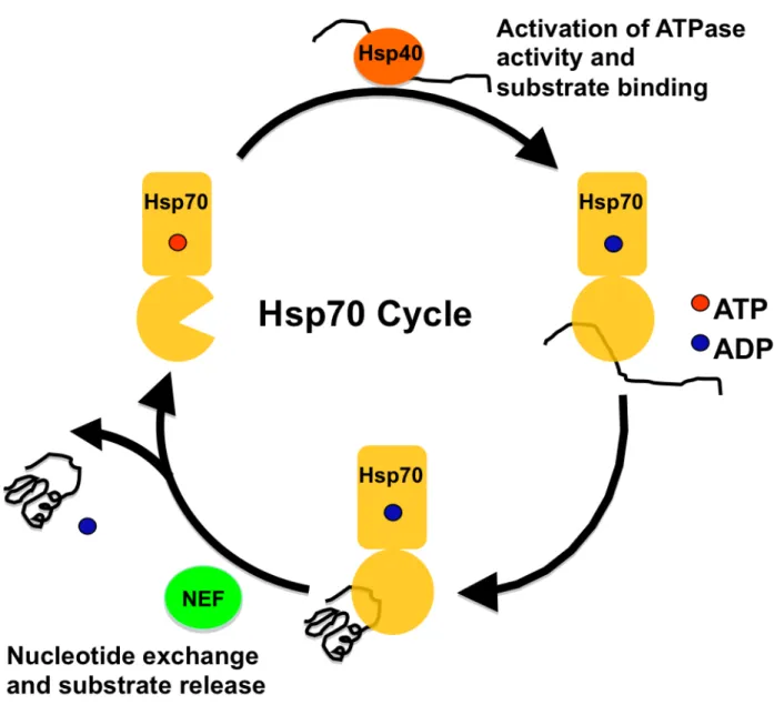
The Hsp70s - As mentioned earlier, the Hsp70s are highly conserved and play an essential role in maintaining protein homeostasis [1, 38]. Hsp70 functions through a nucleotide dependent cycleto protect exposed hydrophobic regions of polypeptides from the aqueous environment of the cytosol, while they fold into their native structure as seen in Figure 1.1 [39]. Hsp70 switches from a low affinity to a high affinity substrate binding conformation using the energy from binding, hydrolysis and release of ATP (Fig. 1.2) [40, 41]. This cycle repeats as necessary to complete protein folding [42]. Hsp70 has a very low innate ATPase activity, 0.04 ATP hydrolyzed min-1, resulting in a low protein folding capacity. To improve folding efficiency,
Hsp70 interacts with a myriad of co-chaperones [43]. The conserved Hsp40s or J-domain proteins interact with Hsp70 and can both activate its ATPase activity at least 200-fold as well as deliver substrate to Hsp70 in some cases [44-46]. The folding cycle is further regulated by the nucleotide exchange factors (NEFs) that bind the Hsp70 nucleotide binding domain (NBD) promoting the release of ADP and in turn substrate [47-51].
The architecture of Hsp70 includes a 44 kd nucleotide binding domain (NBD) and a 25 kd carboxyl-terminal substrate binding domain (SBD) [52]. The SBD is further composed of an α-helical lid domain and β-sandwich domain [53]. Substrates interact with the β sandwich domain in the high affinity, ADP bound state, and the α-helical lid closes over the substrate providing optimal protection [54]. The domains communicate nucleotide status through allosteric coupling of the NBD and SBD via an interdomain linker [40, 41, 53, 55]. Structure and co-chaperone interactions have afforded Hsp70 the functional flexibility to be involved in
Figure 1.1. Model of Hsp70-mediated folding. Hsp70 promotes folding into a native active structure and prevents misfolding and aggregation.
Figure 1.1. Model of Hsp70-mediated folding
Figure 1.2. Hsp70 Cycle. This schematic depicts the nucleotide dependent mechanism by which Hsp70 binds and releases substrate. In the ATP bound conformation, Hsp70 transiently interacts with polypeptide with low affinity. Interaction with the Hsp40 family of co-chaperones activates Hsp70’s innate ATPase activity hydrolyzing ATP to ADP and switching Hsp70 to a high affinity substrate binding conformation. Then the interaction between Hsp70 and the nucleotide exchange factors (NEFs) alters the structure to release ADP and the substrate.
protein folding/biogenesis, maturation, transport, and degradation [1, 38].
The Hsp70 superfamily includes Ssa, Ssb, Sse, and the atypical Ssz (Stress Seventy A, B, E, and Z) (Table 1.3) [22]. In yeast there are two families of cytosolic Hsp70s, Ssa and Ssb. There are two proteins, Ssb1 and Ssb2, in this non-essential, ribosome-associated family of Hsp70s; they are 99% identical and believed to be functionally redundant [56]. Ssb is a fungal specific Hsp70, not present in metazoans; in mammalian cells, cytosolic Hsp70s are recruited to the ribosome for co-translational protein folding [57, 58]. Ssb1/2 are considered to be functionally distinct from the Ssa family proteins due to the inability of either family to completely complement the absence of the other [22, 59]. In contrast to the stress inducible Ssa family of Hsp70s, Ssb is regulated similar to ribosomal proteins in that its transcription is decreased in stress conditions [60, 61]. Ssb primarily interacts with translating ribosomes in a ribosome-associated complex (RAC) dependent fashion, independent of nascent chain association; however, nascent chain interaction stabilizes the Ssb/ribosome complex [62, 63]. RAC exists as a ribosome-bound heterodimer, which includes the J-domain protein, Zuo1, and the atypical Hsp70, Ssz1, and acts as a co-chaperone for Ssb, activating its ATPase activity [64]. The specific function of Ssz1 is unclear, but it is required for Zuo1 activity. In addition, Zuo1 contains the interaction domain required for RAC association with the ribosomal exit tunnel [64, 65]. Deletion of SSB, ZUO1, or SSZ1 results in identical phenotypes, slow growth, cold sensitivity, and hypersensitivity to aminoglycosides (translation inhibitors) suggesting they act together in one process. Deletion of SSB also results in accumulation of aggregated proteins as well as a decrease in small and large ribosomal subunits, indicating roles in both co-translational protein folding and ribosome biogenesis [19] [66]. In addition to RAC, the highly conserved nascent chain associated complex (NAC) also binds the ribosome at the exit tunnel, but unlike RAC it interacts directly with nascent chains [67]. In yeast, NAC is composed of three subunits, Egd1, Egd2, and Btt1, but the protein is a heterodimer of Egd1 with either Btt1 or Egd2, which allows for differential substrate specificity [68, 69]. NAC has also been
Table 1.1. Yeast Cytosolic Chaperones [22].
Class Protein(s) Function(s)
Hsp70
Hsp70 Ssa1, Ssa2 Constitutively expressed protein foldase Ssa3, Ssa4 Stress inducible protein foldase
Ssb1, Ssb2 Ribosome localized, constitutively expressed nascent protein foldase Ssz1 Atypical Hsp70, involved in ATPase
activation of Ssb, part of the ribosome associated complex
Hsp70 co-chaperones
Hsp70 NEF
Hsp110 Sse1 Constitutively expressed Hsp70
nucleotide exchange, substrate binding. Sse2 Stressed inducible, Hsp70 nucleotide
exchange, substrate binding.
HspBP1 Fes1
Bag-domain Snl1 ER localized, Hsp70 nucleotide exchange factor
Hsp70 J protein
Ydj1 Hsp70 ATPase activator, substrate
binding
Sis1 Hsp70 ATPase activator, substrate
binding
Zuo1 Ribosome localized, Hsp70 ATPase
activator, part of the ribosome associated complex
Jjj1 Hsp70 ATPase activator, substrate
binding, ribosome biogenesis
Swa2 Hsp70 ATPase activator, substrate
binding, vesicular transport
Hsp100 Hsp104 Disaggregase, unfoldase
sHsps Hsp42 Antiaggregase
Hsp26 Antiaggregase
Chaperonin TriC/Cct1-Cct8
shown to assist in de novo protein folding and ribosome biogenesis and has overlapping substrate interactions with Ssb [66]. Together these proteins affect proteostasis through folding and stabilizing proteins being released from the ribosome during translation.
The Ssa family includes four homologs, two of which are constitutively expressed, Ssa1/2 and the other two of which are expressed only during stress conditions, Ssa3/4. The Ssa family is essential. Upon loss of Ssa1 and Ssa2, the cells exhibit a slow growth defect, heat sensitive phenotype, and derepression of Hsf1, which results in upregulation of the HSR [70, 71]. SSA3/4 are upregulated in response to loss of Ssa1/2, likely due to derepression of the HSR, but despite high levels of similarity cannot fully complement the loss of Ssa1/2 [71]. Ssa1 and Ssa2 are 98% identical, but still maintain some unique functions, which has been tied to a single residue difference in the NBD[72]. It is thought that the promiscuity of the SBD and the specificity that can be imparted through protein interactions with the NBD allow Hsp70 to contribute to a multitude of distinct functions as seen in Figure 1.3. Elizabeth Craig’s laboratory at University of Wisconsin performed work identifying phenotypes associated with the Ssa proteins using an SSA1 and SSA2 deletion strain or a strain lacking all four Ssa genes and expressing only a temperature sensitive allele (ssa1-45), which is inactivated after 30 min at 37°C [73]. Current findings using this construct have to be interpreted with some caution because the results could reflect phenotypes associated with loss of cell viability compared to the loss of the Ssa proteins alone. Many of the in vivo and in vitro folding studies have been performed using well-established model proteins, such as firefly luciferase (FFL) and ornithine transcarbamylase, which have been shown to require Ssa1/2 for both biogenesis and refolding after denaturation [74, 75]. Studies performed in my laboratory and by others have shown that both Ssa1 and its co-chaperones, Ydj1 and Sse1, were necessary for the processing of pre-pro-α factor to the mature form of the mating type specific protein, α factor [76, 77]. Ssa has also been shown to be involved in transport of mitochondrial, vacuolar, and nuclear precursors [78, 79]. Within the cytosol, Ssa is involved in maintaining solubility of immature proteins
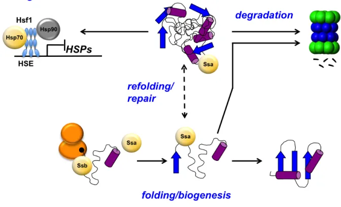
Figure 1.3. Hsp70-mediated functions. In yeast the cytosolic Hsp70s are Ssa and Ssb. Both Ssa and Ssb are involved in protein biogenesis, but in some cases misfolding occurs and the Ssa can triage aberrant protein to the ubiquitin proteasome pathway or refold it. Depending on the level of misfolding in the cell, the heat shock response can be triggered, resulting in release of the Hsp70s and Hsp90s from Hsf1 bound to the heat shock element (HSE) in the promoter of heat shock genes allowing for expression of heat shock proteins (HSPs).
and delivering them to the endoplasmic reticulum (ER) for translocation and processing. Studies have also been performed relating Hsp70 to degradation of terminally misfolded proteins as well as proteins programmed with short half-lives. In vivo and in vitro studies using well-characterized client proteins such as cystic fibrosis transmembrane conductance regulator (CFTR), von Hippel-Lindau (VHL) protein variants, as well as α-synuclein and poly-Q expanded proteins, have shown that Hsp70 is necessary for efficient protein degradation [81-84]. In mammals the Hsp70 machine along with the NEF, Hsp110, and the J-domain proteins have been shown to act in disaggregation, a process important for clearance of misfolded proteins [85, 86]. In yeast, disaggregation is the primary function of the Hsp104 chaperone, a member of the Hsp100 family that is absent from metazoans, but Hsp70 interacts with Hsp104 to activate disaggregation and promote refolding of the liberated protein [87]. It is interesting that mammalian cells have evolved so Hsp70 and its co-chaperones can perform a function that yeast manages using an alternate family of chaperones. The diversity of co-chaperones establish them as important modulators of Hsp70 function and specificity.
The Hsp40 co-chaperones - Aside from their role in modulating the folding cycle through activation of ATPase activity, the Hsp40 co-chaperones can also recruit Hsp70 for specific functions. The Hsp40s bind to the Hsp70 NBD to enhance the relatively low innate ATPase activity [38]. Yeast cells encode for at least 22 J-domain-containing proteins, or Hsp40s, which vary in substrate binding capabilities and process specificity. Ydj1 is a general Hsp40 and one of the most highly expressed genes in the family. This J-protein is one of the primary co-chaperones involved in Hsp70-mediated protein folding. Jjj1, on the other hand is a highly specialized Hsp40 protein involved in ribosome biogenesis [88]. There are also Hsp40s that have no substrate binding capabilities, such as Zuo1, which is a component of the RAC complex involved in nascent chain folding [38, 89]. In spite of the structural and functional diversity of the J-domain proteins, they all perform the same biochemical function, which is to
activate the ATPase activity of Hsp70. This provides a mechanism by which co-chaperones can delicately modulate a highly promiscuous and ubiquitous chaperone in order to contribute to a plethora of distinct processes.
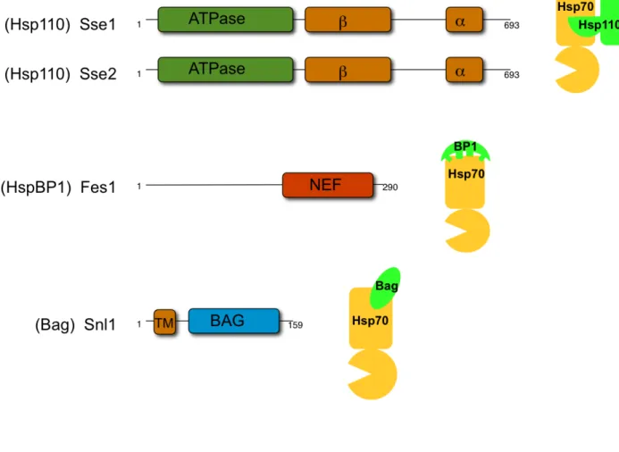
Cytosolic Hsp70 nucleotide exchange factors - Unlike the Hsp40s, which all have a common J-domain, the NEFs are completely distinct in sequence and structure and are thought to have emerged via convergent evolution (Fig 1.4). The three families that exist in the cytosol of mammalian cells are identified as:Hsp110, HspBP1, and Bag domain-containing proteins [90]. In yeast, the Hsp110 family is represented by the two paralogs, Sse1 and Sse2, which are considered to be atypical members of the Hsp70 family because ATPase activity is limited and not required for their known functions [91]. Analogous to the Hsp70 family, Sse1/2 are composed of an NBD and an SBD. Within the SBD, these proteins also have α and β subdomains important for substrate binding. One striking difference between the Hsp110 and the Hsp70 families is an extended linker region between the two subdomains of the SBD [92]. The extended linker results in a structural change that causes the α domain to play a role in Hsp70 binding and likely plays less of a role in substrate interaction. Due to these structural differences Sse1/2 act independently as holdases, which are proteins that bind to substrate and maintain it in a folding competent conformation, as opposed to foldases, which are proteins that bind and release substrate in iterative cycles to promote folding [93-95]. Co-crystal structures have been solved for the Hsp110/Hsp70 heterodimer, which shows the interaction interface lying between the two NBDs, and the α subdomain of the Hsp110 wraps around the Hsp70 NBD [92, 96]. The structure also shows that the β-sandwich domain of Hsp110 remains exposed and available for substrate binding [77, 92, 96, 97]. HspBP1, or Fes1 in yeast interacts with the NBD of Hsp70 through its armadillo repeat domain, or NEF domain, which distorts the two lobes of the Hsp70 NBD resulting in nucleotide release (Fig. 1.4) [98, 99]. The Bag domain-containing family is made up of six homologs in humans and a single protein in yeast, Snl1, which is ER-membrane bound through its N-terminal transmembrane domain (Fig. 1.4).
Figure 1.4. Cytosolic Hsp70 nucleotide exchange factor (NEF) architectures and interaction interfaces. Schematic representation of the architecture of each of the cytosolic Hsp70 NEFs as well as cartoon figures showing the interaction interfaces between Hsp70 and the NEFs. NEF family classifications are shown in parentheses.
Figure 1.4. Cytosolic Hsp70 nucleotide exchange factor (NEF) architectures and interaction interfaces
The Bag family of proteins interacts with Hsp70 through a triple helical bundle that contacts the Hsp70 NBD, slightly tilting one lobe to release ADP [38, 95, 100]
All of the NEFs are present at different levels in the cytosol: Sse1 is the most abundant NEF, present at 70,000 molecules per cell (compared to approximately 270,000 molecules of the Hsp70, Ssa), which is approximately five times that of Fes1, the next most abundant NEF, and levels of Sse2 and Snl1 are 15-20 times lower than Sse1 [101]. However Sse2 is the only NEF upregulated more than 20 fold in response to a number of stress conditions [61, 102]. Loss of Sse1 results in a slow-growth phenotype exacerbated by temperature stress and deletion of both of the Hsp110s, Sse1 and Sse2, results in a lethal phenotype [102, 103]. FES1 deletion cells exhibit mild slow growth and heat sensitivity [104]. No growth phenotypes associated with loss of Snl1 or Sse2 alone have been identified.
Similar to the cytosol, the ER also houses two distinct families of Hsp70 NEFs. Kar2, the ER-resident Hsp70, interacts with two proteins Lhs1 and Sil1, that both result in the release of ADP from the Hsp70 NBD. Lhs1 is a member of the Hsp110 family, but there are a few differences between Lhs1 and Sse1. Lhs1 has a more extended linker between the α and β subdomains of the SBD [105]. Furthermore, Kar2 activates the ATPase activity of Lhs1, which is considered to be important for coordinated substrate interaction between the two proteins [106]. This linked chaperone/co-chaperone activity is also observed between the human ER Hsp70 and NEF, BiP and Grp170 [107, 108]; this type of chaperone interaction is thought to be possible between Sse1/2 and the Ssa proteins, but the function of the Sse1 SBD in Hsp70-mediated folding is currently unknown. Other than these two differences, there are many similarities between the Sse1 and Lhs1 including the dual NEF and holdase activities of Lhs1 [77, 109, 110]. Sil1 is thought to be a member of the HspBP1 family, and in humans, a mutation in this protein is associated with the neurodegenerative disorder, Marinesco-Sjögren syndrome [111]. Although Sil1 acts as an equally potent NEF, it does not have completely redundant activities with Lhs1, mirroring the findings that will be later discussed in Chapter 4 between Sse1 and Fes1. Deletion of both SIL1 and LHS1 results in synthetic lethality, but loss
of either gene leads to protein folding defects in the ER [112, 113], suggesting these two proteins play unique roles in proteostasis. Lhs1 was also shown to be important in protein translocation into the ER, which is not a function requiring Sil1 [113]. In addition, Lhs1 contributes to Kar2-mediated folding and secretion of the heat denatured substrate, Hsp105Δ-β-lactamase, but was not involved in initial folding of the protein. These findings are similar to the work presented in this thesis, in that the NEFs contribute differentially to Hsp70-mediated functions, and in the finding that ER de novo folding and refolding of heat denatured proteins are distinct processes [114]. The ER-resident NEFs discussed above present a chaperone network very similar to the cytosol and show functional differentiation between two proteins, Lhs1 and Sil1, that perform the same biochemical function to release ADP from the Hsp70, Kar2. This theme will be reiterated throughout the work presented in this thesis.
Several studies have identified a number of individual NEF contributions to cytosolic Hsp70 function. Sse1, the most highly studied yeast cytosolic NEF, has been shown to be important for a number of Hsp70-mediated processes. Sse1 plays a significant role in the flux of nascent substrate through Ssa and Ssb proteins [97, 115]. In addition, Sse1 is necessary for biogenesis of the FFL protein in vivo or refolding in vitro [48]. Multiple experiments have also illustrated Sse1’s contribution to Hsp70-dependent co-translational folding, Hsp90-dependent signaling and post-translational translocation of pre-pro-α factor [77, 91, 97, 116]. Both Sse1 and Fes1 have been implicated in prion propagation and curing. Sse1 is essential for [PSI+] propagation, and cells lacking Sse1 or Fes1 are cured for [URE3] [117, 118]. Sse1 and Fes1 also contribute to Hsp70-mediated degradation via the ubiquitin proteasome pathway [119-123]. It was recently shown that Snl1 associates with ribosomes through basic residues located near the Hsp70 binding Bag domain, but the functional ramifications are yet to be determined [124]. Work performed in the last decade has greatly augmented our understanding of the individual contributions of the cytosolic NEFs, but now the functions of these proteins must be studied at the network level to determine their relative contributions in vivo.
In this study I have investigated differential contributions of the cytosolic Hsp70 NEFs to proteostasis, examined the functional significance of Sse1 and Snl1 synthetic growth phenotypes, and developed a coupled assay to address the relationship between protein aggregation and activation. I concluded that Sse1 is the primary NEF involved in most Hsp70 mediated processes. However, it appears that none of the individual NEFs nor the two most abundant NEFs, Sse1 and Fes1, is necessary for protein refolding and repair. The role of Sse1 as the major NEF involved in cellular viability is reflected in the growth assays, which show that an SSE1 deletion strain exhibits a slow-growth phenotype with temperature sensitivity suggesting it is likely important in protein biogenesis as well as quality control. Loss of Fes1 only showed a mild growth defect and heat sensitivity, suggesting it might be primarily important in protein quality control mechanisms. In addition, cells lacking Fes1 resulted in high de-repression of the HSR. Induction of the HSR also results in an increase in the cellular chaperone load. It is possible that this phenotype could mask Fes1’s contributions to Hsp70 function due to the availability of other chaperones. This study did not reveal any unique functions of the Snl1 or Sse2 proteins, but previous studies performed in my laboratory with Snl1, as well as findings in this study, suggest that Snl1 may be involved in regulating some aspect of translation. In addition, it is possible that Sse2 is conditionally needed because it is the only NEF highly induced in response to stress.
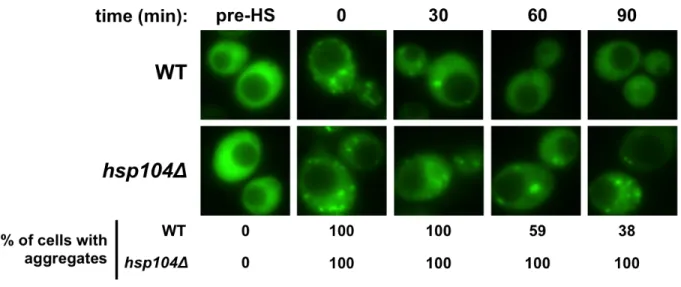
In several experiments I compared solubility of a protein to its enzymatic activity or its rate of degradation; these two phenomena are closely connected. In a coupled assay to monitor near real time fluorescence microscopy with automated enzymatic assays, I discovered that the dynamics of an aggregate is directly related to the ease at which the protein is liberated from the aggregate for refolding. In addition, the presence of a disaggregase system is essential for efficient restoration of proteostasis.
Finally, phenotypes identified in my earlier work, as well as the synthetic deletion phenotype identified in the NEF study, led me to examine the functional relationship between Sse1 and Snl1. This line of investigation uncovered that the levels of Snl1 become very important for
viability in the absence of Sse1, which suggests that Sse1 antagonizes the Snl1-mediated activity. Due to the cold sensitivity (known to be associated with translation defects) observed upon deletion of SSE1 and SNL1, the ER localization of Snl1, and the induction of the HSR in the presence of high levels Snl1 in an SSE1 null strain, I hypothesized that this unknown function of Snl1 is associated with translation regulation. Ultimately, all of these proteins have homology to human cytosolic proteins and therefore these studies can be applied to understand the division of labor among the NEFs in human cells, which can be used to develop treatments to modulate proteostasis.
SIGNIFICANCE OF THIS STUDY
Understanding cytosolic Hsp70 NEFs - Previous findings mentioned above were performed in different strain backgrounds using multiple substrates, which contribute to the understanding of how the NEFs function. However, the NEFs do not function alone in the cell and it is important to investigate how they work within the context of the chaperone network. I have performed comprehensive genetic, biochemical, and cell biological analyses in isogenic single and combinatorial cytosolic NEF deletion strains. This thesis work aims to address a more global question of why have the three families of NEFs been maintained throughout eukaryotic evolution upon converging to perform the same biochemical function, which is to release ADP from Hsp70. All three families have similar levels of NEF activity in vitro but are completely distinct structurally and in sequence. Understanding how these NEFs contribute relatively to the chaperone network is important for two reasons: first, understanding specific functions can lead to the discovery of molecules that modulate Hsp70 function, which is becoming important in treatment for both neurodegenerative diseases and cancer. In addition, there are disorders directly associated with NEF dysfunction such as Marinesco-Sjøgren syndrome, which is an autosomal recessive cerebellar ataxia caused by a mutation in the Fes1 homolog in the ER, Sil1 (BAP) [125]. Second, loss of Hsp110 is associated with tau pathology in a mouse model and huntingtin-related neurodegeneration in a Drosophila model [126, 127]. The NEFs provide both direct and indirect targets for the development of drugs to treat a number of diseases.
Human diseases related to proteostasis - Protein misfolding and aggregation in humans have been associated with the formation of highly structured amyloid deposits common to many neurodegenerative disorders including Alzheimer’s, Parkinson’s, and Huntington’s diseases [128]. These neurodegenerative diseases have a common theme in which normally existing proteins with either known or unknown functions adopt an alternative fold. This can lead to the formation of disordered and ordered aggregates that can further develop into highly structured amyloid fibers. Understanding which of these proteins is most toxic and important in
neurodegeneration and what promotes its formation will require many more years of study. In Alzheimer’s disease, both accumulation of amyloid beta (Aβ) and hyperphosphorylated tau protein are associated with neuroinflammation and progression of the disease [129]. α-synuclein, the major component of Lewy Bodies, is a neuronal protein that misfolds leading to the formation of oligomers and ultimately fibrils, a mature ordered form of aggregate associated with Parkinson’s disease [130-132]. In Huntington’s disease and other poly-Q pathologies, disease onset and severity are highly dependent on the number of glutamine residues found in the protein, which have a direct relationship with the tendency toward forming ordered aggregates [133, 135, 136]. There are also a number of neurodegenerative diseases associated with the human prion protein PrP, which switches to the infectious scrapie form (PrPSC) through an unknown mechanism. PrPSC is associated with a number of human
diseases including Creutzfeldt-Jakob disease (CJD), Gerstmann-Straussler syndrome (GSS), fatal familial insomnia (FFI), and Kuru [134, 135]. Mechanisms for how the alternate forms of each of these proteins are formed and how they lead to toxicity are mostly unknown. Because protein misfolding seems to be central to all of these human diseases, it is important that the field identifies how chaperone activity can be altered to help correct folding or improve degradation of these aberrant proteins [136].
Treatments and therapies - There are currently no treatments or therapies that slow the progression or reverse the effects of neurodegenerative diseases. Primarily small molecules or genetic modifiers have been identified and tested for their ability to modulate proteostasis [137]. The chief goals for treating these types of diseases are to stabilize proteins to prevent misfolding and aggregation, or once the protein is misfolded, to promote protein refolding or degradation. Both of these can be addressed through activation of stress responses or through direct modulation of molecular chaperones. Hsp90 and Hsf1 have been primary targets for small molecule treatments, but more recently Hsp70 has become a desirable target [137, 138]. Multiple compounds were identified through a high-throughput screen that either increased or
decreased Hsp70 ATPase activity. The group that decreased activity were shown to specifically lower the levels of both total and hyperphosphorylated tau species in HeLa cells, and clear both normal and aberrant forms of the protein from mouse brains [139-141]. In addition, treatment with a compound that increased Hsp70 ATPase activity improved Hsp70-mediated luciferase refolding and suppressed the temperature-dependent slow growth defect observed in ydjΔ
yeast cells [142]. One major issue with modulating chaperones, such as Hsp70 and Hsp90, is that these proteins act in a wide variety of proteostasis pathways, so it is important to regulate them as specifically as possible. One way to do this is to target the co-chaperones. Although the J-domain proteins are well known to provide substrate and process specificity to Hsp70, this work suggests that the cytosolic NEFs may provide an additional level of control. Gestwicki and associates have identified a family of small molecules that affect the interaction between Hsp70 and the Bag-domain containing family of proteins, and I am now testing these compounds for specificity and efficacy. Targeting distinct families of co-chaperones is promising for developing treatment and therapies of proteostasis diseases.
MATERIALS AND METHODS
Strains, plasmids and growth conditions - All strains in these studies are listed in Table 2.1 and are isogenic to BY4741 (MATa/his3Δ1/leu2Δ0/met15Δ0/ura3Δ0), BY4742 (MATα/his3Δ1/leu2Δ0/lys2Δ0/ura3Δ0), or DS10 (MAT a/leu2-3/112trp1-1/ura3-52/lys1/lys2/his3-11,15). Cells were grown on media containing 2% dextrose. Media used in these studies include the nutrient rich media, yeast extract-peptone-dextrose (YPD) or synthetic complete (SC) media lacking only amino acids necessary for plasmid selection and supplemented with yeast nitrogen base (YNB) at 6.71 mg/ml (Sunrise Science Products, San Diego, CA). In addition, for some experiments additives were used including azetidine-2-carboxylic acid (2 mM) (Sigma Aldrich, St. Louis, MO), cycloheximide (100 µg/ml) (Acros Organics, Geel, Belgium), CdSO4 (200 µM) (Sigma) and diamide (300 µM) (Research
Organics, Cleveland, OH).
Plasmids used in these studies are listed in Table 2.2. Deletion strains were constructed using cassettes built in pBluescriptII carrying the marker genes KANMX4, LEU2, or HIS3, flanked upstream and downstream by non-coding regions of SSE1, SSE2, FES1, and SNL1. Descriptions of the plasmids, including restriction sites used for cloning, are provided in Table 2.2. Folding analysis was done in cells expressing the 425MET25-FFL-GFP–leu2::URA3 plasmid. The original plasmid, p425MET25-FFL-GFP, was a generous gift from John Glover’s laboratory at University of Toronto. The plasmid used in this study was constructed by amplifying the URA3 gene from p426 [143] using oligonucleotides containing homologous regions of the LEU2 gene (5’-ATGTCTGCCCCTAAGATCGTCGTTTTGCCAGGTGACTAACTG AGAGTGCACCATACCACAGC-3’, 5’-TTAAGCAAGGATTTTCTTAACTTCTTCGGCGACAGCA TCACCCACCGCATAGGGTAATAACTG-3’). For recombination of URA3 into the LEU2 locus, the amplicon and p425MET25-FFL-GFP were transformed into BY4741 cells. The construct was rescued from Leu+ Ura- transformants (Geitz laboratory protocol; University of Manitoba) and transformed into E. coli for amplification. The plasmid was purified from E.coli using standard procedures and confirmed using PCR. The p425MET25-FFL-GFP-leu2::URA3
construct was transformed into NEF deletion strains using the rapid yeast transformation protocol [144]. For Hsf1 activity assays, pSSA3HSE-lacZ, previously described in [145], was transformed into wild-type cells and each of the single deletion strains. For degradation analysis, strains were constructed using pRH2081 (from Randy Hampton’s Laboratory, UC San Diego), which is an integrative plasmid that carries TDH3-driven ssCPY*-GFP [120]. The plasmid was linearized using restriction endonuclease Van91I and transformed into wild-type and NEF deletion strains. For immunoprecipitation analyses, yeast cells were transformed with either p413TEF-FLAG-SSE1 or p413TEF-FLAG-FES1, which were constructed by subcloning the FLAG tagged SSE1 or FES1 using SpeI/XhoI restriction sites into the 413TEF plasmid as previously described in [124, 143]. For overexpression studies, all SSE1 and SNL1 alleles were cloned into parent vectors at SpeI/XhoI restriction sites.
Yeast growth assays - For NEF deletion strain growth analysis, cultures were diluted to A600= 1;
1:10 serial dilutions were spotted on YPD plates. To identify growth phenotypes in both optimal and stress-inducing growth conditions, plated cells were incubated at 15, 25, 30 and 37°C for up to 5 days. To test azetidine-2-carboxylic acid (AZC) toxicity, strains were grown overnight and 1:10 serial dilutions were plated on SC media or SC+2 mM AZC and incubated at 30°C for 3-5 days. For de novo folding analyses, NEF deletion strains containing p425MET25-FFL -GFP-leu2::URA3 were grown overnight in SC-URA media containing 200 µM additional methionine (addition of methionine represses expression of the FFL-GFP fusion under the MET25 promoter), subcultured in the same media, and grown to early log phase (A600=
0.4-0.5), then induced in SC-URA-MET media. Refolding assays were performed using NEF deletion strains containing p425MET25-FFL-GFP-leu2::URA3 and cells were grown overnight in SC-URA and subcultured to log phase A600= 0.8-1.0. Cells were induced in SC-URA-MET
for 1 hour at 30°C. Prior to heat shock cells were treated with 100 µg/ml of cycloheximide and then incubated at 42°C for 25 minutes, and recovered for 60 minutes at 30°C. For degradation analysis log phase cells were treated with 100 µg/ml cycloheximide (CHX).
Protein isolation and visualization - For folding and immunoprecipitation analyses, proteins were isolated using glass bead lysis as described in [124]. Western blot analysis was performed using α-Ssa1/2 polyclonal antibody (from M. Ptashne, Sloan Kettering Institute) at a 1:100,000 dilution, α-Ssb1/2 polyclonal antibody (from E. Craig, University of Wisconsin) at a 1:10,000 dilution, α-GFP monoclonal antibody (Roche; Nutley, NJ) at a 1:1000 dilution, or α-FLAG monoclonal antibody (Sigma). For degradation analyses and Hsf1 derepression assays, denaturing extractions were performed. Cells were resuspended in 200 µl SUME buffer (sodium dodecyl sulfate, 1%; urea, 8 M; MOPS, 10 mM, EDTA, 10 mM) + protease inhibitors (aprotinin, 2µg/ml; pepstatin A, 2 µg/ml; leupeptin, 1µg/ml; phenylmethylsulfonyl fluoride, 1mM; chymostatin, 2 µg/ml). Glass beads were added and cells were lysed by vortexing for 3 minutes and then centrifuging cells at approximately 4,600 x g for 5 minutes at room temperature. Supernate was transferred to a new tube and 6x SDS sample buffer (Tris-HCl, pH 6.8, 350 mM; glycerol, 36%(v/v); SDS, 10% (w/v); β-mercaptoethanol, 5% (w/v); bromophenol blue, 0.012% (w/v)) was added and the sample was incubated at 65°C for 10 min. Proteins were separated by 15% SDS-PAGE and transferred to a PVDF membrane (Millipore Corporation, Billerica MA). For degradation analysis α-GFP (Roche; 1:1000) or α-Pgk (Invitrogen, Carlsbad, CA; 1:1000) primary antibodies were used. For Hsf1 derepression assay proteins were incubated with α-Cpr6 (Jill Johnson Laboratory, University of Idaho; 1:1000), αHsp104 (Assay Designs / Stressgen, Farmingdale, NY; 1:1000), α-Sti1 (David Toft laboratory, Mayo Clinic, Rochester, NY; 1:1000), or α-Pgk (Invitrogen; 1:1000). Translocation analysis used anti-pre-pro-α factor (α-ppαF) (Randy Schekman laboratory, University of California Berkeley; 1:2000). To visualize proteins, membranes were exposed to enhanced chemiluminescence reagents and developed on X-ray film using a developer or a LI-COR C-DiGit Blot Scanner and Image Studio Digits software (Lincoln, NE). Coomassie staining was performed by placing the SDS-PAGE gel into Coomassie Brilliant Blue Stain (Coomassie Blue 0.076% (w/v); methanol 45% (v/v); acetic acid 10% (v/v); water 45% (v/v)) for 20 minutes with rocking. Gels were rinsed in water and destain (acetic acid 5% (v/v); methanol 16.5% (v/v);
water 78.5% (v/v)), microwaved for 30 seconds, and allowed to incubate until the desired staining of the protein bands was achieved.
Fluorescence microscopy - Cells were collected and visualized using an Olympus IX81-ZDC inverted microscope as described previously in [146] as well as in Chapter 3. To test steady-state protein solubility, log phase cells were visualized. For refolding analysis, samples were collected prior to heat shock, immediately following heat shock, and 60 minutes post heat shock to be visualized using fluorescence microscopy. To perform degradation assays, samples were collected immediately after cycloheximide treatment and one and two hours post treatment. Quantification was performed by counting approximately 100 cells and dividing the number of cells containing aggregates by the total number of cells.
FFL-GFP activity assay - To test steady-state FFL activity, light unit production was monitored once cells reached log phase exactly as described in Abrams 2013 and Chapter 3. In short, an automated plate reader protocol (BioTek Synergy MX; Winooski, VT) was used to inject 100 µl of cells with 50 µl of D-luciferin reagent (~23 µg) (Sigma) in a 96-well white plate (Greiner; Monroe, NC) followed by mixing and a luminescence reading. For refolding analysis, the activity was measured after cycloheximide treatment and heat shock. In addition, an automated protocol described in Abrams 2013 and Chapter 3 was programmed using the Synergy MX plate reader (BioTek) to measure luminescence immediately after heat shock and at 60 minutes into recovery at 30°C.
Hsf1 de-repression assay - Cells expressing the HSE-lacZ reporter construct were grown to log phase. Activity of Hsf1 was determined by adding 50 µl of cells to 50 µl of Beta-Glo reagent (D-luciferin-o-β-galactopyranoside, Promega; Madison, WI) in wells of a white 96-well plate. After a 30 minute incubation at 30°C, the Synergy MX plate reader was used to measure luminescence.
Immunoprecipitation - The in vitro immunoprecipitation (IP) was performed using purified FLAG-Fes1 (3 µg/µl) incubated with no lysate or whole cell lysate at 4.5 µg/µl, or 7.5 µg/µl for two hours. Protein was eluted using 30 µl of M2 FLAG resin (Sigma). For in vivo FLAG-Fes1 IP strains expressing empty vector p413TEF or p413TEF-FLAG-FES1 were grown to mid-log phase and the protein was isolated using glass bead lysis. 10 µl of supernate was mixed with 10 µl of 2X SDS-PAGE sample buffer and incubated at 65°C for 10 minutes. For the IP, the remaining supernate was transferred to a new tube and 30 µl of FLAG resin was added along with 700 µl of TEGN (Tris-HCl, pH 7.9, 20mM; ethylenediaminetetraacedic acid (EDTA), 0.5 mM; glycerol 10%(v/v); NaCl, 50 mM) + PI. The IP was incubated for 2 hours at 4°C with rocking, followed by 8 washes with 500 µl of TEGN + PI. After the beads were washed, 40 µl of FLAG peptide solution (7 µg total) was added and the mixture was incubated at 37°C for 25 minutes. Protein solution was centrifuged at 4,600 x g and 40 µl of the supernate was transferred to a new tube, SDS-PAGE sample buffer was added and samples were incubated at 65°C for 10 minutes.
Statistical analysis – The statistical analysis pertains to Chapter 4 only and was adapted from the publication denoted at the beginning of the chapter. All experiments were performed in triplicate and the results are represented as mean ± SD. Significance determinations were performed using the two-tailed Student t test. P values are represented as follows: *, p < 0.05; **, p < 0.005; ***, p < 0.0005. Differences in data sets were considered to be statistically significant for all comparisons where p < 0.05.
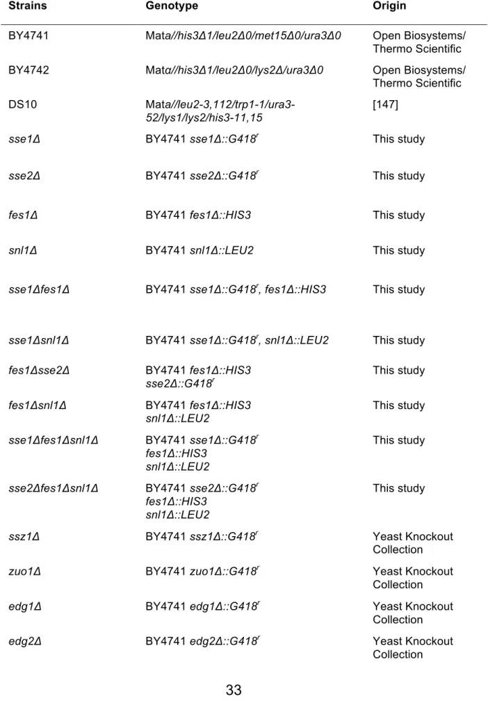
Table 2.1. Strains used in these studies
Strains Genotype Origin
BY4741 Mata//his3Δ1/leu2Δ0/met15Δ0/ura3Δ0 Open Biosystems/ Thermo Scientific BY4742 Matα//his3Δ1/leu2Δ0/lys2Δ/ura3Δ0 Open Biosystems/
Thermo Scientific DS10 Mat
a//leu2-3,112/trp1-1/ura3-52/lys1/lys2/his3-11,15
[147]
sse1Δ BY4741 sse1Δ::G418r This study
sse2Δ BY4741 sse2Δ::G418r This study
fes1Δ BY4741 fes1Δ::HIS3 This study
snl1Δ BY4741 snl1Δ::LEU2 This study
sse1Δfes1Δ BY4741 sse1Δ::G418r, fes1Δ::HIS3 This study
sse1Δsnl1Δ BY4741 sse1Δ::G418r, snl1Δ::LEU2 This study fes1Δsse2Δ BY4741 fes1Δ::HIS3
sse2Δ::G418r This study
fes1Δsnl1Δ BY4741 fes1Δ::HIS3 snl1Δ::LEU2
This study sse1Δfes1Δsnl1Δ BY4741 sse1Δ::G418r
fes1Δ::HIS3 snl1Δ::LEU2
This study
sse2Δfes1Δsnl1Δ BY4741 sse2Δ::G418r
fes1Δ::HIS3 snl1Δ::LEU2
This study
ssz1Δ BY4741 ssz1Δ::G418r Yeast Knockout
Collection
zuo1Δ BY4741 zuo1Δ::G418r Yeast Knockout
Collection
edg1Δ BY4741 edg1Δ::G418r Yeast Knockout
Collection
edg2Δ BY4741 edg2Δ::G418r Yeast Knockout
ssb1Δssb2Δ DS10 ssb1Δ::HIS3 ssb2Δ::LEU2 [56] ssb1Δssb2Δsse1Δ DS10 ssb1Δ::HIS3 ssb2Δ::LEU2
sse1Δ::G418r
Table 2.2. Plasmids used in these studies
Plasmid Description
pBluescriptII – LEU2 BluescriptII plasmid with ~2173 bp of the LEU2 gene cloned with BamHI/SpeI
pBluescriptII – HIS3 BluescriptII plasmid with ~1200 bp of the LEU2 gene cloned with BamHI
pBluescriptII – KANMX4 BluescriptII plasmid with ~1440 bp of the LEU2 gene cloned with EcoRI/BamHI
p413TEF Low copy; HIS+, constitutive promoter
[143]
pRS426 High copy; URA+ [143]
pRH2081TDH3 – ss – CPY* - GFP Integrative plasmid containing the soluble CPY mutant with a C-terminal GFP tag (Hampton, UC San Diego)
p425MET25-FFL-GFP High copy; Met repressible promoter containing firefly luciferase fused to green fluorescent protein [148]
p425MET25-FFL-GFP – leu2::URA3 Contains a LEU2 deletion with a URA3 inserted through homologous
recombination
pSSA3HSE-lacZ Contains a heat shock response element from the promoter of SSA3 [145]
p413TEF-FLAG-FES1 N-terminally FLAG-tagged SSE1 ORF inserted into MCS (SpeI/XhoI)
p413TEF-FLAG-SSE1 N-terminally FLAG-tagged FES1 ORF inserted into MCS (SpeI/XhoI)
p423GPD High copy; HIS+ constitutive promoter
p423GPD-SNL1-FLAG C-terminally FLAG-tagged SNL1 ORF inserted into the MCS (SpeI/XhoI)
![Table 1.1. Yeast Cytosolic Chaperones [22].](https://thumb-us.123doks.com/thumbv2/123dok_us/1992031.2795960/27.918.128.755.103.1077/table-yeast-cytosolic-chaperones.webp)