Loyola University Chicago
Loyola eCommons
Dissertations Theses and Dissertations
2010
The Role of IGF-1 and Notch Signaling in Thoracic
Malignancies.
Sandra Eliasz
Loyola University Chicago
This Dissertation is brought to you for free and open access by the Theses and Dissertations at Loyola eCommons. It has been accepted for inclusion in Dissertations by an authorized administrator of Loyola eCommons. For more information, please contactecommons@luc.edu.
This work is licensed under aCreative Commons Attribution-Noncommercial-No Derivative Works 3.0 License. Copyright © 2010 Sandra Eliasz
Recommended Citation
Eliasz, Sandra, "The Role of IGF-1 and Notch Signaling in Thoracic Malignancies." (2010).Dissertations.Paper 83. http://ecommons.luc.edu/luc_diss/83
LOYOLA UNIVERSITY CHICAGO
THE ROLE OF IGF-1 AND NOTCH SIGNALING IN THORACIC MALIGNANCIES
A DISSERTATION SUBMITTED TO THE FACULTY OF THE GRADUATE SCHOOL
IN CANDIDACY FOR THE DEGREE OF DOCTOR OF PHILOSOPHY
PROGRAM IN MOLECULAR AND CELLULAR BIOCHEMISTRY
BY
SANDRA ELIASZ CHICAGO, ILLINOIS
Copyright by Sandra Eliasz, 2010 All rights reserved.
!
! """!
ACKNOWLEDGEMENTS
I would like to express my gratitude to Dr Maurizo Bocchetta, my mentor, for his invaluable guidance and support. His ideas have influenced all my work, his dynamism and enthusiasm have allowed me to progress quickly and with passion, his trust and honesty I valued deeply; he has made my work on this dissertation a truly rewarding and gratifying experience.
I would also like to thank members of my dissertation committee: Dr Mary Manteuffel, Dr Caroline Le Poole, Dr Clodia Osipo, Dr Manuel Diaz and Dr Maurizio Bocchetta, for their insightful comments and encouragement. I am greatly indebted to all members of Dr Bocchetta laboratory for their help and assistance: Dr Yuanbin Chen, Melissa De Marco, Ovidiu Machek and Sylvia Skucha, and specifically to Dr Irene Graziani for her contributions to the research on the role of Notch in mesothelioma, which are part of this dissertation, and to Shuang Liang for her essential work on ChIP and in vivo experiments in Adenocarcinoma of the lung I present herein. I also thank our neighboring labs – of Dr Miele and Dr Osipo, for their expert advice and assistance in research on Notch.
My gratitude also goes to the Graduate School of Loyola University, which first endowed me with the necessary knowledge in biochemistry, and next provided a very
!
!
! "#!
stimulating environment to pursue my research interests. I am very grateful to Dr Manteuffel, the graduate program director, for her advice and assistance with all matter pertaining to the program. I would also like to thank the administrative staff, in particular Ashyia Paul and Elayne Grzeda, for all their help.
Finally, I would like to express my deepest gratitude to my family, especially my husband Piotr, my sons Ignacy and Iwo, and my parents and parents in law, who have always supported me and gave a lot of encouragement. Having them close has been very important to me.
!
! "!
TABLE OF CONTENTS
ACKNOWLEDGEMENTS... iii
LIST OF TABLES... vii
LIST OF FIGURES ... viii
LIST OF ABBREVIATIONS... xi
ABSTRACT... xvi
Chapter 1. INTRODUCTION...1
2. REVIEW OF RELATED LITERATURE 2.1 Thoracic Malignancies...6
2.2 Molecular Pathogenesis of Malignant Mesothelioma...6
2.3 Molecular Pathogenesis of Lung Cancer ...8
2.4 Notch Signaling ...10
2.5 Notch and Cancer...13
2.6 Notch in Malignant Mesothelioma ...15
2.7 Notch in Lung Cancer...15
2.8 Notch and Hypoxia ...17
2.9 IGF-1R pathway...18
3. THE SV40 LATE T ANTIGEN REGULATES IGF-1 EXPRESSION IN TRANSFORMED MESOTHELIAL CELLS Abstract ...20
!
!
! "#!
Materials and Methods...24
Results...33
Discussion ...49
4. THE ROLE OF NOTCH SIGNALING IN MALIGNANT MESOTHELIOMA Abstract ...53
Introduction...54
Materials and Methods...57
Results...64
Discussion ...76
5. THE ROLE OF NOTCH SIGNALING IN ADENOCARCINOMA OF THE LUNG Abstract ...79
Introduction...80
Materials and Methods...82
Results...88
Discussion ...110
6. GENERAL CONCLUSIONS...113
REFERENCE LIST ...116
!
! "##!
LIST OF TABLES
Table Page
1. A list of all antibodies used in Chapter 3...28
2. A list of all oligonucleotides used in Chapter 3...30
3. A list of oligonucleotides used for ChIP experiment on the IGF-1 promoter ...32
4. A list of all oligonucleotides used in Chapter 4...59
5. A list of all antibodies used in Chapter 4...60
6. A list of oligonucleotides used for ChIP experiment on the PTEN promoter ...74
7. A list of oligonucleotides used for ChIP experiment on the IGF-1R promoter...82
8. A list of all oligonucleotides used in Chapter 5...83
! ! ! "###! LIST OF FIGURES Figure Page 1. Dissertation outline ...5 2. Notch signaling ...12
3. E6 induction down-regulates p53 and p53 regulated gene products ...34
4. E6 induction in S-HML suppresses DNA synthesis and causes cell growth arrest. Critical components of the IGF-1 signaling pathway are down-regulated upon either E6 or mdm2 induction ...36
5. Both p53 down-regulation through RNAi and transfection of S-HML with dominant negative p53 cause reduced expression of p21, mdm2, IGF-1 and IGF-1R ...38
6. E6-mediated inhibition of DNA synthesis in S-HML is mediated through the IGF-1/IGF-1R pathway...39
7. IGF-1R expression in S-HML and in 1R cells...41
8. IGF-1 regulates IGF-1R expression levels in S-HML...42
9. E6 induction in S-HML causes transcriptional repression at p53-regulated promoters. Tag and p53 bind to the rat IGF-1 promoter in vitro...44
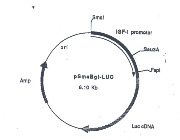
10.Map of pSmaBgl-LUC plasmid ...45
11.Tag and p53 associate with the IGF-1 promoter in a multiprotein complex ...46
12.Model of Tag/p53 regulation of the IGF-1 promoter...50
13.Expression of Notch signaling components in primary human mesothelial cell cultures (HM), mesothelioma cell lines (ME), microdissected lung pleura (HP) and microdissected mesothelioma cells (MM) ...65
!
! "#!
14. Down-regulation of Notch-1 in ME leads to cell death both in normoxia and
hypoxia...67
15. Active Akt-1 rescues apoptosis caused by Notch-1 inhibition ...69
16. Notch-1 and Notch-2 have opposite effects on PTEN expression both at the protein and at the mRNA level ...72
17. Notch-1 directly interacts with the PTEN promoter within a region between position -602 and -387 (+1 is the PTEN start codon) ...75
18. Notch-1 activation in ACL cells under hypoxia is HIF-1! dependent...81
19. Notch-1 negatively regulates PTEN expression in ACL cells...89
20. Notch-1 regulates Akt-1 phosphorylation in ACL cells under hypoxia ...90
21. Akt-1 activation protects ACL cells from apoptosis triggered by Notch inhibition under hypoxia ...91
22. Constitutively active Akt-1 rescues ACL cells from cell death even at very high GSI concentrations (100 µM) under hypoxia ...92
23. Notch-1 inhibition leads to reduced Bcl2-A1 and increased caspase-1 expression ...94
24. Notch-1 modulates the activation levels of PDK-1, Akt-1 and mTOR in PTEN null ACL cells ...96
25. Hypoxia/Notch-1 regulates IGF-1R expression; manipulation of IGF-1R signaling modulates the rate of GSI induced cell death under hypoxia...97
26. Notch-1 regulates IGF-1R expression ...100
27. Artificial down-regulation of HIF-1! causes reduced expression of the IGF-1R in ACL cells under hypoxia...101
28. Notch-1 regulates IGF-1R expression in cell lines obtained from different tissues...102
29. Region of the IGF-1R that we analyzed by ChIP for the indicated proteins ...103
!
!
! "!
31. Notch-1 associates with the +1478 region of the IGF-1R gene alongside MAML-1 and p300; transfection of ACL cells with dominant negative
MAML-1 disrupts such assiociation...105 32. Metastatic ACL model in immunocompromized mice...107 33. GLUT-1 is an effective marker of hypoxia in ACL cells ...108 34. Notch-1IC is coexpressed in hypoxic tumor cells together with IGF-1R and
phosphorylated Akt-1...110 35. Schematic of Notch-1 regulation of Akt-1 activation under hypoxia in ACL...111
!
! "#!
LIST
OF
ABBREVIATIONS
ACL adenocarcinoma of the lung B-CLL B-cell chronic lymphoid leukemia bHLH basic helix-loop-helix
BKV baculovirus
BrDU bromodeoxy uridine CDS coding sequence
ChIP chromatin immunoprecipitation CMV cytomegalovirus
CSL CBF1-Suppressor of Hairless-Lag1 ECL enhanced chemiluminescence EGFR epithelial growth factor receptor EPO erythropoethin
FACS fluorescence activated cell sorting FBS fetal bovine serum
GFP green fluorescence protein GSI !-secretase inhibitor
!
!
! "##!
HDAC histone deacetylase HES hairy enhancer of split HIF-1! hypoxia inducible factor 1! HM human mesothelial cells HP human pleura
HPV human papilloma virus HSP70 heat shock protein 70 IGFBPs IGF binding proteins IGF-1 insulin like growth factor-1
IGF-1R insulin like growth factor-1 receptor iNOS inducible nitric oxide synthase IR insulin receptor
IRS-1 insulin receptor substrate-1 JCV John Cunningham virus MAML-1 mastermind like-1
MAPK mitogen activated protein kinase ME mesothelioma cell lines
MEF mouse embryonic fibroblasts MM malignant mesothelioma
mTOR mammalian target of rapamycin NEC Notch extracellular domain
!
! "###!
NTM Notch transmembrane domain
NSCLC non-small cell lung cancer
PDK-1 phosphoinositide-dependent kinase-1
PI propidium iodide
PI3K phosphatidylinositol 3-kinase
PIP2 phosphoinositol-2,3-bisphosphate
PIP3 phosphoinositol-2,3,4-trisphosphate
PP2A protein phosphatase 2A
PTEN phosphatase and tensin homolog
Rb retinoblastoma
ROS reactive oxygen species
RTK receptor tyrosine kinase
RT-PCR reverse transcription polymerase chain reaction
SCLC small cell lung cancer
SHC Src homology 2 domain containing transforming protein
S-HML SV40 transformed human mesothelial cells
siRNA small interfering RNA
STAT signal transducers and activators of transcription
SV40 simian virus 40
TAD transactivation domain
tag small tumor antigen
!
!
! "#$!
T-ALL T cell acute lymphoblastic leukaemia
!
! "#$!
ABSTRACT
Thoracic malignancies are one of the deadliest of all cancers, being the leading cause of cancer death in the Western world. Thoracic malignancies arise from different tissues; however the most common are of epithelial (commonly referred to as non-small cell lung cancer, or NSCLC), neuroendocrine (small cell lung cancer, or SCLC) and mesothelial origin (malignant mesothelioma, or MM). The DNA oncogenic virus Simian Virus 40 (SV40) has been shown to cooperate with environmental oncogenic fibers in the onset of MM (Bocchetta et al., 2000, Kroczynska et al., 2006). Insulin like growth factor-1 (IGF-factor-1) signaling plays a central role in all thoracic malignancies and in the process of mediated malignant transformation of human cells. We have found that in SV40-transformed human mesothelial cells (HM) the Large T antigen (Tag), p53, pRb and p300 function in a multi-protein complex to promote transcription of the IGF1 gene. Depletion of p53 in these cells causes growth arrest because of lack of IGF-1 synthesis. These results provide a novel mechanistic and biological interpretation of the p53/Tag complexes and of DNA tumor virus transformation in general. It was generally believed that one of the major functions of Tag was to bind and inactivate the tumor suppressor p53. Our data, instead, support a model in which the Tag/p53 complexes are not inert, but rather play an active, essential role in the process of SV40-mediated transformation of HM, hence in the pathogenesis of MM.
!
!
! "#$!
Aside from the well-established role played by the IGF-1/Akt-1 axis in thoracic malignancies, we focused our research on the role of Notch signaling in cancers of the thorax. Notch-1 signaling has been shown to be required for the growth of SV40-transformed human mesothelial cells (Bocchetta et al., 2003). We therefore expanded the studies of the role of Notch signaling in MM and NSCLC. We have found that, under hypoxia, the condition that best recapitulates solid tumors microenvironment, both MM and NSCLC cells have an elevated Notch signaling pathway as compared to normal human mesothelial (HM) and lung bronchioalveolar cells. Genetic and chemical modulation of the Notch pathway indicated that these tumor cells are dependent on Notch signaling. More specifically, MM and NSCLC cell survival was Notch-1 dependent. Notch-1 through its negative regulation of phosphatase and tensin homolog (PTEN) and positive regulation of the IGF-1 receptor (IGF-1R) expression causes activation of the pro-survival IGF-1/Akt-1 signaling pathway. These results provide new insight into the role of Notch in MM and lung cancer, and strongly implicate that Notch pathway inhibitors may be useful in the treatment of those deadly diseases. Our data indicate that targeting Notch-1 signaling using !-secretase inhibitors (GSI) may represent a novel, promising therapeutic approach for thoracic malignancies treatment, by specifically targeting hypoxic tumor microenvironment. This is especially important because hypoxic tumor microenvironment is responsible for poor response to standard anticancer treatment, tumor recurrence and ultimately death. These results also identify additional molecular targets that may snergize with Notch-1 inhibition.
! !
! "!
CHAPTER 1 INTRODUCTION
Since its discovery in 1960 as a contaminant of polio vaccines, SV40 has been the object of extensive studies to assess whether this DNA virus plays a role in human carcinogenesis. Although this issue has met a broad skepticism in the past two decades, an increasing amount of data has accumulated linking SV40 to specific types of human cancer, especially malignant mesothelioma (MM).
Primary human mesothelial cells (HM), as compared to other cells (e.g. fibroblasts), are uniquely susceptible to SV40 infection/transformation. HM are able to survive SV40 infection since viral replication takes place at the low rate, which renders them subjected to the transforming activities of the SV40 oncogenes for a prolonged time. This translates into a uniquely high rate of SV40 mediated transformation in these cells (Bocchetta et al., 2000). HM transformation by SV40 is enhanced by environmental agents, with asbestos being a known etiological agent for MM (Cicala et al., 1993; Kroczynska et al., 2006).
SV40 major oncogene, large T antigen (Tag), besides its role in an initiation of viral replication (Wobbe et al., 1985) and regulation of SV40 transcription (Tjian, 1981), also interacts with host cellular proteins. Among those, interaction with heat shock protein 70 (HSP70; Sullivan et al., 2000), pRb protein family members (DeCaprio et al.,
!
!
2! 1988) and cellular p53 (Pinhasi-Kimhi et al., 1986) appears to be the most intimately linked to SV40 promotion of cell cycle progression and cell transformation. Tag trans-activates a number of genes, which protein products promote cell cycle progression, of which regulation of insulin like growth factor 1 (IGF-1) signaling appears to be required for SV40 mediated transformation of human mesothelial cells (HM; Porcu et al., 1992). SV40 is not able to transform mouse embryonic fibroblasts (MEF) in the absence of insulin like growth factor-1 receptor (IGF-1R; Sell et al., 1993). The IGF-1R docking protein insulin receptor substrate-1 (IRS-1; White, 1998) was found in complex with Tag, which resulted in protection from apoptosis (Zhou-Li et al., 1997). In Aim 1 (results presented in Chapter 3) we hypothesize that there is a direct transcriptional regulation of the IGF1 by Tag. Specifically, we tested the involvement of Tag in regulation of the transcription of the IGF1 in SV40 transformed HM. We sought to determine whether Tag mediated transactivation of IGF1 requires p53, and if so whether Tag/p53 complexes can associate with the IGF1 promoter. We also wanted to characterize the composition of the transactivator complex and determine its location in the promoter region.
During the process of HM cell transformation, SV40 transcriptionaly up-regulates Notch-1 signaling (Bocchetta et al., 2003). Interfering with Notch-1 signaling causes growth arrest of SV40-transformed HM (Bocchetta et al., 2003). Increasing number of reports suggest pro-oncogenic role of Notch in many solid tumors, however the role of Notch signaling in MM is not characterized. The family of Notch receptors includes four isoforms, all of which can play a different role in the malignant setting. It is imperative to
!
understand what is the role of all Notch isoforms in pathogenesis of MM. Our objective in Aim 2 (presented in Chapter 4) is to determine what is the outcome of Notch signaling in MM in general (e.g., irrespective of SV40). Notch signaling can play a pleiotropic role, and affect signaling of other important pro-survival pathways during malignant transformation. We hypothesized that given the importance of IGF-1R signaling in malignant transformation of HM by SV40, Notch can affect the IGF-1/Akt-1 signaling pathway promoting the pro-survival events. Since Notch signaling is dose, time and context dependent, in our studies we take into consideration all of these variables.
Notch signaling has been studied more in thoracic malignancies of different histological derivation as compared to MM. In lung cancer there are reports suggesting that Notch signaling plays a role in non-small cell lung cancer (NSCLC). Our previous work provided evidence of the oncogenic role of Notch-1 in adenocarcinoma of the lung (ACL; Chen et al., 2007), the most frequently occurring type of lung cancer. Targeting Notch using !-secretase inhibitor (GSI) caused ACL cell death under hypoxia, the
condition of low oxygen concentration, which characterizes majority of solid tumor mass. In Aim 3 (presented in Chapter 5) we try to understand which molecular pathways are affected by Notch-1 signaling in ACL. Given our previous findings that the primary isoform that mediates ACL pro-survival effects is Notch-1 (Chen et al., 2007), we focused on mechanisms that mediate Notch-1 signaling. In T-cell acute lymphoblastic leukemia (T-ALL) Notch-1 affects Akt-1 signaling by repressing phosphatase and tensin homolog expression (PTEN; Palomero et al., 2007). Based on our gene expression studies, we hypothesized that Notch-1 can affect Akt-1 signaling pathway in ACL. In our
!
!
4! studies we wanted to validate whether expression of PTEN, the major inhibitor of Akt-1 signaling, was affected by 1. Given the evidence that hypoxia can enhance Notch-1 transcriptional regulation (Gustafsson et al., 2005, Chen et al., 2007), and that hypoxia was shown to regulate IGF1 expression (Joung et al., 2007), we also looked if Notch-1 can affect other components of the IGF-1/Akt-1 pathway.
Understanding the molecular pathways that underlie malignant transformation of the two thoracic malignancies of different cell origin (epithelial versus mesothelial) would provide a better understanding of these malignancies, the role played by Notch signaling in tumors of different histological derivation, and possibly suggest novel treatment strategies. If indeed Notch and IGF-1/Akt-1 pathways are interconnected, targeting Notch signaling pathway using GSI or monoclonal antibodies may represent a novel and attractive treatment approach for MM and lung cancer patients. Also, elucidating the mechanisms that are affected by Notch signaling may reveal new therapeutic targets for the treatment of those deadly malignancies.
In conclusion, our studies have unveiled a molecular circuitry, which involves SV40/Notch/PTEN/IGF-1 signaling that leads to MM cell survival and malignant growth. Surprisingly, a similar molecular network takes place in NSCLC leading to similar physiological outcomes. We can summarize those relationships in the following figure:
!
!" CHAPTER 2
REVIEW OF THE RELATED LITERATURE
2.1.THORACIC MALIGNANCIES
Thoracic malignancies are among the most prevalent and the most rapidly expanding in incidence worldwide. Lung cancer is the most common cause of cancer-related death in both men and women in the United States and it originates from different regions in the epithelial component of the airways. Malignant mesothelioma (MM) is relatively rare, but it is a very aggressive tumor of the cells that form the lining of the chest, heart and abdomen. Both malignancies differ in the incidence rate, however both are very aggressive and respond poorly to chemotherapy. Understanding the molecular mechanisms that underlie the pathogenesis of those deadly malignancies, as well as finding novel therapeutic strategies appears to be imperative.
2.2.MOLECULAR PATHOGENESIS OF MALIGNANT MESOTHELIOMA
MM is an aggressive tumor of the serosal lining of pleural (lungs and internal chest wall), pericardial (heart) and peritoneal (abdomen) cavities. MM is among the tumors with the shortest median survival after diagnosis, with little benefit provided by current chemotherapies (Carbone et al., 2002). MM arises after malignant transformation of human mesothelial cells (HM), and its pathogenicity has been traditionally linked to
environmental fibers exposure (asbestos and erionite; Gazdar et al., 2002). The mechanisms through which asbestos promotes cellular transformation are unclear, and may potentially involve: generation of reactive oxygen species (ROS) caused by asbestos exposure (Heintz et al., 1993); asbestos induced auto-phosphorylation of epidermal growth factor receptor (EGFR; Robledo and Mossman, 1999) or asbestos mediated immunosuppressive effects (Rosenthal et al., 1999). More recently, the DNA oncogenic virus SV40 has been linked to MM (Gazdar et al., 2002). SV40 oncogenic Tag was found to be expressed in some mesothelioma specimens in complex with p53 (Carbone et al., 1997) and pRb protein family members (DeLuca et al., 1997). Expression of Tag in HM without cell lysis can lead to cellular transformation by mechanisms that involve inactivation of tumor suppressors, activation of pro-survival pathways including IGF-1/IGF-1R (Porcu et al., 1994) and other mechanisms. SV40 appears to be required for MM cell growth and survival. Targeting Tag using antisense technologies in SV40-positive MM cell lines caused cell growth inhibition and apoptosis (Weheed et al., 1999). Other evidences suggest an active role for SV40 in MM pathogenesis. SV40-positive MM display a characteristic inactivation of the tumor suppressor gene RASSF1A achieved through methylation of its promoter. Accordingly, progressive methylation of the RASSF1A promoter is one of the events promoted during the process of SV40-mediated HM transformation (Toyooka et al., 2002). Furthermore, SV40 cooperates with asbestos in inducing MM in vitro and in vivo (Bocchetta et al., 2000; Kroczynska et al., 2006) and exacerbates DNA damage caused by asbestos (Pietruska and Kane, 2007). The
! 8! presence of a MM epidemic in parts of Turkey suggests that genetic predisposition may also play a major role in MM onset (Carbone et al., 2007, Dogan et al., 2006).
2.3. MOLECULAR PATHOGENESIS OF LUNG CANCER
Lung cancer develops from normal bronchoepithelial cells through a multistep process that involves successive genetic and epigenetic changes that lead to inactivation of tumor suppressor genes, and/or activation of proto-oncogenes, and is usually associated with cigarette smoking (Sato et al., 2007).
Some of the most important abnormalities in growth-stimulatory signaling pathways, which activate proto-oncogenes, work through deregulation of receptor tyrosine kinase (RTK) activity. Among those, over-expression of EGFR is found in the majority of NSCLC (Bunn and Franklin, 2002) and similarly another member of the ErbB RTKs HER2/neu is highly expressed in 30% of NSCLCs (Bunn et al., 2001). MYC phosphoprotein is amplified in 18-31% small cell lung cancers (SCLCs) and 8-20% in NSCLCs (Richardson and Johnson, 1993). Most oncogenic mutations in lung cancer are KRAS mutations (Sato et al., 2007), with RAS activating mutations found in 10-15% of NSCLCs, especially in ACL (Sekido et al., 2003). The catalytic subunit of Phosphatidylinositol 3-kinase (PI3K) is mutated in 3% of NSCLCs, and results in elevated kinase levels (Samuels et al., 2004).
Signal Transducers and Activators of Transcription (STAT) family members STAT3 and STAT5 contribute to lung cancer by stimulating proliferation and inhibiting apoptosis through activation of transcription of the relevant genes (Karamouzis et al.,
2007). NSCLCs with mutated EGFR show enrichment of STAT3 activation, which mediate oncogenic effects of mutant EGFR (Alvarez et al., 2006).
Other important mechanisms that lead to lung cancer pathogenesis involve inactivation of major tumor suppressor genes including p53 and pRb. The transcription factor p53 is a protein that is stabilized in response to multiple stimuli including hypoxia, DNA damage and oncogenic stress. Activation of p53 leads to the expression of genes that are involved in cell cycle arrest or apoptosis (P21, BAX, PUMA etc.; Vousden and Lu, 2002). P53 is inactivated by mutation in 90% of SCLCs and 50% of NSCLC (Takahashi et al., 1989). Most inactivating mutations of P53 are point mutations (adducts at p53 critical residues) and to a lesser extent homozygous deletions, and are frequently caused by tobacco smoke carcinogens (benzo[a]pyrene; Hainaut et al., 1998). MDM2 is an important p53 regulator involved in its degradation by polyubiquitination and is amplified in 6% of NSCLCs, resulting in loss of p53 function (Higashiyama et al., 1997). p14ARF, which is another relevant p53 regulator, is lost in 65% of SCLCs and 40% of
NSCLCs and is associated with the loss of p53 expression and activation (Gazzeri et al., 1998; Vonlanthen et al., 1998). Mutations in the gene coding for p14ARF may result in
deregulated expression of p16INK4A which is derived from the same locus.
Majority of lung cancers carry the mutation in one of the components of p16INK4A
– cyclin D1 - CDK4 – pRb cell cycle regulatory pathway, which in turn causes abnormal functioning of its other components. In effect, majority of SCLCs have inactivated tumor suppressor gene pRb (found in almost 90% of SCLCs and only in 15-30% of NSCLCs; Reissmann et al., 1993) whereas p16INK4a, a protein that regulates pRb function and
! 10! keeps it in the tumor suppressor state, is frequently inactivated in NSCLCs (70%; Belinsky et al., 1998). Decreased activation of p16INK4a is due to homozygous deletion, mutation or promoter hypermethylation. pRb pathway can also be inactivated by overexpression of CDK4 (amplified in a subset of NSCLCs) or Cyclin D1 (overexpressed in 40% NSCLCs) by blocking the growth suppressive activity of p16INK4a (Sherr and McCormick, 2002).
2.4. NOTCH SIGNALING
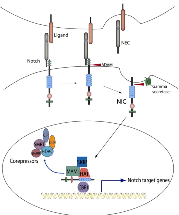
Notch is an evolutionary conserved family of single transmembrane receptors that participate in the development of multicellular organisms by regulating the following critical processes: lateral inhibition, lineage specification and boundary formation (Radtke and Raj, 2003). To date, four Notch receptor paralogs (Notch 1-4) and five Notch ligands have been identified in mammals (Delta-like-1, Delta-like-3, Delta-like-4, Jagged-1 and Jagged-2; Lai 2004). Notch is synthesized as a single precursor protein, and during post-transcriptional processing in trans-Golgi becomes cleaved by furin-like proteases into heterodimer comprising of an extracellular domain (NEC) non-covalently
associated with transmembrane domain (NTM; Fig. 2; Miele, 2006). NEC contains multiple
EGF-like repeats, whereas NTM consists of extracellular ‘Lin-Notch’ repeats, a
single-pass transmembrane domain and an intracellular domain, the latter composed of ‘RAM23’ (which participates in CBF-1/RBPJ! binding, see below), six ankyrin repeats and a polyglutamine stretch and a PEST region, possibly involved in ubiquitination (Nickoloff et al., 2003). Upon ligand binding NEC dissociates from NTM, and becomes
endocytosed along with the ligand by ligand expressing, neighboring cells. This event activates cleavage of extracellular fragment of NTM catalyzed by ADAM10 or ADAM17
metalloproteases, followed by intracellular cleavage mediated by !-secretase (aspartyl
protease; Brou et al., 2000). This second cleavage releases the intracellular fragment (NIC), which is able to translocate into the nucleus, where it binds CBF-1 and regulates
transcription of its target genes (Miele, 2006). The mammalian member of CBF1-Suppressor of Hairless-Lag1 (CSL) transcription factors CBF-1 binds to a consensus sequence cGtGGGAA and acts as a transcriptional repressor by binding to a corepressor complex that includes SMRT or N-coR, SKIP, CIR, HDAC, SHARP, CtBP and CtIP (Miele, 2006). NIC binding to the CBF-1-corepressor complex replaces SMRT and
histone deacetylase (HDAC) for transcriptional coactivators including mastermind like-1 (MAML1) and histone acetyl transferases (HAT) and results in activation of target genes (Lai, 2004). The number of genes that are directly or indirectly regulated by Notch is large and depends on the cellular context. The most common are the bHLH (basic helix-loop-helix) transcription factors families: HEY, HES (hairy/enhancer of split in Drosophila melanogaster) and HERP. Other Notch target genes include p21Cip/Waf, CyclinD1, CyclinA, transcription factors of the NF"B family, poly (ADP-ribose)
polymerase and SKP2 ubiquitin ligase which triggers degradation of p27Kip1 (Sarmento et al., 2005). Some of the Notch transcriptional targets have been shown to be also MYC targets in primary human T-cell lymphoblastic leukemia and include genes that regulate cell growth (Palomero et al., 2006); in the same system cMYC itself is a direct target of Notch (Weng et al., 2006).
! 12!
Notch signaling is involved in many developmental events that are highly context-dependent. Notch activation determines cell fate by controlling responsiveness to either proliferative or differentiation stimuli, and allows for their proper interpretation.
Depending on the conditions Notch can have differential effects on cell’s proliferation and apoptosis. Notch-1 promotes differentiation of keratinocytes (Nickoloff et al., 2002) and myeloid progenitor cells (Schroeder and Just, 2000). On the other hand, induction of Notch stimulates proliferation of mammary epithelial cells (Lee et al., 1999) and hematopoietic progenitor cells (Carlesso et al., 1999).
2.5. NOTCH AND CANCER
The oncogenic role of Notch was first identified in T-ALL (Reynolds et al., 1987) and is associated with chromosomal translocation resulting in deregulated expression of truncated form of Notch-1, which corresponds to NIC (Radtke and Raj, 2003). Other
studies showed that activating mutations of Notch-1 that stabilize NIC or facilitate the
cleavage of it are found in almost 80% of T-ALL (Mansour et al., 2007). There is an increasing evidence that aberrant Notch signaling plays a role in many cancers. Deregulated expression of Notch pathway components was demonstrated in other cancers of hematopoietic origin, like B-cell chronic lymphoid leukemia (B-CLL; Hubmann et al., 2002), as well as in Hodgkin's and large cell anaplastic lymphomas (Jundt et al., 2002). Altered expression of Notch receptors and ligands was demonstrated in many solid tumors. Truncated, constitutively active Notch-1 and Notch-4 cause mammary tumors in mice (Callahan and Raafat, 2001; Dievart et al., 1999; Gallahan and Callahan, 1997) and
! 14! high expression of Notch-1 and Jagged-1 has been associated with poor prognosis in breast cancer (Reedijk et al., 2005). Overexpression of intact Notch receptors has been documented in cervical, pancreatic, endometrial, renal, lung, colon, head and neck carcinomas (Zagouras et al., 1995; Miele et al., 2006) and melanoma (Balint et al., 2005).
A large line of evidence supports the role of Notch in contributing to tumor progression. However in certain types of cancer Notch has been shown to have a tumor suppressor function. Notch-1 induces growth arrest and promotes differentiation of keratinocytes in humans and mice (Nickoloff et al., 2002; Rangarajan et al., 2001). Overexpression of NIC inhibits proliferation of myeloid progenitor cells (Schroeder and
Just, 2000). In B cells apoptosis caused by NIC expression is mediated through HES
family of proteins, whereas growth arrest is mediated independently (Morimura et al., 2000). Activated Notch-1 expression induces cell cycle arrest in SCLC by up-regulating cyclin-dependent kinase inhibitors WAF1 (p21) and KIP1 (Sriuranpong et al., 2001). However, since the effects of Notch signaling are strictly dose- and time-dependent, experiments with over-expressed active form of Notch could lead to artifacts and should be interpreted bearing this caviat in mind. Our results in ACL cell lines reveal that the effects of Notch signaling depend on oxygen concentration (Chen et al., 2007). We found that in normoxic environment Notch-1 suppresses growth, whereas under low oxygen concentration Notch-1 promotes survival of ACL cells. Thus, when studying biological functions of Notch signaling in cancer, many variables need to be considered including dose, time and environment.
2.6. NOTCH IN MALIGNANT MESOTHELIOMA
Increasing number of reports indicate oncogenic role of Notch in solid tumors. The role of Notch signaling in MM is poorly understood. Notch-1 expression is elevated in MM biopsies as compared to stromal tissue. Notch-1 is required for the maintenance of Simian Virus 40 (SV40) transformed phenotype of HM, and inhibition of Notch-1 resulted in SV40-transformed HM cell growth arrest (Bocchetta et al. 2003). Expression of Notch-1 in those cells is regulated primarily by small tumor antigen (tag), which inactivates protein phosphatase 2A (PP2A; Testa and Giordano, 2001), resulting in an increased phosphorylation of the components of the ERK pathway. There are no reports indicating the role of Notch signaling in MM that is independent of SV40.
2.7. NOTCH IN LUNG CANCER
Over-expression of Notch-1IC caused growth arrest of SCLC cell lines
(Sriuranpong et al., 2001). Hence, this study suggested a tumor suppressive role in SCLC. Three studies proposed an oncogenic role for Notch-3 in NSCLC. Dang et al. (2000) found a chromosomal translocation t(15;19) in an aggressive lung carcinoma. Similar translocations were found in two separate thoracic malignancies case reports (Kees et al., 1991; Lee et al., 1993) including a thymic carcinoma (Kubonishi et al., 1991). The likely gene to be affected by such translocation was proposed to be Notch-3 because the NSCLC cell line HCC2429 (harboring such translocation) expressed high levels of the Notch-3 mRNA (Dang et al., 2000). Moreover, 7 out of 44 NSCLC cell lines expressed the Notch-3 mRNA as determined by Northern blot hybridization (Dang et al., 2000).
! 16! The Dang group further expanded on these early findings. The expression of a truncated Notch-3 receptor (missing the intracellular portion of the protein, and therefore considered as a Notch-3 dominant negative form) caused reduced growth of NSCLC cells and increased “growth factor dependence” (Haruki et al., 2005). In a more recent study, treatment of NSCLC cell lines with a GSI (MRK-003) reduced tumor formation in xenografts mouse models. When Notch-3 was genetically inhibited through RNA interference, the same cells became GSI insensitive, indicating that Notch-3 was the target of the GSI used (Konishi et al., 2007). These results, and their interpretation, are somewhat controversial. In Haruki et al. (2005) the authors have not explored the possibility that the truncated, dominant negative Notch-3 over-expressed in their NSCLC cells could have saturated the cell membrane with a non-functional Notch receptor, ultimately resulting in the expression of a Notch pan-inhibitor. This possibility is reinforced by the fact that the authors did not verify that in their experimental conditions activation of Notch-1, -2 and -4 was still attainable. The study of Konishi et al. (2007)is also difficult to interpret. The authors propose an oncogenic role for Notch-3 in NSCLC. However, when Notch-3 was down-regulated genetically, NSCLC cells grew and formed tumors. If Notch-3 was the main oncogene in NSCLC cells, Notch-3 siRNA should have yielded similar effects as dominant negative Notch-3 and chemical inhibition. Therefore it does not seem to be the case that the role of Notch-3 in NSCLC is yet well understood. A recent study (Zheng et al., 2007)showed that forced expression of constitutively active Notch-1 inhibited growth of the ACL line A549 cultured in standard conditions and interfered with tumor growth in vivo. We obtained analogous results in the same cell line
cultured in normoxia (Chen et al., 2007). However, experiments generated from Notch-1 over-expression are difficult to interpret and may lead to artifactual results. Dissimilar conclusions drawn by Zheng et al. (2007)were proposed by a Chinese study based on immunohistochemical analysis of NSCLC tumor biopsies compared to normal “bronchi mucosa” (Jiang et al., 2007). These authors observed strong Notch-1 immunoreactivity in NSCLC compared to normal lung, which correlated with Jagged-1 and VEGF expression. The intensity of the immunostaining appeared to increase with more advanced tumor stages. This was not observed in the study by Chen et al. (2007) performed on snap-frozen tissue samples. In conclusion, a better understanding of the role played by Notch receptors in lung cancer still awaits further study.
2.8. NOTCH AND HYPOXIA
Due to the high cell expansion and insufficient neo-vascularization, solid tumors, including MM and lung cancer, are highly hypoxic. Rapid proliferation of tumor cells requires increased glucose metabolism. The lack of oxygen shifts glucose metabolism from oxidative phosphorylation to anaerobic glycolysis, which results in accumulation of pyruvate and lactate. Tumor cells adapt to the hypoxic microenvironment by inducing the expression of genes that promote cell survival, motility and angiogenesis (Keith and Simon, 2007). Hypoxia Inducible Factor-1" (HIF-1") is the major transcription factor responsible for regulation of expression of genes under hypoxia. Genes that are directly regulated by HIF-1" include: Erythropoetin (EPO), Vascular Endothelial Growth Factor (VEGF), and Inducible Nitric Oxide Synthase (iNOS; Gordan and Simon, 2007). Under
! 18! normoxic conditions HIF-1" becomes degraded by ubiquitin mediated proteasomal pathway. Under hypoxia HIF-1" is stabilized and can promote tumor progression. In B progenitor cells forced expression of Notch leads to acute lymphoblastic leukemia (Pear and Aster, 2004). In many cell types, including neuronal progenitors, hypoxia relies on Notch to maintain the undifferentiated state. Hypoxia promotes Notch-1 stabilization and induces transcription of Notch responsive genes by recruiting HIF-1" to the target gene promoters (Gustafsson et al., 2005). These findings suggest that oxygen concentration is an important determinant of Notch function, and needs to be accounted for in studying Notch signaling pathways in MM and lung cancer.
2.9. IGF-1R PATHWAY
The insulin like growth factor-1 (IGF-1) signaling pathway is a complex system involved in regulation of processes that lead to cell growth, proliferation and development. Uncontrolled transduction of signals through IGF-1R may result in pathological conditions including transformation, metastasis and evasion of apoptosis. The IGF-1R system is composed of IGF-1 ligands (IGF-1, IGF-2), transmembrane receptors (IGF-1R, IGF-2R and insulin receptor-IR) composed of " and # subunits, and IGF binding proteins (IGFBPs) which affect bioavailability of IGFs in extracellular fluids (LeRoith and Roberts, 2003). Upon ligand binding to the extracellular domain of the receptor, conformational change of the receptors occurs, which leads to the auto-phosphorylation of the receptor’s tyrosine residues on the # subunit. This, through docking molecules such as members of the insulin receptor substrate (IRS) family
(IRS-1, IRS-2, IRS-3, IRS-4) and or Src homology 2 domain containing transforming protein (SHC), results in activation of either PI3K or mitogen activated protein kinase (MAPK) pathways. PI3K is a major downstream effector of RTKs. It is a lipid kinase, and catalyzes the phosphorylation of PIP2 to form phosphatidylinositol-3,4,5-triphosphate (PIP3). PIP3 is a second messenger at the plasma membrane where it recruits Akt-1 (serine-threonine protein kinase, protein kinase B; Cantley, 2002). This event ultimately leads to activation of downstream effectors, and expression of target genes that mediate the proliferative and anti-apoptotic effects of IGF-1 induced Akt-1 signaling (LeRoith et al., 1995). The major supressor of the Akt-1 pathway is a lipid phosphatase PTEN, which dephosphorylates PIP3 to PIP2 at the plasma membrane to prevent the recruitment of Akt-1, and progression of the proliferative signal.
! "#!
CHAPTER3
THESV40LARGETANTIGENREGULATESIGF-1EXPRESSIONIN
TRANSFORMEDMESOTHELIALCELLS
ABSTRACT
During SV40 mediated malignant transformation of human mesothelial cells (HM), oncogenic viral antigen Tag promotes cell proliferation by interfering with pro-survival and tumor suppressive pathways. Tag is found in complex with p53, and the current view is that those complexes are the way to sequester and inactivate cellular p53. On the other front, Tag promotes cell survival by inducing insulin like growth factor-1 (IGF-1) signaling which leads to cell proliferation. Using primary human cells and SV40-transformed human cells, we show that in addition to inactivating p53 tumor suppressor activities, the Tag-p53 complex has growth stimulatory activities that are required for malignant cell growth. We found that in human cells, Tag/p53 complexes regulate
transcription of the IGF1 gene by binding to the IGF1 promoter together with pRb and
p300. Depletion of p53 leads to structural rearrangement of this multi-protein complex, resulting in IGF1 promoter transcriptional repression and growth arrest. Our data provide a novel mechanistic and biological interpretation of the p53/Tags complexes and of DNA tumor virus transformation in general. In the model we propose, p53 is not a passive
inactive partner of Tag. Instead thep53/Tag complex promotes malignant cell growth through its ability to activate the IGF-1 signaling pathway.
INTRODUCTION
IGF-1 pathway plays a critical role in regulating normal cell growth and transformation events. IGF1R-/- mice are less than half the size of normal counterparts at birth and are viable for only a few hours (Liu et al., 1993). Mice lacking insulin receptor substrate-1 (IRS-1) are severely growth impaired, although being viable and reaching fertility (Araki et al., 1994; Tamemoto et al., 1994).
In malignant setting IGF-1R is frequently over-expressed, and it functions as a potent anti-apoptotic agent supporting cell survival of neuronal, hematopoietic and fibroblast cells, to the point that IGF-1R signaling is commonly referred to as “cell survival factor” (Gluckman et al., 1992; Rodriguez-Tarduchy et al., 1992; Harrington et al., 1994). IGF-1R is absolutely required for the successful transformation of a number of cell lines (reviewed in Baserga, 1995). Furthermore, fibroblasts obtained from IGF1R
-/-mice are virtually resistant to cell transformation operated by a variety of chemical carcinogens, as well as viral and cellular oncogenes (Sell et al., 1994). SV40 mediated cell proliferation requires the interaction of IGF-1 with its receptor (Porcu et al., 1992). Intact IGF-1R is required for SV40 mediated transformation of primary HM (Sell et al., 1993). Tag can promote cell cycle progression in BALB/c 3T3 cells cultured in low serum only when IGF-1/IGF-1R pathway is active (Valentinis et al., 1994).
! 22! During malignant transformation DNA viral oncoproteins can bind and inactivate p53 tumor suppressor functions (Tag of human polyomaviruses JCV, BKV and SV40, the E1b of adenoviruses; Ali and DeCaprio, 2001). This allows infected cells to survive, and prevent p53-mediated apoptosis or cell growth arrest. Inactivation of p53 impairs DNA repair thus favoring the early steps of carcinogenesis. The latter effect is mediated mostly by the ability of p53 to induce p21 expression, a CDK inhibitor that in turn causes cell cycle arrest, allowing DNA repair to take place. By inducing p21 expression, p53 prevents cells that have accumulated genetic damage from undergoing mitosis and propagating the damaged DNA to the progeny. Due to its critical role in regulating the cell cycle, and DNA damage, which results in proper cell growth, cancer cells need to find ways to inactivate wild-type p53. Accordingly, inactivation of p53 must occur to transform primary cells in vitro (Hahn et al., 1999). Oncogenic viruses other than SV40 have developed unique mechanisms for inactivating p53; HPV16 oncoprotein E6, binds to cellular p53 promoting its ubiquitylation and degradation (Wernes et al., 1990; Scheffner et al., 1990).
The current hypothesis is that, upon Tag binding, p53 loses its ability to work as a transcription factor and as a tumor suppressor gene (Bargonetti et al., 1992; Jiang et al., 1993; Segawa et al., 1993). A few studies have shown, however, that Tag in complex with p53 does not prevent p53 from binding to its DNA binding sites (Long et al., 1995; Sheppard et al., 1999; Technau et al., 2001). Moreover, p53 complexed with Tag was able to stimulate transcription of a p53-regulated promoter in cell free extracts from monkey and human cells (Sheppard et al., 1999). SV40-mediated transformation of
fibroblasts was enhanced by wild type mouse p53 (Michalovitz et al., 1986). Similarly, transformation of rat fibroblasts required both Tag and stabilized p53 (Deppert et al., 1989). Animal studies have shown that SV40 is more efficient in promoting tumor growth in the presence of wild type p53 (Herzig et al., 1999). These data did not fit with the generally accepted view that the major role of DNA viral Tags is to bind to and inhibit cellular p53.
Primary HM have high levels of p53, and in such environment SV40 requires IGF-1R signaling for cellular transformation. Here we investigated the possible biological effects of Tag-p53 complexes on IGF-1/IGF-1R pathway mediated cell growth and transformation.
! 24! MATERIALS AND METHODS
CELLS AND GENE TRANSFER PROCEDURES
Primary human mesothelial cell (HM) cultures were obtained from non-cancerous donors, cultured and characterized as described in Bocchetta et al. 2000. These cells contain wild-type p53 (Bocchetta et al., 2000). SV40-transformed HMs (S-HML) were obtained through HM infection with SV40 virions (10 plaque forming units –pfu/cell). Six to eight weeks after infection tridimensional foci of transformed cells were hand picked and cultured into cell lines. The latter were grown in DMEM supplemented with 5% fetal bovine serum (FBS). Critical results were confirmed in three independent S-HMLs.
Retroviral packaging was performed with Phoenix cells using standard procedures. S-HML transduced with the retrovirus expressing the tetracycline regulator (TR; TET-ON system, Clontech Laboratories Inc., Mountain View, CA) were selected using 600 µg/ml of G418. After selection, the functionality of the system was assayed as recommended by the manufacturer. These cells were transduced with the retrovirus expressing HPV16 E6 (in the presence of doxycycline). Transduced cells were then selected with 600µg/ml of hygromycin and the resulting clone was named S-HML/E6.
Western blot and immunostaining experiments were performed as described in Bocchetta et al. 2000.
DNA SYNTHESIS ANALYSES
Detection of proliferating cells by the measurement of the DNA replication was performed using the 5-bromo-2'-deoxyuridine (BrdU) Flow kit (BD PharMingen, Franklin lakes, NJ) according to the manufacturer’sinstructions. Briefly, 1 x 10⁶ cells
were stimulated withIGF-1 (5 nM) in culture medium. A total of 100 $l of BrdU solution (1 mM BrdU in PBS) was added to each dish containing 10 ml culture medium and was incubated for 8 hours at 37°C. The cells were then fixed and permeabilized with Cytofix/Cytoperm buffer (BD PharMingen). To prevent cells form clumping (which would interfere with FACS analysis) the cellular DNAwas digested with DNAse for 1 h at 37°C. The cells were stained with an anti-BrdU FITC-conjugated antibody and analyzed by FACSCanto Flow Cytometer (BD Bioscience, San Jose, CA). Cells that stained positively for BrdU indicated actively proliferating ones.
Apoptosis was assayed using Annexin V/7-aminoactinomycin D staining according to standard procedures.
RNA STUDIES
Nuclear run-on assays were performed as described in Bocchetta et al. 2003. Each slot of alkali-denatured probes contained 5 µg of probe DNA, which corresponded to: a 133 bp PCR fragment of the human 18S rRNA gene; a 654 bp PCR fragment of the human p21 cDNA; a 378 bp PCR fragment of the human Bax cDNA; a 593 bp PCR fragment of the human IGF1 cDNA; # phage DNA digested with Hind III. After
! 26! hybridization to 32P-labelled nuclear transcripts, membranes were washed and exposed
to both X-ray film and a phosphoimager.
RT-Real time PCR was performed using standard protocols. Briefly, cells were dissociated with trypsin/EDTA, harvested, and snap frozen. Total cellular mRNA was obtained using the RNeasy kit (Qiagen, Valencia, CA) in the presence of RNAse-free DNAse I. The concentration of RNA in each sample was measured using spectrophotometer (GE Healthcare, Uppsala, Sweden), and the quality of the mRNA was assayed in 1% formaldehyde agarose gels. Two µg of total RNA from each sample was reverse transcribed using a first strand synthesis kit (MBI-Fermentas, Hanover, MD) in the presence of 10 pmol of random primers. Real Time PCR was performed as follows: 1/5 of the reverse transcription reaction from each sample was diluted serially in H2O to
determine the optimal range of dilution for the samples (CT between 15 and 25), using a
Gene Amp 5700 (PE-Applied Biosystems, Wellesley, MA). Oligo pair combinations (see Table 2) were chosen using the Primer Express 1.0 software (Applied Biosystems). Reactions were performed using the SYBR GREEN PCR Master Mix (Applied Biosystems). After estimating the sample with higher levels of GAPDH mRNA, 1:2 serial dilutions were made in H2O (range 1 to 128) to construct a calibration curve for each
mRNA. Similar calibration curves were run along with each experiment. “No reverse transcription” of each sample was run as the negative control. mRNA values were normalized for GAPDH amounts, and plotted as a percentage of the control sample.
Reporter assays were performed using the Dual-Glow Luciferase Assay system (Promega, San Luis Obispo, CA) and measured with a luminometer (Veritas; Turner Biosystems, Sunnyvale, CA).
CHROMATIN IMMUNOPRECIPITATION ASSAYS
Crosslinking of 1 x 10⁷ cells was performed for 10 mins at a room temperature
using 1% formaldehyde in PBS. The reactionwas stopped, by adding 0.25M glycine in PBS. The cells were harvested and the DNA was sheared by sonication in lysis buffer (20 mM Tris-HCl pH 7.4, 150 mM NaCl, 1% NP-40) supplemented with 1% SDS, using a Branson SONIFIER 250 (Wolf Laboratories Lmd, UK) togenerate DNA fragments $600
bp. Cell lysates were diluted ten times with lysis buffer to reduce the SDS final concentration to 0.1%. Lysates were precleared with protein A/G agarose beads (Pierce, Rockford, IL) for 4 hr at 4°C, and were then incubated overnight at 4°C with the respective primaryantibodies. Immunecomplexes were collected on Protein A/G agarose beads overnight at 4°C. Samples were dialyzed twice against dialysis buffer (2 mM EDTA pH 8.0, 50 mM Tris-HCl pH 8.0). Following 4 washes in 1 ml wash buffer (100 mM Tris-HCl pH 8.0, 500 mM LiCl, 1% NP-40); immune complexes were elutedfrom the beads by vigorously shaking twice for 15 minutes in 150 µl of 50mM NaHCO3, 1%
SDS. Cross-linkingwas reversed by incubation in 0.15 M NaCl overnight at 65°C.Each sample’s DNA was ethanol precipitated and purified using QIAprep spin miniprep kit (Qiagen).DNA was resuspended in 100 µl of TE buffer. Finally, each sample was PCR
! 28! amplified using appropriate oligonucleotides as primers (Table 2), and the results were visualized on 2% agarose gels.
ANTIBODIES
Target Type Company
HPV16 E6 Ab-1 mouse monoclonal EMD Bioscience anti Xpress mouse monoclonal Invitrogen IGF-1R 2C8 mouse monoclonal Santa Cruz Biotech
IGF-1 H-70 rabbit polyclonal Santa Cruz Biotech p300 H-272 rabbit polyclonal Santa Cruz Biotech p300 D-12 mouse monoclonal Santa Cruz Biotech Tag pAb419 mouse monoclonal Santa Cruz Biotech p53 DO-1 mouse monoclonal Santa Cruz Biotech pRb IF-8 mouse monoclonal Santa Cruz Biotech mdm2 D-12 mouse monoclonal Santa Cruz Biotech P21 187 mouse monoclonal Santa Cruz Biotech GAPDH MAB374 mouse monoclonal Chemicon Table 1. A list of all antibodies used in Chapter 3
Annexin V PE-conjugated was from BD Biosciences (Franklin Lakes, NJ). Agarose-conjugated protein A/G was from Pierce (Rockford, IL).
PLASMIDS
HPV16 E6 was PCR amplified from pE6E7 (Dr Martin W. Kast, University of Southern California, CA) and ligated into different plasmids. The constructs used here were the following: pE6CDNA4, in which E6 was cloned into the Kpn I/Not I sites of pcDNA4/HisMax (Invitrogen); pREVE6, in which the recombinant E6 obtained after the construction of pE6cDNA4 was PCR amplified and cloned into the Hind III/Cla I sites of pREV-TRE (Clontech Laboratories, Mountain View, CA). In retroviral transduction experiments the Tet-On Gene Expression System (Clontech) was used. It included pTet-ON, encoding the tetracycline transactivator, pTRE-d2EGFP, used to validate the functionality of the TET-ON clones, and pREV-TRE, which expresses proteins under the control of a CMV minimal promoter that contains four tetracycline responsive elements. All vectors can be used as ordinary plasmids, or they can be packaged in retroviral particles. pSmaBgl-LUC was provided by Dr Renato Baserga (Kimmel Cancer Center, PA). pMDM2 was provided by Dr Zhuo Zhang (University of Alabama, AL). pGRI5, expressing wild type human IGF1R under the control of a CMV promoter was a gift from Dr Clodia Osipo (Loyola University Chicago, IL). p53mt135, expressing dominant negative p53 was purchased from BD Biosciences. Oligonucleotides 5’GATCCCGCAATGGTTCACTGAAGACCCAGGTCCAGAGAAGCTTTCTGGACC TGGGTCTTCAGTGAACCATTGTTTTT-3’and 5’CTAGAAAAACAATGGTTCACT GAAGACCCAGGTCCAGAAAGCTTCTCTGGACCTGGGTCTTCAGTGAACCATT GCGG-3’ were annealed in 20 mM Tris-HCl pH 7.4, 1 mM EDTA pH 8.0, 100 mM NaCl by incubating 2 nmoles of each primer at 100°C for 5’. The reaction was then left
! 30! to slowly cool down to room temperature in a heat block. Annealing was verified by running the products before and after annealing onto a 10%, non-denaturing polyacrylamide gel. The annealed products were ligated into the Bam HI and Xba I sites of pGE-1 (Gene Eraser, Stratagene, La Jolla, CA) to give rise to plasmid shp53.
Table 2. A list of oligonucleotides used in Chapter 3 Gene Primer Sequence
P21-A 5’-CCTGCCGAAGTCAGTTCCTTG- 3’ P21-B 5’- GCGGCAGACCAGCATGACAG -3’ P21-C 5’- AAACTGAGACTAAGGCAGAAGATGTAGAGC -3’ Bax-A 5’- ATGGACGGGTCCGGGGAGC -3’ Bax-B 5’- GCAGAGGATGATTGCCGCCG -3’ Bax-C 5’- GCACAGGGCCTTGAGCACCA -3’ GAPDH-F 5’- CAATGACCCCTTCATTGACC -3’ GAPDH-R 5’- TTGATTTTGGAGGGATCTCG -3’ IGF-1 A 5’- CTTCTGTTTGCTAAATCTCACTGTC -3’ IGF-1 B 5’- TTTTATTTCAACAAGCCCACAGGG -3’ IGF-1 C 5’-GGGCTGATACTTCTGGGTCTTGG-3’ MDM2-F 5’- GATCCTGGAAGTGTCCCTGA-3’ MDM2-R 5’- AAGGACCGTTCTGTTTGTGG-3’ 18S F 5’-TGATTAAGTCCCTGCCCTTTGT-3’ 18S R 5’-TCAAGTTCGACCGTCTTCTCAG-3’
p21: Primers A and C were used to amplify a 654 bp fragment used as probe in nuclear run-on experiments, while primers B and C were used in RT-Real time experiments (positions 504-654 of the p21 cDNA).
Bax: Primers A and C were used to amplify a 378 bp fragment used as probe in nuclear run-on experiments, while primers B and C were used in RT-Real time experiments (positions 228-378 of the Bax cDNA).
IGF-1: Primers A and C were used to amplify a 593 bp fragment used as the probe in nuclear run-on experiments, while primers B and C were used in RT-Real time experiments (positions 393-593 of the IGF1 cDNA).
! 32! OLIGONUCLEOTIDES USED IN CHIP ASSAYS
Table 3. A list of oligonucleotides used for ChIP experiments on the IGF-1 promoter
Region Primer sequence Position A-F 5’-GGTACCCCAAAGCCTCTCATG-3’ -1952 to -1700 A-R 5’- CCCAACTACAACATCCCTAGG-3’ -1952 to -1700 B-F 5’-GCCCCTGAAGGGACTTGACC-3’ -1764 to -1414 B-R 5’-GACTCTCAGGGGACTGACAC-3’ -1764 to -1414 C-F 5’-CCAGAGTAGGATTTCAAGCAG-3’ -1472 to -1078 C-R 5’-CTGGCTAGCAATACCCTCTTG-3’ -1472 to -1078 D-F 5’-AGAACCGTGAATTCTCAATGGC-3’ -1215 to -921 D-R 5’-GCAAACAATTTTCCTGTTGTTTG-3’ -1215 to -921 E-F 5’-CTGGCACACAGACTCCCTCTG-3’ -1031 to -659 E-R 5’-GGAAGACAGCACTCGGGTGAC-3’ -1031 to -659 F-F 5’-ACCAATCCAATGCTGCCTGCC-3’ -757 to -464 F-R 5’-TTTCTGCTGGGCATGAAGACAC-3’ -757 to -464 G-F 5’-TAGAATCTAAAATTGCTCTC-3’ -555 to -322 G-R 5’-AAATAACATCATACCTTTGC-3’ -555 to -322 cont-F 5’-CAGTCTTCTGTGTCCTGTTC-3’ N/A cont-R 5’-AGAGGCTGATGGGAAGGAAC-3’ N/A
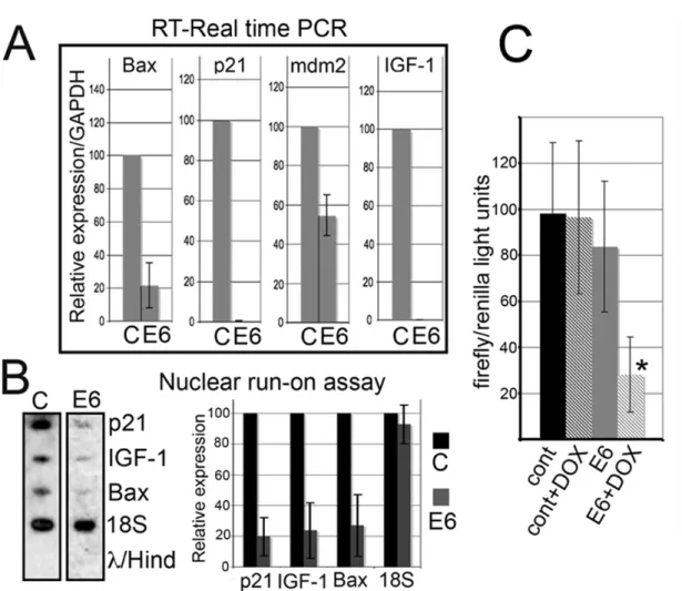
IGF1 was artificially downregulated using Hs_IGF1_5_HP Validated siRNA (Qiagen). The negative control for ChIP assays was a 266 bp region (part of BAC RP11-25I15, locus AC089982, a sequence far from known genes located on chromosome 12q12, while the IGF1 gene is located on chromosome 12q22).
RESULTS
S-HML/E6 CELLS EXPRESS FUNCTIONALLY ACTIVE RECOMBINANT HPV16 E6
S-HM are characterized by high levels of endogenous p53. We wanted to study the effects of lowering p53 in cells expressing the SV40 Tag. To achieve this goal, we expressed HPV16 E6, an oncoprotein which binds and targets p53 for ubiquitin mediated degradation (Wernes et al., 1990; Scheffner et al., 1990) in S-HML. We used a stable, tetracycline inducible transduced cell clone expressing a fusion protein consisting of 6 histidines at its N-terminal portion (for conjugation to Ni-charged carriers), an Xpress peptide flag and the full length HPV16 E6. These cells (named S-HML/E6) express the SV40 Tag and upon doxycycline treatment also expressed HPV16 E6 that bound and degraded p53 (Fig. 3 and Fig. 4).
! 34!
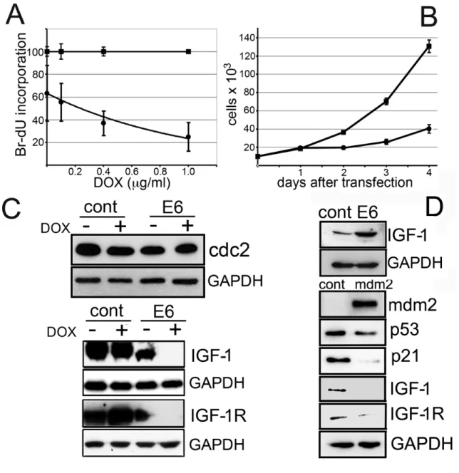
Figure 3. E6 induction down-regulates p53 and p53 regulated gene products. (A) Expression of Tag and p53 in S-HML expressing E6 (which degrades p53, Fig.4). (B) Reduced p53 expression is paralleled by decreased p21 and mdm2 expression. Cells were treated with or without doxycycline for 48 hr to induce E6 expression. E6: S-HML/E6 cells. CONT: control cells. (C) Transient transfection of S-HML with pE6CDNA4 causes marked p53 downregulation. C24: control 24 hr after transfection; E624: E6 24 hr after transfection; C48: control 48 hr after transfection; E648: E6 48 hr after transfection.
DECREASED AMOUNTS OF P53 IN S-HML/E6 CORRESPOND WITH DECREASED EXPRESSION OF PROTEINS TRANSCRIPTIONALLY REGULATED BY P53 AND CAUSE CELL GROWTH ARREST
We measured p53 and Tag expression levels at different time-points after doxycycline induction in S-HML/E6 cells. 48 hr after doxycycline-mediated induction of HPV16 E6, S-HML/E6 had reduced protein expression of p53, and is followed by the reduction in expression of its targets p21 and mdm2 (Gudkov and Komarova, 2003). Tag expression was not influenced by doxycycline treatment (Fig. 3A, B). We hypothesized that these effects could have caused apoptosis/mitotic catastrophe, or a proliferative advantage. Instead, Annexin V/ propidium iodide (PI) staining followed by FACS analysis did not show evidence of apoptosis or the appearance of aberrant DNA peaks (an indication of mitotic catastrophe), and S-HML/E6 showed a doxycycline, dose-dependent reduction in DNA synthesis as compared to controls (Fig. 4A). Since doxycycline has pleiotropic effects that might have influenced these findings, we expressed recombinant E6 protein in transiently transfected S-HML. We obtained identical results: E6-transfected S-HML had undetectable p53, and were growth arrested as compared to controls (Fig. 3C and Fig. 4B). The reciprocal experiments (growth curves for the inducible system and DNA incorporation assay for S-HML transiently transfected with E6) gave identical results (Bocchetta et al., 2008). To understand why depletion of cellular p53 in S-HML resulted in growth arrest, we investigated genes that are transactivated by Tag (Chen et al., 1996, Porcu et al., 1994). We found no variation in
! 36! cdc2 protein expression after doxycycline treatment of S-HM/E6, while the same treatment virtually abolished IGF-1 precursor and IGF-1R expression (Fig. 4C).
Figure 4. E6 induction in S-HML suppresses DNA synthesis and causes cell growth arrest. Critical components of the IGF-1 signaling pathway are downregulated upon either E6 or mdm2 induction. (A) Bromodeoxyuridine (BrdU) incorporation assay conducted on control cells (squares) and SV40 transformed human mesothelial cells
transfected with plasmid expressing E6 (S-HML/E6; bullets) performed at increasing doxycycline concentrations. The histogram is the average of 4 independent experiments; each measurement was determined on 30,000 events. The percentages of DNA incorporation of ES-HML/E6 versus control cells were (at concentrations of doxycycline in µg/ml): 0, 62.78 ± 24.40%; 0.1, 55.00 ± 16.67%; 0.4, 37.22 ± 11.11%; 1.0, 22.44 ± 13.33%. The apparent lowered DNA incorporation measured in S-HML/E6 versus control cells at 0 µg/ml of doxycycline may be explained by “leaky” transcription at the “tet-on” retroviral promoter. (B) Cell growth curves of S-HML transfected with a control plasmid (pcDNA4/His-Max; squares) and with the same plasmid expressing recombinant E6 (bullets). The histogram is the average of three independent experiments. Using a plasmid expressing green fluorescent protein followed by FACS analysis we determined that the efficiency of transfection was > 95% (using electroporation). (C) Western blot analysis performed in S-HML/E6 and control cells in the presence and absence of doxycycline. Representative experiment. Western blot was performed 48 hr after antibiotic exposure (+ lanes). E6: S-HML/E6. (D; Top) Western blot analysis performed on HM transfected with the control plasmid (“cont” lane) or with the plasmid expressing recombinant E6 (“E6” lane). (D; Bottom) Western blot analysis conducted on S-HML transfected with a control plasmid (“cont” lanes) and with a plasmid expressing human mdm2 (mdm2 lanes). Note mdm2 expression and downregulation of p53, p21, IGF-1 and IGF-1R.
The effects observed upon E6-transfection appeared to be dependent on the presence of Tag, because E6-transfection of primary HM (which do not contain SV40) caused the opposite effect: a 4.3 fold increase of IGF-1 expression (Fig. 4D; Top). Since E6 did not influence Tag expression but influenced p53 expression in S-HML (Fig. 3A) we speculated that the E6 activities in Tag containing cells were mediated through the degradation of p53. To test our hypothesis that the decreased expression of p21, mdm2, IGF-1 and IGF-1R upon E6 induction was related to p53 down-regulation, we deregulated p53 in S-HML using a shRNA against p53, a dominant negative p53 (p53mt135, which interferes with proper p53 complex formation; Fig 5A and 5B, respectively) or over-expressing mdm2 in S-HML (Fig. 4D; Bottom): all these
! 38! approaches yielded reproducible p21, IGF-1 and IGF-1R down-regulation. The most effective and reproducible way to down-regulate p53 expression in S-HML was expressing HPV16 E6 in these cells (Fig. 3C). Combined together, these results suggested that the decreased expression of p21, IGF-1 and IGF-1R was related to p53 depletion independently of how it was achieved.
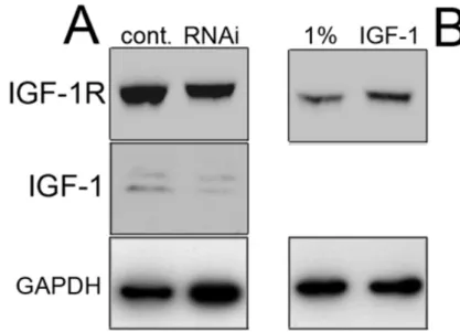
Figure 5. Both p53 down-regulation through RNAi and transfection of S-HML with dominant negative p53 cause reduced expression of p21, mdm2, IGF-1 and IGF-1R. (A) Western blot analysis of the indicated gene products after transfection of S-HML with the plasmid encoding shp53. (B) Western blot analysis of the indicated gene products after transfection of S-HML with the plasmid encoding dominant negative p53. !
E6-MEDIATED P53 DOWN-REGULATION CAUSES S-HML CELL GROWTH ARREST THROUGH THE IGF-1/IGF-1R SIGNALING PATHWAY
To confirm that p53 depletion in S-HML causes cell growth arrest through IGF-1/IGF-1R signaling, we designed the experiment summarized in Fig. 6A.
Figure 6. E6-mediated inhibition of DNA synthesis in S-HML is mediated through the IGF-1/IGF-1R pathway. 1%, cells grown in 1% FBS; IGF-1, cells grown in 1% FBS plus IGF-1. (A) Schematic of the experiment performed to verify the hypothesis that E6-mediated impairment of DNA synthesis in S-HML is exerted through the IGF-1 signaling pathway. (B) BrdU/FACS analysis performed on 1Cc cells grown in 1% FBS containing medium (red graph) or grown in medium supplemented with 1% FBS and 5 nM IGF-1 (blue graph). Note that IGF-1 can resume DNA synthesis in these cells. (C) Same as in (B) performed on 1C6 cells. Note that these cells are unable to respond to IGF-1 stimulation. (D) Same as in (B) performed on 1R6 cells. Note that forced expression of the IGF-1R resumes the ability of S-HML to respond to IGF-1 stimulation.