80 Journal of Dental Sciences and Research
Pyogenic granuloma –A case report
Dr. Sanjay Venugopal1, Dr. Shobha K.S2, Dr. Netravathi T.D3 1 Associate Professor , 2 Assistant Professor , 3 P.G.Student Sri Siddhartha Dental College and Hospital, Tumkur
Journal of Dental Sciences & Research 1:1: Pages 80-85
Abstract:
Pyogenic granuloma (also known as a "Granuloma gravidarum," and "Pregnancy tumor") is a primarily oral disease which appears in the mouth as an overgrowth of tissue to due irritation, physical trauma or hormonal factors.
Pyogenic granuloma was first originally described in 1897 by two French surgeons, Poncet and Dor, who named this lesion otyomycosis hominis. The name for pyogenic granuloma is misleading because it is not a true granuloma. In actuality, it is a capillary
hemangioma of lobular subtype which is the reason they are often quite prone to
bleeding. The growth is typically seen in young adults, it may occur in any age,
especially in individuals with poor oral hygiene. Females are far more susceptible than males because of the hormonal changes that occur in women during puberty, pregnancy, and menopause.
The purpose of this article is to describe a case of pyogoenic granuloma.
Keywords: pyogenic granuloma, gingiva, trauma,
Introduction
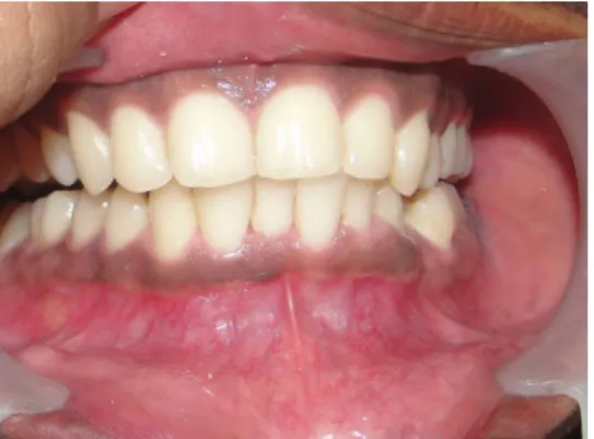
A male patient aged 24 years was referred to the department of periodontics, Sri Siddhartha dental collage, Tumkur, with a complaint of
growth of gum in the lower front teeth since 2 years. Soft tissue growth was small in size and was stable for 1½ years, but it started increasing in size slowly since past 6 months.Hegave history of using a pin to clean the affected area. The patient had not taken any treatment
81 Journal of Dental Sciences and Research for the same and had no relevant medical
history.
The patient brushed his teeth once daily using a toothpaste and brush using a horizontal stroke and consumed a mixed diet.
Clinical examination
On Intraoral examination, a growth was seen in the gingiva in relation to
mandibular right central incisor which was small in size, but has now gradually started to increase in size since past 6 months and is painless.
The growth was 0.7 × 0.8 cms in size, roughly oval in shape, reddish, ulcerated, soft and was associated with bleeding on probing. The growth covered
approximately 2/3 of the crown. The oral hygiene status was fair and width of attached gingiva was adequate. The overjet was decreased and overbite was increased.
Intra oral periapical radiograph revealed mild marginal bone loss.
Blood examination revealed normal values.
The treatment comprised of oral
prophylaxis and surgical excision of the
growth by gingivectomy procedure under local anesthesia.
Histopathological
examination
Histopathological examination of the growth revealed Stratified squamous orthokeratinized epithelium covering cellular connective tissue. The epithelium shows area of ulceration below which can be seen inflammation in the connective tissue. the connective tissue shows proliferating fibroblasts and collagen fibres interposed in which can be seen lot of epithelial lined spaces with in the connective tissue can be seen patchy distribution of lyphocytes and plasma cells. There was no evidence of atypia or malignancy. The clinical and histopathological findings confirmed it to be a case of pyogenic granuloma.
Discussion
The pyogenic granuloma is a relatively common, tumor like, exuberant tissue response to localized irritation or trauma. The name pyogenic granuloma is a misnomer since the condition is not associated with pus and does not
82 Journal of Dental Sciences and Research represent a granuloma histologically. It
is a reactive inflammatory process filled with proliferating vascular channels, immature fibroblastic connective tissue, and scattered inflammatory cells. The surface usually is ulcerated, and the lesion exhibits a lobular architecture.
The pyogenic granuloma most frequently develops on the buccal gingiva in the interproximal tissue between teeth. Three quarters of all oral pyogenic granulomas occur on the gingiva, with the lips, tongue (especially the dorsal surface), and buccal mucosa also affected. A history of trauma is common in extragingival sites, whereas most lesions of the gingiva are a
response to irritation. Individuals with poor oral hygiene and chronic oral irritants (eg, overhanging restorations, calculus) most frequently are affected.
Females are far more susceptible than males because of the hormonal changes that occur in women during puberty, pregnancy, and menopause. The pyogenic granuloma has been called a "pregnancy tumor" and does occur in 1% of pregnant women. When possible, wait until after delivery to remove the lesion in pregnant women because of a greater
tendency for recurrence during pregnancy. In a number of cases,
mastication on the lesion causes
bleeding and pain and requires surgical intervention before parturition.Some pyogenic granulomas regress after childbirth without surgical intervention Pyogenic granulomas occur at any age, but they most frequently affect young adults
Early lesions bleed easily due to extreme vascularity.
Pyogenic granulomas can have a rapid growth pattern that can cause alarm. If left alone, a number of pyogenic granulomas undergo fibrous maturation and resemble and/or become fibromas. The typical lesion involves the
interproximal gingiva and increases in size to cover a portion of the adjacent teeth.
The maxillary gingiva (especially in the anterior region) is involved more frequently than the mandibular gingiva; the facial gingiva is involved more than the lingual gingiva.
A number of lesions affect both the facial and lingual gingivae.
Pyogenic granulomas usually present as smooth or lobulated red-to-purple
83 Journal of Dental Sciences and Research masses that may be either pedunculated
or sessile.
As lesions mature, the vascularity decreases and the clinical appearance is more collagenous and pink.
Pyogenic granulomas vary in size from a few millimeters to several centimeters and are painless.
These tumors are soft to palpation A history of trauma is common in extragingival sites, whereas most lesions of the gingiva are a response to irritation Individuals with poor oral hygiene and chronic oral irritants (eg, overhanging restorations, calculus) most frequently are affected.
Histologic examination reveals sectioned soft tissue consisting of a lesion
composed of ulcerated mucosa covering a core of cellular fibrous connective tissue admixed with proliferating vascular channels and a mixed
inflammatory infiltrate. This lesion is a reactive/inflammatory process
The treatment of choice is conservative surgical excision.
For gingival lesions, excising the lesion down to the periosteum and scaling adjacent teeth to remove any calculus and plaque that may be a source of
continuing irritation is recommended. Pyogenic granuloma occasionally recurs, and a reexcision is necessary. The
recurrence rate is higher for pyogenic granulomas removed during pregnancy The only outpatient care is observation of the surgical healing 1 week after removal.
Prevention consists of routine dental cleanings and home care, especially during pregnancy.
No complications are anticipated with removal of this lesion other than the chance of a cosmetic gingival defect. The prognosis is excellent, and the lesion usually does not recur unless inadequately removed.
Lesions removed during pregnancy may have a higher recurrence rate.
Focus patient education on better oral hygiene, and consider recommending pulsating mechanical toothbrushes with timers. These toothbrushes encourage better hygiene.
84 Journal of Dental Sciences and Research
Conclusion
The pyogenic granuloma most frequently develops on the buccal gingiva in the interproximal tissue between teeth. A history of trauma is common in extragingival sites, whereas most lesions of the gingiva are a
response to irritation. Individuals with poor oral hygiene and chronic oral irritants (eg, overhanging restorations, calculus) most frequently are affected.
In the present case as there was gradual increase in the size of the growth covering almost 2/3of the crown resulting in esthetic concern to the patient. Patient was also unable to keep the area clean andwas using pin to keep the affected area clean resulting in trauma to the growth and aggravating the condition further. A gingivectomy procedure was performed and the growth was excised and send for biopsy. Both the clinical and histopathological findings showed it to be a case of pyogenic granuloma.The case was followed up for six months and growth did not recur.
References
1. Sills ES, Zegarelli DJ, Hoschander MM, Strider WE. Clinical diagnosis and management of hormonally responsive oral pregnancy tumor (pyogenic
granuloma). J Reprod Med. Jul 1996;41(7):467-70.
2. Vilmann A, Vilmann P, Vilmann H. Pyogenic
granuloma: evaluation of oral conditions. Br J Oral
Maxillofac
Surg. Oct 1986;24(5):376-82.
3. Bhaskar SN, Jacoway JR. Pyogenic granuloma--clinical features, incidence, histology, and result of treatment: report of 242 cases. J Oral
Surg. Sep 1966;24(5):391-8. ].
4. Shafer WG.“benign and malignant tumors of the oral cavity”chapter 2,textbook of oral pathology,shafer,hine and levy, 4th edition,USA, W.B. Saunders 1983;89-95
85 Journal of Dental Sciences and Research 5. Zain RB, Fei YJ. “Fibrous
lesion of the gingiva:a histopathologic analysis of
204 cases”.oral surg oral med oral pathol 1990;70:466-470.
Figure 1: Pre-Operative View
Figure 2: After Excision
Figure 3: Excised Tissue
Figure 4: 3-Month Post-Operative View
Figure 5: Histological view