ISSN: 1524-4539
Copyright © 2010 American Heart Association. All rights reserved. Print ISSN: 0009-7322. Online
72514
Circulation is published by the American Heart Association. 7272 Greenville Avenue, Dallas, TX
DOI: 10.1161/CIRCULATIONAHA.110.971101
2010;122;S876-S908
Circulation
der Jagt and Arno L. Zaritsky
Lester T. Proctor, Faiqa A. Qureshi, Kennith Sartorelli, Alexis Topjian, Elise W. van
Fink, Eugene B. Freid, Robert W. Hickey, Bradley S. Marino, Vinay M. Nadkarni,
Mary Fran Hazinski, Dianne L. Atkins, Marc D. Berg, Allan R. de Caen, Ericka L.
Monica E. Kleinman, Leon Chameides, Stephen M. Schexnayder, Ricardo A. Samson,
Care
http://circ.ahajournals.org/cgi/content/full/122/18_suppl_3/S876
located on the World Wide Web at:
The online version of this article, along with updated information and services, is
http://www.lww.com/reprints
Reprints: Information about reprints can be found online at
[email protected]
410-528-8550. E-mail:
Fax:
Kluwer Health, 351 West Camden Street, Baltimore, MD 21202-2436. Phone: 410-528-4050.
Permissions: Permissions & Rights Desk, Lippincott Williams & Wilkins, a division of Wolters
http://circ.ahajournals.org/subscriptions/
2010 American Heart Association Guidelines for Cardiopulmonary
Resuscitation and Emergency Cardiovascular Care
Monica E. Kleinman, Chair; Leon Chameides; Stephen M. Schexnayder; Ricardo A. Samson;
Mary Fran Hazinski; Dianne L. Atkins; Marc D. Berg; Allan R. de Caen; Ericka L. Fink;
Eugene B. Freid; Robert W. Hickey; Bradley S. Marino; Vinay M. Nadkarni; Lester T. Proctor;
Faiqa A. Qureshi; Kennith Sartorelli; Alexis Topjian; Elise W. van der Jagt; Arno L. Zaritsky
I
n contrast to adults, cardiac arrest in infants and children
does not usually result from a primary cardiac cause. More
often it is the terminal result of progressive respiratory failure
or shock, also called an asphyxial arrest. Asphyxia begins
with a variable period of systemic hypoxemia, hypercapnea,
and acidosis, progresses to bradycardia and hypotension, and
culminates with cardiac arrest.
1Another mechanism of cardiac arrest, ventricular
fibrilla-tion (VF) or pulseless ventricular tachycardia (VT), is the
initial cardiac rhythm in approximately 5% to 15% of
pediatric in-hospital and out-of-hospital cardiac arrests;
2–9it
is reported in up to 27% of pediatric in-hospital arrests at
some point during the resuscitation.
6The incidence of VF/
pulseless VT cardiac arrest rises with age.
2,4Increasing
evidence suggests that sudden unexpected death in young
people can be associated with genetic abnormalities in
myo-cyte ion channels resulting in abnormalities in ion flow (see
“Sudden Unexplained Deaths,” below).
Since 2010 marks the 50th anniversary of the introduction
of cardiopulmonary resuscitation (CPR),
10it seems
appropri-ate to review the progressive improvement in outcome of
pediatric resuscitation from cardiac arrest. Survival from
in-hospital cardiac arrest in infants and children in the 1980s
was around 9%.
11,12Approximately 20 years later, that figure
had increased to 17%,
13,14and by 2006, to 27%.
15–17In
contrast to those favorable results from in-hospital cardiac
arrest, overall survival to discharge from out-of-hospital
cardiac arrest in infants and children has not changed
sub-stantially in 20 years and remains at about 6% (3% for infants
and 9% for children and adolescents).
7,9It is unclear why the improvement in outcome from
in-hospital cardiac arrest has occurred, although earlier
rec-ognition and management of at-risk patients on general
inpatient units and more aggressive implementation of
evidence-based resuscitation guidelines may have played a
role. Implementation of a formal pediatric medical
emer-gency team (MET) or rapid response team (RRT) as part of an
emergency response system for a deteriorating inpatient has
been shown to significantly decrease the incidence of cardiac
and respiratory arrests, as well as hospital mortality rates in
some large children’s hospitals.
18 –21Such teams, often
con-sisting of providers with expertise in assessment and initial
management of acutely ill patients (critical-care nurses,
respiratory therapists, and critical-care physicians), decreased
the number of cardiac and respiratory arrests by as much as
72%
18and hospital mortality by as much as 35% in
institu-tions where the effect was studied.
19Although it is possible
that most of the impact is due to a decrease in respiratory
arrests, this cannot be confirmed by the available published
data. Implementation of a pediatric MET/RRT may be
beneficial in facilities where children with high risk illnesses
are present on general inpatient units (Class IIa, LOE B).
Despite the improved outcome of in-hospital CPR, a
majority of children with in-hospital cardiac arrest and an
even larger percentage of children with out-of-hospital
car-diac arrest do not survive, or they are severely incapacitated
if they do. Several studies, discussed later in this document,
showed that the presence of family members during
resusci-tation has helped them deal with the inevitable trauma and
grief following the death of a child. Therefore, whenever
possible, provide family members with the option of being
present during resuscitation of an infant or child (Class I,
LOE B).
BLS Considerations During PALS
Pediatric advanced life support (PALS) usually takes place
in the setting of an organized response in an advanced
healthcare environment. In these circumstances, multiple
responders are rapidly mobilized and are capable of
simulta-neous coordinated action. Resuscitation teams may also
have access to invasive patient monitoring that may
provide additional information during the performance of
basic life support (BLS).
The American Heart Association requests that this document be cited as follows: Kleinman ME, Chameides L, Schexnayder SM, Samson RA, Hazinski MF, Atkins DL, Berg MD, de Caen AR, Fink EL, Freid EB, Hickey RW, Marino BS, Nadkarni VM, Proctor LT, Qureshi FA, Sartorelli K, Topjian A, van der Jagt EW, Zaritsky AL. Part 14: pediatric advanced life support: 2010 American Heart Association Guidelines for Cardiopulmonary Resuscitation and Emergency Cardiovascular Care. Circulation. 2010;122(suppl 3):S876 –S908.
(Circulation. 2010;122[suppl 3]:S876 –S908.) © 2010 American Heart Association, Inc.
Circulationis available at http://circ.ahajournals.org DOI: 10.1161/CIRCULATIONAHA.110.971101
S876
Simultaneous Actions
BLS (whether for a child or adult) is presented as a series of
sequential events with the assumption that there is only one
responder, but PALS usually takes place in an environment
where many rescuers are rapidly mobilized and actions are
performed simultaneously. The challenge is to organize the
rescuers into an efficient team. Important considerations for
the greatest chance of a successful resuscitation from cardiac
arrest include the following:
●
Chest compressions should be immediately started by one
rescuer, while a second rescuer prepares to start
ventila-tions with a bag and mask. Ventilation is extremely
important in pediatrics because of the large percentage of
asphyxial arrests in which best results are obtained by a
combination of chest compressions and ventilations.
8Un-fortunately ventilations are sometimes delayed because
equipment (bag, mask, oxygen, airway) must be mobilized.
Chest compressions require only the hands of a willing
rescuer. Therefore, start CPR with chest compressions
immediately, while a second rescuer prepares to provide
ventilations (Class I, LOE C).
●
The effectiveness of PALS is dependent on high-quality
CPR, which requires an adequate compression rate (at least
100 compressions/min), an adequate compression depth (at
least one third of the AP diameter of the chest or
approx-imately 1
1⁄2
inches [4 cm] in infants and approximately 2
inches [5 cm] in children), allowing complete recoil of the
chest after each compression, minimizing interruptions in
compressions, and avoiding excessive ventilation. Reasons
for not performing high-quality CPR include rescuer
inat-tention to detail, rescuer fatigue, and long or frequent
interruptions to secure the airway, check the heart rhythm,
and move the patient.
22Optimal chest compressions are
best delivered with the victim on a firm surface.
23,24●
While one rescuer performs chest compressions and
an-other performs ventilations, an-other rescuers should obtain a
monitor/defibrillator, establish vascular access, and
calcu-late and prepare the anticipated medications.
Monitored Patients
Many in-hospital patients, especially if they are in an ICU, are
monitored and some have an advanced airway and are
receiving mechanical ventilation. If the patient has an
in-dwelling arterial catheter, use the waveform as feedback to
evaluate hand position and chest compression depth. A minor
adjustment of hand position or depth of compression can
significantly improve the amplitude of the arterial waveform,
reflecting better chest compression-induced stroke volume.
The arterial waveform may also be useful in identification of
return of spontaneous circulation (ROSC). If the patient’s
end-tidal CO
2(P
ETCO2) is being monitored, it can be used to
evaluate the quality of chest compressions; it can also provide
an indication of ROSC (see below).
Respiratory Failure
Respiratory failure is characterized by inadequate ventilation,
insufficient oxygenation, or both. Anticipate respiratory
fail-ure if any of the following signs is present:
●
An increased respiratory rate, particularly with signs of
distress (eg, increased respiratory effort including nasal
flaring, retractions, seesaw breathing, or grunting)
●
An inadequate respiratory rate, effort, or chest excursion
(eg, diminished breath sounds or gasping), especially if
mental status is depressed
●
Cyanosis with abnormal breathing despite supplementary
oxygen
Shock
Shock results from inadequate blood flow and oxygen
deliv-ery to meet tissue metabolic demands. The most common
type of shock in children is hypovolemic, including shock due
to hemorrhage. Distributive, cardiogenic, and obstructive
shock occur less frequently. Shock progresses over a
contin-uum of severity, from a compensated to a decompensated
state. Compensatory mechanisms include tachycardia and
increased systemic vascular resistance (vasoconstriction) in
an effort to maintain cardiac output and perfusion pressure
respectively. Decompensation occurs when compensatory
mechanisms fail and results in hypotensive shock.
Typical signs of compensated shock include
●
Tachycardia
●
Cool and pale distal extremities
●
Prolonged (
⬎
2 seconds) capillary refill (despite warm
ambient temperature)
●
Weak peripheral pulses compared with central pulses
●
Normal systolic blood pressure
As compensatory mechanisms fail, signs of inadequate
end-organ perfusion develop. In addition to the above, these
signs include
●
Depressed mental status
●
Decreased urine output
●
Metabolic acidosis
●
Tachypnea
●
Weak central pulses
●
Deterioration in color (eg, mottling, see below)
Decompensated shock is characterized by signs and
symp-toms consistent with inadequate delivery of oxygen to tissues
(pallor, peripheral cyanosis, tachypnea, mottling of the skin,
decreased urine output, metabolic acidosis, depressed mental
status), weak or absent peripheral pulses, weak central pulses,
and hypotension.
Learn to integrate the signs of shock because no single sign
confirms the diagnosis. For example:
●
Capillary refill time alone is not a good indicator of
circulatory volume, but a capillary refill time
⬎
2 seconds is
a useful indicator of moderate dehydration when combined
with decreased urine output, absent tears, dry mucous
membranes, and a generally ill appearance. Capillary refill
time is influenced by ambient temperature,
25site, and age
and its interpretation can be influenced by lighting.
26●
Tachycardia is a common sign of shock, but it can also
result from other causes, such as pain, anxiety, and fever.
●
Pulses are weak in hypovolemic and cardiogenic shock, but
may be bounding in anaphylactic, neurogenic, and septic
shock.
●
Blood pressure may be normal in a child with compensated
shock but may decline rapidly when the child
decompen-sates. Like the other signs, hypotension must be interpreted
within the context of the entire clinical picture.
There are several sources of data that use large populations
to identify the 5th percentile for systolic blood pressure at
various ages.
27,28For purposes of these guidelines,
hypoten-sion is defined as a
systolic
blood pressure:
●
⬍
60 mm Hg in term neonates (0 to 28 days)
●
⬍
70 mm Hg in infants (1 month to 12 months)
●
⬍
70 mm Hg
⫹
(2
⫻
age in years) in children 1 to 10 years
●
⬍
90 mm Hg in children
ⱖ
10 years of age
Airway
Oropharyngeal and Nasopharyngeal Airways
Oropharyngeal and nasopharyngeal airways help maintain an
open airway by displacing the tongue or soft palate from the
pharyngeal air passages. Oropharyngeal airways are used in
unresponsive victims who do not have a gag reflex. Make
sure to select the correct size: an oropharyngeal airway that is
too small may push the base of the tongue farther into the
airway; one that is too large may obstruct the airway.
Nasopharyngeal airways can be used in children who do
have a gag reflex. Pay careful attention to proper diameter
and length. A nasopharyngeal airway that is too short may not
maintain an open airway, while one that is too long may
obstruct it. A small-diameter nasopharyngeal airway may be
obstructed easily by secretions. It may therefore require
frequent suctioning.
Laryngeal Mask Airway (LMA)
Although several supraglottic devices have been used in
children, clinical studies of devices other than the LMA in
pediatric patients are limited. When bag-mask ventilation (see
“Bag-Mask Ventilation,” below) is unsuccessful and when
endotracheal intubation is not possible, the LMA is
accept-able when used by experienced providers to provide a patent
airway and support ventilation (Class IIa, LOE C).
29 –37LMA
insertion is associated with a higher incidence of
complica-tions in young children compared with older children and
adults.
38 – 43Oxygen
It is reasonable to ventilate with 100% oxygen during CPR
because there is insufficient information on the optimal
inspired oxygen concentration (Class IIa, LOE C). Once the
circulation is restored, monitor systemic oxygen saturation. It
may be reasonable, when the appropriate equipment is
available, to titrate oxygen administration to maintain the
oxyhemoglobin saturation
ⱖ
94%. Provided appropriate
equipment is available, once ROSC is achieved, adjust the
F
IO2to the minimum concentration needed to achieve an
arterial oxyhemoglobin saturation at least 94%, with the goal
of avoiding hyperoxia while ensuring adequate oxygen
de-livery. Since an arterial oxyhemoglobin saturation of 100%
may correspond to a PaO
2anywhere between
⬃
80 and
500 mmHg, in general it is appropriate to wean the F
IO2when
saturation is 100%, provided the oxyhemoglobin saturation
can be maintained
ⱖ
94% (Class IIb, LOE C). Remember that
adequate oxygen delivery requires not only adequate arterial
oxyhemoglobin saturation but also adequate hemoglobin
concentration and cardiac output.
Pulse Oximetry
If the patient has a perfusing rhythm, monitor oxyhemoglobin
saturation continuously with a pulse oximeter because
clini-cal recognition of hypoxemia is not reliable.
44Pulse oximetry
may, however, also be unreliable in patients with poor peripheral
perfusion, carbon monoxide poisoning, or methemoglobinemia.
Bag-Mask Ventilation
Bag-mask ventilation can be as effective, and may be safer,
than endotracheal tube ventilation for short periods during
out-of-hospital resuscitation.
45–52In the prehospital setting it
is reasonable to ventilate and oxygenate infants and children
with a bag-mask device, especially if transport time is short
(Class IIa, LOE B). Bag-mask ventilation requires training
and periodic retraining in selecting a correct mask size,
main-taining an open airway, providing a tight seal between mask and
face, providing ventilation, and assessing effectiveness of
ven-tilation (see Part 13, “Pediatric Basic Life Support”).
Precautions
Use only the force and tidal volume needed to just make the
chest rise visibly (Class I, LOE C); avoid delivering excessive
ventilation during cardiac arrest (Class III, LOE C). Evidence
shows that cardiac arrest victims frequently receive excessive
ventilation.
22,53–55Excessive ventilation during cardiac arrest
increases intrathoracic pressure, which impedes venous
re-turn, thus reducing cardiac output and cerebral and coronary
blood flow. These effects will reduce the likelihood of
ROSC.
54In addition, excessive ventilation may cause air
trapping and barotrauma in patients with small airway
ob-struction. It also increases the risk of stomach inflation,
regurgitation, and aspiration.
If the infant or child is not intubated, pause after 30 chest
compressions (1 rescuer) or after 15 chest compressions (2
rescuers) to give 2 ventilations (mouth,
mouth-to-mask, or bag-mask). Deliver each breath with an inspiratory
time of approximately 1 second. If the infant or child is
intubated, ventilate at a rate of about 1 breath every 6 to 8
seconds (8 to 10 times per minute) without interrupting chest
compressions (Class I, LOE C). It may be reasonable to do
the same if an LMA is in place (Class IIb, LOE C).
In the victim with a perfusing rhythm but absent or
inadequate respiratory effort, give 1 breath every 3 to 5
seconds (12 to 20 breaths per minute), using the higher rate
for the younger child (Class I, LOE C). One way to achieve
that rate with a ventilating bag is to use the mnemonic
“squeeze-release-release” at a normal speaking rate.
45,56Two-Person Bag-Mask Ventilation
A 2-person ventilation technique may be preferable when
personnel are available and may be more effective than
ventilation by a single rescuer if the patient has significant
airway obstruction, poor lung compliance, or the rescuer has
difficulty in creating a tight mask-to-face seal.
57,58One
rescuer uses both hands to maintain an open airway with a
jaw thrust and a tight mask-to-face seal while the other
compresses the ventilation bag. Both rescuers should observe
the victim’s chest to ensure chest rise.
Gastric Inflation
Gastric inflation may interfere with effective ventilation
59and
cause regurgitation, aspiration of stomach contents, and
further ventilatory compromise. The risk of gastric inflation
can be decreased by
●
Avoiding excessive peak inspiratory pressures by
ventilat-ing slowly and givventilat-ing only enough tidal volume to just
achieve visible chest rise.
45●
Applying cricoid pressure in an unresponsive victim to
reduce air entry into the stomach (Class IIa, LOE B).
60 – 62This may require a third rescuer if cricoid pressure cannot
be applied by the rescuer who is securing the bag to the
face. Avoid excessive cricoid pressure so as not to obstruct
the trachea (Class III, LOE B).
63●
Passing a nasogastric or orogastric tube to relieve gastric
inflation, especially if oxygenation and ventilation are
compromised. Pass the tube after intubation because a
gastric tube interferes with gastroesophageal sphincter
function, allowing regurgitation during intubation. If a
gastrostomy tube is present, vent it during bag-mask
ventilation to allow gastric decompression.
Ventilation With an Endotracheal Tube
Endotracheal intubation in infants and children requires
special training because the pediatric airway anatomy differs
from that of the adult. The likelihood of successful
endotra-cheal tube placement with minimal complications is related to
the length of training, supervised experience in the operating
room and in the field,
64,65adequate ongoing experience,
66and
use of rapid sequence intubation (RSI).
67,68Rapid Sequence Intubation (RSI)
To facilitate emergency intubation and reduce the incidence
of complications, skilled, experienced providers may use
sedatives, neuromuscular blocking agents, and other
medica-tions to rapidly sedate and neuromuscularly block the
pedi-atric patient.
69Use RSI only if you are trained, and have experience using
these medications and are proficient in the evaluation and
management of the pediatric airway. If you use RSI you must
have a secondary plan to manage the airway in the event that
you cannot achieve intubation.
Actual body weight, rather than ideal body weight,
should be used for some non-resuscitation medications (eg,
succinylcholine).
70 – 85Cricoid Pressure During Intubation
There is insufficient evidence to recommend routine cricoid
pressure application to prevent aspiration during endotracheal
intubation in children. Do not continue cricoid pressure if it
interferes with ventilation or the speed or ease of intubation
(Class III, LOE C).
86,87Cuffed Versus Uncuffed Endotracheal Tubes
Both cuffed and uncuffed endotracheal tubes are acceptable
for intubating infants and children (Class IIa, LOE C). In the
operating room, cuffed endotracheal tubes are associated with
a higher likelihood of correct selection of tube size, thus
achieving a lower reintubation rate with no increased risk of
perioperative complications.
88 –90In intensive care settings the
risk of complications in infants and in children is no greater
with cuffed tubes than with noncuffed tubes.
91–93Cuffed
endotracheal tubes may decrease the risk of aspiration.
94If
cuffed endotracheal tubes are used, cuff inflating pressure
should be monitored and limited according to manufacturer’s
instruction (usually less than 20 to 25 cm H
2O).
In certain circumstances (eg, poor lung compliance, high
airway resistance, or a large glottic air leak) a cuffed
endotracheal tube may be preferable to an uncuffed tube,
provided that attention is paid to endotracheal tube size,
position, and cuff inflation pressure (Class IIa, LOE B).
88,91,92Endotracheal Tube Size
Length-based resuscitation tapes are helpful and more
accu-rate than age-based formula estimates of endotracheal tube
size for children up to approximately 35 kg,
77,95,96even for
children with short stature.
97In preparation for intubation with either a cuffed or an
uncuffed endotracheal tube, confirm that tubes with an internal
diameter (ID) 0.5 mm smaller and 0.5 mm larger than the
estimated size are available. During intubation, if the
endotra-cheal tube meets resistance, place a tube 0.5 mm smaller instead.
Following intubation, if there is a large glottic air leak that
interferes with oxygenation or ventilation, consider replacing the
tube with one that is 0.5 mm larger, or place a cuffed tube of the
same size if an uncuffed tube was used originally. Note that
replacement of a functional endotracheal tube is associated with
risk; the procedure should be undertaken in an appropriate
setting by experienced personnel.
If an uncuffed endotracheal tube is used for emergency
intubation, it is reasonable to select a 3.5-mm ID tube for infants
up to one year of age and a 4.0-mm ID tube for patients between
1 and 2 years of age. After age 2, uncuffed endotracheal tube
size can be estimated by the following formula:
Uncuffed endotracheal tube ID (mm)
⫽
4
⫹
(age/4)
If a cuffed tube is used for emergency intubation of an infant
less than 1 year of age, it is reasonable to select a 3.0 mm ID
tube. For children between 1 and 2 years of age, it is
reasonable to use a cuffed endotracheal tube with an internal
diameter of 3.5 mm (Class IIa, LOE B).
89,98 –100After age 2 it
is reasonable to estimate tube size with the following formula
(Class IIa, LOE B:
89,98 –101):
Cuffed endotracheal tube ID (mm)
⫽
3.5
⫹
(age/4)
Verification of Endotracheal Tube Placement
There is a risk of endotracheal tube misplacement (ie, in the
esophagus, the pharynx above the vocal cords, or a mainstem
bronchus) and an ongoing risk of displacement or
obstruc-tion,
45,102especially during patient transport.
103Since no
single confirmation technique, including clinical signs
104or
the presence of water vapor in the tube,
105is completely
reliable, use both clinical assessment and confirmatory
de-vices to verify proper tube placement immediately after
intubation, again after securing the endotracheal tube, during
transport, and each time the patient is moved (eg, from
gurney to bed) (Class I, LOE B).
The following are methods for confirming correct position:
●
Look for bilateral chest movement and listen for equal
breath sounds over both lung fields, especially over the
axillae.
●
Listen for gastric insufflation sounds over the stomach.
They should
not
be present if the tube is in the trachea.
104●
Check for exhaled CO
2(see “Exhaled or End-Tidal CO
2Monitoring,” below).
●
If there is a perfusing rhythm, check oxyhemoglobin
saturation with a pulse oximeter. Remember that following
hyperoxygenation, the oxyhemoglobin saturation detected
by pulse oximetry may not decline for as long as 3 minutes
even without effective ventilation.
106,107●
If you are still uncertain, perform direct laryngoscopy and
visualize the endotracheal tube to confirm that it lies
between the vocal cords.
●
In hospital settings, perform a chest x-ray to verify that the
tube is not in a bronchus and to identify proper position in
the midtrachea.
After intubation, secure the tube; there is insufficient
evidence to recommend any single method. After securing the
tube, maintain the patient’s head in a neutral position; neck
flexion may push the tube farther into the airway, and
extension may pull the tube out of the airway.
108,109If an intubated patient’s condition deteriorates, consider the
following possibilities (mnemonic DOPE):
●
D
isplacement of the tube
●
O
bstruction of the tube
●
P
neumothorax
●
E
quipment failure
Exhaled or End-Tidal CO
2Monitoring
When available, exhaled CO
2detection (capnography or
colorimetry) is recommended as confirmation of tracheal tube
position for neonates, infants, and children with a perfusing
cardiac rhythm in all settings (eg, prehospital, emergency
department [ED], ICU, ward, operating room) (Class I,
LOE C)
110 –114and during intrahospital or interhospital
trans-port (Class IIb, LOE C).
115,116Remember that a color change
or the presence of a capnography waveform confirms tube
position in the airway but does not rule out right mainstem
bronchus intubation. During cardiac arrest, if exhaled CO
2is
not detected, confirm tube position with direct laryngoscopy
(Class IIa, LOE C)
110,117–120because the absence of CO
2
may
reflect very low pulmonary blood flow rather than tube
misplacement.
Confirmation of endotracheal tube position by colorimetric
end-tidal CO
2detector may be altered by the following:
●
If the detector is contaminated with gastric contents or
acidic drugs (eg, endotracheally administered epinephrine),
a consistent color rather than a breath-to-breath color
change may be seen.
●
An intravenous (IV) bolus of epinephrine
121may
tran-siently reduce pulmonary blood flow and exhaled CO
2below the limits of detection.
120●
Severe airway obstruction (eg, status asthmaticus) and
pulmonary edema may impair CO
2elimination below the
limits of detection.
120,122–124●
A large glottic air leak may reduce exhaled tidal volume
through the tube and dilute CO
2concentration.
Esophageal Detector Device (EDD)
If capnography is not available, an esophageal detector device
(EDD) may be considered to confirm endotracheal tube
placement in children weighing
⬎
20 kg with a perfusing
rhythm (Class IIb, LOE B),
125,126but the data are insufficient
to make a recommendation for or against its use in children
during cardiac arrest.
Transtracheal Catheter Oxygenation
and Ventilation
Transtracheal catheter oxygenation and ventilation may be
considered for patients with severe airway obstruction above
the level of the cricoid cartilage if standard methods to
manage the airway are unsuccessful. Note that transtracheal
ventilation primarily supports oxygenation as tidal volumes
are usually too small to effectively remove carbon dioxide.
This technique is intended for temporary use while a more
effective airway is obtained. Attempt this procedure only
after proper training and with appropriate equipment (Class
IIb, LOE C).
127Suction Devices
A properly sized suction device with an adjustable suction
regulator should be available. Do not insert the suction
catheter beyond the end of the endotracheal tube to avoid
injuring the mucosa. Use a maximum suction force of -80 to
-120 mm Hg for suctioning the airway via an endotracheal
tube. Higher suction pressures applied through large-bore
noncollapsible suction tubing and semirigid pharyngeal tips
are used to suction the mouth and pharynx.
CPR Guidelines for Newborns With Cardiac
Arrest of Cardiac Origin
Recommendations for infants differ from those for the newly
born (ie, in the delivery room and during the first hours after
birth) and newborns (during their initial hospitalization and in
the NICU). The compression-to-ventilation ratio differs
(newly born and newborns – 3:1; infant two rescuer - 15:2)
and how to provide ventilations in the presence of an
advanced airway differs (newly born and newborns – pause
after 3 compressions; infants – no pauses for ventilations).
This presents a dilemma for healthcare providers who may
also care for newborns outside the NICU. Because there are
no definitive scientific data to help resolve this dilemma, for
ease of training we recommend that newborns (intubated or
not) who require CPR in the newborn nursery or NICU
receive CPR using the same technique as for the newly born
in the delivery room (ie, 3:1 compression-to-ventilation ratio
with a pause for ventilation). Newborns who require CPR in
other settings (eg, prehospital, ED, pediatric intensive care
unit [PICU], etc.), should receive CPR according to infant
guidelines: 2 rescuers provide continuous chest compressions
with asynchronous ventilations if an advanced airway is in
place and a 15:2 ventilation-to-compression ratio if no
ad-vanced airway is in place (Class IIb, LOE C). It is reasonable
to resuscitate newborns with a primary cardiac etiology of
arrest, regardless of location, according to infant guidelines,
with emphasis on chest compressions (Class IIa, LOE C). For
further information, please refer to Part 13, “Pediatric Basic
Life Support,” and Part 15, “Neonatal Resuscitation.”
Extracorporeal Life Support (ECLS)
Extracorporeal life support (ECLS) is a modified form of
cardiopulmonary bypass used to provide prolonged delivery
of oxygen to tissues. Consider early activation of ECLS for a
cardiac arrest that occurs in a highly supervised environment,
such as an ICU, with the clinical protocols in place and the
expertise and equipment available to initiate it rapidly. ECLS
should be considered only for children in cardiac arrest
refractory to standard resuscitation attempts, with a
poten-tially reversible cause of arrest (Class IIa, LOE C).
128 –154When ECLS is employed during cardiac arrest, outcome for
children with underlying cardiac disease is better than the
outcome for children with noncardiac disease. With
underly-ing cardiac disease, long-term survival when ECLS is
initi-ated in a critical-care setting has been reported even after
⬎
50
minutes of standard CPR.
128,129,139,147Monitoring
Electrocardiography
Monitor cardiac rhythm as soon as possible so both normal
and abnormal cardiac rhythms are identified and followed.
Continuous monitoring is helpful in tracking responses to
treatment and changes in clinical condition.
Echocardiography
There is insufficient evidence for or against the routine use of
echocardiography in pediatric cardiac arrest. When
appropri-ately trained personnel are available, echocardiography may
be considered to identify patients with potentially treatable
causes of the arrest, particularly pericardial tamponade and
inadequate ventricular filling (Class IIb, LOE C).
155–162Mini-mize interruption of CPR while performing echocardiography.
End-Tidal CO
2(P
ETCO2)
Continuous capnography or capnometry monitoring, if
avail-able, may be beneficial during CPR, to help guide therapy,
especially the effectiveness of chest compressions (Class IIa,
LOE C). Animal and adult studies show a strong correlation
between P
ETCO2and interventions that increase cardiac output
during CPR or shock.
53,163–169If the P
ETCO2
is consistently
⬍
10 to 15 mm Hg, focus efforts on improving chest
com-pressions and make sure that the victim does not receive
excessive ventilation. An abrupt and sustained rise in P
ETCO2in adults
170,171and animals
110is observed just prior to clinical
identification of ROSC, so use of P
ETCO2may spare the
rescuer from interrupting chest compressions for a pulse
check. P
ETCO2must be interpreted with caution for 1 to 2
minutes after administration of epinephrine or other
vasocon-strictive medications because these medications may decrease
the end-tidal CO
2level by reducing pulmonary blood flow.
Vascular Access
Vascular access is essential for administering medications
and drawing blood samples. Obtaining peripheral venous
access can be challenging in infants and children during an
emergency; intraosseous (IO) access can be quickly
estab-lished with minimal complications by providers with varied
levels of training.
172–179Limit the time spent attempting to
establish peripheral venous access in a critically ill or injured
child.
180Intraosseous (IO) Access
IO access is a rapid, safe, effective, and acceptable route for
vascular access in children,
172–179,181and it is useful as the
initial vascular access in cases of cardiac arrest (Class I,
LOE C). All intravenous medications can be administered
intraosseously, including epinephrine, adenosine, fluids,
blood products,
182,183and catecholamines.
184Onset of action
and drug levels for most drugs are comparable to venous
administration.
185IO access can be used to obtain blood
samples for analysis including for type and cross match and
blood gases during CPR,
186but acid-base analysis is
inaccu-rate after sodium bicarbonate administration via the IO
cannula.
187Use manual pressure or an infusion pump to
administer viscous drugs or rapid fluid boluses;
188,189follow
each medication with a saline flush to promote entry into the
central circulation.
Venous Access
Peripheral IV access is acceptable during resuscitation if it
can be placed rapidly, but placement may be difficult in a
critically ill child. Although a central venous catheter can
provide more secure long-term access, its placement requires
training and experience, and the procedure can be
time-consuming. Therefore central venous access is not
recom-mended as the initial route of vascular access during an
emergency. If both central and peripheral accesses are
avail-able, administer medications into the central circulation since
some medications (eg, adenosine) are more effective when
administered closer to the heart, and others (eg, calcium,
amiodarone, procainamide, sympathomimetics) may be
irri-tating when infused into a peripheral vein. The length of a
central catheter can contribute to increased resistance, making it
more difficult to push boluses of fluid rapidly through a
multilumen central than a peripheral catheter.
Endotracheal Drug Administration
Vascular access (IO or IV) is the preferred method for drug
delivery during CPR, but if it is not possible, lipid-soluble
drugs, such as lidocaine, epinephrine, atropine, and naloxone
(mnemonic “LEAN”)
190,191can be administered via an
endo-tracheal tube.
192However, the effects may not be uniform
with tracheal as compared with intravenous administration.
One study of children in cardiac arrest
193demonstrated
similar ROSC and survival rates regardless of the method of
drug delivery, while three studies of adults in cardiac
ar-rest
194 –196demonstrated reduced ROSC and survival to
hos-pital discharge with tracheal administration of epinephrine
compared to vascular delivery. If CPR is in progress, stop
chest compressions briefly, administer the medications, and
follow with a flush of at least 5 mL of normal saline and 5
consecutive positive-pressure ventilations.
197Optimal
endo-tracheal doses of medications are unknown; in general expert
consensus recommends doubling or tripling the dose of
lidocaine, atropine or naloxone given via the ETT. For
epinephrine, a dose ten times the intravenous dose (0.1 mg/kg
or 0.1 mL/kg of 1:1000 concentration) is recommended (see
Table 1).
The effectiveness of endotracheal epinephrine during
car-diac arrest is controversial. Some studies showed it to be as
effective as vascular administration
193,198,199while other
stud-ies have not found it to be as effective.
194 –196,200Animal
studies
201–206suggested that a higher dose of epinephrine is
required for endotracheal than for intravascular
administra-tion because the lower epinephrine concentraadministra-tions achieved
when the drug is delivered by the endotracheal route may
produce predominant transient peripheral

2-adrenergic
va-sodilating effects. These effects can be detrimental, and cause
hypotension, lower coronary artery perfusion pressure and
flow, and a reduced potential for ROSC.
Non-lipid-soluble drugs (eg, sodium bicarbonate and
cal-cium) may injure the airway; they should not be administered
via the endotracheal route.
Emergency Fluids and Medications
Estimating Weight
In the out-of-hospital setting, a child’s weight is often
unknown, and even experienced personnel may not be able to
estimate it accurately.
74Tapes with precalculated doses
printed at various patient lengths have been clinically
vali-dated
74,77,95and are more accurate than age-based or observer
(parent or provider) estimate-based methods in the prediction
of body weight.
70 –77Body habitus may also be an important
consideration.
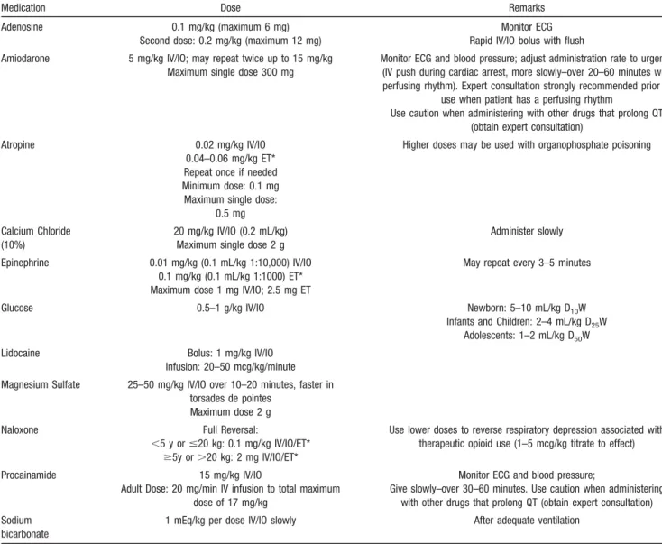
70,72,78,79Table 1.
Medications for Pediatric Resuscitation
Medication Dose Remarks
Adenosine 0.1 mg/kg (maximum 6 mg) Second dose: 0.2 mg/kg (maximum 12 mg)
Monitor ECG Rapid IV/IO bolus with flush Amiodarone 5 mg/kg IV/IO; may repeat twice up to 15 mg/kg
Maximum single dose 300 mg
Monitor ECG and blood pressure; adjust administration rate to urgency (IV push during cardiac arrest, more slowly–over 20–60 minutes with perfusing rhythm). Expert consultation strongly recommended prior to
use when patient has a perfusing rhythm
Use caution when administering with other drugs that prolong QT (obtain expert consultation)
Atropine 0.02 mg/kg IV/IO
0.04–0.06 mg/kg ET* Repeat once if needed Minimum dose: 0.1 mg Maximum single dose:
0.5 mg
Higher doses may be used with organophosphate poisoning
Calcium Chloride (10%)
20 mg/kg IV/IO (0.2 mL/kg) Maximum single dose 2 g
Administer slowly Epinephrine 0.01 mg/kg (0.1 mL/kg 1:10,000) IV/IO
0.1 mg/kg (0.1 mL/kg 1:1000) ET* Maximum dose 1 mg IV/IO; 2.5 mg ET
May repeat every 3–5 minutes
Glucose 0.5–1 g/kg IV/IO Newborn: 5–10 mL/kg D10W
Infants and Children: 2–4 mL/kg D25W Adolescents: 1–2 mL/kg D50W Lidocaine Bolus: 1 mg/kg IV/IO
Infusion: 20–50 mcg/kg/minute Magnesium Sulfate 25–50 mg/kg IV/IO over 10–20 minutes, faster in
torsades de pointes Maximum dose 2 g
Naloxone Full Reversal:
⬍5 y orⱕ20 kg: 0.1 mg/kg IV/IO/ET*
ⱖ5y or⬎20 kg: 2 mg IV/IO/ET*
Use lower doses to reverse respiratory depression associated with therapeutic opioid use (1–5 mcg/kg titrate to effect)
Procainamide 15 mg/kg IV/IO
Adult Dose: 20 mg/min IV infusion to total maximum dose of 17 mg/kg
Monitor ECG and blood pressure;
Give slowly–over 30–60 minutes. Use caution when administering with other drugs that prolong QT (obtain expert consultation) Sodium
bicarbonate
1 mEq/kg per dose IV/IO slowly After adequate ventilation IV indicates intravenous; IO, intraosseous; and ET, via endotracheal tube.
Medication Dose Calculation
To calculate the dose of resuscitation medications, use the
child’s weight if it is known. If the child’s weight is unknown,
it is reasonable to use a body length tape with precalculated
doses (Class IIa, LOE C).
70 –77It is unclear if an adjustment in the calculation of
resusci-tation medications is needed in obese children. Use of the
actual body weight in calculation of drug doses in obese
patients may result in potentially toxic doses. Length-based
tapes estimate the 50th percentile weight for length (ie, ideal
body weight), which may, theoretically, result in inadequate
doses of some medications in obese patients. Despite these
theoretical considerations, there are no data regarding the
safety or efficacy of adjusting the doses of resuscitation
medications in obese patients. Therefore, regardless of the
patient’s habitus, use the actual body weight for calculating
initial resuscitation drug doses or use a body length tape with
precalculated doses (Class IIb, LOE C).
For subsequent doses of resuscitation drugs in both
non-obese and non-obese patients, expert providers may consider
adjusting doses to achieve the desired therapeutic effect. In
general, the dose administered to a child should not exceed
the standard dose recommended for adult patients.
Medications (See Table 1)
Adenosine
Adenosine causes a temporary atrioventricular (AV) nodal
conduction block and interrupts reentry circuits that involve
the AV node. The drug has a wide safety margin because of
its short half-life. Adenosine should be given only IV or IO,
followed by a rapid saline flush to promote drug delivery to
the central circulation. If adenosine is given IV, it should be
administered as close to the heart as possible. (See also
“Arrhythmia.”)
Amiodarone
Amiodarone slows AV conduction, prolongs the AV
refrac-tory period and QT interval, and slows ventricular conduction
(widens the QRS). Expert consultation is strongly
recom-mended prior to administration of amiodarone to a pediatric
patient with a perfusing rhythm. (See also “Arrhythmia.”)
Precautions
Monitor blood pressure and electrocardiograph (ECG) during
intravenous administration of amiodarone. If the patient has a
perfusing rhythm, administer the drug as slowly (over 20 to
60 minutes) as the patient’s clinical condition allows; if the
patient is in VF/pulseless VT, give the drug as a rapid bolus.
Amiodarone causes hypotension through its vasodilatory
property, and the severity is related to the infusion rate;
hypotension is less common with the aqueous form of
amiodarone.
207Decrease the infusion rate if there is
prolon-gation of the QT interval or heart block; stop the infusion if
the QRS widens to
⬎
50% of baseline or hypotension
devel-ops. Other potential complications of amiodarone include
bradycardia and torsades de pointes ventricular tachycardia.
Amiodarone should not be administered together with
an-other drug that causes QT prolongation, such as
procain-amide, without expert consultation.
Atropine
Atropine sulfate is a parasympatholytic drug that accelerates
sinus or atrial pacemakers and increases the speed of AV
conduction.
Precautions
Small doses of atropine (
⬍
0.1 mg) may produce paradoxical
bradycardia because of its central effect.
208Larger than
recommended doses may be required in special
circum-stances such as organophosphate poisoning
209or exposure to
nerve gas agents.
Calcium
Calcium administration is not recommended for pediatric
car-diopulmonary arrest in the absence of documented
hypocalce-mia, calcium channel blocker overdose, hypermagnesehypocalce-mia, or
hyperkalemia (Class III, LOE B). Routine calcium
administra-tion in cardiac arrest provides no benefit
210 –221and may be
harmful.
210 –212If calcium administration is indicated during cardiac arrest,
either calcium chloride or calcium gluconate may be
consid-ered. Hepatic dysfunction does not appear to alter the ability
of calcium gluconate to raise serum calcium levels.
222In
critically ill children, calcium chloride may be preferred
because it results in a greater increase in ionized calcium
during the treatment of hypocalcemia.
222AIn the nonarrest
setting, if the only venous access is peripheral, calcium
gluconate is recommended because it has a lower osmolality
than calcium chloride and is therefore less irritating to the
vein.
Epinephrine
The
␣
-adrenergic-mediated vasoconstriction of epinephrine
increases aortic diastolic pressure and thus coronary
perfu-sion pressure, a critical determinant of successful
resuscita-tion from cardiac arrest.
223,224At low doses, the

-adrenergic
effects may predominate, leading to decreased systemic
vascular resistance; in the doses used during cardiac arrest,
the vasoconstrictive
␣
-effects predominate.
Precautions
●
Do not administer catecholamines and sodium bicarbonate
simultaneously through an IV catheter or tubing because
alkaline solutions such as the bicarbonate inactivate the
catecholamines.
●
In patients with a perfusing rhythm, epinephrine causes
tachycardia; it may also cause ventricular ectopy,
tachyarrhythmias, vasoconstriction, and hypertension.
Glucose
Because infants have a relatively high glucose requirement
and low glycogen stores, they may develop hypoglycemia
when energy requirements rise.
225Check blood glucose
concentration during the resuscitation and treat hypoglycemia
promptly (Class I, LOE C).
226Lidocaine
Lidocaine decreases automaticity and suppresses ventricular
arrhythmias,
227but is not as effective as amiodarone for
improving ROSC or survival to hospital admission among
adult patients with VF refractory to shocks and
epineph-rine.
228Neither lidocaine nor amiodarone has been shown to
improve survival to hospital discharge.
Precautions
Lidocaine toxicity includes myocardial and circulatory
de-pression, drowsiness, disorientation, muscle twitching, and
seizures, especially in patients with poor cardiac output and
hepatic or renal failure.
229,230Magnesium
Magnesium is indicated for the treatment of documented
hypomagnesemia or for torsades de pointes (polymorphic VT
associated with long QT interval). There is insufficient
evidence to recommend for or against the routine
adminis-tration of magnesium during cardiac arrest.
231–233Precautions
Magnesium produces vasodilation and may cause
hypoten-sion if administered rapidly.
Procainamide
Procainamide prolongs the refractory period of the atria and
ventricles and depresses conduction velocity.
Precautions
There is limited clinical data on using procainamide in infants
and children.
234 –236Infuse procainamide very slowly (over 30
to 60 minutes) while monitoring the ECG and blood pressure.
Decrease the infusion rate if there is prolongation of the QT
interval, or heart block; stop the infusion if the QRS widens
to
⬎
50% of baseline or hypotension develops. Do not
administer together with another drug causing QT
prolonga-tion, such as amiodarone, without expert consultation. Prior
to using procainamide for a hemodynamically stable patient,
expert consultation is strongly recommended.
Sodium Bicarbonate
Routine administration of sodium bicarbonate is not
recom-mended in cardiac arrest (Class III, LOE B).
212,237,238Sodium
bicarbonate may be administered for treatment of some
toxidromes (see “Toxicological Emergencies,” below) or
special resuscitation situations such as hyperkalemic cardiac
arrest.
Precautions
During cardiac arrest or severe shock, arterial blood gas
analysis may not accurately reflect tissue and venous
acido-sis.
239,240Excessive sodium bicarbonate may impair tissue
oxygen delivery;
241cause hypokalemia, hypocalcemia,
hy-pernatremia, and hyperosmolality;
242,243decrease the VF
threshold;
244and impair cardiac function.
Vasopressin
There is insufficient evidence to make a recommendation for
or against the routine use of vasopressin during cardiac arrest.
Pediatric
245–247and adult
248,249case series/reports suggested
that vasopressin
245or its long-acting analog,
terlipres-sin,
246,247may be effective in refractory cardiac arrest when
standard therapy fails. A large pediatric NRCPR case series,
however, suggested that vasopressin is associated with lower
ROSC, and a trend toward lower 24-hour and discharge
survival.
250A preponderance of controlled trials in adults do
not demonstrate a benefit.
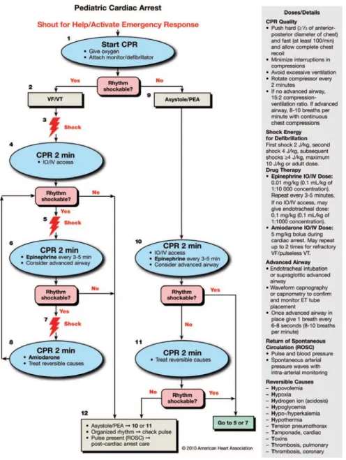
251–256Pulseless Arrest
In the text below, box numbers identify the corresponding
step in the algorithm (Figure 1).
●
(Step 1) As soon as the child is found to be unresponsive
with no breathing, call for help, send for a defibrillator
(manual or AED), and start CPR (with supplementary
oxygen if available). Attach ECG monitor or AED pads as
soon as available. Throughout resuscitation, emphasis
should be placed on provision of high-quality CPR
(pro-viding chest compressions of adequate rate and depth,
allowing complete chest recoil after each compression,
minimizing interruptions in compressions and avoiding
excessive ventilation).
●
While CPR is being given, determine the child’s cardiac
rhythm from the ECG or, if you are using an AED, the
device will tell you whether the rhythm is “shockable” (eg,
VF or rapid VT) or “not shockable” (eg, asystole or PEA).
It may be necessary to temporarily interrupt chest
compres-sions to determine the child’s rhythm. Asystole and
brady-cardia with a wide QRS are most common in asphyxial
arrest.
1VF and PEA are less common
13but VF is more
likely to be present in older children with sudden witnessed
arrest.
“Nonshockable Rhythm”: Asystole/PEA (Step 9)
PEA is an organized electric activity—most commonly slow,
wide QRS complexes—without palpable pulses. Less
fre-quently there is a sudden impairment of cardiac output with
an initially normal rhythm but without pulses and with poor
perfusion. This subcategory, formerly known as
electrome-chanical dissociation (EMD), may be more reversible than
asystole. For asystole and PEA:
●
(Step 10) Continue CPR with as few interruptions in chest
compressions as possible. A second rescuer obtains
vascu-lar access and delivers epinephrine, 0.01 mg/kg (0.1 mL/kg
of 1:10 000 solution) maximum of 1 mg (10 mL), while
CPR is continued. The same epinephrine dose is repeated
every 3 to 5 minutes (Class I, LOE B). There is no survival
benefit from high-dose epinephrine, and it may be harmful,
particularly in asphyxia (Class III, LOE B).
257–261High-dose epinephrine may be considered in exceptional
circum-stances, such as

-blocker overdose (Class IIb, LOE C).
●
Once an advanced airway is in place, 1 rescuer should give
continuous chest compressions at a rate of at least 100 per
minute without pause for ventilation. The second rescuer
delivers ventilations at a rate of 1 breath every 6 to 8
seconds (about 8 to 10 breaths per minute). Rotate the
compressor role approximately every 2 minutes to prevent
compressor fatigue and deterioration in quality and rate of
chest compressions. Check rhythm every 2 minutes with
minimal interruptions in chest compressions. If the rhythm
is “nonshockable” continue with cycles of CPR and
epi-nephrine administration until there is evidence of ROSC or
you decide to terminate the effort. If at any time the rhythm
becomes “shockable,” give a shock (Step 7) and
immedi-ately resume chest compressions for 2 minutes before
rechecking the rhythm. Minimize time between chest
compressions and shock delivery (ie, check rhythm and
deliver shocks immediately after compressions rather than
after rescue breaths, if possible) and between shock
deliv-ery and resumption of chest compressions.
●
Search for and treat reversible causes.
“Shockable Rhythm”: VF/Pulseless VT (Step 2)
Defibrillation is the definitive treatment for VF (Class I,
LOE B) with an overall survival rate of 17% to 20%.
4,262,263Survival is better in primary than in secondary VF.
6In adults,
the probability of survival declines by 7% to 10% for each
minute of arrest without CPR and defibrillation.
264Survival is
better if early, high-quality CPR is provided with minimal
interruptions. Outcome of shock delivery is best if rescuers
minimize the time between last compression and shock
delivery, so rescuers should be prepared to coordinate (brief)
interruptions in chest compressions to deliver shocks, and
should resume compressions immediately after shock
delivery.
Defibrillators
Defibrillators are either manual or automated (AED), with
monophasic or biphasic waveforms. For further information
see Part 6, “Electrical Therapies: Automated External
Defi-brillators, Defibrillation, Cardioversion, and Pacing.”
AEDs in institutions caring for children at risk for
arrhyth-mias and cardiac arrest (eg, hospitals, EDs) must be capable
of recognizing pediatric cardiac rhythms and should ideally
have a method of adjusting the energy level for children.
The following should be considered when using a manual
defibrillator:
Paddle Size
In general, manual defibrillators have two sizes of hand-held
paddles: adult and infant. The infant paddles may slide over
or be located under the adult paddles. Manual defibrillators
can also be used with hands-free pads that are self adhesive.
Use the largest paddles or self-adhering electrodes
265–267that
will fit on the child’s chest without touching (when possible,
leave about 3 cm between the paddles or electrodes). Paddles
and self-adhering pads appear to be equally effective.
268Self-adhering pads should be pressed firmly on the chest so
that the gel on the pad completely touches the child’s chest.
An appropriate paddle or self-adhesive pad size is
●
“Adult” size (8 to 10 cm) for children
⬎
10 kg
(
⬎
approximately 1 year)
●
“Infant” size for infants
⬍
10 kg
Interface
The electrode– chest wall interface is part of the self-adhesive
pad; in contrast, electrode gel must be applied liberally on
manually applied paddles. Do not use saline-soaked pads,
ultrasound gel, bare paddles, or alcohol pads.
Paddle Position
Follow package directions for placement of self-adhesive
AED or monitor/defibrillator pads.
Place manual paddles over the right side of the upper chest
and the apex of the heart (to the left of the nipple over the left
lower ribs) so the heart is between the two paddles. Apply
firm pressure. There is no advantage in an anterior-posterior
position of the paddles.
268Energy Dose
The lowest energy dose for effective defibrillation and the
upper limit for safe defibrillation in infants and children are
not known; more data are needed. It has been observed that in
children with VF, an initial monophasic dose of 2 J/kg is only
effective in terminating ventricular fibrillation 18% to 50% of
the time,
269,270while similar doses of biphasic shocks are
effective 48% of the time.
268Children with out-of-hospital
VF cardiac arrest often receive more than 2 J/kg,
271,272and
one in-hospital cardiac arrest study
268showed that children
received doses between 2.5 and 3.2 J/kg to achieve ROSC.
Energy doses
⬎
4 J/kg (up to 9 J/kg) have effectively
defibrillated children
272–274and pediatric animals
275with
negligible adverse effects. Based on data from adult
stud-ies
276,277and pediatric animal models,
278 –280biphasic shocks
appear to be at least as effective as monophasic shocks and
less harmful.
It is acceptable to use an initial dose of 2 to 4 J/kg (Class
IIa, LOE C), but for ease of teaching an initial dose of 2 J/kg
may be considered (Class IIb, LOE C). For refractory VF, it
is reasonable to increase the dose to 4 J/kg (Class IIa, LOE C).
Subsequent energy levels should be at least 4 J/kg, and higher
energy levels may be considered, not to exceed 10 J/kg or the
adult maximum dose (Class IIb, LOE C).
AEDs
Many AEDs can accurately detect VF in children of all
ages.
271,281–283They can differentiate “shockable” from
“non-shockable” rhythms with a high degree of sensitivity and
specificity.
281,282It is recommended that systems and
institu-tions that have AED programs and that care for children
should use AEDs with a high specificity to recognize
pedi-atric shockable rhythms and a pedipedi-atric attenuating system
that can be used for infants and children up to approximately
25 kg (approximately 8 years of age).
274,284If an AED with an
attenuator is not available, use an AED with standard
elec-trodes (Class IIa, LOE C).
In infants
⬍
1 year of age a manual defibrillator is
pre-ferred. If a manual defibrillator is not available, an AED with
a dose attenuator may be used. An AED without a dose
attenuator may be used if neither a manual defibrillator nor
one with a dose attenuator is available (Class IIb, LOE C).
Integration of Defibrillation With Resuscitation
Sequence
The following are important considerations:
●
Provide CPR until the defibrillator is ready to deliver a
shock; after shock delivery, resume CPR, beginning with
chest compressions. Minimize interruptions of chest
com-pressions. In adults with prolonged arrest
285,286and in
animal models,
287defibrillation is more likely to be
suc-cessful after a period of effective chest compressions.
Ideally chest compressions should be interrupted only for
ventilations (until an advanced airway is in place), rhythm
check, and shock delivery. If a “shockable” rhythm is still
present, continue chest compressions after a rhythm check
(when possible) while the defibrillator is charging (so chest
compressions are delivered until shock delivery).
●
(Step 3) Give 1 shock (2 J/kg) as quickly as possible and
immediately resume CPR, beginning with chest
compres-sions. If 1 shock fails to eliminate VF, the incremental
benefit of another immediate shock is low, and resumption
of CPR is likely to confer a greater value than another
shock. CPR may provide coronary perfusion, increasing
the likelihood of defibrillation with a subsequent shock. It
is important to minimize the time between chest
compres-sions and shock delivery and between shock delivery and
resumption of postshock compressions.
●
(Step 4) Continue CPR for about 2 minutes. In in-hospital
settings with continuous invasive monitoring, this sequence
may be modified at the expert provider’s discretion (see,
also Part 8.2: “Management of Cardiac Arrest”). If sufficient
rescuers are present, obtain vascular (IO or IV) access.
●
After 2 minutes of CPR, check the rhythm; recharge the
defibrillator to a higher dose (4 J/kg).
●
(Step 5) If a “shockable” rhythm persists, give another
shock (4 J/kg). If rhythm is “nonshockable,” continue with
the asystole/PEA algorithm (Steps 10 and 11).
●
(Step 6) Immediately resume chest compressions. Continue
CPR for approximately 2 minutes. During CPR give
epinephrine 0.01 mg/kg (0.1 mL/kg of 1:10 000
concen-tration), maximum of 1 mg (Class I, LOE B)
every 3 to 5
minutes.
It is helpful if a third rescuer prepares the drug
doses
before
the rhythm is checked so epinephrine can be
administered as soon as possible. Epinephrine should be
administered during chest compressions, but the timing of
drug administration is less important than the need to
minimize interruptions in chest compressions. Just prior to
the rhythm check, the rescuer operating the defibrillator
should prepare to recharge the defibrillator (4 J/kg or more
with a maximum dose not to exceed 10 J/kg or the adult
dose, whichever is lower).
●
Check the rhythm
●
(Step 7) If the rhythm is “shockable,” deliver another shock
(4 J/kg or more with a maximum dose not to exceed 10 J/kg
or the adult dose, whichever is lower) and immediately
resume CPR (beginning with chest compressions).
●
(Step 8) While continuing CPR, give amiodarone (Class
IIb, LOE C)
228,288 –290or lidocaine if amiodarone is not
available.
●
If at any time the rhythm check shows a “nonshockable”
rhythm, proceed to the “Pulseless Arrest” sequence (Steps
10 or 11).
●
Once an advanced airway is in place, 2 rescuers no longer
deliver cycles of CPR (ie, compressions interrupted by
pauses for ventilation). Instead, the compressing rescuer
gives continuous chest compressions at a rate of at least
100 per minute without pause for ventilation. The rescuer
delivering ventilation provides about 1 breath every 6 to 8
seconds (8 to 10 breaths per minute). Two or more rescuers
should rotate the compressor role approximately every 2
minutes to prevent compressor fatigue and deterioration in
quality and rate of chest compressions.
●
If defibrillation successfully restores an organized rhythm
(or there is other evidence of ROSC, such as an abrupt rise
in P
ETCO2or visible pulsations on an arterial waveform),
check the child’s pulse to determine if a perfusing rhythm
is present. If a pulse is present, continue with
postresusci-tation care.
●
If defibrillation is successful but VF recurs, resume CPR
and give another bolus of amiodarone before trying to
defibrillate with the previously successful shock dose.
●